Severe community-acquired meningitis
Christophe Boisson, Sophie Arnaud, Renaud Vialet and Claude Martin
Addresses: Department of Anesthesia and Intensive Care, and Trauma Center, Marseilles University Hospital System, Marseilles School of Medicine, Marseilles, France
Correspondence: Intensive Care Unit and Trauma Center, Hôpital Nord, Marseilles University Hospital System, Marseilles School of Medicine, Marseilles, France
Received: 6 July 1999 Accepted: 13 July 1999 Published: 6 August 1999
Crit Care1999, 3:R55–R65
The original version of this paper is the electronic version which can be seen on the Internet (http://ccforum.com). The electronic version may contain additional information to that appearing in the paper version.
© Current Science Ltd ISSN 1364-8535
Introduction
Community-acquired meningitis is associated with high morbidity and mortality. The epidemiology of community-acquired meningitis has changed over the past 15 years with the use of new vaccines and with the development of resistance to antibiotics. Bacterial meningitis would appear to be the most frequent by far, but most viral aetiologies are very often poorly recognized. The annual incidence of purulent community-acquired meningitis in France was estimated at 22 cases per million inhabitants in 1993. Age is a major risk factor in bacterial meningitis. The most affected group is children under 2 years of age, in whom the infection rate ranges from 10 to 110 cases per 100 000 infants per year. Such cases must be considered as an absolute medical emergency. This review is limited to meningitis in immunocompetent patients.
Bacterial epidemiology
From birth until the first month of life, Streptococcus agalactiae, Listeria monocytogenes and Enterobacteriaceae (especially Escherichia coli) are the main pathogens. From 2 months until 4 years of age, meningitis is principally due to Haemophilus influenzaetype B (the incidence of which is dramatically decreasing because of widespread vaccina-tion), Neisseria meningitidisand Streptococcus pneumoniae. In children, teenagers and young adults, N. meningitidisand S. pneumoniae represent more than 80% of the cases. Above the age of 60 years, S. pneumoniaeand H. influenzae are the most frequently isolated bacteria [1,2].
When all of these bacteria are taken together, the ele-ments that indicate a poor prognosis are as follows: age
less than 1 month or greater than 45 years; worsening level of consciousness on admission; and forms with purpura fulminans [3–6]. In the absence of a specific treatment, purulent meningitis is ultimately fatal.
Pneumococcal meningitis
Pneumococcal meningitis is the most frequent cause of bacterial meningitis in those older than 30 years, and was responsible for 20% of cases of purulent community meningitis in children and 60% of adult cases in France in 1993. It is also more severe in terms of mortality (15–30%) and morbidity (deafness).
The increasing resistance of S. pneumoniaeto penicillin (in more than 10% of isolates) has created a problem for therapy, which at present cannot be completely optimized and requires a knowledge of the sensitivity phenotype of the bacteria in question. Penicillin-resistant pneumococci (PRP) include pneumococci with an intermediate sensi-tivity [minimum inhibitory concentration (MIC) of between 0.12 and 1 mg/l] and highly resistant pneumo-cocci (MIC > 1 mg/l).
Resistance to β-lactams is related to modifications of the genes coding for penicillin-linked proteins (PLPs), which are the active sites for all β-lactams, especially PLP2b and PLP2x [7]. Such genomic change is due either to a genetic transfer or to on-off mutations. The main serotypes involved in the resistance to β-lactams are 6A, 6B, 14, 19A, 19F and 23F, and 99% of the serotypes are present in antipneumococcal vaccine of 23 valences. This evolution in resistance should lead to a renewed interest in antipneumococcal vaccination.
Resistance to β-lactam antibiotics in pneumococci has been increasing in a disturbing manner, especially in Europe. The first strains resistant to penicillin were described in Boston in 1964 and in Chicago in 1974, and the first case of pneumococcal meningitis resistant to ceftriaxone was reported in 1991. The progression of resistance has been constant in the USA; from 1979 to 1987 the rate of resis-tance was below 10%, but since 1987 there has been an increase of up to 30% in PRP. In Europe, Spain has a rate of almost 60% of PRP, followed by Bavaria with 50% resis-tance, but the rate is lower for the rest of Germany. France is next with 36% in children and 25% in adults in 1994. The magnitude of resistance was high in half of the cases.
Among the cephalosporins, cefaclor, cefuroxime and cefixime are no longer active on strains resistant to
penicillin, and a direct relationship has even been noted between cefaclor and penicillin resistance. Of PRP strains 10% are cross-resistant to macrolides: erythromycin, as well as clarithromycin and azithromycin.
In children, the risk factors for acquiring infection with resistant S. pneumoniaeare hospitalization, living in closed populations, β-lactam treatment in the preceding months and recurring otitis media. In adults the risk factors are immunodeficiency, nosocomial infections and β-lactam treatment in the preceding months. Limiting the spread of S. pneumoniae resistance to antibiotics therefore depends on the following: more rational utilization of antibiotics and perhaps reduction of the systematic prophylaxis of acute otitis media in children with recurrent otitis; tion of the optimal duration of antibiotic therapy; evalua-tion of posologies and the intake required in order to avoid long periods with serum levels nearly at subinhibition con-centrations and favouring the selection of resistant mutants; alternating different classes of antibiotics; and development of the antipneumococcal vaccine.
Meningococcal meningitis
Meningococcal meningitis occurs in endemics, with cases that are usually sporadic causing minor endemia that are limited in time and space, particularly in closed popula-tions of children and young adults. There is a distinct pre-ponderance of group B (60%) over group C (30%) organisms. The mean mortality rate of 10% is principally linked to the occurrence of purpura fulminans [8].
Haemophilus influenzaemeningitis
Widespread vaccination is currently disrupting the epidemi-ology of H. influenzaemeningitis, which is caused by type B strains. Approximately 50% produce β-lactamases, making them resistant to amino-penicillins. Despite this increase in resistance, the mortality rate has gone from 10 to 3%.
Physiopathology
The bacteria responsible for purulent meningitis are of the extracellular multiplication type, and have properties that make them resistant to nonspecific defence factors and allow them to develop in the host tissues. They also have specific properties that enable them to invade the meninges and cause inflammation [9,10].
Bacterial penetration in the cerebrospinal fluid
There are various arguments that support the hypothesis of penetration of bacteria into the cerebrospinal fluid (CSF) by the haematogenous route, with secondary penetration through the haematomeningeal barrier. In the case of experimental infection with H. influenzae, rhinopharyngeal administration of bacteria is followed by bacteremia and then invasion of the CSF. Similarly, intraperitoneal injec-tion of N. meningitidis in newborn rats or intravenously in macaques causes secondary colonization of the CSF [11].
The haematomeningeal barrier is only one of the ele-ments of the haematoencephalic barrier, and pathogenic bacteria are hypothesized to invade the CSF via the blood by two different routes: direct penetration of the meningeal capillary endothelium; or by direct penetration at the level of the choroid plexus, which produces CSF by secretion. The latter of these would appear to be the most plausible hypothesis following postmortem and experi-mental observations of major and selective concentrations of bacteria in the choroid plexus capillaries.
Inflammatory mechanisms of the subarachnoid space Once the bacterium has invaded the CSF, little can prevent it from multiplying because there is almost no complement fractions and the immunoglobulin concentra-tion is very low. On the other hand, poor nutriconcentra-tion slows bacterial multiplication.
In order to understand the cascade of events that occurs as soon as the bacterium has invaded the CSF, experimental meningitis models have been designed by direct injection of bacteria into rabbit and rat cisterna magna, making it possible to short-circuit the haematomeningeal barrier.
In-situ production of cytokines
The most important event that follows bacterial penetra-tion into the CSF is the producpenetra-tion of cytokines [12]. In animal experiments, intracisternal injection of lipopolysac-charide triggers production of tumour necrosis factor (TNF)-α, interleukin (IL)-1 and IL-6, preceding the appearance of inflammatory exudate, which can be par-tially prevented by the injection of anti-TNF-α and/or anti-IL-1 antibodies. Injection of TNF-α and IL-1 into the cisterna magna is followed by an elevation in the protein concentration in the CSF, an afflux of polynuclear cells and an increase in the weight of the brain, reflecting oedema. It would therefore follow that the production of cytokines in the CSF is a prerequisite to episodes of meningitis. This production occurs in situ, is independent of all systemic cytokine production because the molecules cannot penetrate the haematomeningeal barrier and there-fore comes from the macrophage cells within the meninges itself [13,14].
Polynuclear afflux
polynuclear cells and the endothelium, leading to polynu-clear extravasation in the infected tissue [15,16].
Alteration of the haematomeningeal barrier
The other consequence of cytokine production is a reduc-tion in the tightness of the hematomeningeal barrier, which is essentially related to local production of IL-1 and synergistically assisted by TNF-α, creating a loosening of the tight cerebral capillary junctions.
Consequences of bacterial meningitis
All of the events that occur during an episode of bacterial meningitis are the result of polynuclear afflux and alter-ation of the haematomeningeal barrier. The result is a mixed cerebral oedema (ie both interstitial, because it is linked to the resorption of CSF at the level of the arach-noid villi, and vasogenic, because it is linked to an increase in the permeability of the haematomeningeal barrier). In addition, this major meningeal inflammation can lead to major vascular alterations of the meningeal vessels, as occurs in vasculitis, facilitating microthromboses and par-ticipating in profound alterations in cerebral blood flow and anoxia [17].
Meningococcus
N. meningitidisis a Gram-negative, oxidase-positive aerobic diplococcus that has a polysaccharide capsule. It is a sapro-phyte, and colonizes most humans at some point in their lives without producing any particular symptoms. The development of disease after exposure to meningococci is the result of a poorly known mechanism of host factors that have an influence on bacterial invasion of mucosal surfaces. Among the elements that influence host response, genetic control of the response to the acute stage and cytokine secretion (TNF-α, IL-1, IL-6) is of importance in making the prognosis. Moreover, a defi-ciency in complement predisposes the patient to a meningococcus infection with unusual serogroups (X, Y, Z, W135, 29E) [18,19].
Listeria monocytogenes
L. monocytogenes is an occasional intracellular Gram-posi-tive bacteria that is capable of multiplying in the macrophages and in most of the cells of the infected host tissue. From the sites of intracellular multiplication, L. monocytogenes can spread through the blood and reach numerous target organs, particularly the placenta and the central nervous system. The bacterium first enters via direct contact with the host cells through an 80 kDa protein called internalin, coded by chromosomal gene InlA. Internalin is a protein located at the surface of the bacterium and has a structural organization that is charac-teristic of numerous Gram-positive bacteria proteins (such as Streptococcusand Staphylococcusspp.). Internalin interacts with receptors of unknown nature (probably integrins) located at the surface of the infected cells. After
penetrat-ing the cells, the bacteria are capable of escappenetrat-ing the phagosomes and of multiplying in the cytoplasm. In the infected host, orally ingested bacteria go through the intestine via Peyer’s patches and reach the lymphatic gan-glions and the blood circulation. The principal target organ is the liver, where the bacteria multiply inside the hepatocytes. Hepatocyte lysis induced by the early recruitment of polynuclear cells triggers a liberation of intercellular bacteria and results in prolonged bacteraemia. At this stage, the disease is often managed well by immunocompetent patients, who may contract an infec-tion that is often totally asymptomatic. This is probably the most frequent scenario if the frequency of exposure to L. monocytogenes is low. However, if the inoculum is massive, in a pregnant woman, or in patients who are immunocompromised (patients with acquired immunode-ficiency syndrome, patients with neutropenia or who are undergoing chemotherapy) or who present with hepatic anomalies (cirrhosis, hemochromatosis), infection of the hepatocytes is not controlled and the bacteria are released into the bloodstream. Metastatic localizations are possible at this stage, particularly in the placenta or the central nervous system [20].
Therapeutic implications
Bacterial meningitis is an infection of a defined space in which the host is compromised because of the absence of complement fractions and the low levels of specific anti-bodies. For this reason phagocytosis is ineffective and bac-terial multiplication is rapid. Optimal antibiotic treatment requires that the selected antibiotic has a bactericidal effect in the CSF.
Passage of antibiotics into the cerebrospinal fluid
Antibiotics that strongly bind to proteins and those with a high molecular weight diffuse poorly into the cerebral parenchyma and the CSF. Meningeal inflammation par-tially improves this situation by modifying the permeabil-ity of the haematomeningeal barrier (increase in vesicular transport, and separation of the tight junctions between the endothelial cells and the meningeal vessels) [21].
Antibacterial synergy in cerebrospinal fluid in vivo
In a model of L. meningitisinfection, an ampicillin–gentam-icin combination (in vitrosynergy) turned out to be more rapidly bactericidal than either when used alone [22].
Antibiotic concentration in the cerebrospinal fluid
demonstrated with other bacteria and other antibiotics [9]. One explanation could be that infected CSF reduces antibiotic activity. For example, the low pH of infected CSF (between 6.7 and 7.1) reduces the activity of amino-glycosides and the increase in protein concentration decreases the concentration of the active drug, particularly that of the strongly bound β-lactamase (cephalosporins). This implies the use of high doses of antibiotics, espe-cially when the MIC is elevated.
Immunotherapy
Because of the excessive inflammation of the meningeal spaces and its experimental aggravation by bacterial lysis caused by the antibiotics, it can be useful to modify the various players in this immunoinflammatory cascade by attempting to inhibit the polynuclear interactions with the haematomeningeal barrier. Inflammation, however, plays a positive role in the penetration of certain antibiotics at the infection site. The use of methylprednisolone reduces the spinal penetration of ampicillin and gentamicin by approximately half.
Diagnosis
Clinical diagnosisThe infectious syndrome is always present. It has a brutal aspect with a high fever (> 38.5°C), shivers and myalgia, sometimes accompanied by the following: pneumopathy (in cases of pneumococcus infection), purpura and septic shock. At times, it is subtle and is unfortunately diagnosed as an ear–nose–throat infection, which are often degener-ated by antibiotic therapy.
The meningeal syndrome is quasi constant. Cephalalgia is diffuse, intense and continuous, and can radiate towards the nape of the neck and the back. It is increased by light, noise and coughing. Vomiting is frequent.Meningeal stiff-ness corresponds to the extension of the neck muscles during massive flexion of the head. It is responsible for the coiled position. It can sometimes be subtle, especially in an early stage or in elderly patients or in cases of coma. It can be evaluated by means of certain manoeuvres. Kernig’s sign is a symptom of meningitis that is demon-strated when patients cannot keep their legs extended while sitting on a bed. In the prone position, the patient cannot raise and extend his arms because flexing occurs at a certain height. Brudzinski’s sign is a spontaneous flexion of the thighs and legs during flexion provoked by the head (when the neck is flexed from a supine position). The meningeal syndrome can also include signs of cutaneous hyperesthesia or pyramidal irritation such as in osteoten-don hyperreflexia.
Signs of encephalitis are erratic but must be considered as having a poor clinical outcome; they include change in mental status that ranges from mental confusion to coma. They may also include local or generalized convulsions.
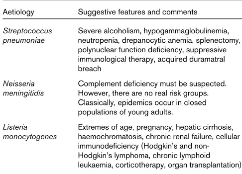
The factors that should raise suspicion of certain aetiologic diagnoses are listed Table 1.
Biological diagnosis Lumbar puncture
When the clinical examination only reveals meningeal and infectious signs (60–70% of cases) lumbar puncture is the first examination to perform [23]. If, on the other hand, onset is progressive and there appears to be signs of defi-ciency without change in mental status, followed by signs of intracranial hypertension, a cerebral computed tomogra-phy (CT) scan must be urgently performed in order to exclude the presence of an expansion process. Fundus-copic examination is completely useless.
In cases of comatose meningitis, the relationship between lumbar puncture and herniation is, in any case, very debat-able. Such an important examination should therefore not be put off.
Macroscopically, the CSF has an elevated opening pres-sure, is cloudy (rice water) or purulent, confirming puru-lent meningitis and the need for subsequent antibiotic therapy (Fig. 1). Cytochemically, the CSF contains numerous polynuclear neutrophils (≥1000/mm3); protein
concentration is high (≥1 g/l); and the CSF glucose con-centration is low or even collapsed but, in any case, is less than 70% of the blood glucose concentration (ie the CSF:blood glucose concentration ratio is < 0.3).
[image:4.609.312.555.118.293.2]Particular forms can be encountered. The CSF can have minimal cytochemical modifications but be swarming with bacteria in cases of fulminant meningococcus meningitis, necessitating Gram’s staining in all cases with purpura, whatever the cytological results of the CSF. Moreover, an Table 1
Aetiologic diagnoses and suggestive features Aetiology Suggestive features and comments
Streptococcus Severe alcoholism, hypogammaglobulinemia, pneumoniae neutropenia, drepanocytic anemia, splenectomy,
polynuclear function deficiency, suppressive immunological therapy, acquired duramatral breach
Neisseria Complement deficiency must be suspected. meningitidis However, there are no real risk groups.
Classically, epidemics occur in closed populations of young adults.
Listeria Extremes of age, pregnancy, hepatic cirrhosis, monocytogenes haemochromatosis, chronic renal failure, cellular
intense cellular reaction without micro-organisms on direct examination is possible, with bacteriologic confir-mation by culture of CSF.
Lymphocytosis is possible if lumbar puncture is per-formed very early, but this condition above all indicates Listeriameningoencephalitis [24,25].
Blood cultures
Blood cultures provide an indispensable diagnostic com-plement. Nonspecific examination results such as polynu-cleosis and elevated C-reactive protein may be suggestive of a bacterial aetiology.
Bacteriology
Gram’s staining will provide an aetiologic diagnosis in 60–90% of the cases of bacteriologic meningitis. The per-centage of positive direct examinations is highest for S. pneumoniaeand H. influenzae, is lower for N. meningitidis, and is especially low for L. monocytogenes. Culturing is sys-tematic and allows for isolation and identification of the causative bacteria. An antibiogram may be directly per-formed with the CSF if it is sufficiently rich in bacteria. The results obtained after 18–24 h must always be con-firmed by classic study performed after the culture. In cases of pneumococcus infection, resistance to β -lacta-mases is sought by means of an oxacillin disk. Detection is completed by parallel determination of the MIC, per-formed either using classic dilution methods or using E-test with penicillin G and the β-lactams habitually used for the treatment of pneumococcus meningitis.
In patients with H. influenzae infection it is essential to look for β-lactamase production. Some strains of N. menin-gitidis with reduced sensitivity to penicillin G have been observed in France and Spain, but without therapeutic consequences (at the time of writing).
The interest of soluble antigen exploration has been subject to diverse evaluations. Performed using a particle agglutination of latex sensitivity technique, it can rein-force a diagnosis when the Gram’s staining is question-able, but is of little use when the Gram’s staining is negative [23]. Polymerase chain reaction can be a better means to detect bacteria in the CSF. Intraspinal lactate concentration alone does not have sufficient diagnostic value [26].
During the initial work-up, the diagnosis of bacterial meningitis is confirmed if the bacteriologic examinations are positive. In the case of negative results on direct exam-ination, the medical probabilities should be determined in order to differentiate the bacterial or viral nature of an episode of meningitis. This can be achieved by using valid decision models that take several cytological and biochem-ical parameters of the CSF into account, in addition to the
clinical data. Subsequent biological examinations are dic-tated by the clinical evolution and the initial bacterial data. A possible second lumbar puncture is performed 36–48 h after treatment, enabling the clinician to judge the therapeutic efficacy of CSF sterilization, as well as the rise in glycorrhachia. The other cytological and biochemical parameters of the CSF are of no diagnostic or prognostic value. In cases of therapeutic failure, an antibiotic may be administered in the CSF.
Radiological examinations
The indications for scanning or imaging should be very limited when a patient with purulent meningitis is admit-ted. Performing a CT scan before the lumbar puncture exposes the patient to the risks of delaying an antibiotic therapy that has reliable results. Lumbar puncture must therefore precede a CT scan, even in cases of coma. This diagnostic approach should only be changed if there are localized neurological signs that suggest another diagnosis or if there is a risk of an intracranial complication, possibly making lumbar puncture dangerous. Haemocultures and antibiotic therapy must therefore be immediately under-taken before imaging results are obtained (Fig. 1). In an Figure 1
Management algorithm in case of suspicion of bacterial meningitis. Directed
antibiotherapy
Suspicion of bacterial meningitis Localized neurological deficit
Absent Present
Blood cultures Blood cultures
Lumbar puncture
Empiric antibiotic therapy Positive Gram staining
or positive antigen test
No Yes
Cerebral computed tomography scan:
No lesion: lumbar puncture Lesion: abscess, empyema Empiric
emergency situation, cerebral CT scan enables the clini-cian to resolve most diagnostic problems. Magnetic reso-nance imaging is more accurate than CT, but it is less often available and does not provide decisive supplemen-tary information.
Once antibiotic therapy has been started, the diagnosis of intracranial complications will depend on imaging. Cere-bral CT scan is adequate for most intracranial complica-tions (hydrocephalus, abscess, empyema, cerebral infarct, haemorrhage, ventriculitis). However, in cases of cerebral thrombophlebitis CT scanning does not reveal direct signs and an angiography is required. Magnetic resonance imaging is the examination of choice when it is available and will provide a diagnosis of all intracranial complica-tions with sensitivity and specificity that are superior to those of CT scanning, particularly for the diagnosis of cerebral thrombophlebitis. In infants, transfontanel ultra-sonography is easy to perform and enables the clinician to detect abscess, ventriculitis and hydrocephalus.
A search for a portal of entry is especially justified in pneumococcal meningitis. Portals of entry are often ear–nose–throat-related in cases of both acute and chronic pathology. Imaging relies on CT scanning, which has better bone definition than MRI. It is not indicated for clinically diagnosed acute otitis. On the other hand, it is justified when mastoiditis or an unsatisfactory evolution of meningitis is suspected.
Imaging is indispensable for the preoperative work-up of all chronic pathologies and for the diagnosis of most cases of acute sinusitis.
The existence of an osteodural breach must be considered in cases of post-traumatic pneumococcal meningitis and recurring meningitis. In rare cases, the breach can be a congenital ear and petrosa malformation that is clearly revealed by CT scan, or an anterior meningoencephalo-cele or lumbosacral dermic sinus, which are better explored using magnetic resonance imaging. More often, the breach is secondary to a recent or old head trauma or to surgery at the base of the skull. At the level of the ear, exploration for a breach must be performed by CT scan-ning in frontal and axial millimeter sections. For the ante-rior floor, the CT scan should be used to investigate for a lack of continuity, essentially at the level of the lamina cribrosa and the roof of the ethmoid bone. CT scanning is the technique of choice because it best examines the small bone dehiscences. Isotropic transit of the CSF is no longer an indication [23].
Differential diagnosis Atypical cerebrospinal fluid
It can be difficult to differentiate between bacterial menin-gitis “beheaded” by an antibiotic treatment and viral
meningitis which is diagnosed early due to the context, briefness of the signs and normal glycorrhachia, reinforced by the disappearance of polynucleosis and the appearance of lymphocytosis in the CSF sampled 24–48 h later.
In the absence of bacterial meningitis, the presence or even the predominance of polynuclear cells in the CSF can be encountered in several situations: bacterial patholo-gies suppurated from the brain or parameningeal spaces (subdural empyema, brain abscess), fungal infections (cryptococcosis, candidiasis, histoplasmosis, coccid-ioidomycosis), rare cases of amoebic meningitis, neoplastic meningitis, chemical meningitis (co-trimoxazole, nons-teroidal anti-inflammatory drugs, azathioprine), systemic recurrent aseptic puriform meningitis, disseminated lupus erythematosus, sarcoidosis, Behçet’s disease. Rupture of an epidermoid cyst in the arachnoidea space and Mol-laret’s benign recurrent pleocytic meningitis are very rare and their diagnosis is favoured by recurrence with a sudden onset, rapidly favourable evolution and the poly-morphic character of the CSF.
Acute clear cerebrospinal fluid meningitis
Acute clear CSF meningitis is benign in most cases. It is due to a great number of viruses and evolves sponta-neously towards resolution. Measles and German measles are in very distinct decline due to systematic vaccination.
The major problem is the possibility of a herpetic aetiology leading to emergency empiric treatment with acyclovir. The other emergency diagnostic hypothesis is Listeria neu-romeningeal infection, which also requires emergency empiric antibiotic treatment. The possibility of a tubercu-lous infection should not be ignored, but therapy is often less urgent. The various diagnostic hypotheses are often formulated from the epidemiological data gathered from the context, because the microbiological results often arrive too late for emergency treatment [9].
Herpetic meningoencephalitis
Herpetic meningitis is usually due to herpes simplex virus I. In 30% of the cases, the encephalitis is primary, but in 70% it is preceded by a past history of benign herpes. Such pathology represents direct viral aggression located at the frontal and temporal level and leading to necrosis of the affected tissue. The clinical picture is not very spe-cific, but the patient will present with increasingly severe mental confusion, convulsions, cranial nerve impairment and olfactory hallucinations. However, because these symptoms will appear too late for effective treatment, her-petic aetiology must be presumed for encephalitis. If untreated, death occurs in 60–80% of the cases with very severe neurological sequelae in the surviving patients.
emergency diagnosis because antibody intrathecal secre-tion (estimated by comparing its level with serum level) is specific but inconsistently detected. The electroen-cephalograph is more reliable with pseudo-periodic rhyth-micity in the frontotemporal regions. However, this assessment is often delayed in comparison with therapy, the need for which is urgent; its absence must not delay antivi-ral treatment. The same is true for the cerebantivi-ral CT scan with iodine injection. The images are typically hypodense zones with peripheral contrast in the temporal and frontal regions. Such images are often bilateral and symmetrical.
The currently accepted treatment for herpetic encephali-tis has considerably improved outcome: acyclovir, 10 mg/kg every 8 h by intravenous infusions of approxi-mately 1 h. The duration of treatment is classically 10 days, but there have been reports in the literature of rare relapses due to a short duration of therapy, and conse-quently 3 weeks of treatment are often recommended.
Listeriameningoencephalitis
Listeria meningoencephalitis is certainly the most fre-quently encountered at present. The clinical signs are not very specific. It is, however, possible to stress the frequent association of a straightforward meningeal syndrome and impairment of one or several cranial nerves. Diagnosis is confirmed by isolation of the bacterium in blood cultures and/or CSF culture. Given the small size of this Gram-positive bacillus and the low inoculum, direct examination of the CSF is very often negative. The cytorachia is typi-cally mixed with polynuclear cells and lymphocytes, but a predominantly lymphocytic composition is observed just as fequently. Cerebral CT scan will reveal diverse features ranging from simple oedema to necrosis. On the other hand, a temporal lesion location is suggestive. Encephali-tis is diffuse when there are intracerebral abscesses pre-dominantly located in the rhombencephalon with multiple and necrotic abscesses. More rarely, there can be pure encephalitic forms without meningitis, which must be treated as an emergency (see the section on antibiotic therapy for L. monocytogenes, below).
Such impairment of the central nervous system is singular. Although it is frequently observed with a virus, it is rarely encountered with pathogenic bacteria, except Mycobac-terium tuberculosis meningoencephalitis and rare neu-romeningeal infections with Nocardia asteroides, or Brucella or Leptospira spp. Certain parasites such as Toxoplasm gondiiare also capable of triggering very severe encephali-tis, especially in acquired immunodeficiency patients.
Tuberculous meningoencephalitis
Neurological locations account for 0.5% of the cases of tuberculosis. It should nevertheless be considered when the predisposing factors suggest it (disadvantaged social background, recent immigrant, tuberculosis in family
circle) or when neurological impairment is not sensitive to antibiotic or antiviral therapies. Diagnosis is difficult: the CSF reveals a mixed composition (but could be lympho-cytic), hypoglycorrhachia and hypochlorurorachia (but can be normal), hyperproteinorachia (which only expresses the chronic inflammatory aspect); the growth of M. tuber-culosis(Koch’s bacillus) is slow and cannot support a rapid diagnosis; and direct examination of the CSF by Ziehl staining is exceptionally positive. There is hope with the detection of bacterial antigens in the CSF (using a gene amplification technique, polymerase chain reaction), but this is still in the research stage and is not routine. Cere-bral imaging is only useful in cases with localized lesions: abscess or evolved tuberculoma, or granulomas that are difficult to detect. Apart from these cases, the images are less specific and show inflammation: oedema or inflam-matory filling of the cisterna. More often than not, the clinician is therefore reduced to prescribing a presump-tive therapy following a series of arguments, which can easily lead to therapeutic failure.
Evolution and prognosis
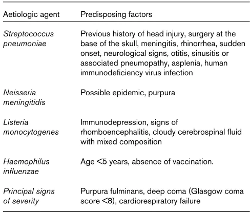
In the review by Durand et al[3] of 493 cases of bacterial meningitis in adults the mortality rate was 25%, and in a recent study of Gram-negative meningitis 61% of the chil-dren had neurological sequelae [27]. Predisposing factors and signs of severity for meningitis caused by several aeti-ologic agents are outlined in Table 2.
Pneumococcal meningitis
[image:7.609.312.556.116.323.2]Prognosis depends on the severity of both encephalitic impairment and associated systemic signs. Mortality is high (16–33%) with otoneurological sequelae (17–29%) [10]. Table 2
Predisposing factors and signs of severity Aetiologic agent Predisposing factors
Streptococcus Previous history of head injury, surgery at the pneumoniae base of the skull, meningitis, rhinorrhea, sudden
onset, neurological signs, otitis, sinusitis or associated pneumopathy, asplenia, human immunodeficiency virus infection
Neisseria Possible epidemic, purpura meningitidis
Listeria Immunodepression, signs of
monocytogenes rhomboencephalitis, cloudy cerebrospinal fluid with mixed composition
Haemophilus Age <5 years, absence of vaccination. influenzae
Meningococcal meningitis
In such cases severity is indicated by the infectious signs of purpura fulminans (6–13% of meningococcal infections, of which there is a 50% mortality) than to neurological signs, which have a low mortality rate (3–10%) and rare sequelae (6–10%) including deafness, impairment of another cranial nerve, hydrocephalus and epilepsy.
Listeriameningitis
The prognosis is poor, with 33% mortality and 33% sequelae including upper function, tonic and deglutination disorders.
Treatment
Antibiotic therapyThere is no justification for delaying antibiotic therapy (Tables 3 and 4), because early treatment is associated with better outcome in cases of meningococcal and pneumococ-cal meningitis. Treatment must therefore be started imme-diately after the lumbar puncture and the initial blood culture, and sometimes before if the CSF examination has been deferred (patient far from a hospital, decision to perform a CT scan) and/or if the symptoms appear to be
fulminant (fever at 40°C, meningeal syndrome and worsen-ing level of consciousness in < 24 h or purpura; Fig. 1) [23].
Neisseria meningitidis
N. meningitidisis sensitive to penicillins and third-genera-tion cephalosporins. Amoxicillin (200 mg/kg per day), cefotaxime (200 mg/kg per day), or ceftriaxone (70–100 mg/kg per day) can be used for 7–10 days [28].
Listeria monocytogenes
The reference treatment for L. monocytogenesis amoxicillin (200 mg/kg per day) plus gentamicin (6 mg/kg per day), which, despite the weak meningeal diffusion of these compounds, is justified because of their in-vitro synergis-tic effect and the frequency of initial bacteraemic forms. Another excellent possibility, supported by its diffusion into the CSF, is co-trimoxazole: 960 mg every 12 h intra-venously for 2–3 weeks [29].
Haemophilus influenzae
[image:8.609.54.557.392.474.2]H. influenzae meningitis is treated by cefotaxime (200 mg/kg per day) or ceftriaxone (70–100 mg/kg per day).
Table 3
Initial treatment of purulent meningitis with negative direct examination, absence of predisposing factors and signs of severity [1] Antibiotic Posology (mg/kg per day) Administration route
Child > 3 months Cefotaxime 200–300 Four infusions
or ceftriaxone 70–100* One or two intravenous injections
Adult Amoxicillin 200 Four to six infusions
or cefotaxime 200–300 Four infusions
or ceftriaxone 70–100 One or two intravenous injections
*Maximum 4 g/day
Table 4
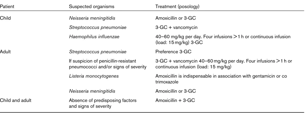
Initial treatment of purulent meningitis with negative direct examination, according to predisposing factors and/or signs of severity [1]
Patient Suspected organisms Treatment (posology)
Child Neisseria meningitidis Amoxicillin or 3-GC
Streptococcus pneumoniae 3-GC + vancomycin
Haemophilus influenzae 40–60 mg/kg per day. Four infusions > 1 h or continuous infusion (load: 15 mg/kg) 3-GC
Adult Streptococcus pneumoniae Preference 3-GC
If suspicion of penicillin-resistant 3-GC + vancomycin 40–60 mg/kg per day. Four infusions > 1 h or pneumococci and/or signs of severity continuous infusion (load: 15 mg/kg)
Listeria monocytogenes Amoxicillin is indispensable in association with gentamicin or co trimoxazole
Neisseria meningitidis Amoxicillin or 3-GC Child and adult Absence of predisposing factors Amoxicillin + 3-GC
and signs of severity
[image:8.609.57.557.546.731.2]Streptococcus pneumoniae
Antibiotic therapy for S. pneumoniaeis confronted with two difficulties: the virulence of the micro-organism and increasing resistance to the antibiotics usually prescribed for purulent community-acquired meningitis. The major antibiotics that have the best in-vitro activity on PRP are cefotaxime, ceftriaxone, vancomycin, amoxicillin and imipenem. Rapid determination of the MIC for the above β-lactams is indispensable in order to best adjust the treat-ment. The therapy is administered by the intravenous route for 10–14 days and is prolonged in cases of slow response and/or a strain with reduced sensitivity. The choice of antibiotic therapy is governed by the age of the patient, the presence of signs of severity and/or risk factors for PRP [23,30,31].
In children under 3 months of age or in adults with risk factors for PRP and/or presenting with signs of severity, initial treatment must include a third-generation cephalosporin such as cefotaxime (300 mg/kg per day in four infusions) or ceftriaxone (70–100 mg/kg per day in one or two injections, maximum 4 g/day), and vancomycin (60 mg/kg per day either in four infusions of at least 1 h or in continuous infusion with an initial load of 15 mg/kg) [32]. Re-evaluation is performed at 36–48 h and is based on the clinical data and lumbar puncture.
If the evolution is favourable, further treatment will depend on the MIC of the cephalosporin that is used. If the MIC is inferior to 0.5 mg/l, one can discontinue the vancomycin and possibly reduce the dosage of the third-generation cephalosporin (to 200 mg/kg per day) or pre-scribe amoxicillin (150–200 mg/kg per day) if the amoxicillin MIC is inferior to 0.12 mg/l.
If the third-generation cephalosporin MIC is superior or equal to 0.5 mg/l, initial treatment must be continued. In case of clinical and/or microbiological failure, treatment must be modified by taking into account the results of the second lumbar puncture, the MIC of the antibiotics and the concentration of vancomycin in the CSF [21,23]. The drug combination should be decided with the microbiologist and chosen among a list of antibiotics for which the MIC are known: imipenem, meropenem, rifampin and fosfomycin.
The initial treatment for an adult without a risk factor for PRP or signs of severity is cefotaxime (200–300 mg/kg per day in four infusions), but amoxicillin remains an option at a dose of 200 mg/kg per day in four to six infusions, espe-cially in regions where the prevalence of PRP is low. Clin-ical re-evaluation is performed at 36–48 h. For subsequent treatment, if the evolution is favourable and if the strain has normal sensitivity to these β-lactams, one can reduce the dose of the third-generation cephalosporin or pre-scribe amoxicillin (150–200 mg/kg per day). When the MIC of the third-generation cephalosporin is superior or
equal to 0.5 mg/l, lumbar puncture is indispensable to confirm an improvement in the CSF; such improvement mandates continuation of the initial treatment. In cases of clinical and/or microbiological failure, the treatment must be modified by adding vancomycin to the initial treatment (see doses above).
It is recommended to treat H. influenzaeand meningococ-cal meningitis for 7–10 days, but longer periods of 10–21 days are required for other pathogens: 10–14 days for S. pneumoniae; 14–21 days for L. monocytogenesand group B streptococci; and 21 days for Gram-negative bacilli, except for H. influenzae.
Empirical choice of an antibiotic therapy
When lumbar puncture is delayed or direct examination of the CSF is not helpful, empiric treatment is essential. A broad-spectrum cephalosporin (cefotaxime or ceftriaxone) is recommended for most patients, combined with ampi-cillin in children under 3 months and adults over 50 years (two populations in which S. agalactiaeand L. monocytogenes are frequently found). In patients with recent head trauma or who have undergone neurosurgery, the clinician must choose a broad-spectrum antibiotic treatment that is active on Gram-positive and Gram-negative bacteria, such as the combination of ceftazidime and vancomycin. In immuno-compromised patients (chemotherapy, corticotherapy, haematologic tumours), treatment must include ampicillin (for possible listeriosis) and a cephalosporin, such as cef-tazidime, which is active on Gram-negative bacilli.
Intensive care
Given the number of cases of life-threatening secondary or associated visceral impairment, it is important to treat respiratory failure, septic shock, intracranial hypertension or disseminated intravascular coagulation. As for the latter syndrome, a recent study has demonstrated the interest of administering concentrated Protein C for purpura fulmi-nans in cases of meningococcemia.
Treatment of the portal of entry
Otorhinologic treatment of otitis and sinusitis is an emer-gency that can be put off for a few days if the clinical evo-lution during the first few days is favourable. It can include atticomastoidectomy, exeresis of a cholesteatoma or sinus drainage. Detection of traumatic osteodural breaches and their treatment are not considered as emergencies.
Immunotherapy Corticotherapy
mortality of a short corticoid treatment administered in the initial phase of bacterial meningitis. These studies have demonstrated a possible reduction in mortality that has, however, only been found in open studies performed in populations of patients whose clinical presentation was immediately severe with treatment often delayed. A reduction in auditory sequelae, even neuromotor seque-lae, in children treated with dexamethasone (double-blind studies), especially in children with H. influenzaepurulent meningitis, has been noted; there is probably a similar effect for pneumococcal meningitis, but cannot be evalu-ated from the available data for meningococcal meningitis. In adults there is only partial information on the efficacy of initial corticotherapy, but there are potential disadvan-tages of corticosteroid use. Through the rapid sympto-matic modifications that they can trigger, corticosteroids can interfere with assessment of evolution and lead to pos-sible errors in clinical interpretation. Consequently, they must not be used for a meningitis that is insufficiently documented. Complications due to corticotherapy when used in this context are very rare. On the other hand, one can observe a return of fever when corticosteroid treat-ment is stopped. The use of dexamethasone in animal models has demonstrated a reduction in the penetration of certain antibiotics, including vancomycin, into the CSF. This could create a problem for treatment with certain antibiotics with relatively weak meningeal diffusion in cases of PRP infection.
On the whole, the favourable benefit:risk ratio for early corticoid therapy would advocate the prescription of dex-amethasone at the beginning of treatment of purulent meningitis in children when it is probably due to the usual community bacteria. For adults, however, additional clini-cal studies will be necessary to draw conclusions on this point. The adults who could benefit the most are those who present with a high bacterial concentration in the CSF and signs of intracranial hypertension [35].
When it is used, corticotherapy must be instituted early. It is usually administered a few minutes before the first dose of antibiotic [ie, dexamethasone intravenously (0.6 mg/kg per day in two to four injections)]. The duration of treat-ment is debatable, but a 2-day treattreat-ment would appear to be as effective as a 4-day treatment. If dexamethasone is chosen, it is better to associate it with rifampicin rather than vancomycin in cases of PRP.
Antimediators
The use of agents that block the action of polynuclear cells or cytokines has been promising in experimental meningitis, but has yet to be validated in humans. A recent clinical study would seem to have found an inter-esting effect of a protein that increases bacterial perme-ability (rBPI21) in 26 children aged from 1 to 18 years and presenting with severe meningococcaemia.
Prophylaxis
Pneumococcal meningitis
Continuous cyclic chemoprophylaxis with penicillin G is used in certain adults (following splenectomy, osteodural breach victims), but is not based on well established data [36]. The emergence of resistant strains should put this practice into question.
Vaccination is recommended for patients at risk. It is debat-able for patients aged over 65 years or under certain circum-stances for elderly or handicapped patients living in groups. Its efficacy has not been demonstrated, especially given the poor vaccination response in groups at risk. Vaccination is ineffective before the age of 2 years and not advisable in pregnant women. A booster vaccination is performed after 5 years or earlier in immunocompromised patients.
Meningococcal meningitis
In France, a Public Health Service declaration is obligatory once a case has been identified. Prophylaxis involves those in contact with the patient before diagnosis (living under the same roof in the 10 days before hospitalization, close friends, immediate neighbours in class, cafeteria, room) and after curative treatment, as well as clinical staff exposed to air contamination (mouth-to-mouth resuscitation, intuba-tion). It is extended to an entire school class if several cases have occurred in the same class, and to an entire school if three or more cases have occurred in different classes in less than a month. Casual contacts (coworkers, other students who were not close to the patient, health care personnel in general) are not involved. Except for secondary cases, mea-sures must be taken within 8 days of the diagnosis and are organized by the family doctor (in household cases) or, in group cases, by the Public Health Service and the doctor responsible for the group. Contact subjects and contacts from the same group as the patient must be informed about the disease and the measures to be taken. Medical observa-tion of the contact subjects must be undertaken for 2 weeks after the institution of prophylactic measures. Rhinopharyn-geal disinfection and sampling are not useful. Suspension from school or isolation of the contact subjects is not recom-mended. Given the fragility of meningococcus, disinfection or closure of an establishment, including a school, are mea-sures that are unnecessary and unjustified.
Chemoprophylaxis consists of rifampicin per os for 2 days: in adults, 600 mg twice a day; in children aged 1 month to 12 years, 10 mg/kg three times a day; in children aged under 1 month, 5 mg/kg twice a day. The wearing of soft contact lenses must be stopped during treatment due to the risk of irreversible dyeing. Moreover, subjects taking oral contraception must be given another contraceptive solution due to the risk of enzymatic induction.
the latter situation, use spiramycin per os for 5 days: in adults, 3 million IU twice a day; in children, 75 000 IU/kg twice a day.
Vaccination (meningococcal vaccine A+C) is recom-mended in addition to chemoprophylaxis, beginning at the age of 3 months in the case of meningococcus A and from 1 year of age in the case of meningococcus C. It can also be offered to those who have stayed in a region with an epidemic: peace corps workers and members of medical expeditions in the sahel. Professional or academic suspension of contact subjects and disinfection of premises are not useful.
Haemophilus influenzaetype B meningitis
The probability of contracting meningitis is markedly increased in children aged under 4 years living in the same domicile as an index case. Prophylaxis relies on the administration of rifampicin (30 mg/kg per day) for 4 days from the age of 1 month. It provides an eradication rate of approximately 95% for carriers of H. influenzae. The emer-gence of type B strains that are resistant to rifampicin is rare (0.2% in France). H. influenzaetype B vaccination and protective and immunogenic vaccines in very young chil-dren have greatly modified the epidemiology of meningi-tis in countries where these vaccines are used on a large scale. In France, according to the National Reference Center data, 635 strains of H. influenzae were isolated in CSF between 1984 and 1992, 71 strains in 1993, and 34 strains in 1994.
Listeria monocytogenesmeningitis
The prevention of listeriosis essentially depends on food hygiene. This applies to pregnant women, immunocom-promised subjects and the elderly.
References
1. Sigurdardottir B, Bjornsson OM, Jonsdottir KE, Erlendsdottir H, Gud-mundsson S: Acute bacterial meningitis in adults. A 20-year overview.Arch Intern Med1997, 157:425–430.
2. Miller LG, Choi C: Meningitis in older patients: how to diagnose and treat a deadly infection.Geriatrics1997, 52:43–44, 47–50, 55. 3. Durand ML, Calderwood SB, Weber DJ, et al: Acute bacterial
menin-gitis in adults.N Engl J Med1993; 328:21–28.
4. Schaad UB: Current concepts of bacterial meningitis.Eur J Pediatr
1995, Suppl 4:S20–S22.
5. Richardson M: Bacterial meningitis. Br J Hosp Med 1996, 55: 685–688.
6. Segreti J, Harris AA: Acute bacterial meningitis.Infect Dis Clin North Am1996, 10:797–809.
7. Grebe T, Hakenbeck R: Penicillin-binding proteins 2b and 2x of
Streptococcus pneumoniaeare primary resistance determinants
for different classes of ββ-lactam antibiotics. Antimicrob Agents Chemother 1996; 40:829–834.
8. Booy R, Kroll JS: Bacterial meningitis and meningococcal infection. Curr Opin Pediatr1998, 10:13–18.
9. Infections cérébro-méningées in Monographies [in French]. La Revue du Praticien1994, 44:2145–2224.
10. Kragsbjerg P, Källman J, Olcén P: Pneumococcal meningitis in adults.Scand J Infect Dis1994, 26:659–666.
11. de Saint-Martin L, Nassif X: Physiopathologie des méningites bac-tériennes aigües purulentes [in French]. Méd Thér 1995, 1: 527–532.
12. Waage A, Halstensen A, Shalaby R, Brandtzaeg P, Kierulf P, Espevik T: Local production of tumor necrosis factor αα, interleukin 1 and interleukin 6 in meningococcal meningitis.J Exp Med1989, 170: 1859—1867.
13. Kornelisse RF, Savelkoul HF, Mulder PH, et al: Interleukin-10 and soluble tumor necrosis factor receptors in cerebrospinal fluid of chil-dren with bacterial meningitis.J Infect Dis1996, 173:1498–1502. 14. Kornelisse RF, Hack CE, Savelkoul HF, et al: Intrathecal production
of interleukin-12 and gamma interferon in patients with bacterial meningitis.Infect Immun1997, 65:877–881.
15. Fassbender K, Ries S, Schminke U, Schneider S, Hennerici M:
Inflammatory cytokines in CSF in bacterial meningitis: association with altered blood flow velocities in basal cerebral arteries. J Neurol Neurosurg Psychiatry1996, 61:57–61.
16. Sprenger H, Rosler A, Tonn P, et al: Chemokines in the cere-brospinal fluid of patients with meningitis.Clin Immunol Immuno-pathol1996, 80:155–161.
17. Fassbender K, Schminke U, Ries S, et al: Endothelial-derived adhe-sion molecules in bacterial meningitis: association to cytokine release and intrathecal leukocyte recruitment. J Neuroimmunol
1997, 74:130–134.
18. van Deuren M, van der Ven Jongekrijg J, Vannier E, et al: The pattern of interleukin-1beta (IL-1beta) and its modulating agents IL-1 receptor antagonist and IL-1 soluble receptor type II in acute meningococcal infections.Blood1997, 90:1101–1108.
19. Ernst T, Spath PJ, Aebi C, Schaad UB, Bianchetti MG: Screening for complement deficiency in bacterial meningitis.Acta Paediatr1997,
86:1009–1010.
20. Calder JA: Listeria meningitisin adults.Lancet1997, 350:307–308. 21. Paris MM, Ramilo O, McCracken GH Jr: Management of meningitis caused by penicillin-resistant Streptococcus pneumoniae. Antimi-crob Agents Chemother1995, 39:2171–2175.
22. Scheld WM, Fletcher DD, Fink FN, Sande MA: Response to therapy in an experimental model of meningitis due to Listeria monocyto-genes.J Infect Dis1979, 140:287–294.
23. 9° Conference de Consensus en Therapeutique Anti-infectieuse: Les méningites purulentes communautaires [in French]. Méd Mal Infect. 1996, 26, Spécial: 1–8.
24. Garty BZ, Berliner S, Liberman E, Danon YL: Cerebrospinal fluid leukocyte aggregation in meningitis. Pediatr Infect Dis J 1997,
16:647–651.
25. Azuma H, Tsuda N, Sasaki K, Okuno A: Clinical significance of cytokine measurement for detection of meningitis.J Pediatr1997,
131:463–465.
26. Pavese P, François P, Lafond JL, Kayemba-Kay SS, Bosson JL:
Dosage de l’acide lactique dans le liquide céphalorachidien pour le diagnostic des méningites bactériennes. Stratégies pour le choix du seuil discriminant [in French].Presse Med1997, 26:551–554. 27. Grimwood K, Nolan TM, Bond L, et al: Risk factors for adverse
out-comes of bacterial meningitis.J Paediatr Child Health1996, 32: 457–462.
28. Kennedy NJ, Duncan AW: Acute meningococcaemia: recent advances in management.Anaesth Intens Care1996, 24:197–216. 29. Hof H, Nichterlein T, Kretschmar M: Management of listeriosis.Clin
Microbiol Rev1997, 10:345–357.
30. Astruc J. Méningites à pneumocoque de l’enfant. Propositions thérapeutiques [in French]. Méd Mal Infect 1994, 24 Spécial: 982–985.
31. Decazes J-M, Casin I, Kitzis M-D, et al: Méningite à pneumocoque résistant à la pénicilline: bases expérimentales de la thérapeu-tique [in French].In: Infections à Pneumocoque de Sensibilité Dimin-uée aux Béta-lactamines. Edited by Carbon C, Chastang C, Decazes J-M. Springer-Verlag, Paris, 1993.
32. Viladrich PF, Cabellos C, Pallares R, et al: High doses of cefotaxime in treatment of adult meningitis due to Streptococcus pneumoniae with decreased susceptibilites to broad-spectrum cephalo-sporins.Antimicrob Agents Chemother 1996, 40:218–220. 33. Quagliarello VJ, Scheld WM. Treatment of bacterial meningitis.
N Engl J Med1997, 336:708–716
34. Lauritsen A, Oberg B: Adjunctive corticosteroid therapy in bacterial meningitis.Scand J Infect Dis1995, 27:431–434.
35. Townsend GC, Scheld WM: The use of corticosteroids in the man-agement of bacterial meningitis in adults.J Antimicrob Chemother
1996, 37:1051–1061.