Selection of our books indexed in the Book Citation Index in Web of Science™ Core Collection (BKCI)
Interested in publishing with us?
Contact [email protected]
Numbers displayed above are based on latest data collected. For more information visit www.intechopen.com Open access books available
Countries delivered to Contributors from top 500 universities
International authors and editors
Our authors are among the
most cited scientists
Downloads
We are IntechOpen,
the world’s leading publisher of
Open Access books
Built by scientists, for scientists
12.2%
116,000
120M
TOP 1%
154
Acoustics and Biological
Structures
Mariana Alves-Pereira, Bruce Rapley, Huub Bakker
and Rachel Summers
Abstract
Within the context of noise-induced health effects, the impact of airborne acoustical phenomena on biological tissues, particularly within the lower frequency ranges, is very poorly understood. Although the human body is a
viscoelastic-composite material, it is generally modeled as Hooke elastic. This implies that acoustical coupling is considered to be nonexistent at acoustical frequencies outside of the human auditory threshold. Researching the acoustical properties of mam-malian tissue raises many problems. When tissue samples are investigated as to their pure mechanical properties, stimuli are not usually in the form of airborne pressure waves. Moreover, since the response of biological tissue is dependent on frequency, amplitude, and time profile, precision laboratory equipment and relevant physi-ological endpoints are mandatory requirements that are oftentimes difficult to achieve. Drawing upon the viscoelastic nature of biological tissue and the tensegrity model of cellular architecture, this chapter will visit what is known to date on the biological response to a variety of different acoustic stimuli at very low frequencies.
Keywords: infrasound, low frequency noise, health, cellular biology, tissue
morphology
1. Introduction
Airborne pressure waves are ubiquitous in all human environments and have played vital roles in the survival, evolution, and development of the human species. Under certain conditions, airborne pressure waves can be perceived as “sound” by the human auditory system. Under other conditions, they may be perceived as a whole-body or partial-body vibration. Some airborne pressure waves are not consciously perceived at all. As human societies developed and became more tech-nological, airborne pressure waves emanating from human-made devices became ubiquitous and “noise” became a more serious issue. By the late nineteenth century, noise and health studies began to flourish. In the early twentieth century, the telephone and growing industrialization led to more in-depth studies of the human hearing function. In 2011, a WHO document on the burden of diseases reflected the seriousness of the ongoing “noise problem” [1].
The only airborne pressure waves considered of consequence for human health were those that could be heard, i.e., “what you can’t hear can’t hurt you” (Figure 1).
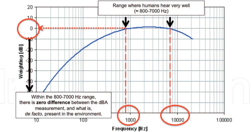
Within the audible segment (20–20,000 Hz), human auditory acuity is not evenly distributed, and is more sensitive within the 800–7000 Hz range than it is to air-borne acoustic events occurring below 500 Hz or above 15,000 Hz. Thus, early on, scientists understood that in order to protect human hearing function and speech intelligibility, the entire audible segment need not be considered, but rather, only the frequencies at which the acuity was highest: 800–7000 Hz range. The development of the A-frequency weighting and the resulting deciBel-A (dBA) metric allowed acousticians and health professionals to assess acoustical environments simulating this variability of human auditory acuity.
Figure 2 shows the frequency response curve for the dBA metric, clearly
follow-ing the human auditory response to airborne acoustic pressure waves.
While the dBA metric proved to be key for the protection of hearing and speech intelligibility, it was insufficient for the assessment of airborne pressure waves occurring outside of the 800–7000 Hz range. Figure 3 emphasizes the 800–7000 Hz
range within the dBA metric, and Figure 4 shows its application at 10 Hz. The dBA
[image:3.612.135.477.336.519.2] [image:3.612.90.526.565.795.2]metric is, therefore, unsuited for evaluating airborne pressure waves occurring at frequencies below 800 Hz. Health effects that may be developing due to exposures
Figure 1.
Acoustical spectrum showing the classical three segments (infrasound, audible, and ultrasound) with the frequency and wavelength indicated at the cutoff of each segment.
Figure 2.
at these lower frequencies cannot be properly studied if the dBA metric is being used to characterize acoustical environments.
There is a shortage of studies that properly evaluate the biological response to infrasonic (≤20 Hz) or lower frequency (≤200 Hz) airborne pressure waves. Three
important reasons for this have been provided above: the rudimentary segmenta-tion of the entire acoustical spectrum into merely three “blocks” (compare to
[image:4.612.92.526.66.294.2]segmentation of the electromagnetic spectrum), the unsuitability of the dBA metric to quantify airborne acoustical pressure waves at these lower frequencies, and the ingrained notion that “what you can’t hear can’t hurt you.” These major hindrances have been crystallized into mainstream science [3] and have served to significantly impede scientific inquiry and human health protection.
Figure 3.
[image:4.612.94.524.374.618.2]Frequency response curve for the dBA metric applied to the range of highest human auditory acuity. Within this frequency range, the dBA measurement will accurately reflect the airborne acoustical energy present in the environment.
Figure 4.
The goal of this chapter is to consolidate what is known on the biological response to airborne pressure waves occurring within the infrasonic and lower frequency ranges. A biomedical engineering approach is taken, whereby biological organisms are viewed as structures of composite materials, with significant visco-elastic components and organized in accordance with the principles of tensegrity architectures. When airborne pressure waves impact these types of structures, the biological response will depend on the type of biomaterial under study, it will exhibit anisotropic properties, and it will vary nonlinearly with exposure time. Depending on the physical properties of the airborne pressure waves (including time profiles) and on the biostructure under study, mechanical perturbations are relayed into cells and tissues through a variety of different pathways that, to date, still remain unclear.
2. Biomaterials and human anatomy
2.1 Viscoelasticity
Viscoelasticity is an attribute given to bodies that exhibit both viscous and elastic behaviors beyond the classical Hooke’s elastic model [4]. Viscoelastic materials have three distinct properties not contemplated by Hookean models: creep, stress relaxation, and hysteresis. Most biological materials have viscoelastic behaviors.
In a Hookean (or purely elastic) material, total deformation depends on total load, and no further deformation occurs even if load is maintained. In viscoelastic materials, however, when sufficient stress is applied and maintained, they may continue to deform, even though stress load remains unaltered. This property is called creep.
In a purely elastic material, the strain within the material is constant throughout the application of the load; it does not vary with time, but only with the amount of applied stress. In viscoelastic materials, when stress is applied and maintained, strain can decrease with time. This property is called stress relaxation.
Consider repetitive or cyclical loads on materials. In purely elastic materials, peri-odic loads will not alter the stress-strain curve. The pathway taken by the material to deform is exactly the same pathway it takes to return to its original, equilibrium position. In viscoelastic materials, however, the return to equilibrium may be dif-ferent than the pathway used to get to the point of deformation (The word pathway is here loosely used, and is meant to encompass all spatial, temporal and energetic components of these types of movements.) This property is called hysteresis.
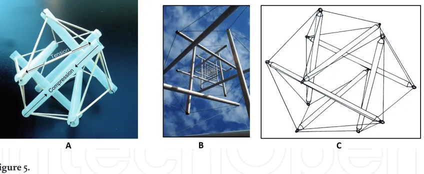
2.2 Tensegrity structures
Many structures in the natural world are organized in accordance with the principles of tensegrity architecture—elements providing discontinuous compres-sion are held together through elements of continuous tencompres-sion [5]. Figure 5 shows several examples of tensegrity structures.
Depending on the properties of the airborne pressure waves and biomaterial under study, the propagation of mechanical perturbations throughout these types of structures can reach long distances, without loss of structural integrity.
2.3 Cellular and tissue mechanotransduction
cells also communicate with their surroundings through mechanical signals. Mechanosensitive receptors exist on cell surfaces, and mechanosensitive junctions interconnect cells, thus forming tissues. Depending on the physical properties of the airborne pressure waves and biomaterials under study, external airborne mechanical perturbations can elicit a mechanical response, which, in a larger, macroscopic view, can lead to clinically pathological situations.
2.4 The fasciae
The fascia is a sheet of connective tissue that uninterruptedly extends from head to toe, suspended from the skeleton, and that provides the integrated supporting framework for maintaining anatomical and structural form [10, 11]. That external mechanical perturbations elicit responses at large distances away from the point of entry is a well-known concept among scientists and health professionals who study fasciae. When presented with external airborne pressure waves, fasciae can respond by changing their structural properties: from a mechanical point of view, the fasciae are organized in chains to defend the body against restrictions. When a restriction goes beyond a specific threshold, the fasciae respond by modifying their viscoelasticity, chang-ing the collagenic fibers, and transformchang-ing healthy fascial chains into lesioned chains [10]. One of the fascia’s key roles is that of shock absorption.
Connective tissue structures are ubiquitous forming all external surfaces of vessels, nerves, organs, and muscles, and at the cellular level, the extra-cellular matrix that surrounds and communicates with each individual cell. In addition to maintaining structural integrity, the fasciae are the first line of defense against external perturbations, playing important physiological roles in mobilizing the immune system.
3. Laboratorial studies, field studies, and biological outcomes
[image:6.612.97.521.69.243.2]Studying the effects of infrasonic or lower-frequency airborne pressure waves on biological structures is a very complex undertaking, whether it be on cell cul-tures, on animal models, or on human populations. Laboratorial studies, occu-pational field studies, and residential field studies all have their own strengths and weaknesses. When the latter go unrecognized, however, experimental design flaws can ensue. In this section, the attributes of these different experimental setups are discussed, and their weaknesses and strengths are explored. Together Figure 5.
with the preceding section, this serves as a preamble to Section 4, where the results of experimental studies are described in detail.
3.1 Laboratorial studies
Laboratories where infrasonic and lower frequency airborne pressure waves can be applied in a controlled manner are in short supply worldwide, and those that do exist are mostly associated with military installations. Laboratories emitting airborne pressure waves with infrasonic and lower frequency components cannot be randomly placed within residential environments; issues with neighbor disturbance and public health would curtail its use. Moreover, the equipment used to generate the airborne pressure waves is, typically, very large and very expensive, and few sectors of society (other than military or space exploration industries) would have the need for an extensive use of these types of installations.
In these laboratory settings, continuous or pulsed-trains of single-tone air-borne pressure waves can be applied, as well as, broadband exposures that can be accurately characterized. The fact that exposure times and acoustic parameters can be precisely controlled is one of the strengths of laboratorial studies, allowing for continuous time exposures, or occupationally simulated exposure schedules. Immediate (hours or days) versus long-term (weeks or months) effects can also be explored.
There are numerous types of biological outcomes that can be studied under lab-oratorial conditions. Light-, electron- and atomic-force microscopy can be used to study cellular and tissue structural properties, as well as their chemical composition and content of bio-reactive elements. Polymerase chain reaction (PCR) techniques can provide information on messenger RNA (mRNA) expression, allowing for the identification of key pathways. With pharmacological intervention or gene knock-out specimens, specific signaling molecules and pathways involved in the elicited responses can be pinpointed. Additionally, control populations for comparison are fairly easy to achieve—they are simply not subjected to the laboratorial exposures.
3.2 Occupational field laboratories
Occupational environments are exceptional field laboratories, as both short-term (several months) and long-short-term (years) effects can be investigated in more realistic acoustic environments. Typically, different workstations have different acoustical features that can greatly depend on different machinery regimens. For occupational field laboratories, acoustical characterizations of the workplace(s) must be comprehensively undertaken and time exposures to each type of environ-ment should be scored.
Exposure times at work must be differentiated from exposure times away from work, i.e., when the work shift ends, workers leave the field laboratory, but additional exposures to infrasonic or lower frequency airborne pressure waves may be incurred (e.g., recreational, transportation). These must be documented. Significant confounding factors may be introduced unless each subject’s residential area is scrutinized and prior-exposure histories probed for fetal, childhood, and adolescent exposures.
Survivorship bias is a well-known confounding factor in human population studies. In occupational environments, workers with more time on-the-job are those who have survived throughout the years of professional activity, while workers with less time in professional activity may exhibit more severe biological outcomes. This phenomenon is often misinterpreted leading to inconclusive or erroneous conclusions.
Control populations for exposures to infrasonic and lower frequency airborne pressure waves have been a very difficult proposition, given the ubiquitous nature of this stressor. One of the solutions to this profound problem is the scoring of subjects into different groups as per their exposure. Within this context, control groups are composed of individuals who have the least amount of cumulative (prior and present) exposure, and not of individuals with zero exposure.
Different professions can provide different field laboratories, both in terms of acoustic environment and time exposure schedules. For example, long-haul truck drivers are typically exposed for more than 8 hours daily and, oftentimes, sleep in the truck while it is idling, or while refrigeration systems are continu-ously operating. Workers onboard ships, submarines, offshore oilrigs, aircraft, and spacecraft (for example) can be exposed to significant amounts of infrasonic and lower frequency airborne pressure waves for weeks or months at a time. The wealth of information waiting to be gleaned from these types of field laboratories is breathtaking.
3.3 Residential field laboratories
Field laboratories in urban, suburban, and rural residential settings are generally designed to investigate environmental health effects due to human-made infrasonic and lower frequency airborne pressure waves. Typically, these sources are associ-ated with industrial complexes or infrastructure that, in turn, are usually linked with important economic interests. In general, the amount and type of infrasonic and lower frequency airborne pressure waves contaminating a home will depend on the machine operation and/or the use of the infrastructure. For example, in most urban and suburban areas, airports must close down between the hours of midnight and 5 am. Some factories do not have night shifts and therefore also have daily shutdown periods. Large refrigeration units, hydroelectric dams, and large volume highways, however, must be kept running 24/7 and can also be viewed as continu-ous sources of infrasonic and lower frequency airborne pressure waves. Wind turbines are the latest addition to these type of sources although they are almost exclusively within rural areas.
Comprehensive characterization of the acoustic environments in the different residential areas must be undertaken (e.g., master bedroom, children’s bedrooms, living-lounge areas), since room-resonance phenomena can significantly modify the acoustic environment that is originally being induced and driven by external, incoming airborne pressure waves. Additionally, wind can also influence the spectrum, intensity and type of infrasonic and lower frequency airborne pressure waves that exist within a room. This differentiation is readily achieved with proper acoustic evaluations.
Short-, medium- and long-term effects can be studied in residential settings when the implementation of a new infrastructure or industrial complex is known to be coming to the area. Biological outcomes should strive to be either noninvasive or minimally invasive, and prior-exposure histories are fundamental for achieving useful statistical data.
4. Past relevant studies
Numerous studies conducted over the decades have shed light on the biological response to infrasonic and lower frequency airborne pressure waves and associated symptomatic complaints. Due to space limitations, this discussion will only deal with some of the vascular and collagenous abnormalities, cardiomyocyte changes, and the hippocampus responses, as induced by different types of exposures. For reasons explained in the section “Introduction,” all studies using the dBA metric have been eliminated from consideration (with one exception in an occupational setting). Selected studies mostly focus on the cellular and tissue changes observed in laboratory, occupational, and residential settings, using light and electron microscopy. The sequence in which the studies are presented does not follow the classical anatomical order.
4.1 Vascular changes
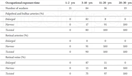
In the mid-1960s, within a military setting, the immediate exposure to 10–60 Hz, at 118–140 dB, for 2 minutes, induced disturbances of the visual field as reported by all five human subjects [12]. In 1985, laboratorial animal studies exposed rats to tonal 8 Hz at 100–140 dB, 3 hours daily, for 5, 10, 15, or 25 days, and examined the blood and lymph networks of the palpebral (eyelid) and bulbar (eye globe) conjunctiva. Day 5: narrowing of all parts of the conjunctiva blood network was observed, with decreased blood capillary lumens. Capillaries, precapillaries, and arterioles were twisted, and blood component agglomerations were identified in venous vessels. Day 10: conjunctiva capillaries were twisted and large vessel diameters were decreased. Day 15: blood and lymph vessel tonus had changed, and stagnation was present. Day 25: failure of tissue homeostasis was aggravated. Capillary penetration was increased, as seen through tissue enlargement, and significant agglutination was observed in the large vessels [13].
areas. Vascular changes as seen previously were more expressed: edema, paresis state in capillaries (erythrocyte stasis), and extra-vascular erythrocytes. The iris exhibited narrower vessels. Day 30: narrowed and twisted vessels were clini-cally detected, with ocular fundus arteries and veins significantly narrowed and twisted, more pronounced in the 16-Hz group. In the eyelid conjunctiva, derma exhibited the same vascular changes seen before: edema and erythrocyte stasis. Sclera arteries and veins were larger, overfilled with blood, and with the presence of extra-vascular focal and diffuse hemorrhages with conjunctiva involvement. At all time points, the 16-Hz group disclosed more destruction than the 8-Hz group. Day 60 (30 days post-exposure): clinical evaluations revealed less twisted and nar-row arteries and veins, but morphological recovery was slower. In the 8-Hz group, moderate regeneration was observed in the eyelid conjunctiva epithelium. In the 16-Hz group, predominant retinal damage persisted. Day 90: no clinical changes were observed in either group [14].
Within an occupational setting (reinforced concrete factory), vessel changes in the palpebral and bulbar conjunctiva, and in the retina, were investigated among 214 workers (age range: 20–58 years), with 1–30 years of employment. Workers were divided into two groups:
• Control group (n = 54): not occupationally exposed to significant levels of infra-sonic and lower frequency airborne pressure waves.
• Exposed group (n = 160): tonal 8 and 16 Hz at 96–100 dB, simultaneously with non-tonal 20–500 Hz at 91–93 dBA.
The exposed group was divided into subgroups as per years of professional activity. Table 1 describes each subgroup and the vessel abnormalities found. No such abnormalities were found in the control population [14].
Within a different occupational setting (aircraft industry), ocular changes were studied in 23 male workers (average age: 42, range: 32–58 years). Lesions
Occupational exposure time 1–2 yrs 3–10 yrs 11–20 yrs 20–30 yrs
Number of workers 21 84 36 19
Palpebral and bulbar arteries (%)
Enlarged 0 82 8 0
Narrow 0 17 91 100
Twisted 0 80 100 100
Retinal arteries (%)
Enlarged 0 0 0 0
Narrow 0 91 100 100
Twisted 0 90 100 100
Retinal veins (%)
Enlarged 0 87 11 0
Narrow 0 13 88 100
[image:10.612.89.521.529.780.2]Twisted 0 75 97 100
Table 1.
were observed in the blood-retinal barrier in 19 workers (lesion types: 13 inactive, 2 active, 4 mixed). Choroidal circulation was altered in 14 workers (late perfusion with chronic features). Changes in retinal circulation were observed in four workers (type: 1 occlusive, 1 exudative, 2 mixed). Three workers presented with optic neu-ropathy (1 papillitis, 2 optic atrophy), and one exhibited sensorial retinal macular detachment [15]. The immediate effects of tonal exposures with 8 Hz at 130 dB, 2 hours daily, for 1, 7, 14 and 21 days, also revealed a breakdown of the blood-retinal barrier in the rat eye [16].
These studies strongly suggest that under the impact of infrasonic and lower frequency airborne pressure waves, a vascular response is mounted by ocular struc-tures and could be related to decreased visual acuity in workers. Data in Table 1
seem to indicate that, as exposure time progressed, vessels that were initially enlarged ceased to exist, apparently being replaced with narrower and twisted vessels. Enlarged vessels usually suggest the need for an increased blood supply. However, given the sustained mechanical insult, making the vessels narrower and twisting them throughout the structures may, in fact, reflect a more efficient blood delivery system.
This concept is further reinforced by the observation of narrow and twisted blood vessels in the gastric mucosa of rats, exposed to non-tonal, occupationally simulated (aircraft industry) acoustic environments characterized as 6.3–25 Hz at 70–90 dB and 40–500 Hz at 90–100 dB. Continuous exposure was applied, and evaluations occurred at 1, 3, 5, 9, and 13 weeks. In 3–5 weeks, the gastric submucosal layer exhibited significantly increased thickness, when compared to non-exposed controls. This increased thickness was due to the proliferation of type IV col-lagen. Arterial walls disclosed significant intima and media thickening, ruptured internal elastic lamina, and thrombotic changes. In 9–13 weeks, neoangiogenesis was observed, with the appearance of tortuous and twisted vessels. The authors concluded that, in the stomach, continuous exposure induced fibrosis that could be linked with neoangiogenesis, since collagen type IV is also an early marker of neoangiogenesis [17]. One of the earliest studies investigating the long-term effects of airborne pressure waves on gastric complaints was conducted in 1968, in a residential setting where changes in gastric function were associated with aircraft noise [18]. Within occupational settings, an increase in gastric complaints was documented among boiler-plant workers, 2 years after the implementation of mandatory hearing protection devices [19]. Among aircraft industry workers, gastrointestinal problems were among the earliest to appear after 1–4 years of professional activity [20].
Vascular changes were also identified in the liver structures of animals exposed to 2, 4, 8, or 16 Hz, at 90–140 dB, 3 hours daily, for 5–40 days. Exposures to 2 or 4 Hz induced less damage than exposures to 8 and 16 Hz. Single, 3-hour exposures: with 2 or 4 Hz and 90 dB, no changes were observed in the hepatic structures, while at 100–110 dB, liver parenchyma disclosed single fine hemorrhages. At 120 dB, increased arterial wall diameters were observed, as well as capillary lumen expansion, indicating the development of ischemia. At 130–140 dB, the number of hemorrhagic events increased, as did the number of affected hepatocytes. With 8 or 16 Hz exposures, damaged hepatocytes were present in the ischemic and non-ischemic areas. Days 5–15: more pronounced hepatocyte changes were seen. Days 25–40: a gradual death of changed hepatocytes was observed [21].
exceeded 3 mm in diameter. Microscopic analyses of the hemorrhagic sections dis-closed ruptured capillaries and larger blood vessels [22]. In a laboratory setting, rats received tonal exposures to 2, 4, 8, or 16 Hz at 90–140 dB, 3 hours daily, for 40 days. Analysis time points were conducted after 3 hours, at 5, 10, 15, 24, and 40 days of exposure, as well as during post-exposure times. Single, 3-hour exposures: with 2 or 4 Hz at 90–110 dB, mosaic hemorrhages were observed under the pleura, covering the entire lung surface. With 8 Hz at 110 dB, more hemorrhagic expression was observed. With 8 or 16 Hz at 120–140 dB, larger hemorrhagic foci were disclosed. Within the alveolar capillary network and postcapillary venules, vessel diameters were increased with 2 or 4 Hz at 90–110 dB, leading to large hemorrhages and peri-vascular edema. Erythrocyte overflow in alveolar capillaries was observed with 8 or 16 Hz at 110 dB. With 8 or 16 Hz at 120 or 140 dB, lung tissue exhibited large hemor-rhagic foci in the connective tissue septa of the bronchi-pulmonary segments. In all exposure types, capillary changes were followed by alveolar epithelium desquama-tion and basal membrane denudadesquama-tion. Longer exposures: with 8 Hz at 120 dB, acinuses became filled with erythrocytes, and interstitial hemorrhagic foci caused a strong deformation of the respiratory bronchioles. With 8 or 16 Hz at 140 dB, ruptured vascular walls were observed leading to decreased alveolar lumen [23].
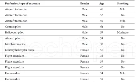
The highly invasive bronchoscopic evaluation with biopsy was performed among a group of volunteer subjects, with occupational or residential exposures to infrasonic and lower frequency airborne pressure waves, as detailed in Table 2.
Bronchoscopic observations in all patients revealed small submucosal, vascular-like lesions (“pink” lesions), located distally in both tracheal and bronchial trees, and uniformly distributed bilaterally near the spurs. Biopsies were performed on the abnormal mucosa (pink lesions) and on the apparently normal mucosa (outside of the pink lesions). In the non-pink areas, some vessel wall thickening was visible. In the pink areas, the basal membrane disclosed abnormal neovascularization, with thickened blood vessel walls and scarce lumen. No gender differences were identified [24].
4.2 Collagen and connective tissue
Collagen, composed of triple-helix tropocollagen chains, is the most abundant protein in the human body, a key component of the fasciae, and is produced by fibroblast cells. It has long since been considered as the “steel” of the human body [25], but its energy storage capacity has been shown to be 10 orders higher than in spring steel [26]. Different types of collagen have different mechanical properties. Type IV collagen (increased in the exposed gastric mucosa [17]—see above), is organized into X-shaped structures and is commonly found in the basal membrane of arterial walls, hence its increased expression during angiogenesis.
In day 5 of the eyelid-and-bulbar-conjunctiva animal studies (see above [13]), collagen fibers in the connective tissue were enlarged, as were some fibroblast nuclei; on day 10, adipose cells in the connective tissue had been redistributed and positioned in the vascular areas of the conjunctiva. In the second animal study described above [14], day 3 included edema of the sclera causing separation of colla-gen filaments in the 16 Hz group, and by day 7, this was observed in the 8-Hz group as well; day 15: focal and disseminated disorganization of sclera collagen fibers was observed in both groups; day 30: homogenization and disorganization of collagen in the derma while, in the sclera, collagen fibers were persistently separated due to edema, with some undergoing dystrophic and necrotic changes. Slow regeneration was observed during the post-exposure periods.
structures of alveoli walls. In the biopsy images of the bronchoscopic study (see above [24]), non-pink areas disclosed a thickened basement membrane with abnor-mal amounts of collagen, while the pink areas disclosed an even thicker membrane with very large amounts of collagen. The abnormal neovascularization was embed-ded within collagen bundles. Retraction of structures neighboring the collagen fibers was not observed. A marked reinforcement of the cytoskeleton and intercel-lular junctions was seen in the pink areas, as compared to non-pink areas. The five individuals that disclosed images of collagen fiber degeneration and disruption also tested positive for antinuclear antibodies.
Under an occupationally simulated acoustic environment, characterized as 20–200 Hz at 70–90 dB (aircraft industry), and occupationally simulated exposure schedules (8 hours daily, 5 days weekly, weekends in silence), focal interstitial fibrosis was found in the lung parenchyma of rats after a cumulative 4000-hour exposure. Additionally, thickened alveoli walls and dilated alveoli were observed [27]. Tracheal epithelium in similarly exposed rats disclosed significant subepithelial fibrosis [28, 29], and with longer occupationally simulated exposures, the sub-epithelial layer became composed of hyperplastic collagen bundles, some with a degenerative pattern. Cellular edema was also observed [28, 30].
Within an occupational setting (aircraft industry) and investigating long-term outcomes, high-resolution CT scans of the lungs and respiratory function tests were provided to 21 nonsmoker male workers, who were divided into two groups: with (n = 7, average age: 42) and without (n = 15, average age: 36) complaints of airflow limitations. There was a significant relationship between the presence of symptoms and images of lung fibrosis through the CT scan. No differences existed among the groups when comparing the percentage of predicted values of lung function [31].
Fasciae abnormalities have been most prominently studied in the pericardia of exposed workers, subsequent to autopsy findings in an aircraft industry worker that disclosed a grossly thickened pericardium [32]. Pericardial morphological changes were studied among 12 male workers: three aircraft technicians, four fixed-wing aircraft pilots, four helicopter pilots, and one long-haul truck driver. Pericardial samples were removed with informed consent of the patient and Ethics Committee
Profession/type of exposure Gender Age Smoking
Aircraft technician Male 48 Mild
Aircraft technician Male 52 No
Aircraft technician Male 59 Mild
Combat pilot Male 61 No
Helicopter pilot Male 59 Moderate
Aircraft pilot Male 54 No
Merchant marine Male 37 No
Military helicopter nurse Female 56 No
Flight attendant Female 36 No
Flight attendant Female 39 No
Flight attendant Female 40 No
Homemaker Female 54 Mild
[image:13.612.91.524.65.330.2]Homemaker Female 59 No
Table 2.
approval, at the beginning of cardiac surgery (prescribed for other reasons by the National Healthcare Service). In all cases, there were no visual adherences, or inflammatory aspects and pericardia were grossly thickened. The classical, three pericardial layers were identified: serosa, fibrosa, and epipericardium. However, in all cases, the fibrosa had split in two and, in between, a new layer of loose tissue was observed, consisting of vessels, nerves, arteries, and lymphatics surrounded by adipose tissue. Both fibrosa layers were composed almost entirely by wavy, interwoven collagen bundles, surrounded by numerous cytoplasmic extensions (whose mother cell was difficult to identify), and interspersed with some elastic fibers. The new, loose tissue layer sandwiched in between the split fibrosa contained blood and lymphatic vessels, adipose tissue, and nerves. Both the loose tissue layer and the fibrosa layers contained macrophages and vascular hyperplasia, also seen in lymphatic vessels [33–36]. Pericardial and cardiac valve thickening has also been confirmed through echocardiography studies in occupational settings (aircraft [37] and commercial-airline industries [38]), with thickness increasing with increasing exposure time. In residential settings, pericardial and valve thickening [39] and increased arterial stiffness [40] were observed in populations chronically exposed to military-training exercises [39], and transportation systems [40].
4.3 Heart cells and tissues
In 1983, electron microscopy techniques were used to study animal myocardia exposed to single and multiple infrasonic exposures of 4–16 Hz at 90–150 dB, 3 hours daily, for 45 days, and post-exposure time points were included. No changes were observed with single exposures at 4–6 Hz and at less than 100 dB, when
compared to non-exposed controls. Single exposure with 4–10 Hz at 120–125 dB: induced decreased arterial diameter and capillary expansion, with resulting focal ischemia. Images of intracellular myocytolysis were frequently found. These pro-cesses were reversible. Multiple exposures with 4–10 Hz at 120–125 dB for 5–25 days: ventricle fibrillation and subsegmental contractures in ischemic foci were identi-fied. Myofibril fragmentation was observed in the Z-line, sarcoplasmic reticulum structures were absent, cell nuclei were deformed, and chromatin was found accu-mulated under the nuclear membrane. post-exposure: intracellular regeneration was concomitant with damaged cells. In surviving cells, mitochondria were increased in number and size, and both myofilaments and sarcoplasmic reticulum elements were being created. Intracellular regeneration was slow and ended with the creation of Z-lines, after which myofibrils became normal and myocardiocytes completely recovered. Single exposure with 10–15 Hz at 135–145 dB: more pronounced myo-cardial damage, with partial death of myocardiocytes, resulting in myocardiocyte dystrophy. Damaged cells included chromatin condensation and redistribution to the nuclei membrane. Less damaged cells regenerated after 5–10 days post-exposure. Multiple exposures with 10–15 Hz at 135–145 dB: persistent myocardial ischemia related to vascular changes and accompanied by cardiocyte damage. After 15–25 days post-exposure, recovered cells began functioning normally despite the presence of abnormal structures within the cellular cytoplasm, namely, giant mitochondria [41].
Cardiac injury was studied in rat cardiomyocytes exposed to tonal 5 Hz at
increased with increasing exposure time [42]. With similar exposure protocols, another study repeated the SERCA2 and intercellular Ca2+ concentrations, but also included evaluations of the expression of whole cell L-type Ca2+ currents (WLCC) and the mRNA expression of a subunit of the L-type Ca2+ channel (LCC). SERCA2 and intercellular Ca2+ concentrations behaved as described immediately above, while the expression of WLCC and mRNA expression of LCC increased with increasing exposure time [43].
For three continuous months, rats were exposed to non-tonal, occupationally simulated (aircraft industry) acoustical environments characterized as 6.3–25 Hz at 70–90 dB and 40–500 Hz at 90–100 dB. Ventricular cardiac muscle and interstitial fibrosis were quantified and compared to non-exposed controls. Exposed rats disclosed a 97.5% increase in fibrosis in the left ventricle, an 81.5% increase in the interventricular septum, and an 83.7% increase in the right ventricle. No significant differences were found in the mean values of cardiac muscle in the left and right ventricles, when compared to non-exposed controls. However, the fibrosis-to-muscle ratio was significantly higher in the exposed rats, indicating significant ventricular myocardial fibrosis [44].
In another study, rats were exposed to a non-tonal, occupationally simulated (textile mill) environment rich in infrasonic and lower frequency components, under an occupationally simulated schedule (8 hours daily, 5 days weekly, weekends in silence), for 1, 3, 5, and 7 months. Ventricular coronary artery caliber, artery wall thickness, and size of arterial perivascular tissue were quantified in a total of 130 arteries (61 exposed and 69 controls). No changes were observed in arterial lumen caliber, and in arterial wall thickness, when compared to non-exposed controls. Perivascular tissue was more prominent in the exposed samples and seemed to exhibit fibrotic development. Lumen-to-wall ratio showed no differences, while wall-to-perivascular-tissue ratio showed a significant increase, as compared to non-exposed controls [45].
In animals exposed to 2–20 Hz peaking at 114 dB, for 28 continuous days, ventricular arteries were studied as to the dimensions of lumen, wall, and perivascular space. An additional group of animals received the same exposure but were treated with dexamethasone (a corticosteroid). Blind evaluation of 31 arteries disclosed increased perivascular spaces in the exposed groups, reflected in the significantly reduced wall-to-perivascular-space ratio, as compared to non-exposed controls. No changes were observed in the lumen-to-wall ratio. With dexamethasone treatment and exposure, no differences were observed in the wall-to-perivascular-space ratio, as compared to controls, suggesting an underly-ing inflammatory mechanism [46].
4.4 The hippocampus
Prior studies have shown that the hippocampus is involved in learning and memory impairment, such as that seen in rodents after infrasound exposure [49]. The hippocampus—located between the cerebral hemispheres and the brainstem—was classically considered as part of the limbic system. The hippo-campus proper is divided into four regions (CA1, CA2, CA3, and CA4), each with different input and output pathways. The Dentate Gyrus (DG) is an additional hippocampus structure and that contributes to the formation of new episodic memories, and spontaneous exploration of novel environments. In the central nervous system (CNS), neuroglia consists of the non-neuronal cells (oligodendro-cytes, astro(oligodendro-cytes, ependymal cells, and microglia) and is often referred to as the connective tissue of the brain. Glial cells surround neurons to hold them in place, supply them with oxygen and nutrients, insulate them from one another, destroy pathogens, and remove dead neurons.
Glial fibrillary acidic protein (GFAP) is an intermediate filament protein expressed by numerous cells within the CNS, and although its exact function
remains unknown, it appears to be involved in maintaining the mechanical strength of astrocytes. The expression of GFAP was studied in the brains of mice exposed to 16 Hz at 130 dB, 2 hours daily, for 1, 7, 14 21, or 28 days. GFAP expression was increased in the hippocampus, cortex, and hypothalamus in a time-dependent manner [50].
Corticotrophin releasing hormone (CHR) is a peptide hormone involved in the stimulation of the pituitary synthesis of ACTH (adrenocorticotropic hormone) as part of the hypothalamic-pituitary-adrenal axis’ response to stress. Corticotrophin releasing hormone-receptor 1 (CHR-R1) has wide expression in the CNS. It plays important roles in fear learning and consolidation in the amygdala, in stress-related modulation of memory function in the hippocampus, and in arousal regulation in the brainstem. Prior studies showed that infrasound exposures caused an upregula-tion of CRH and CRH-R1 in neurons of the hypothalamic paraventricular nucleus [51]. Recent studies have also shown that CRH is expressed in activated microglial cells [52]. Within this context, rats and in vitro cultured microglial cells were exposed to 16 Hz at 130 dB for 2 hours, after which changes in CHR-R1 were examined. In vivo expo-sure disclosed activation of microglial cells and an upregulation in the expression of CRH-R1 in the hypothalamic periventricular nucleus. In vitro exposure disclosed that, in the absence of neurons, microglial cells were activated and CRH-R1 expres-sion was upregulated. These data suggest that both neurons in the hypothalamic periventricular nucleus and microglial cells are effector cells for infrasound-elicited responses [51].
TRPV4 were increased as compared to non-exposed controls. Pharmacological or knock-out intervention of TRPV4 in cultured glial cells decreased the levels of inflammatory cytokines and attenuated neuronal apoptosis. This study also demonstrated the involvement of calmodulin and protein kinase C signaling pathways in the response to infrasonic exposures. These data suggest that TRPV4 expressed by glial cells is potentially a key factor in infrasound-induced neuronal impairment [53].
Neonatal rat hippocampal astrocyte cultures were exposed to 16 Hz at 130 dB for 15, 30, 60, 90, 120, and 240 minutes. Extra-cellular glutamate levels increased with increasing exposure time, and at 90 min, there was a 100% increase over baseline. The astroglial expression of Cx43 (connexin43—see above) was increased, as compared to non-exposed controls, as was the synthesis of Cx43 mRNA. Through additional evaluations using pharmacological and knock-out interventions, the authors concluded that infrasonic exposures induced astrocytes to release mate, and that Cx43 gap junctions were required for the exposure-induced gluta-mate release [54].
The endocannabinoid system includes lipid-based retrograde neurotransmit-ters, expressed throughout the CNS, and involved in fertility, pregnancy, pre-and postnatal development, appetite, pain-sensation, mood, and memory. Animals were exposed to 16 Hz at 130 dB, 2 hours daily, for 14 days. Cannabinoid (CB) receptors 1 and 2 in the CA1 hippocampal region of the exposed rats were down-regulated in a time-dependent manner, as compared to non-exposed controls. Apoptotic cells in the CA1 only became obvious after day 5, and cell death coincided with the decreased expression of CB receptors. Through pharmacological interven-tion, activation of CB receptors significantly reduced the number of apoptotic cells, ameliorated the behavior performance of exposed rats, and reduced the infrasound-elevated levels of proinflammatory cytokines. These data suggest that CB receptors could potentially serve as promising targets for future treatments against infrasound-induced injury [55].
Fibroblasts synthesize extracellular matrix (glycosaminoglycans, reticular, and elastic fibers) and collagen, and, in addition to their structural role, fibro-blasts are also important for mounting the immune response to tissue damage. Fibroblast growth factors (FGF) signal through fibroblast growth factor recep-tors (FGFR). The fibroblast growth factor 2/fibroblast growth factor receptor 1 (FGF2/FGFR1) signaling pathway was investigated in animals and in cultured astrocytes, exposed to 16 Hz at 150 dB, 2 hours daily, for 1, 3, or 7 days. In both experimental models, astrocyte activation increased with exposure time and astrocyte-expressed FGFR1 was downregulated as compared to non-exposed controls. Pharmacological intervention using FGF2 exerted an inhibitory effect on infrasound-induced astrocyte activation, inhibited the elevation of proinflammatory cytokines, upregulated the expression of FGFR1, and allevi-ated neuron loss in CA1 hippocampus region. Inhibition of the FGF2/FGFR1 pathway aggravated astrocyte-mediated inflammation after infrasonic exposure. The authors concluded that astrocyte-mediated inflammation was involved in infrasound-induced neuronal damage and that the FGF2/FGFR1 pathway played a key role [56].
5. Conclusions
Exposure to infrasonic and lower frequency airborne pressure waves can cause cellular and tissue damage depending on frequency, dB-level, and exposure time, while the viscoelastic properties inherent to biological tissues impart a nonlinear response to this type of acoustic stressor. The complex mechanosensitive and
biochemical cellular signaling pathways mediating this cellular damage have not yet been pinpointed, although fasciae structures and connective tissues (including the neuroglia) seem to be the most sensitive under longer term exposures. Immediate exposures appear to induce inflammatory processes that do not seem to be main-tained with longer exposures.
Widespread vascular involvement (not limited to the biological structures addressed herein) was observed in palpebral and bulbar conjunctiva and retina, gastric mucosa, liver structures, lungs, pleura and tracheae, alveoli, pericardia, and coronary arteries. This vascular response may (unsuspectingly) be the underlying cause of many symptomatic complaints. Cognitive deficits oftentimes documented within residential field laboratories may not merely be due to sleep deprivation, but also to hippocampal neuronal damage. Fasciae morphogenesis speaks to the demand on the whole-body structural integrity elicited by this type of external mechanical insult, while collagenous growths and hemorrhagic events of a focal nature may reflect concomitant resonance phenomena.
Recovery periods are not linear, and 2-hour daily exposures imply a 22-hour nonexposure period. This presents a problem for continuous exposures, such as those encountered in some professional activities and most residential environ-ments. The underlying objectives of most of the studies discussed herein are related to occupational exposures and do not consider continuous exposures at less than 90 dB, nor are pressure pulsed trains presented within the laboratorial acoustic environments. In residential environments, however, these attributes are often present. The simulation of residential exposures does not appear to have yet been integrated into laboratory settings and protocols.
The whole-body response also elicits the immune system, affects organs of the reproductive system, changes receptor cells in the vestibular semicanals and auditory cochlea, and induces genotoxic effects, including teratogenesis. This is a pioneering field of science, still in its infancy and urgently requiring scientists from multidisciplinary areas of study because, ultimately, the health of human popula-tions and their offspring must be protected.
Conflict of interest
© 2019 The Author(s). Licensee IntechOpen. This chapter is distributed under the terms of the Creative Commons Attribution License (http://creativecommons.org/licenses/ by/3.0), which permits unrestricted use, distribution, and reproduction in any medium, provided the original work is properly cited.
Author details
Mariana Alves-Pereira1*, Bruce Rapley2, Huub Bakker3 and Rachel Summers3 1 Lusófona University, Lisbon, Portugal
2 Atkinson and Rapley Consulting, Palmerston North, New Zealand
3 Massey University, Palmerston North, New Zealand
References
[1] WHO. The Burden of Disease from Environmental Noise. Copenhagen: WHO Europe; 2011
[2] Dirac Dirac Delta Science &
Engineering Encyclopedia. A-weighting. 2017. Availble from: diracdelta.co.uk
[3] WHO. Environmental Noise
Guidelines for the European Region. Copenhagen: WHO Europe; 2018
[4] Ross Ethier C, Simmons CA.
Introductory Biomechanics: From Cells to Organisms. Cambridge: Cambridge University Press; 2007
[5] Motro R. Tensegrity: Structural
Systems for the Future. London: Hermes Science Publishing; 2003
[6] WikiCommons. Needle Tower by Kenneth Snelson in Hirshhorn Sculpture Garden. Photo by Ben Stephenson. https://commons.wikimedia.org/wiki/ File:Converging_Pattern.jpg
[7] WikiCommons. Tensegrity Icosahedron. Design first exhibited by Buckminster Fuller in 1949. Line Drawing by Bob Burkhardt. https:// commons.wikimedia.org/wiki/ File:Tensegrity_Icosahedron.png
[8] Ingber DE. The architecture of life. Scientific American. 1998;278:48-57
[9] Ingber DE. Mechanobiology and diseases of mechanotransduction. Annals of Medicine. 2003;35:1-14
[10] Paoletti S. The Fasciae: Anatomy, Dysfunction and Treatment. English edition. Seattle: Eastland Press; 2006
[11] Lindsay M, Robertson C. Fascia: Clinical Applications for Health and Human Performance. Delmar: Clifton Park; 2008
[12] Mohr GC, Cole JN, Guild E, von Gierke HE. Effects of low-frequency
and infrasonic noise on man. Aerospace Medicine. 1965;36:817-824
[13] Svigovyi VI, Kuklina OI. State of
the hemolymph circulatory bed of the conjunctiva as affected by infrasound. Gigiena Truda i Professional'nye Zabolevaniya. 1985;6:51-52. [Article in Russian]
[14] Kosacheva TI, Svidovyi VI, Alekseev VN, Kovalenko VI. Influence of noise and infrasound on the vision organs. Meditsina Truda i Promyshlennaia Ekologiia. 2001;(6):34-38. [Article in Russian]
[15] Van Zeller P, Tavares C, Mackay Freitas A, Oliveira A, Castelo Branco NAA. Fluorangiographic study of the ocular fundus in systemic vibration disease. Revista Portuguesa de Medicina Militar. 1991;39:67-70. ISSN 0482-7171 [Article in Portuguese]
[16] Qiu P, Zhang Z, Jiang Y, Gou Q,
Wang B, Gou L, et al. Effect of infrasound on ultrastructure and permeability of rat's blood-retinal barrier. Zhonghua Yan Ke Za Zhi. 2002;38:499-501. [Article in Chinese]
[17] Fonseca J, Martins dos Santos J, Oliveira P, Laranjeira N, Águas A, Castelo Branco NAA. Noise-induced gastric lesions: A light and electron microscope study of the rat gastric wall exposed to low frequency noise. Arquivos de Gastroenterologia (Brazil). 2012;49:82-88
[18] Kim CY, Ryu JS, Hong SS. Effect of aircraft noise on gastric
function. Yonsei Medical Journal. 1968;9:149-154
[20] Castelo Branco NAA. The clinical
stages of vibroacoustic disease. Aviation, Space and Environmental Medicine. 1999;70(Suppl):A32-A39
[21] Nekhoroshev AS, Glinchikov VV.
Morphological research on the liver structures of experimental animals under the action of infrasound. Aviakosmicheskaia i Ekologicheskaia Meditsina. 1992;26:56-59. [Article in
Russian]
[22] Ponomarkov VI, Tysik A,
Kudryavtseva VI. Biological action of intense wide-band noise on animals. Problems of Space Biology. NASA TT F-529. 1969;7:307-309
[23] Svidovyi VI, Glinchikov VV.
The effect of infrasound on
pulmonary structure. Gigiena Truda i Professional'nye Zabolevaniya. 1987;1:34-37. [Article in Russian]
[24] Reis Ferreira JM, Monteiro MB, Tavares F, Serrano I, Monteiro E, Mendes CP, et al. Involvement of central airways in vibroacoustic
disease patients. Revista Portuguesa de Pneumologia. 2006;12:93-105. [Thomé
Villar/Boehringer Ingelheim Award]
[25] Fung YC. Biomechanics: Mechanical Properties of Living Tissues. New York: Springer-Verlag; 1993
[26] Shewry PR, Tatham AS, Bailey A, editors. Elastomeric Proteins.
Cambridge, UK: Cambridge University Press; 2003
[27] Grande NR, Águas AP, Sousa Pereira A, Monteiro E, Castelo Branco NAA. Morphological changes in the rat lung parenchyma exposed to low frequency noise. Aviation, Space and Environmental Medicine. 1999;70(Suppl):A70-A77
[28] Castelo Branco NAA, Alves-Pereira M, Martins dos Santos J, Monteiro E. SEM and TEM study of
rat respiratory epithelia exposed to low frequency noise. In: Mendez-Vilas A, editor. Science and Technology Education in Microscopy: An Overview. Vol. II. Badajoz (Spain): Formatex; 2003. pp. 505-533. ISBN: 84-607-6699-3
[29] Castelo Branco NAA,
Gomes-Ferreira P, Monteiro E, Costa e Silva A, Reis Ferreira JM, Alves-Pereira M. Respiratory epithelia in Wistar rats after 48 hours of continuous exposure to low frequency noise. Revista Portuguesa de Pneumologia. 2003;IX:473-479
[30] Castelo Branco NAA, Monteiro E, Costa e Silva A, Reis Ferreira JM, Alves-Pereira M. Respiratory epithelia in Wistar rats born in low frequency noise plus varying amount of additional exposure. Revista Portuguesa de
Pneumologia. 2003;IX:481-492
[31] Reis Ferreira JM, Couto AR, Jalles-Tavares N, Castelo Branco MSN, Castelo Branco NAA. Airway flow limitation in patients with vibroacoustic disease. Aviation, Space and Environmental Medicine. 1999;70(Suppl):A63-A69
[32] Castelo Branco NAA. A unique case of vibroacoustic disease. A tribute to an extraordinary patient. Aviation, Space and Environmental Medicine. 1999;70(Suppl):A27-A31
[33] Castelo Branco NAA, Águas AP,
Sousa Pereira A, Monteiro E, Fragata JIG, Tavares F, et al. The human pericardium in vibroacoustic disease. Aviation, Space and Environmental Medicine. 1999;70(Suppl):A54-A62
[35] Castelo Branco NAA. Fragata JI,
Marques MC, Monteiro E, Alves-Pereira M. The pericardium in vibroacoustic disease. II. Cellular death pathways. In: Proceedings of the 12th International Congress on Sound & Vibration (ICSV12); Lisbon, 11-14 July 2005; Lisbon. Red Hook (NY): Curran Associates; 2005. pp. 1380-1387. ISBN: 978-1-62748-149-6
[36] Alves-Pereira M, Fragata JI, Monteiro E, Sousa Silva D, Castelo Branco NAA. The pericardium in vibroacoustic disease. III—A new structure. In: Proceedings of the 12th International Congress on Sound & Vibration (ICSV12); Lisbon, 11-14 July 2005. Red Hook (NY): Curran Associates; 2005. pp. 1372-1379. ISBN: 978-1-62748-149-6
[37] Marciniak W, Rodriguez E,
Olsowska K, Botvin I, Araujo A, Pais F, et al. Echocardiography in 485 aeronautical workers exposed to different noise environments. Aviation, Space and Environmental Medicine. 1999;70(Suppl):A46-A53
[38] Araujo A, Pais F, Lopo Tuna JMC, Alves-Pereira M, Castelo Branco NAA. Echocardiography in noise-exposed flight crew. In: Proceedings of Internoise 2001; The Hague, 27-30 Aug 2001; Reston (VA); INCE-USA. 2001. pp. 1007-1010. ISBN: 9080655422
[39] Torres R, Tirado G, Roman A, Ramirez R, Colon H, Araujo A, et al. Vibroacoustic disease induced by long-term exposure to sonic booms. In: Proceedings of Internoise 2001; The Hague, 27-30 Aug 2001; Reston (VA); INCE-USA. 2001. pp. 1095-1098. ISBN: 9080655422
[40] Foraster M, Eze IC, Schaffner E, Vienneau D, HÉritier H, Endes S, et al. Exposure to road, railway and aircraft noise and arterial stiffness in the SAPALDIA study: Annual
average noise levels and temporal noise
characteristics. Environmental Health Perspectives. 2017;125:097004. DOI: 10.1289/EHP1136
[41] Alexeev SV, Glinchikov VV, Usenko VR. Infrasound induced myocardial ischemia in rats. Gigiena Truda i Professional'nye Zabolevaniya. 1983;8:34-38. [Article in Russian]
[42] Pei Z, Sang H, Li R, Xiao P, He J,
Zhuang Z, et al. Infrasound-induced hemodynamics, ultrastructure, and molecular changes in the rat myocardium. Environmental Toxicology. 2007;22:169-175. DOI:
10.1002/tox.20244
[43] Pei Z, Zhuang Z, Xiao P, Chen J, Sang H, Ren J, et al. Influence of infrasound exposure on the whole L-type calcium currents in rat
ventricular myocytes. Cardiovascular Toxicology. 2009;9:70-77. DOI: 10.1007/ s12012-009-9037-3
[44] Antunes E, Oliveira P, Borrecho G, Oliveira MJR, Brito J, Águas A, et al. Myocardial fibrosis in rats exposed to low frequency noise. Acta Cardiologica. 2013;68:241-245
[45] Antunes E, Oliveira P, Oliveira MJR, Brito J, Águas A, Martins dos Santos J. Histomorphometric evaluation of the coronary arterial vessels in rats submitted to
industrial noise. Acta Cardiologica. 2013;68:285-289
[46] Lousinha A, Oliveira MJ, Borrecho G, Brito J, Oliveira P, Oliveira de
Carvalho A, et al. Infrasound induces coronary perivascular fibrosis in rats. Cardiovascular Pathology. 2018;37:39-44
with increased fibrosis. Circulation. 2008;118:S494
[48] Antunes E, Borrecho G, Oliveira P, Brito J, Águas A, Martins dos Santos J. Immunohistochemical evaluation of cardiac connexin43 in rats exposed to low-frequency noise. International Journal of Clinical and Experimental Pathology. 2013;6:1874-1879
[49] Yuan H, Long H, Liu J, Qu L, Chen J, Mou X. Effects of infrasound on
hippocampus-dependent learning and memory in rats and some underlying mechanisms. Environmental Toxicology and Pharmacology. 2009;28:243-247. DOI: 10.1016/j.etap.2009.04.011
[50] Mou X, Chen J, Li L, Jia KY, Qiu JY.
Expression and distribution of glial fibrillary acidic protein in the brain of the mouse exposed to infrasound. Chinese Journal of Physical Medicine and Rehabilitation. 2001;2:76-78
[51] Du F, Yin L, Shi M, Cheng H, Xu X, Liu Z, et al. Involvement of microglial cells in infrasonic noise-induced stress via upregulated expression of corticotrophin releasing hormone type 1 receptor. Neuroscience. 2010;167:909-919. DOI: 10.1016/j.
neuroscience.2010.02.060
[52] Kritas SK, Saggii A, Cerulli G, Caraffa A, Antinolfi P, Pantalone A, et al. Corticotropin-releasing
hormone, microglia and mental disorders. International Journal of Immunopathology and Pharmacology. 2014;(2):163-167
[53] Shi M, Du F, Liu Y, Li L, Cai J, Zhang GF, et al. Glial cell-expressed mechanosensitive channel TRPV4 mediates infrasound-induced neuronal impairment. Acta Neuropathologica. 2013;126:725-739. DOI: 10.1007/ s00401-013-1166-x
[54] Jiang S, Wang YQ, Xu CF, Li YN,
Guo R, Li L. Involvement of
connexin43 in the infrasonic noise-induced glutamate release by cultured astrocytes. Neurochemical Research. 2014;39:833-842. DOI: 10.1007/
s11064-014-1277-3
[55] Ma L, He H, Liu X, Zhang G, Li L, Yan S, et al. Involvement of cannabinoid receptors in infrasonic noise-induced neuronal impairment. Acta Biochimica et Biophysica Sinica (Shanghai).
2015;(8):647-653. DOI: 10.1093/abbs/ gmv049
[56] Shi YJ, Shi M, Xiao LJ, Li L, Zou LH,
Li CY, et al. Inhibitive effects of FGF2/ FGFR1 pathway on astrocyte-mediated inflammation in vivo and in vitro after infrasound exposure. Frontiers in Neuroscience. 2018;12:582. DOI:
10.3389/fnins.2018.00582
[57] Zhang MY, Chen C, Xie XJ, Xu SL,
Guo GZ, Wang J. Damage to hippocampus of rats after being
Massey Documents by Type Book chapters
Acoustics and Biological Structures
Alves-Pereira, M
2019-04-25http://hdl.handle.net/10179/14755