0022-538X/08/$08.00⫹0 doi:10.1128/JVI.00434-08
A Conserved Sequence within the H2 Subunit of the Vaccinia Virus
Entry/Fusion Complex Is Important for Interaction with the A28
Subunit and Infectivity
䌤
Gretchen E. Nelson, Timothy R. Wagenaar, and Bernard Moss*
Laboratory of Viral Diseases, National Institute of Allergy and Infectious Diseases, National Institutes of Health, Bethesda, Maryland 20892-3210
Received 27 February 2008/Accepted 8 April 2008
The recently described vaccinia virus entry/fusion complex (EFC) comprises at least eight polypeptides that are conserved in all poxviruses. Neither the structure of the complex nor the roles of individual subunits are known. Here we provide evidence for an interaction between the H2 and A28 subunits in the context of a virus infection as well as in uninfected cells transfected with plasmids expressing the corresponding genes. We focused on a highly conserved 21-amino acid-segment in H2 that is flanked by cysteine residues. The effect of amino acid substitutions within the 21-amino-acid segment was determined by an infectivity complementation assay using a conditional H2-null mutant of vaccinia virus. Mutations that had no, moderate, or large negative effects on complementation were found. The latter group included glutamic acid substitutions of leucine and individual glycines and alanine substitution of both glycines within a LGYSG sequence. Mutations with the most pronounced effect on infectivity disrupted the interaction of H2 with A28 to the greatest extent in both infected and uninfected cells. These data indicate that the LGYSG sequence is important for the interaction of H2 with A28 and suggest that this sequence is buried within the EFC complex.
Enveloped viruses may enter cells by fusion with the cell membrane following attachment to a receptor and activation of a fusion protein (33). Substantial progress has been made in determining the molecular mechanisms by which enveloped RNA viruses enter cells, but much less is known about the entry of enveloped DNA viruses. Poxviridae, which are large, complex viruses that replicate entirely in the cytoplasm, have a particularly elaborate system for entry (23). Vaccinia virus (VACV), the prototypic poxvirus, produces two related forms of infectious virus particles (22, 34). The mature virion (MV) consists of a nucleoprotein core surrounded by a viral mem-brane containing at least 20 proteins, whereas the extracellular form (EV) contains an additional outer membrane with a different set of six or more proteins. The MV can be released by cell lysis, but the EV is released from live cells by exocytosis. Two lines of evidence indicate that only the MV membrane is fusogenic. First, images show the MV fusing with the plasma membrane, while the outer EV membrane is disrupted (20). Second, the viral proteins required for membrane fusion are localized in the MV membrane (29). Nevertheless, the EV is important for cell-to-cell spread of virus, and deletion of most individual EV membrane proteins greatly hinders this step (3). Attachment of MVs may be mediated in part by glycosamino-glycans and laminins on the cell surface (7, 8, 16, 21), and entry can occur either by fusion with the plasma membrane or by endocytosis (6, 38). For both routes, the components of a multiprotein entry/fusion complex (EFC) consisting of A16, A21, A28, G3, G9, H2, J5, and L5 (18, 24, 28, 31, 36, 37) and
the associated protein F9 (5) are essential. The same proteins are also required for virus-induced syncytium formation, sug-gesting that they are either activators or mediators of mem-brane fusion. The proteins of the EFC are not required for virus assembly and have no other known roles in virus repli-cation. In vitro studies also suggest that the A17 protein in conjunction with A27 can mediate cell-cell fusion (19). How-ever, A17 is required for virus assembly (26, 41), making it difficult to determine a role in entry.
Many viruses contain one or two proteins that mediate entry and fusion (12). The identification of a multitude of VACV proteins that are necessary for fusion suggests that poxviruses accomplish this in a unique way. Our laboratory is interested in determining the protein-protein interactions that stabilize the EFC as well as the roles of the individual subunits. The pres-ence of a fusion peptide segment is a characteristic feature of the entry proteins of other viruses. Fusion peptide segments are hydrophobic sequences of about 15 amino acids, usually containing glycine residues, that may be located at the N ter-minus following maturational cleavage or at an internal loca-tion. In some cases, such as Ebola virus and avian sarcoma leukosis virus (9, 14, 40), cysteine residues flanking the internal fusion peptide join to form a disulfide-bonded loop. We noted the presence of a highly conserved sequence flanked by cys-teine residues near the C terminus of the VACV H2 protein. Using complementation-of-infectivity assays, we demonstrated that mutation of the leucine or glycines of a LGYSG sequence resulted in a loss of function. Further studies showed that H2 interacts with the A28 subunit of the EFC and that mutations of the peptide segment that impaired infectivity also abrogated this protein-protein interaction.
(This research was conducted by G.E.N. in partial fulfillment of the requirements for a doctorate from the Johns Hopkins
* Corresponding author. Mailing address: Laboratory of Viral Dis-eases, NIAID, NIH, 33 North Drive, MSC 3210, Bethesda, MD 20892-3210. Phone: (301) 496-9869. Fax: (301) 480-1535. E-mail: bmoss@nih .gov.
䌤Published ahead of print on 16 April 2008.
6244
on November 8, 2019 by guest
http://jvi.asm.org/
University/National Institutes of Health graduate training pro-gram.)
MATERIALS AND METHODS
Cell and virus propagation.BS-C-1 cells were grown in minimum essential medium with Earle’s balanced salt supplement (Quality Biologicals, Gaithers-burg, MD) containingL-glutamine and 10% fetal bovine serum. Viral stocks were prepared essentially as described elsewhere (11).
Expression plasmids.For expression in infected cells, the H2R open reading frame with the promoter region and a V5 epitope tag at the 3⬘end was amplified by PCR using DNA from VA28-HA/H2-V5 (28) as the template. A V5 epitope tag sequence was included at the 5⬘end of the reverse PCR primer. The PCR product was then cloned, using TOPO TA cloning, into the pCR2.1 vector (Invitrogen, Carlsbad, CA). For expression in uninfected cells, codon-optimized H2R and A28L sequences with the three putative N-glycosylation sites of A28 mutated were synthesized by GeneArt (Regensburg, Germany) and cloned into the pVRC8400 vector (2) for expression under the control of the cytomegalovi-rus promoter. PCR was then used to add a hemagglutinin (HA) and a V5 epitope tag sequence to the 3⬘ends of the A28L and H2R open reading frames, respec-tively. Truncations of H2R were generated by PCR using reverse primers that included a V5 epitope tag at the 3⬘ ends, and the DNA was inserted into pVRC8400. Site-specific mutations of the H2R gene in both pCR2.1 and pVRC8400 were made using the QuikChange site-directed mutagenesis kit (Stratagene, Los Angeles, CA).
Complementation assay.BS-C-1 cells in a 6-well plate were infected with 5 PFU of vH2i containing an intact D8 gene (28) per cell; then, 1 h later, the cells were transfected with 1g of plasmid that had been incubated with 8l of Lipofectamine 2000 (Invitrogen) according to the manufacturer’s protocol. After 5 h, the medium was changed. After 24 h, the cells were collected and infectivity determined by plaque titration on BS-C-1 cells in the presence of 100M isopropyl-D-thiogalactoside (IPTG) (Sigma, St. Louis, MO), or the cells were lysed in 2⫻sodium dodecyl sulfate (SDS) loading buffer with dithiothreitol (Quality Biological), resolved by SDS-polyacrylamide gel electrophoresis (PAGE), transferred to a nitrocellulose membrane, analyzed with an anti-V5 mouse monoclonal antibody (Invitrogen) followed by a peroxidase-conjugated goat anti-mouse antibody (Pierce, Rockford, IL), and detected with a chemilu-minescence detection kit (Pierce).
Immunoaffinity purification.For combined infection and transfection exper-iments, BS-C-1 cells in a 6-well plate were infected with 5 PFU per cell of vH2i and then, 1 h later, were transfected with 1g of pCR2.1 or derivatives con-taining wild-type or mutated H2 that had been incubated with 8l of Lipo-fectamine 2000 (Invitrogen). After 5 h, the medium was changed, and after 24 h, the cells were collected and lysed in phosphate-buffered saline containing 1% Nonidet P-40 detergent for 30 min at 4°C. Insoluble material was removed by centrifugation for 10 min at 10,000⫻g, and the supernatant was incubated with agarose conjugated to a mouse monoclonal antibody against V5 (Sigma, St. Louis, MO) for 1.5 h at 4°C. The agarose beads were washed with phosphate-buffered saline, and proteins were eluted in loading buffer, resolved by SDS-PAGE, transferred to a nitrocellulose membrane, probed with a polyclonal rabbit antibody against A28 followed by a peroxidase-conjugated anti-rabbit antibody (Pierce), and detected with a chemiluminescence detection kit (Pierce). For cotransfection experiments, BS-C-1 cells in a 6-well plate were transfected with 1g of pVRC8400 or pVRC8400/A28HA together with 1g of pVRC8400 or versions of pVRC8400/H2V5 encoding V5-tagged wild-type, truncated, or mutated H2 that had been incubated with 8l of Lipofectamine 2000 (Invitro-gen). After 5 h, the medium was changed, and after 24 h, the cells were collected and lysed in 0.2% deoxycholate for 30 min at 4°C. Insoluble material was removed by centrifugation for 10 min at 14,000 rpm, and the supernatant was incubated with agarose beads conjugated to an anti-HA antibody (Pierce) for 3 h at room temperature. The agarose beads were washed with phosphate-buffered saline, and proteins were eluted in 2⫻nonreducing sample buffer with dithio-threitol (Pierce), resolved by SDS-PAGE, transferred to a nitrocellulose mem-brane, analyzed with an anti-V5 mouse monoclonal antibody (Invitrogen) along with the anti-HA mouse monoclonal antibody 12CA5 (Roche) followed by a peroxidase-conjugated goat anti-mouse antibody (Pierce), and detected with a chemiluminescence detection kit (Pierce).
Confocal microscopy.HeLa cells, seeded onto glass coverslips in a 24-well plate, were transfected with 250 ng of pVRC8400 or pVRC8400/A28HA to-gether with 250 ng of pVRC8400 or versions of pVRC8400/H2V5 encoding V5-tagged wild-type or mutated H2 that had been incubated with 8l of Lipo-fectamine 2000 (Invitrogen). After 24 h, the cells were fixed with cold 4% paraformaldehyde for 20 min at room temperature, permeabilized in 0.1%
Tri-ton X-100 for 15 min, and blocked for 30 min in 10% heat-inactivated fetal bovine serum. Blocked cells were incubated with an anti-V5 rabbit polyclonal antibody (Covance, Princeton, NJ) and either the anti-HA mouse monoclonal antibody 12CA5 (Roche, Indianapolis, IN) or anti-protein disulfide isomerase (PDI) mouse monoclonal immunoglobulin G2a (IgG2a) (Affinity BioReagents, Golden, CO), followed by an Alexa Fluor 488-conjugated goat rabbit anti-body (Invitrogen) and an Alexa Fluor 594-conjugated goat anti-mouse antianti-body (Invitrogen). Finally, cells were stained with 40g of Hoechst reagent/ml for 15 min. Fluorescence was examined with a 63⫻oil immersion objective (numerical aperture, 1.4) attached to a Leica inverted confocal microscope, and images were collected using Leica confocal SP2 software (Leica Microsystems, Heidelberg, Germany).
RESULTS
Effects of mutations within the C-terminal conserved region of H2 on complementation of a conditional null mutant virus.
The H2R gene is conserved in all sequenced poxviruses and encodes a 189-amino-acid polypeptide component of the EFC with a predicted transmembrane domain located 30 amino acids from the N terminus and four invariant cysteines in the ectodomain. The predicted secondary structure consists of
sev-eral-strands alternating with␣-helices (28). The N terminus
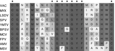
is embedded in the viral membrane, with the long C-terminal domain extending into the cytoplasm during assembly. The cysteines form two intramolecular disulfide bonds through the action of a unique poxvirus-encoded redox system (28, 32). Our attention was captured by a highly conserved 21-amino-acid sequence near the C terminus of H2, which was bounded by cysteine residues 162 and 182 and contained a central hy-drophobic region interrupted by glycine and serine residues (Fig. 1). In particular, the LGYSG sequence was reminiscent of a motif found in the fusion peptide segments of some other viruses (1, 10, 25, 27).
[image:2.585.301.543.69.177.2]We previously constructed a recombinant VACV called vH2i, in which transcription of the H2R gene was regulated by IPTG (28). In the absence of IPTG, noninfectious virus parti-cles are formed. These partiparti-cles bind to cells, but the cores are unable to enter because of a defect in membrane fusion. In order to assess the contribution of individual amino acids to the function of H2, we developed a complementation assay.
FIG. 1. Conservation of a 21-amino-acid sequence near the C ter-minus of H2. Residues 162 through 182 of VACV H2 are aligned with orthologs of other poxviruses. H2 orthologs from a representative of each chordopoxvirus genus and two entomopoxviruses are listed. VAC, VACV; MYX, myxoma virus; LSDV, lumpy skin disease virus; SPV, sheep poxvirus; YMTV, Yaba monkey tumor virus; BPSV, bo-vine papular stomatitis virus; MCV, molluscum contagiosum virus; FPV, fowlpox virus; AMV, Amsacta mooreientomopoxvirus; MSV, Melanoplus sanguinipesentomopoxvirus. Residues are shaded accord-ing to the percentage of amino acid identity. Asterisks indicate VACV amino acids that were mutated for this study.
VOL. 82, 2008 VACCINIA VIRUS ENTRY/FUSION COMPLEX 6245
on November 8, 2019 by guest
http://jvi.asm.org/
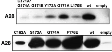
When cells were infected with vH2i in the absence of IPTG, infectious virus formation could be specifically complemented by transfection of a plasmid containing the wild-type H2R gene regulated by its natural promoter (Fig. 2). The background infectivity with the empty vector may represent residual inoc-ulum virus, which was grown in the presence of IPTG and therefore contains H2, and incomplete repression of H2 ex-pression. Complementation values were similar to that of the empty vector when leucine 170, glycine 171, or glycine 174 was changed to glutamic acid, whereas there was still measurable complementation when alanine was substituted individually for either glycine 171 or glycine 174 (Fig. 2). However, replace-ment of both glycines 171 and 174 with alanine led to a severe loss of complementation (Fig. 2). Simultaneous replacement of both cysteines 162 and 182 was less deleterious than replace-ment of cysteine 182 only. This could be explained by aberrant disulfide bond formation as a result of an odd number of cysteines, since our preliminary data are consistent with a di-sulfide bond between cysteines 162 and 182. Despite their complete conservation, replacement of phenylalanine 176 with glutamic acid or serine 173 with alanine had no effect on complementation. Substitution of alanine for tyrosine 172 or tyrosine 175 caused an intermediate reduction in complemen-tation. Western blotting showed that the mutated proteins were expressed at approximately wild-type levels (Fig. 2B).
Effects of mutations within the C-terminal conserved region of H2 on interaction with A28.In view of the presence of eight or more subunits within the EFC, a large number of polypep-tide interactions must occur. Previous studies indicated that the A28 subunit is important for assembling or stabilizing the EFC and suggested that A28 interacts directly or indirectly with H2 (28). The next series of experiments was designed to
[image:3.585.134.449.67.306.2]determine the effects of mutations in the C-terminal conserved region of H2 on the interaction with A28. As in the experi-ments described in the preceding section, cells were infected with vH2i in the absence of IPTG and transfected with plas-mids containing wild-type or mutated H2 under the control of its natural promoter. Similar amounts of the wild-type and mutated H2 proteins, all of which had a C-terminal V5 epitope tag, were captured with an anti-V5 antibody coupled to aga-rose, as determined by Western blotting (data not shown). Similar amounts of A28 were associated with wild-type H2 and with the S173A, G174A, and F176E mutants (Fig. 3), which were all able to complement vH2i infectivity (Fig. 2A). In contrast, very small amounts of A28 were detected in associa-tion with the G171A G174A, G174E, L170E (Fig. 3), and
FIG. 2. Effects of point mutations in a conserved segment of H2 on complementation of infectivity. (A) Cells were infected with vH2i in the absence of IPTG and transfected with an empty plasmid vector or plasmids containing wild-type or mutated H2 with V5 epitope tags. Cell lysates were collected and the infectious virus titers determined by plaque formation in the presence of an inducer. Data averaged from two or more separate experiments are shown. Error bars, standard errors of the means. Amino acid substitutions are shown at the bottom. (B) Expression of H2 was determined by chemiluminescence after Western blotting using an antibody to the V5 epitope.
FIG. 3. Effects of mutations in a conserved segment of H2 on association with A28 in infected cells. Cells were infected with vH2i in the absence of IPTG and transfected with an empty plasmid vector or plasmids containing wild-type or mutated H2 with V5 epitope tags. H2-V5 was then captured with agarose beads coupled to anti-V5 an-tibody. Bound proteins were eluted and detected by chemilumines-cence after Western blotting with anti-A28 antibody. The anti-A28 antibody was prepared from rabbits that had been immunized with a soluble recombinant form of A28 (to be described elsewhere).
on November 8, 2019 by guest
http://jvi.asm.org/
[image:3.585.323.515.554.643.2]G171E (data not shown) mutants, which failed to complement vH2i (Fig. 2A). The intermediate amounts of A28 detected in association with the Y172A, G171A, and C162A mutants (Fig. 3) are consistent with an intermediate level of complementa-tion (Fig. 2A). Thus, for each mutant there was a good corre-lation between vH2i complementation and interaction with A28.
H2 associates A28 specifically.It was not known whether H2 interacts with other EFC polypeptides. Our previous studies had shown a major disruption of the EFC when expression of the A21 subunit was repressed (29). Under these conditions, H2 remained associated with A28 but did not copurify with two other components of the EFC (A16 and L5) for which we had antibodies at the time. To further investigate EFC subunit interactions, we constructed a new recombinant virus, vA21iA28TAP, that has an inducible A21 and a TAP tag on the C terminus of A28. When A21 was expressed in the pres-ence of IPTG, all eight subunits were affinity purified with A28, as determined by mass spectroscopy of the indicated gel slices
(Fig. 4,⫹IPTG). The non-VACV proteins identified were also
[image:4.585.43.285.65.327.2]present in the untagged VACV WR control, indicating non-specific affinity purification. When IPTG was omitted, only H2
FIG. 5. Interaction of full-length and mutated forms of H2 and A28 in the absence of other viral proteins. (A) Interactions of full-length H2 and A28. BS-C-1 cells were cotransfected with a plasmid encoding A28-HA (⫹) or an empty vector (⫺) along with a plasmid encoding H2-V5 (⫹) or an empty vector (⫺). Lysates were prepared, and pro-teins were allowed to bind to agarose beads coupled to HA anti-body. Both the input lysate and immunopurified proteins (IP) were analyzed by SDS-PAGE and probed with V5 and HA anti-bodies. The positions of the H2-V5 and A28-HA bands are indicated. (B) Effects of truncation mutations of H2 on interaction with A28 in transfected cells. Cells were cotransfected with an empty vector plas-mid (⫺) or a plasmid encoding wild-type (wt) or truncated versions of H2-V5 along with a second empty vector plasmid (⫺) or a plasmid expressing A28-HA (⫹). Lysates were prepared, and proteins were allowed to bind to agarose beads coupled to anti-HA antibody. The input lysate and immunopurified proteins were analyzed by Western blotting and probed with anti-V5 and anti-HA antibodies. The posi-tions of A28-HA and H2-V5 are indicated on the right. Arrowheads point to truncated forms of H2-V5. The migration of H2 depended on the length of the truncation. (C) Effects of substitution mutations of H2 on interaction with A28 in transfected cells. Cells were transfected and analyzed as for panel B except that plasmids expressed H2 with the indicated amino acid substitutions instead of truncations. The posi-tions of A28-HA and H2-V5 are indicated on the right.
FIG. 4. Affinity purification of A28 with H2. HeLa S3 cells were infected with 5 PFU per cell of VACV WR or vA21iA28TAP in the presence (⫹) or absence (⫺) of IPTG. At 24 h, cells were lysed with Triton X-100, followed by brief sonication. The postnuclear superna-tant was incubated with streptavidin Sepharose, washed, and eluted with biotin as described elsewhere (39). The eluate was concentrated and separated by SDS-PAGE. The gel was stained with Coomassie blue, and protein bands were cut from the gel, digested with trypsin, and analyzed by mass spectrometry. The Coomassie blue-stained gel is shown. Protein identities are given to the right of the designated band. Peptides belonging to J5, A21, and L5 were identified within the same band. RNP A2/B1 refers to ribonucleoprotein A2/B1, isoform A2 (gi 3329498). The masses (in kilodaltons) of marker proteins are given on the left.
VOL. 82, 2008 VACCINIA VIRUS ENTRY/FUSION COMPLEX 6247
on November 8, 2019 by guest
http://jvi.asm.org/
[image:4.585.324.512.69.507.2]was specifically associated with the TAP-tagged A28 (Fig. 4,⫺ IPTG).
To further investigate the interaction of H2 and A28, we constructed plasmids that express HA epitope-tagged A28 (A28-HA) and V5 epitope-tagged H2 (H2-V5) under the di-rection of the cytomegalovirus promoter. The plasmids were transfected separately or together into uninfected cells. Cell lysates were incubated with anti-HA antibody coupled to aga-rose, washed, and analyzed by Western blotting. A specific association of A28 with H2 was demonstrated in the absence of any other viral proteins (Fig. 5A).
Effects of mutations within the C-terminal conserved region of H2 on the interaction with A28 in uninfected cells. The demonstration of a specific interaction between H2 and A28 allowed us to confirm the effects of mutations within the con-served region between amino acids 162 and 182 of H2. H2-V5 was truncated immediately before the first conserved cysteine
(H2⌬102-189) or immediately before the third conserved
cys-teine (H2⌬162-189) in the absence of other viral proteins. A
plasmid expressing truncated H2 or full-length H2 with a V5 tag was cotransfected with the plasmid expressing A28-HA, and lysates were incubated with beads coupled to the anti-HA antibody. Although each H2 construct was stably expressed, only full-length H2 was able to bind to A28 (Fig. 5B). Thus, the C-terminal conserved region was required for interaction with A28 in this assay. Next, we made point mutations in the H2
protein that had been tested in the infectivity complementation
assay: L170E and G171A⫹G174A, which were unable to
com-plement, and F176E, which complemented as well as wild-type H2 did. Again, each of the mutated proteins was stably ex-pressed. However, only the wild-type and F176E proteins in-teracted with A28-HA (Fig. 5C).
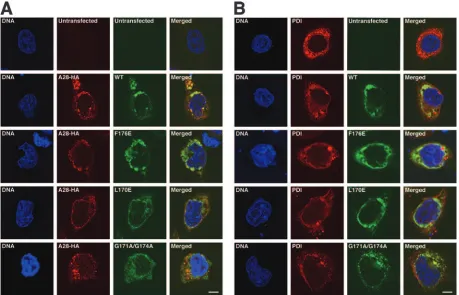
Confocal microscopy was carried out to determine whether the inability of mutated H2 proteins to interact with A28 was specifically due to their cellular mislocalization. Examination of uninfected cells expressing unmutated H2 and A28 indi-cated their colocalization with each other and with PDI, an endoplasmic reticulum-resident protein. Next, we determined that the mutated H2 proteins, regardless of their ability to associate with A28 in the coimmunoprecipitation assay, colo-calized with A28 and PDI (Fig. 6A and B). Therefore, the
failure of the L170E and G171A⫹G174A mutants to interact
with A28 was specific and not due to mislocalization. Taken together, our data show that H2 and A28 interact with each other, that selected mutations within a conserved region near the C terminus interfere with this interaction, and that this interaction is necessary for virus infectivity.
DISCUSSION
[image:5.585.63.522.69.364.2]The elucidation of the mechanism of poxvirus entry is a daunting task, given the large number of proteins involved, the
FIG. 6. Localization of wild-type H2 and mutated forms of H2 in transfected cells. Uninfected cells cotransfected with an empty vector plasmid (untransfected) or a plasmid encoding wild-type (WT) or mutated versions of H2-V5 along with a second empty vector plasmid (untransfected) or a plasmid expressing A28-HA were fixed and permeabilized 24 h after transfection. Cells were stained with a rabbit anti-V5 antibody and either a mouse anti-HA (A) or a mouse anti-PDI (B) antibody, followed by Alexa 488-conjugated anti-rabbit IgG or Alexa 495-conjugated anti-mouse IgG, respectively. Finally, cells were stained with Hoechst reagent and visualized by confocal microscopy. Green, Alexa 488; red, Alexa 594; blue, Hoechst reagent. Bars, 10m.
on November 8, 2019 by guest
http://jvi.asm.org/
existence of two infectious virus forms, and the simultaneous utilization of both low and neutral pH modes of fusion (29). Here we have taken an initial step in defining the interaction between two of the EFC proteins, H2 and A28. The interaction was first inferred by copurification of proteins from cells in which formation of the EFC was perturbed by preventing ex-pression of one subunit. When exex-pression of the A21 subunit was repressed, TAP-tagged A28 and H2 were the only two viral proteins recovered by affinity purification. Nevertheless, for these two proteins to be part of a larger network, at least one of them must interact with a third protein. A21 might be a second binding partner of one of the proteins, since its expres-sion was specifically repressed. Alternatively, the interaction of A28 and H2 with another protein may not have been suffi-ciently stable to survive the purification procedure or was not detected by the mass spectroscopy analysis. Analysis of unin-fected cells transunin-fected with plasmids expressing H2 and A28 confirmed that the interaction of A28 and H2 does not require any other viral proteins. Ordinarily, in infected cells, these two proteins are inserted into unique viral membranes (28, 30). However, in uninfected cells, the two proteins colocalized with an endoplasmic reticulum marker when expressed under the transfection conditions described above, under which no viral membranes were formed. Recent studies suggest that viral proteins may normally traffic from the endoplasmic reticulum to the nascent viral membrane in infected cells (17).
A 21-amino-acid sequence flanked by cysteine residues that may form a disulfide loop in H2 appears to form an interaction site with A28. Our initial interest in this sequence was based on high conservation among chordopoxviruses and entomopoxvi-ruses, as well as the resemblance of this sequence to some internal fusion peptides. In particular, LGYSG is similar to sequences found in the fusion peptides of other viruses: GLFG in influenza virus, GLFG or GFFG in flaviviruses (1), and GFLG in retroviruses (10, 25) and hepatitis B virus (27). In several cases, the importance of this conserved sequence for fusion has been determined by mutagenesis (1, 4, 13, 15, 35). Using a complementation-of-infectivity assay, we found that glutamic acid substitutions within the LGYSG sequence of H2 or the substitution of alanines for both glycines resulted in a loss of function. Interestingly, these mutations also interfered with the interaction of H2 with A28 in both infected and transfected cells. Since the conserved peptide segment of H2 was important for the association with A28, we cannot deter-mine whether it has a second role in fusion. However, since fusion peptides are typically buried in the prefusion state, it is possible that the LGYSG sequence of H2 is concealed within the EFC by interaction with A28 and is exposed during a conformation change initiated by receptor binding or low pH.
ACKNOWLEDGMENTS
We thank Tatiana Senkevich for useful discussions and comments on the manuscript; Norman Cooper and Catherine Cotter for help with tissue culture; Mary Ann Robinson, Carl Hammer, and Raynaldo Martin for mass spectroscopy; and Meggan Czapiga for help with confocal microscopy.
The research was funded by the Intramural Program of the National Institute of Allergy and Infectious Diseases of the National Institutes of Health.
REFERENCES
1.Allison, S. L., J. Schalich, K. Stiasny, C. W. Mandl, and F. X. Heinz.2001. Mutational evidence for an internal fusion peptide in flavivirus envelope protein E. J. Virol.75:4268–4275.
2.Barouch, D. H., Z. Y. Yang, W. P. Kong, B. Korioth-Schmitz, S. M. Sumida, D. M. Truitt, M. G. Kishko, J. C. Arthur, A. Miura, J. R. Mascola, N. L. Letvin, and G. J. Nabel.2005. A human T-cell leukemia virus type 1 regu-latory element enhances the immunogenicity of human immunodeficiency virus type 1 DNA vaccines in mice and nonhuman primates. J. Virol.79: 8828–8834.
3.Blasco, R., and B. Moss.1991. Extracellular vaccinia virus formation and cell-to-cell virus transmission are prevented by deletion of the gene encoding the 37,000 Dalton outer envelope protein. J. Virol.65:5910–5920. 4.Bosch, M. L., P. L. Earl, K. Fargnoli, S. Picciafuoco, F. Giombini, F.
Wong-Staal, and G. Franchini.1989. Identification of the fusion peptide of primate immunodeficiency viruses. Science244:694–697.
5.Brown, E., T. G. Senkevich, and B. Moss.2006. Vaccinia virus F9 virion membrane protein is required for entry but not virus assembly, in contrast to the related L1 protein. J. Virol.80:9455–9464.
6.Carter, G. C., M. Law, M. Hollinshead, and G. L. Smith.2005. Entry of the vaccinia virus intracellular mature virion and its interactions with glycosamino-glycans. J. Gen. Virol.86:1279–1290.
7.Chiu, W. L., C. L. Lin, M. H. Yang, D. L. M. Tzou, and W. Chang.2007. Vaccinia virus 4c (A26L) protein on intracellular mature virus binds to the extracellular cellular matrix laminin. J. Virol.81:2149–2157.
8.Chung, C.-S., J.-C. Hsiao, Y.-S. Chang, and W. Chang.1998. A27L protein mediates vaccinia virus interaction with cell surface heparin sulfate. J. Virol. 72:1577–1585.
9.Delos, S. E., and J. M. White.2000. Critical role for the cysteines flanking the internal fusion peptide of avian sarcoma/leukosis virus envelope glycopro-tein. J. Virol.74:9738–9741.
10.Durell, S. R., I. Martin, J. M. Ruysschaert, Y. Shai, and R. Blumenthal. 1997. What studies of fusion peptides tell us about viral envelope glycopro-tein-mediated membrane fusion (review). Mol. Membr. Biol.14:97–112. 11.Earl, P. L., N. Cooper, S. Wyatt, B. Moss, and M. W. Carroll.1998.
Prepa-ration of cell cultures and vaccinia virus stocks, p. 16.16.1–16.16.3.InF. M. Ausubel, R. Brent, R. E. Kingston, D. D. Moore, J. G. Seidman, J. A. Smith, and K. Struhl (ed.), Current protocols in molecular biology, vol. 2. John Wiley and Sons, New York, NY.
12.Earp, L. J., S. E. Delos, H. E. Park, and J. M. White.2005. The many mechanisms of viral membrane fusion proteins. Curr. Top. Microbiol. Im-munol.285:25–66.
13.Freed, E. O., D. J. Myer, and R. Risser.1990. Characterization of the fusion domain of the human immunodeficiency virus type 1 envelope glycoprotein gp41. Proc. Natl. Acad. Sci. USA87:4650–4654.
14.Gallaher, W. R.1996. Similar structural models of the transmembrane pro-teins of Ebola and avian sarcoma viruses. Cell85:477–478.
15.Gething, M. J., R. W. Doms, D. York, and J. White.1986. Studies on the mechanism of membrane fusion: site-specific mutagenesis of the hemagglu-tinin of influenza virus. J. Cell Biol.102:11–23.
16.Hsiao, J. C., C. S. Chung, and W. Chang.1999. Vaccinia virus envelope D8L protein binds to cell surface chondroitin sulfate and mediates the adsorption of intracellular mature virions to cells. J. Virol.73:8750–8761.
17.Husain, M., A. S. Weisberg, and B. Moss.2006. Existence of an operative pathway from the endoplasmic reticulum to the immature poxvirus mem-brane. Proc. Natl. Acad. Sci. USA103:19506–19511.
18.Izmailyan, R. A., C. Y. Huang, S. Mohammad, S. N. Isaacs, and W. Chang. 2006. The envelope G3L protein is essential for entry of vaccinia virus into host cells. J. Virol.80:8402–8410.
19.Kochan, G., D. Escors, J. M. Gonzalez, J. M. Casasnovas, and M. Esteban. 2008. Membrane cell fusion activity of the vaccinia virus A17–A27 protein complex. Cell. Microbiol.10:149–164.
20.Law, M., G. C. Carter, K. L. Roberts, M. Hollinshead, and G. L. Smith.2006. Ligand-induced and non-fusogenic dissolution of a viral membrane. Proc. Natl. Acad. Sci. USA103:5989–5994.
21.Lin, C. L., C. S. Chung, H. G. Heine, and W. Chang.2000. Vaccinia virus envelope H3L protein binds to cell surface heparan sulfate and is important for intracellular mature virion morphogenesis and virus infection in vitro and in vivo. J. Virol.74:3353–3365.
22.Moss, B.2007.Poxviridae: the viruses and their replication, p. 2905–2946.In
D. M. Knipe, P. M. Howley, D. E. Griffin, R. A. Lamb, M. A. Martin, B. Roizman, and S. E. Straus (ed.), Fields virology, 5th ed., vol. 2. Lippincott Williams & Wilkins, Philadelphia, PA.
23.Moss, B.2006. Poxvirus entry and membrane fusion. Virology344:48–54. 24.Ojeda, S., T. G. Senkevich, and B. Moss.2006. Entry of vaccinia virus and
cell-cell fusion require a highly conserved cysteine-rich membrane protein encoded by the A16L gene. J. Virol.80:51–61.
25.Pe´cheur, E. I., J. Sainte-Marie, A. Bienvenu¨e, and D. Hoekstra.1999. Pep-tides and membrane fusion: towards an understanding of the molecular mechanism of protein-induced fusion. J. Membr. Biol.167:1–17. 26.Rodrı´guez, D., M. Esteban, and J. R. Rodrı´guez.1995. Vaccinia virus A17L
VOL. 82, 2008 VACCINIA VIRUS ENTRY/FUSION COMPLEX 6249
on November 8, 2019 by guest
http://jvi.asm.org/
gene product is essential for an early step in virion morphogenesis. J. Virol. 69:4640–4648.
27.Rodrı´guez-Crespo, I., E. Nunez, J. Gomez-Gutierrez, B. Yelamos, J. P. Albar, D. L. Peterson, and F. Gavilanes.1995. Phospholipid interactions of the putative fusion peptide of hepatitis B virus surface antigen S protein. J. Gen. Virol.76:301–308.
28.Senkevich, T. G., and B. Moss.2005. Vaccinia virus H2 protein is an essential component of a complex involved in virus entry and cell-cell fusion. J. Virol. 79:4744–4754.
29.Senkevich, T. G., S. Ojeda, A. Townsley, G. E. Nelson, and B. Moss.2005. Poxvirus multiprotein entry-fusion complex. Proc. Natl. Acad. Sci. USA 102:18572–18577.
30.Senkevich, T. G., B. M. Ward, and B. Moss.2004. Vaccinia virus A28L gene encodes an essential protein component of the virion membrane with in-tramolecular disulfide bonds formed by the viral cytoplasmic redox pathway. J. Virol.78:2348–2356.
31.Senkevich, T. G., B. M. Ward, and B. Moss.2004. Vaccinia virus entry into cells is dependent on a virion surface protein encoded by the A28L gene. J. Virol.78:2357–2366.
32.Senkevich, T. G., C. L. White, E. V. Koonin, and B. Moss.2002. Complete pathway for protein disulfide bond formation encoded by poxviruses. Proc. Natl. Acad. Sci. USA99:6667–6672.
33.Sieczkarski, S. B., and G. R. Whittaker.2005. Viral entry. Curr. Top. Mi-crobiol. Immunol.285:1–23.
34.Smith, G. L., A. Vanderplasschen, and M. Law.2002. The formation and
function of extracellular enveloped vaccinia virus. J. Gen. Virol.83:2915– 2931.
35.Steinhauer, D. A., S. A. Wharton, J. J. Skehel, and D. C. Wiley.1995. Studies of the membrane fusion activities of fusion peptide mutants of influenza virus hemagglutinin. J. Virol.69:6643–6651.
36.Townsley, A., T. G. Senkevich, and B. Moss. 2005. The product of the vaccinia virus L5R gene is a fourth membrane protein encoded by all pox-viruses that is required for cell entry and cell-cell fusion. J. Virol.79:10988– 10998.
37.Townsley, A., T. G. Senkevich, and B. Moss.2005. Vaccinia virus A21 virion membrane protein is required for cell entry and fusion. J. Virol.79:9458– 9469.
38.Townsley, A. C., A. S. Weisberg, T. R. Wagenaar, and B. Moss.2006. Vac-cinia virus entry into cells via a low-pH-dependent-endosomal pathway. J. Virol.80:8899–8908.
39.Wagenaar, T. R., and B. Moss.2007. Association of vaccinia virus fusion regulatory proteins with the multicomponent entry/fusion complex. J. Virol. 81:6286–6293.
40.Weissenhorn, W., A. Carfi, K. H. Lee, J. J. Skehel, and D. C. Wiley.1998. Crystal structure of the Ebola virus membrane fusion subunit, GP2, from the envelope glycoprotein ectodomain. Mol. Cell2:605–616.
41.Wolffe, E. J., D. M. Moore, P. J. Peters, and B. Moss.1996. Vaccinia virus A17L open reading frame encodes an essential component of nascent viral membranes that is required to initiate morphogenesis. J. Virol.70:2797– 2808.