Virus Type 1 (HIV-1) and Feline Immunodeficiency Virus Reverse
Transcriptases Detected by HIV-1-Infected Subjects
Missa P. Sanou,aShannon R. Roff,aAntony Mennella,bJohn W. Sleasman,cMobeen H. Rathore,dJanet K. Yamamoto,aJay A. Levye Department of Infectious Diseases and Pathology, College of Veterinary Medicine, University of Florida, Gainesville, Florida, USAa
; Primary Care, Student Health Care Center, University of Florida, Gainesville, Florida, USAb
; Division of Allergy, Immunology and Rheumatology, University of South Florida, All Children’s Hospital, St. Petersburg, Florida, USAc
; University of Florida Center for HIV/AIDS Research, Education and Service (UF CARES), Jacksonville, Florida, USAd
; Department of Medicine, University of California San Francisco, San Francisco, California, USAe
Anti-human immunodeficiency virus (HIV) cytotoxic T lymphocyte (CTL)-associated epitopes, evolutionarily conserved on both HIV type 1 (HIV-1) and feline immunodeficiency virus (FIV) reverse transcriptases (RT), were identified using gamma in-terferon (IFN-␥) enzyme-linked immunosorbent spot (ELISpot) and carboxyfluorescein diacetate succinimide ester (CFSE) pro-liferation assays followed by CTL-associated cytotoxin analysis. The peripheral blood mononuclear cells (PBMC) or T cells from HIV-1-seropositive (HIVⴙ) subjects were stimulated with overlapping RT peptide pools. The PBMC from the HIVⴙsubjects had more robust IFN-␥responses to the HIV-1 peptide pools than to the FIV peptide pools, except for peptide-pool F3. In contrast, much higher and more frequent CD8ⴙT-cell proliferation responses were observed with the FIV peptide pools than with the HIV peptide pools. HIV-1-seronegative subjects had no proliferation or IFN-␥responses to the HIV and FIV peptide pools. A total of 24% (40 of 166) of the IFN-␥responses to HIV pools and 43% (23 of 53) of the CD8ⴙT-cell proliferation responses also correlated to responses to their counterpart FIV pools. Thus, more evolutionarily conserved functional epitopes were identified by T-cell proliferation than by IFN-␥responses. In the HIVⴙsubjects, peptide-pool F3, but not the HIV H3 counterpart, induced the most IFN-␥and proliferation responses. These reactions to peptide-pool F3 were highly reproducible and persisted over the 1 to 2 years of testing. All five individual peptides and epitopes of peptide-pool F3 induced IFN-␥and/or proliferation responses in addition to inducing CTL-associated cytotoxin responses (perforin, granzyme A, granzyme B). The epitopes inducing poly-functional T-cell activities were highly conserved among human, simian, feline, and ungulate lentiviruses, which indicated that these epitopes are evolutionarily conserved. These results suggest that FIV peptides could be used in an HIV-1 vaccine.
T
he commercial release of an effective human immunodefi-ciency virus type 1 (HIV-1) vaccine is not imminent even after completion of four major phase IIB-to-III vaccine trials against HIV-AIDS (1). Our limited understanding of the mechanisms of vaccine protection (2) and of the identity of the protective viral epitopes (3,4) further hampers the development of an effective vaccine. Initial studies focused on antibody-based vaccine designs, with an emphasis on generating broadly virus-neutralizing anti-bodies (bNAbs) (5). However, two phase III vaccine trials using envelope (Env) immunogens failed (6,7). Subsequent attention was focused on the T-cell-based vaccines that generate protective cell-mediated immunity (CMI) to global HIV-1 isolates (8). The CMI responses, essential for an effective vaccine, most likely in-clude cytotoxic T lymphocyte (CTL) activities that specifically tar-get HIV-1-infected cells (9–11). Unlike NAb epitopes which re-side exclusively on the Env proteins, the selection of specific vaccine epitopes for the development of T-cell-based vaccines is more difficult to achieve. A vast number of CTL-associated epitopes can be found to span the whole length of most HIV pro-teins (Los Alamos National Laboratory [LANL] database;http://hiv-web.lanl.gov/content/immunology/maps/maps.html) (12).
The goal to develop T-cell-based vaccines is challenged by the capacity of the virus to evade antiviral immunity through a muta-tion(s) for resistance (13,14).
A recent phase III trial consisting of priming with a gag-pr-gp41-gp120canarypox-vectored vaccine and boosting with Env gp120 induced both humoral immunity and CMI and conferred
modest overall efficacy (15). However, phase I and II vaccine trials consisting of cross-subtype-conserved CTL-associated peptide epitopes have shown minimal CMI responses (16–18). Therefore, a thorough selection of potent anti-HIV T-cell-associated epitopes, which are conserved among HIV-1 subtypes and do not mutate without negatively affecting viral fitness (19–21), would be valuable for an effective HIV-1 vaccine. One approach is to select conserved, nonmutable CTL epitopes on essential viral structural proteins or enzymes that also persist on the older subgenera of the lentiviruses which have survived evolutionary pressure (22). Such an approach was successfully used in the development of the ini-tial smallpox vaccines (23). In line with this strategy, the recogni-tion of conserved epitopes on other lentivirus species has been shown by studies using peripheral blood mononuclear cells (PBMC) from HIV-1-positive (HIV⫹) humans (24), HIV-2-vac-cinated and simian immunodeficiency virus (SIV)-challenged nonhuman primates (25), and HIV-1 p24-vaccinated and feline immunodeficiency virus (FIV)-challenged cats (26,27).
The viral enzyme reverse transcriptase (RT) is one of the most
Received5 February 2013Accepted26 June 2013
Published ahead of print3 July 2013
Address correspondence to Janet K. Yamamoto, [email protected].
Copyright © 2013, American Society for Microbiology. All Rights Reserved.
doi:10.1128/JVI.00359-13
on November 7, 2019 by guest
http://jvi.asm.org/
conserved viral proteins, possessing the lowest entropy value among the HIV-1 proteins from various subtypes (28), and con-tains many CTL-associated epitopes (10). The RT proteins of HIV-1 and FIV also share the highest degree of identity in their amino acid sequences (22). The current studies were undertaken to identify the conserved CTL-associated epitopes on FIV and HIV-1 RT proteins which are recognized by the PBMC and T cells from HIV⫹subjects. The major objective of such studies was to identify evolutionarily conserved CMI epitopes that may be more resistant to mutation and thus useful in the development of an effective, T-cell-based HIV-1 vaccine.
MATERIALS AND METHODS
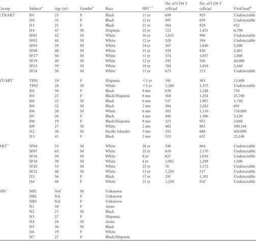
Study population.Blood from HIV-1-infected subjects was obtained from the University of California at San Francisco (UCSF), the University of South Florida in Tampa, and the University of Florida Center for HIV/ AIDS Research, Education and Service (UF CARES) in Jacksonville. These subjects were distributed into three groups according to the length of infection and the antiretroviral therapy (ART) status (Table 1). The HIV-infected (HIV⫹) subjects consisted of long-term survivors (LTS) who had been infected for more than 10 years and remained healthy without anti-retroviral therapy (LTS/ART⫺); subjects with short-term infection with-out ART (ST/ART⫺); and subjects on ART for various amounts of time (ART⫹). T-cell counts and HIV-1 RNA level determinations were per-formed by clinical laboratories at UCSF Medical Center and UF Shands Medical Center (Jacksonville, FL). Blood from HIV-seronegative (HIV⫺) samples was obtained from LifeSouth Community Blood Centers (Gainesville, FL) or randomly selected volunteers at UF. The blood col-lections were performed according to the policies and protocols approved by the Institutional Review Boards at UF and UCSF and processed within 2 to 30 h after collection.
RT overlapping peptides.Overlapping peptides of subtype B HIV-1UCD1and subtype B FIVFC1RT proteins and selected peptides for epitope mapping were produced initially by SynPep (Dublin, CA) and later by RS Synthesis LLC (Louisville, KY) with similar findings. Four to five consec-utive peptides (11 to 16 amino acids [aa] long with an 8-to-10-aa overlap) were grouped into 21 pools: H1 to H21 for HIV and counterparts F1 to F21 for FIV. In addition, 9mer and 15- to 16mer peptides with modified sequences were also synthesized by RS Synthesis LLC and used for peptide epitope mapping as shown inTable 2.
ELISpot assays.Enzyme-linked immunosorbent spot (ELISpot) as-says for IFN-␥(R&D Systems, Minneapolis, MN) were performed with AIM V medium containing 5% heat-inactivated (56°C, 30 min) human serum as previously described (29). The PBMC from HIV⫹subjects were stimulated with either a peptide pool (4 to 5 consecutive peptides per pool at 5g per peptide) or individual peptides (15g/well). The peptides were 11 to 16 aa in length with an 8-to-10-aa overlap. The results were analyzed with an ELISpot reader (MVS Pacific LLC, Minneapolis, MN) and adjusted to determine the number of spot-forming units (SFU) per 106cells, after subtraction of the value corresponding to the average me-dium control for each subject. The PBMC from HIV⫹subjects were stim-ulated with a T-cell mitogen, phytohemagglutinin A (PHA; 5g/ml), as the positive control. At a positive threshold of 70 SFU, HIV⫺subjects had no substantial (⬎50 SFU) IFN-␥responses to HIV and FIV peptide pools.
Flow cytometry (fluorescence-activated cell sorter [FACS]) analysis for CFSE proliferation and intracellular cytotoxin staining (ICS).The carboxyfluorescein diacetate succinimide ester (CFSE) proliferation anal-ysis was performed on PBMC according to the manufacturer’s protocol (Invitrogen, Carlsbad, CA) and processed as previously described (30). Modifications consisted of using 2.0⫻105to 5.0⫻105CFSE-labeled cells stimulated for 5 days (at 37°C and 5% CO2) with 30g/well of total peptides in a pool (15g/well for individual peptides;Table 2) or 5g/ml PHA in AIM V medium containing 25g/ml of gentamicin and 10% heat-inactivated human serum. These cells were subsequently harvested
and labeled with the LIVE/DEAD fixable yellow dye (Invitrogen) and then treated for 5 min with anti-CD16/CD32 antibody (Biolegend, San Diego, CA) for blocking nonspecific binding before analysis using phenotype-specific antibodies. The following antibodies were used for the CFSE pro-liferation analysis: anti-CD4 allophycocyanin (APC), anti-CD3 APC-H7, and anti-CD8 Pacific Blue (BD Biosciences, San Jose, CA).
The ICS analysis (31) involved stimulating 0.5⫻106to 1.0⫻106 freshly isolated PBMC for 6 h with the same peptide stimulant and culture conditions as used in the proliferation analysis in the presence of 1g/ml of Golgi transport inhibitor and monensin followed by labeling with LIVE/DEAD fixable yellow dye and then treatment with anti-CD16/CD32 antibody and T-cell phenotypic antibodies. The cells were subsequently fixed and permeabilized with Cytofix/Cytoperm solution (BD Biosci-ences) before reactions with anti-cytotoxin antibodies were performed. The antibodies consisted of anti-CD3 APC-H7, anti-CD4 BD Horizon V450, and CD8 fluorescein isothiocyanate (FITC) followed by anti-GrzB Alexa 700 and anti-GrzA phycoerythrin (PE) (all from BD Biosci-ences) and anti-perforin peridinin chlorophyll protein (PerCP) (Abcam, Boston, MA).
In both analyses, 1.0⫻104to 2.0⫻104cells were fixed in phosphate-buffered saline (PBS) containing 2% paraformaldehyde and analyzed us-ing a BD LSR II flow cytometer and FACSDiva software (BD Biosciences), with a positive threshold of 3% CFSElowfor CFSE proliferation and 1% T cells expressing cytotoxin for ICS. The final value for each subject was derived after subtraction of the value corresponding to the subject’s me-dium control and the average value for the peptide-stimulated cells from uninfected control subjects.
Statistics.Paired Studentttests with two-tailed distribution (Sigma-Plot version 11.0; Sigma(Sigma-Plot Software Inc., San Jose, CA) were used to evaluate the statistical differences between the results from two time points (seeFig. 3). These results were considered statistically different whenP⬍0.05.
RESULTS
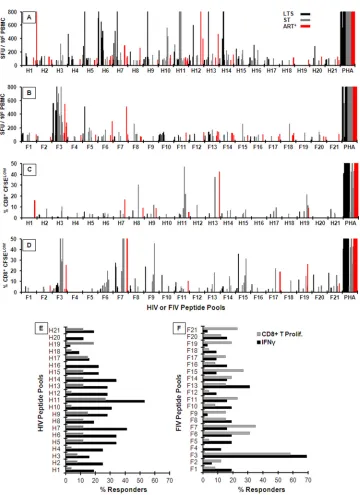
Screening for IFN-␥-inducing epitopes on HIV-1 and FIV RT. As a first step toward identifying the CTL-associated reactive sites on HIV-1 and FIV RT proteins, the PBMC from HIV⫹subjects and HIV⫺subjects were screened by ELISpot analysis for IFN-␥ responses to overlapping RT peptide pools of HIV-1 and FIV. NK cells, CD3⫹CD4⫹T-helper cells, CD3⫹CD4⫹CTLs, and CD3⫹ CD8⫹CTLs generally produce IFN-␥responses to viral peptides (32, 33). In this study, many HIV-1 pools induced IFN-␥ re-sponses of high magnitudes above the threshold level (ⱖ70 SFU) with the PBMC from HIV⫹subjects (Fig. 1A) but none with the PBMC from HIV⫺subjects (data not shown). Therefore, the viral specificity of the IFN-␥responses is associated with HIV-1 infec-tion. The average responder frequency for all 21 pools was 25% (range, 4% to 54%) (Fig. 1E). Of all the HIV peptide pools screened, pool H11 induced the highest and the most frequent IFN-␥responses.
Compared to the HIV peptide-pool responses, the magnitude and the frequency of the IFN-␥responses to the FIV peptide pools were much lower in the PBMC from HIV⫹subjects (Fig. 1Band
F). The average responder frequency for all 21 pools was 17% (range, 4% to 69%) (Fig. 1F). A noticeable exception was observed with pool F3, which induced the highest and the most frequent cellular responses among the FIV pools (Fig. 1BandF). PBMC from only five subjects (SF08, J08, J02, J09, and TP01) responded to a counterpart pool H3 (grouped data only;Fig. 1A), and, re-markably, PBMC from four of these subjects responded to both F3 and H3 (grouped data only;Fig. 1A andB). As expected, the PBMC of HIV⫺subjects had no IFN-␥responses to the FIV pools (data not shown). Overall, HIV pools induced much higher and
on November 7, 2019 by guest
http://jvi.asm.org/
more frequent IFN-␥responses than the FIV counterparts except for pool F3. However, only a few HIV and FIV counterparts were detected by the same individual (4 to 5 responders detected both H3 and F3, H6 and F6, H7 and F7, H11 and F11, and H13 and F13).
The immune responses observed were present in all three clin-ical groups (LTS/ART⫺, ST/ART⫺, and ART⫹). In addition, these groups were not statistically different in terms of cell counts.
How-ever, it is noteworthy that 4 of 5 responders to pool H3 were in the ST group, which may indicate that this response had been lost in the LTS and ART⫹group, possibly due to HIV infection.
[image:3.585.42.545.78.553.2]Screening for T-cell proliferation epitopes on HIV-1 and FIV RTs.The presence of strong T-cell proliferation responses to HIV antigen(s) has been associated with lower viral load and better disease outcome in HIV⫹individuals (34). In the current studies, CD3⫹CD4⫹T cells (referred to from here on as CD4⫹T cells)
TABLE 1Population characteristics
Group Subjecta Age (yr) Genderb Race HIV⫹c
No. of CD4 T cells/l
No. of CD8 T
cells/l Viral loadd
LTS/ART⫺ J01 25 F Black 11 yr 699 935 Undetectable
J10 35 F Black 12 yr 897 659 Undetectable
J11 21 F Black 21 yr 564 829 932
J14 47 M Hispanic 25 yr 722 1,421 6,790
SF01 42 M White 16 yr 1,021 996 Undetectable
SF02 44 M White 12 yr 528 394 Undetectable
SF03 59 M White 24 yr 567 1,040 5,500
SF08 48 M White 31 yr 529 920 3,401
SF17e 56 M White 11 yr 374 1,037 2,000
SF19 40 M White 12 yr 292 556 40,000
SF23 39 M White 10 yr 784 1,018 3,160
SF24 50 M White 11 yr 675 213 Undetectable
ST/ART⫺ TP01 19 F Hispanic ⬍1 yr 391 583 13,400
TP02 28 M White ⬍1 yr 1,280 1,375 Undetectable
J02 50 F Black 9 mo 639 1,248 710
J03 27 F Black/Hispanic 8 mo 368 1,254 25,700
J04 22 M Black 6 mo 537 1,907 1,740
J05 32 M Black 2 mo 384 2,202 691
J06 28 M White 6 mo 501 1,110 134,000
J07 26 F Black 4 mo 448 1,306 5,120
J08 19 F Black/Hispanic 9 mo 323 932 3,040
J09 27 M White 2 mo 482 882 109,168
J12 26 M Pacific Islander 5 mo 352 688 405,000
J13 41 F Black 2 mo 513 632 22,100
ART⫹ SF04 55 M White 28 yr 540 864 Undetectable
SF07 65 M White 25 yr 610 2,170 Undetectable
SF16 50 M White 8 yr 827 1,018 Undetectable
SF18 38 M White 4 yr 1,082 1,298 1,500
SF20 53 M White 23 yr 76 1,172 Undetectable
SF22 48 M White 13 yr 1,205 517 Undetectable
J15 56 F Black 17 yr 291 1,101 Undetectable
J16 48 F White 21 yr 1,250 NAf Undetectable
HIV⫺ NB1 NAf M Unknown
NB2 NA F Unknown
NB3 NA F Unknown
N1 58 F Asian
N2 27 M Black
N3 27 F Hispanic
N4 24 M Asian
N5 36 M Black
N6 19 F White
N7 27 F Black/Hispanic
a
J prefix, HIV⫹subject from the University of Florida (UF) at Jacksonville; SF prefix, HIV⫹subject from the University of California San Francisco; TP prefix, HIV⫹subject from the University of South Florida at Tampa; NB prefix, healthy blood from blood bank; N prefix, normal blood from UF.
b
F, female; M, male. cDuration of HIV infection.
d
Virus loads are shown as copies/ml; undetectable, eitherⱕ50 orⱕ75. eSubject started ART during the study.
f
NA, not available. See Materials and Methods for other abbreviations.
on November 7, 2019 by guest
http://jvi.asm.org/
from HIV⫹subjects (Fig. 2) had fewer proliferation responses than CD3⫹CD8⫹T cells (CD8⫹T cells) to both HIV and FIV pools (Fig. 1CandD). The CD4⫹T cells from only 5 of 26 HIV⫹ subjects responded to at least one HIV pool, whereas 9 of 26 HIV⫹ subjects responded to at least one FIV pool (Fig. 2). In contrast, the CD8⫹T cells from 17 of 26 HIV⫹subjects responded to the HIV pools, and 22 of 26 subjects responded to the FIV pools (Fig.
1CandD).
The most striking results were the high magnitude and the high frequency of CD8⫹T-cell proliferation to the FIV pools in com-parison to HIV pools (Fig. 1CandD). Furthermore, the average frequency of responders to all FIV pools was 17% (range, 0% to 54%) (Fig. 1F), which is higher than the average of 10% (range, 0% to 24%) observed with the HIV pools (Fig. 1E). Only one of the HIV pools (H11) had a responder frequency of⬎20% (Fig. 1E), while the responder frequencies to the six FIV pools (F3, F6, F7, F11, F15, and F21) were⬎20% (Fig. 1E). In addition, only a few HIV and FIV counterparts were detected by the same subject (2 to 4 responders detected both H3 and F3, H6 and F6, H14 and F14, H15 and F15, H17 and F17, and H19 and F19), but 43% (23 of 53) of the total positive proliferation responses to the HIV-1 pools were also positive with respect to the FIV counterparts.
The results presented above support the view that the CD8⫹ T-cell proliferative responses to FIV pools are more robust or possibly more intact than those to HIV pools. This finding is clearly divergent from the results of the IFN-␥studies, where the
IFN-␥responses to the HIV pools were stronger than the FIV pool responses (Fig. 1AandB). These conflicting findings may be par-tially attributed to the difference in the cell types used (PBMC versus CD8⫹T cells). Moreover, 12 of 15 responders showing CD8⫹T-cell proliferation responses to the F3 pool also had robust IFN-␥responses to the F3 pool (Fig. 1BandD). These findings indicate that the samples from these subjects recognized a peptide epitope(s) that induces both responses.
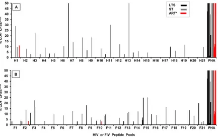
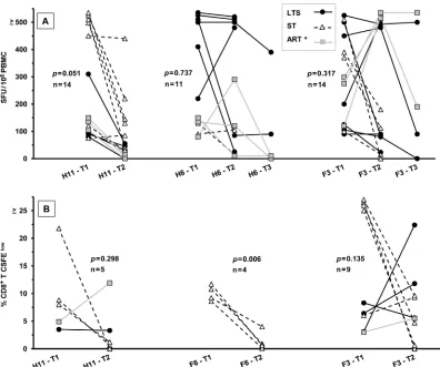
The persistence of IFN-␥and proliferation responses to se-lected HIV and FIV peptide pools.Due to the ability of HIV to quickly escape from immunological pressure (14,19), the PBMC from IFN-␥responders (Fig. 1AandB) were retested at least 1 year later for IFN-␥responses to peptide pools H6, H11, and F3. The majority of the individuals tested at the second time point retained positive IFN-␥responses to H6 (8 of 11 responders) and to F3 (11 of 14 responders) but fewer to H11 (5 of 14 responders) (Fig. 3A). The cells from a few subjects were retested against H6 and F3 for a third time point and demonstrated the persistence of the IFN-␥ responses to the F3 pool (4 of 5) but responded to a lesser extent to the H6 pool (2 of 4) after 3 years (Fig. 3A). More importantly, the persistence of the IFN-␥responses to the F3 pool demonstrated the reproducibility of this activity even though no IFN-␥ re-sponses were observed to the HIV-counterpart H3 by those who were tested after 2 years (Table 2, top, for H3-3 peptide).
[image:4.585.40.545.80.321.2]The CD4⫹and CD8⫹T-cell proliferation responses did not correlate with the IFN-␥responses in general, as only a low
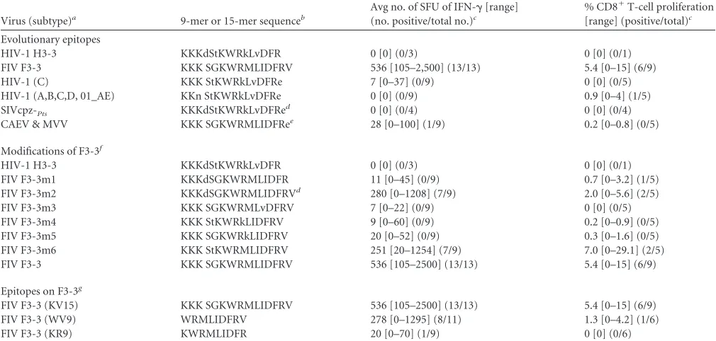
TABLE 2Variation of H3-3/F3-3 amino acid sequences and immunological responses
Virus (subtype)a 9-mer or 15-mer sequenceb
Avg no. of SFU of IFN-␥[range] (no. positive/total no.)c
% CD8⫹T-cell proliferation [range] (positive/total)c
Evolutionary epitopes
HIV-1 H3-3 KKKdStKWRkLvDFR 0 [0] (0/3) 0 [0] (0/1)
FIV F3-3 KKK SGKWRMLIDFRV 536 [105–2,500] (13/13) 5.4 [0–15] (6/9)
HIV-1 (C) KKK StKWRkLvDFRe 7 [0–37] (0/9) 0 [0] (0/5)
HIV-1 (A,B,C,D, 01_AE) KKn StKWRkLvDFRe 0 [0] (0/9) 0.9 [0–4] (1/5)
SIVcpz-Pts KKKdStKWRkLvDFRe
d 0 [0] (0/4) 0 [0] (0/4)
CAEV & MVV KKK SGKWRMLIDFRee 28 [0–100] (1/9) 0.2 [0–0.8] (0/5)
Modifications of F3-3f
HIV-1 H3-3 KKKdStKWRkLvDFR 0 [0] (0/3) 0 [0] (0/1)
FIV F3-3m1 KKKdSGKWRMLIDFR 11 [0–45] (0/9) 0.7 [0–3.2] (1/5)
FIV F3-3m2 KKKdSGKWRMLIDFRVd 280 [0–1208] (7/9) 2.0 [0–5.6] (2/5)
FIV F3-3m3 KKK SGKWRMLvDFRV 7 [0–22] (0/9) 0 [0] (0/5)
FIV F3-3m4 KKK StKWRkLIDFRV 9 [0–60] (0/9) 0.2 [0–0.9] (0/5)
FIV F3-3m5 KKK SGKWRkLIDFRV 20 [0–52] (0/9) 0.3 [0–1.6] (0/5)
FIV F3-3m6 KKK StKWRMLIDFRV 251 [20–1254] (7/9) 7.0 [0–29.1] (2/5)
FIV F3-3 KKK SGKWRMLIDFRV 536 [105–2500] (13/13) 5.4 [0–15] (6/9)
Epitopes on F3-3g
FIV F3-3 (KV15) KKK SGKWRMLIDFRV 536 [105–2500] (13/13) 5.4 [0–15] (6/9)
FIV F3-3 (WV9) WRMLIDFRV 278 [0–1295] (8/11) 1.3 [0–4.2] (1/6)
FIV F3-3 (KR9) KWRMLIDFR 20 [0–70] (1/9) 0 [0] (0/6)
aGenBank numbers are as follows: HIV-1 H3-3, GenBank accession no.K03455.1; FIV F3-3,DQ365597.1; HIV-1 (C),FJ595343; HIV-1 A, B, C, D, and 01_AE,AJ313415,
HM035584,HQ012309,HQ586068, andHE590997; SIVcpz-Pts,ACM63211; CAEV,AAG48629.1; MVV,CAC44543; HERV-K,ABA28284.
bLowercase letters in amino acid sequences indicate differences from FIV F3-3. Many HIV-1 strains have glutamate (E) immediately after the carboxyl end of H3-3.
c
Only responders from the studies described in the legends toFig. 1to3were used. Data represent ranges of responses in SFU and numbers of positive responses versus the total tested (positive/total). Small total participant numbers are due to the use of only the F3 or H3 responders who were still positive at the second or third time point. Only cells from H3 responders were used to test peptide H3-3; cells from F3 responders were used to test other peptides.
dOnly 16-mer sequences.
e
Replacing V15with E15in modifications F3-3m3 to F3-3m6 resulted in an almost total loss of both IFN-␥and T-cell proliferation responses. fSix modifications of 15- to 16-mer F3-3 sequences (F3-3m1 to F3-3m6) with amino acid present on H3-3.
g
Sequence designations are shown in parentheses, with the first and the last amino acids followed by the number of amino acids.
on November 7, 2019 by guest
http://jvi.asm.org/
frequency of CD4⫹T responses was observed. However, more CD4⫹T-cell responses were observed in the production of cy-totoxins. The lack of correlation between IFN-␥ELISpot and CD8⫹T-cell proliferation responses has been described before
in a study performed with p24 proteins. In that study, the ma-jority (64%) of responses were IFN-␥⫹/proliferation⫺and only 30% of the responses were IFN-␥⫹/proliferation⫹(35). Fur-thermore, the use of PBMC in the IFN-␥ analysis may have
FIG 1IFN-␥and CD8⫹T-cell proliferation responses of HIV-infected subjects to HIV and FIV reverse transcriptase (RT) peptide pools. (A to D) The IFN-␥ ELISpot (n⫽32 [12 LTS, 12 ST, and 8 ART⫹]) (A and B) and CD3⫹CD8⫹T-cell proliferation (n⫽26 [11 LTS, 7 ST, and 8 ART⫹]) (C and D) responses to overlapping peptide pools of HIV RT (H1 to H21) (A and C) and FIV RT (F1 to F21) (B and D) are shown. The HIV⫹subjects (see panel A key for panels A to D) consisted of long-term survivors (LTS) who have had HIV infection for over 10 years without antiretroviral therapy (ART) (LTS); subjects recently diagnosed with short-term infection without ART (ST); and subjects on ART at various durations of infection (ART⫹). Each bar represents a positive response by an individual with a threshold of 70 spot-forming units (SFU) per 106peripheral blood mononuclear cells (PBMC) for ELISpot or a threshold of 3% CFSElowfor
CD3⫹CD8⫹T-cell proliferation assay. Cells from each individual were stimulated with a T-cell mitogen, phytohemagglutinin A (PHA), as a positive control. The HIV⫺control subjects (n⫽10) had no responses (data not shown). All responses below the positive threshold are not shown to clearly distinguish those positive values close to the threshold. (E and F) The average frequencies of IFN-␥and proliferation responders to HIV-1 (E) and FIV (F) RT peptide pools are derived from the data represented in panels A to D and are shown as “% Responders.” The solid bar for each peptide represents an average responder frequency for IFN-␥ responses, while the gray bar represents the CD8⫹T-cell proliferation responses.
on November 7, 2019 by guest
http://jvi.asm.org/
[image:5.585.113.473.59.554.2]contributed to the lack of correlation between IFN-␥and T-cell proliferation responses. Cells such as NK cells in PBMC are known to be high producers of IFN-␥(36) and could have also given the IFN-␥responses.
In addition, the CD8⫹T-cell proliferation responses to the F3 pool (7 of 9 responders) persisted but not the responses to the H11 pool (2 of 5) and F6 pool (1 of 4) (Fig. 3B). Overall, these results suggest a major loss of H11-specific IFN-␥and proliferation re-sponses, with maintenance of reactions to the F3 pool. Due to the strong persistent proliferation and IFN-␥responses to the F3 pool, subsequent studies focused on the F3 peptide pool and its five individual peptides and their epitopes.
Identifying the peptide epitope(s) on the F3 region that in-duces IFN-␥and CD8ⴙT-cell proliferation responses.The find-ing that 69% and 58% of the HIV⫹subjects responded to pool F3 with IFN-␥production and CD8⫹T-cell proliferation, respec-tively (Fig. 1EandF), suggested the possibility that the F3 region contains multiple epitopes. The F3 peptide pool has five overlap-ping peptides (F3-1, F3-2, F3-3, F3-4, and F3-5) of 13- to 15mers. All 10 F3 responders tested 1 to 2 years later had IFN-␥responses to the F3-3 peptide, and one individual each had low IFN-␥ re-sponses to individual peptides F3-1 and F3-4 (Fig. 4A). Further-more, 4 of 22 F3 responders had IFN-␥responses to the counter-part H3 pool in the first year (Fig. 1A), but all three of the H3 responders tested at or after the second year lost IFN-␥responses to H3 and had no reactivity to any of the individual 13- to 15mer H3 peptides (Table 2, top; 0/3 IFN-␥responses to H3-3). As ex-pected, all 10 HIV⫺control subjects had no IFN-␥responses to individual peptides of both pools F3 and H3 (data not shown).
Remarkably, the majority of the observed IFN-␥responses to pool F3 were specific for the F3-3 peptide, and the highest re-sponder frequency for CD8⫹T-cell proliferation was observed with F3-3 (5 of 8) as well; slightly lower reaction frequencies were noted with F3-2, F3-4, and F3-5 (3 of 8 each) (Fig. 4B). F3-3 is therefore the predominant FIV RT peptide that gives both IFN-␥ and T-cell proliferation responses.
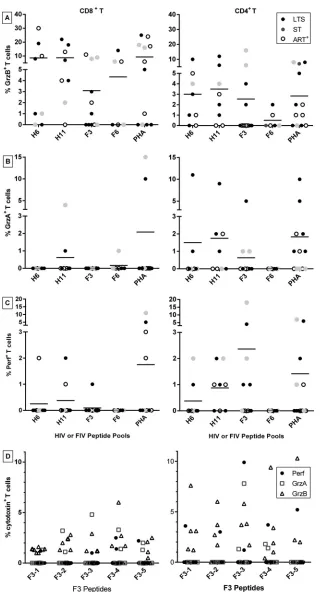
Characterization of CTL-associated activities induced by the F3 pool and F3-3 peptide.One of the most important CMI activ-ities needed to control HIV infection is potent cytotoxicity (37). Both CD4⫹CTLs and CD8⫹CTLs against HIV-1 have been de-tected in HIV⫹subjects (38) and in HIV⫺individuals immunized with a candidate HIV-1 vaccine (39). Although activities with re-spect to H6, H11, and F6 pools were demonstrated, our focus was on CTL-associated activities corresponding to the F3 pool (Fig. 5A
toC) and its five individual peptides (Fig. 5D). A total of 100% (11 of 11) of the F3 responders expressed at least one cytotoxin (GrzA, GrzB, or perforin) in their CD4⫹or CD8⫹T cells, a result similar to the 100% (8 of 8) of H11 responders but higher than the 75% (6 of 8) of H6 responders and 50% (3 of 6) of F6 responders. Hence, a CTL-associated epitope(s) present on F3 and H11 was recog-nized by all the subjects tested. These findings suggest that multi-ple CTL epitopes may reside on each of these regions.
This study showed that all five individual F3 peptides induced expression of GrzA, GrzB, and/or perforin in the CD4⫹and/or CD8⫹T cells of at least one or more HIV⫹subjects tested (Fig. 5D). Based on this finding, different CTL-associated epitopes ap-pear to be present within all five of the individual F3 peptides (data not shown).
FIG 2CD3⫹CD4⫹T-cell proliferation responses to HIV and FIV RT peptide pools. The CD3⫹CD4⫹T-cell proliferation responses to overlapping peptide pools of HIV RT (H1 to H21) (A) and FIV RT (F1 to F21) (B) are shown for HIV⫹subjects (n⫽26 [11 LTS, 7 ST, and 8 ART⫹]). The key in panel A shows the bar color codes for both panels as LTS without ART (LTS), ST without ART (ST), and those on ART (ART⫹). Each bar represents a positive response by an individual with a positive threshold of 70 SFU per 106PBMC for ELISpot or 3% CFSElowfor CD3⫹CD4⫹T-cell proliferation. None of the HIV⫺control subjects (n⫽10) had positive responses (data not shown). Responses below positive thresholds are not shown.
on November 7, 2019 by guest
http://jvi.asm.org/
[image:6.585.78.511.65.337.2]CMI epitopes at H3-3 and F3-3 are conserved among lentivi-ruses.According to LANL QuickAlign analysis, the H3 pool makes up a stretch of amino acids that is highly conserved among lentivi-ruses, as it is identical to the amino acids of 47% of the HIV-1 RTs and 7% of the SIV RTs (http://www.hiv.lanl.gov/content/sequence
/QUICK_ALIGN/QuickAlign.html). Amino acid sequence analysis
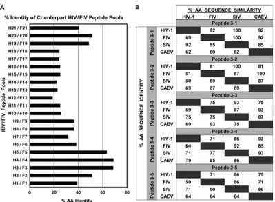
of all HIV and FIV counterpart pairs determined that H3 and F3 had the second highest amino acid identity of 66.7% (Fig. 6A). Furthermore, amino acid sequence analysis of the individual 13-to 15mer peptides shows high amino acid sequence identity and similarity between HIV and FIV (Fig. 6B). The HIV and FIV pairs with the highest similarity in order of the highest to the lowest were as follows: H3-1 and F3-1 (92%), H3-2 and F3-2 (81%), H3-3 and F3-3 (75%), H3-4 and F3-4 (71%), and H3-5 and F3-5 (71%). Considerable similarities in sequences were observed when the H3 and F3 13- to 15mer peptides were compared to SIV and caprine arthritis-encephalitis virus (CAEV) counterpart se-quences. Based on amino acid sequence similarity, both the H3 and F3 peptide-pool regions and their counterpart individual peptides are evolutionarily conserved (Fig. 6).
Due to the CMI responses to F3-3 being consistently higher than the responses to the other four individual F3 peptides (Fig. 4), subsequent studies focused on F3-3 and its HIV-counterpart H3-3. According to LANL QuickAlign analysis, H3-3 has 83% and 35% aa identity with various HIV-1 and SIV sequences, respec-tively. H3-3 and F3-3 peptides have 69% identity and 75% simi-larity, with two gaps (Fig. 6B;Table 2, top). Even with such se-quence similarity, IFN-␥and CD8⫹T-cell proliferation responses greatly differed between these peptides (Table 2).
F3-3 differs from the H3-3 used in the current study (row 1 versus row 2 inTable 2) by lacking one amino acid (Asp on posi-tion 4 of H3-3) and having four amino acid differences at F3-3 positions 5, 9, 11, and 15. The combination of a D4deletion and three changes at K10, V12, and E16of H3-3 with amino acids iden-tical to those of F3-3 resulted in IFN-␥responses approaching those of F3-3 (Table 2, F3-3m6). The addition of D4 to F3-3 (16mer) (F3-3m2) also resulted in IFN-␥responses approaching those of F3-3, whereas the removal of V16from F3-3m2, giving 15mer F3-3m1, caused a major loss in IFN-␥responses and also a modest loss in CD8⫹T-cell proliferation responses. Furthermore,
FIG 3Persistence of IFN-␥and CD8⫹T-cell proliferation responses to selected peptide pools. The IFN-␥(A) and CD8⫹T-cell proliferation (B) responses of HIV⫹subjects who responded the first time (t1) and the second time (t2, at least 1 year later) are shown for peptide pools F3 (both analyses), H11 (both analyses), and H6 (IFN-␥) or F6 (T-cell proliferation). The total numbers of HIV⫹subjects who participated were 22 subjects (9 LTS, 8 ST, and 5 ART⫹) in the IFN-␥study (A) and 11 subjects (3 LTS, 6 ST, and 2 ART⫹) in the CD8⫹T-cell proliferation study (B). However, the numbers of subjects with different clinical status differ among the peptide-pool groups since they are based on the number of responders to the peptide at the first time (t1). In addition, the IFN-␥responses to H6 and F3 have a third time point (t3;ⱖ2 years). ThePvalue of each peptide-pool group indicates that the results from t1 are statistically different from those from t2 whenP⬍0.5. Only the CD8⫹T-cell proliferation responses to F6 at t1 are statistically different from those at t2. The statistical significance values from comparisons between t2 and t3 for H6 (n⫽5) and F3 (n⫽5) wereP⫽0.124 andP⫽0.133, respectively.
on November 7, 2019 by guest
http://jvi.asm.org/
[image:7.585.94.490.65.396.2]a single amino acid change at F3-3 positions 9 (M9¡K9; F3-3m5), 11 (I11¡V11; F3-3m3), and 15 (V15¡E15; CAEV and Maedi-Visna virus [MVV] peptide) caused major losses in both IFN-␥and CD8⫹T-cell proliferation responses. Note that none of the mod-ifications of H3-3 and the peptides tested inTable 2 induced IFN-␥or T-cell proliferation responses in the PBMC or the T cells from HIV⫺control subjects (data not shown).
Peptide F3-3 has a high degree of amino acid identity to those of ungulate lentiviruses (93%; CAEV and MVV) (Table 2). Thus, the F3-3 sequence is greatly conserved among lentiviruses. In this regard, the ungulate peptide counterpart of F3-3 induced IFN-␥ responses in the PBMC from 1 of 9 F3-3 responders tested (Table 2, top). The results presented above demonstrate that the F3-3 sequence contains an evolutionarily conserved epitope(s) that in-duces persistent CMI responses, including strong CTL-associated activity, even when the responses to the counterpart H3-3 are lost.
DISCUSSION
In these studies, the CMI responses to FIV and HIV RT peptides or peptide pools by the HIV⫹subjects resulted in three major obser-vations. First, the CD8⫹ T-cell proliferation responses to FIV pools were more robust, with a frequency of responders higher than the frequency seen with those induced by the HIV pools (Fig. 1EandF). This observation was unexpected, since higher levels of IFN-␥responses were observed with HIV pools than with FIV pools. These proliferation responses to the FIV pools, especially to F3, persisted over a longer time period than those to the HIV pools tested (Fig. 3B). Thus, the few amino acid differences between these viruses might be sufficient for the CD8⫹T cells to recognize the F3 peptides but might not be sufficient for them to recognize the H3 peptides. In fact, three amino acid substitutions in the H3-3 amino acid sequence (V10¡I10; K12¡M12; E16¡V16) with amino acids identical to those of F3-3 led to immunological re-sponses more consistent with that of F3-3 (Table 2). This obser-vation clearly supports our finding that even only a few amino acid changes can substantially alter the responses to a peptide epitope. The robust CD8⫹T-cell responses of the HIV⫹subjects to FIV peptide pools suggest that these peptide regions contain evolu-tionarily conserved epitopes. Importantly, 23 of 53 (43%) total positive CD8⫹T-cell proliferation responses and 40 of 166 (23%)
total positive IFN-␥responses to HIV pools also corresponded to positive responses to their counterpart FIV pools (Fig. 1). This observation suggests that measurement of the T-cell response as the level of CD8⫹T-cell proliferation (43%) was more successful at screening for evolutionarily conserved peptide epitopes than measurement of the IFN-␥response (23%).
The second observation was that of the profound and persis-tent IFN-␥and CD8⫹T-cell proliferation responses to pool F3, which had more responders than any HIV pool (Fig. 3). The F3 pool induced IFN-␥responses in the PBMC from a large propor-tion (69%) of HIV⫹subjects and CD8⫹T-cell proliferation re-sponses in a substantial proportion (58%) of these subjects. These results suggest the presence of multiple CD8⫹T-cell epitopes in the F3 region. One to three F3 responders had IFN-␥or CD8⫹ T-cell proliferation responses to F3-1 and F3-4, and both peptide epitopes induced CTL-associated activities (Fig. 5). In fact, the LANL database shows three CTL-associated epitopes (NTPVFA IKK, NK9; NTPVFAIKKK, NK10; and KLVDFRELNK, KK10) on the counterpart H3. The NK9 and NK10 sequences are identical between FIV and HIV-1 and are found at the carboxy end of both F3-1 and H3-1 13mer peptides. F3-1 differs from H3-1 only by having tryptophan (W3) instead of tyrosine (Y3) at position 3. This finding suggests that this single amino acid difference resulted in the CD8⫹T-cell proliferation response to F3-1 but not to H3-1. In the case of KK10, this epitope resides on H3-4 and differs by three amino acids from its direct counterpart on F3-4 (mLiDFRvLNK [MK10]; differing amino acids are indicated in lowercase charac-ters).
The third major observation was that of the robust IFN-␥ (100%, all 10 of the F3 responders tested) and CD8⫹T-cell pro-liferation (62%, 5 of 8) responses to the F3-3 15mer peptide (Fig. 4). These unusually high frequencies of responders to the F3-3 epitope raised the issue of whether more than one CMI epitope resides on F3-3. In this regard, the current studies, using modified epitopes, identified three CMI epitopes on F3-3 which were not previously described in LANL: KKKSGKWRMLIDFRV (KV15), WRMLIDFRV (WV9), and KWRMLIDFR (KR9) (Table 2, bot-tom). The largest of these epitopes (F3-3; KV15) induced cyto-toxin expression, and thus, one or more of them most likely are
FIG 4F3 peptide epitopes recognized by F3 responders. The peptide-pool F3 consists of five overlapping 13- to 15mer peptides (F3-1, F3-2, F3-3, F3-4, and F3-5) spanning the region from the amino terminus to the carboxyl terminus. IFN-␥(n⫽10 [5 LTS, 3 ST, and 2 ART⫹]) (A) and CD8⫹T-cell proliferation (n⫽8 [3 LTS, 3 ST, and 2 ART⫹]) (B) responses by cells from HIV-infected F3 responders to each of these peptides are shown along with responses to the F3 pool. F3 responders consist of those subjects with long-term HIV-1 infection but not on ART (LTS); those with short-term infection and not on ART (ST); and those on ART with various durations of infection (ART⫹). All responses below positive thresholds are not shown.
on November 7, 2019 by guest
http://jvi.asm.org/
FIG 5Characterization of CTL-associated epitopes on H6, H11, F3, and F6 pools. (A to C) ICS analysis for perforin (Perf), granzyme A (GrzA), and granzyme B (GrzB) expression is shown for CD8⫹T cells (left column) and CD4⫹T cells (right column) from selected HIV⫹responders of designated peptide pools (H6,
n⫽8; H11,n⫽8; F3,n⫽11; F6,n⫽6; and PHA,n⫽12). The HIV⫹subjects (panel A key for panels A to C) consisted of the following individuals: those with long-term infection without ART (LTS); those recently diagnosed with short-term infection without ART (ST); and those on ART with various durations of infection (ART⫹). The numbers of individuals in the clinical status groups and the peptide-pool groups are as follows: H6 (4 LTS, 2 ST, and 2 ART⫹), H11 (4 LTS, 1 ST, and 3 ART⫹), F3 (5 LTS, 3 ST, and 3 ART⫹), F6 (2 LTS, 2 ST, and 2 ART⫹), and PHA (5 LTS, 3 ST, and 4 ART⫹). (D) Six F3-pool responders (5 LTS and 1 ART⫹) were tested for Perf, GrzA, and GrzB responses to the five 13- to 15mer F3 peptides as indicated in the key at the right in panel D. Only three subjects were the same as those described above (A to C), but the blood was collected at a different time point.
on November 7, 2019 by guest
[image:9.585.134.454.29.621.2]CTL-associated epitopes. These epitopes are closely related in se-quence and evolution to those of ungulate lentiviruses (Table 2, top). Therefore, these findings indicate that the F3-3 epitopes are also evolutionarily conserved.
The unique example of pools F3 and H3 (Fig. 6) highlights the existence of an evolutionarily conserved HIV RT epitope region that has been shown to be less immunogenic than its FIV RT counterpart sequence based on analyses of pools F3 and H3 and peptides F3-3 and H3-3. The selection pressure against HIV in humans may explain the lack of responses to the HIV sequence; the same pressure against FIV may not exist in cats. The use of the FIV approach shows that the F3-3 region may be a great target for T-cell responses in an HIV vaccine, since both IFN-␥and proliferation responses to peptide F3-3 by HIV⫹ subjects indicate that they have previously encountered such a sequence or its variant. The approach of using FIV to identify conserved regions for an HIV vaccine is a tool that comple-ments most approaches for developing a T-cell-based vaccine such as in mosaic vaccines (40–42). Computational analyses identify potential conserved epitopes that are later tested for relevant biological activity. These analyses have been used to select conserved HIV/FIV sequences such as the one previously described for HIV/FIV integrase (16). The current FIV ap-proach simultaneously compares both HIV/FIV epitope se-quences and immunological responses.
This cross-recognition of the F3-3 epitope(s) by the HIV⫹ sub-jects demonstrates the polyfunctionality of the T-cell subsets tested. Three patterns with either PBMC or T cells were observed: (i) IFN-␥ production by PBMC (IFN-␥/PBMC), CD8⫹ T-cell proliferation, and CD4⫹or CD8⫹(CD4⫹/CD8⫹) T-cell cytotoxin expression; (ii) IFN-␥/PBMC and CD4⫹/CD8⫹T-cell cytotoxin expression; and (iii) CD8⫹T-cell proliferation and CD4⫹/CD8⫹ T-cell cytotoxin expression. These observations are important since polyfunctional T-cell epitopes are likely to be associated with an effective HIV vaccine (38,43). Although current studies have focused on CD4⫹T-cell responses only minimally, 2 of 3 F3 re-sponders showing CD4⫹T-cell proliferation also expressed CD8⫹ T-cell responses to F3-3 (Fig. 1DandFig. 2B). Moreover, the CD4⫹ T cells from substantial numbers of F3 responders had CTL-associated cytotoxin activities in response to pool F3 (Fig. 5). A vaccine is generally administered to HIV-naive subjects with normal CD4⫹T-cell immunity. Therefore, the importance of the CD4⫹T-cell responses to F3-3 should be considered when iden-tifying CTL-associated epitopes for an HIV vaccine. The vaccine epitopes that induce both anti-HIV CD8⫹and CD4⫹T responses are likely to be needed for effective vaccine protection. The results of these studies using FIV RT peptide pools suggest that evolution-arily conserved immunologic epitopes could be important for an effective HIV vaccine.
FIG 6The amino acid sequence identity between counterpart HIV/FIV peptide pools and between various lentiviruses. (A) The percentages of amino acid
identity between the sequences of each HIV (H) peptide pool and its counterpart FIV (F) peptide pool were obtained by alignment of the sequences using pairwise sequence alignment (http://www.ebi.ac.uk/Tools/psa/emboss_needle/). Note that the highest amino acid identity observed is 68.7% for peptide pools H4 and F4 and that the second highest is 66.7% for peptide pools H3 and F3. (B) The percentages shown on the right of the diagonal divider represent percent amino acid sequence similarity and those on the left represent percent amino acid sequence identity between the two viruses represented by the value at the intersection of each row and column. The lentivirus strains compared are HIV-1HXB2(GenBank accession no.K03455.1), FIVFC1(DQ365597.1), SIVMm251(AAB59906.1), and
CAEV (AAG48629.1).
on November 7, 2019 by guest
http://jvi.asm.org/
[image:10.585.94.493.61.354.2]ACKNOWLEDGMENTS
We thank Ruiyu Pu (UF) and Sue H. Fujimura for their technical help and advice and Kaylynn Peter (UCSF) and Kathy Thoma (UF CARES) for their administrative assistance in organizing the study subjects.
J.K.Y. is the inventor of record on a University of Florida-held patent and may be entitled to royalties from companies that are developing com-mercial products that are related to the research described in this paper.
This work was funded by NIH R01-AI65276 (J.K.Y. and J.A.L.), NIH R01-AI30904 (J.K.Y.), NIH UL1 RR029890 (M.H.R.), and miscellaneous donor funds (J.K.Y.). This work was supported in part by the NIH/NCRR Clinical and Translational Science Award (UL1 RR029890 to M.H.R.).
REFERENCES
1.Saunders KO, Rudicell RS, Nabel GJ.2012. The design and evaluation of HIV-1 vaccines. AIDS26:1293–1302.
2.Plotkin SA. 2008. Vaccines: correlates of vaccine-induced immunity.
Clin. Infect. Dis.47:401– 409.
3.Mothe B, Llano A, Ibarrondo J, Daniels M, Miranda C, Zamarreño J,
Bach V, Zuniga R, Pérez-Álvarez S, Berger CT, Puertas MC, Martinez-Picado J, Rolland M, Farfan M, Szinger JJ, Hildebrand WH, Yang OO, Sanchez-Merino V, Brumme CJ, Brumme ZL, Heckerman D, Allen TM, Mullins JI, Gómez G, Goulder PJ, Walker BD, Gatell JM, Clotet B, Korber BT, Sanchez J, Brander C.2011. Definition of the viral targets of protective HIV-1-specific T cell responses. J. Transl. Med.9:208. doi:10 .1186/1479-5876-9-208.
4.Koff WC.2012. HIV vaccine development: challenges and opportunities towards solving the HIV vaccine-neutralizing antibody problem. Vaccine
30:4310 – 4315.
5.Stamatatos L.2012. HIV vaccine design: the neutralizing antibody co-nundrum. Curr. Opin. Immunol.24:316 –323.
6.Flynn NM, Forthal DN, Harro CD, Judson FN, Mayer KH, Para MF,
rgp120 HIV Vaccine Study Group.2005. Placebo-controlled phase 3 trial of a recombinant glycoprotein 120 vaccine to prevent HIV-1 infection. J. Infect. Dis.191:654 – 665.
7.Pitisuttithum P, Gilbert P, Gurwith M, Heyward W, Martin M, van
Griensven F, Hu D, Tappero JW, Choopanya K, Bangkok Vaccine
Evaluation Group.2006. Randomized, double-blind, placebo-controlled
efficacy trial of a bivalent recombinant glycoprotein 120 HIV-1 vaccine among injection drug users in Bangkok, Thailand. J. Infect. Dis.194:1661– 1671.
8.Buchbinder SP, Mehrotra DV, Duerr A, Fitzgerald DW, Mogg R, Li D,
Gilbert PB, Lama JR, Marmor M, Del Rio C, McElrath MJ, Casimiro DR, Gottesdiener KM, Chodakewitz JA, Corey L, Robertson MN, Step Study Protocol Team.2008. Efficacy assessment of a cell-mediated im-munity HIV-1 vaccine (the Step Study): a double-blind, randomised, pla-cebo-controlled, test-of-concept trial. Lancet372:1881–1893.
9.Ogg GS, Jin X, Bonhoeffer S, Dunbar PR, Nowak MA, Monard S, Segal
JP, Cao Y, Rowland-Jones SL, Cerundolo V, Hurley A, Markowitz M,
Ho DD, Nixon DF, McMichael AJ.1998. Quantitation of HIV-1-specific
cytotoxic T lymphocytes and plasma load of viral RNA. Science279:2103– 2106.
10. Walker BD, Flexner C, Paradis TJ, Fuller TC, Hirsch MS, Schooley RT,
Moss B.1988. HIV-1 reverse transcriptase is a target for cytotoxic T lymphocytes in infected individuals. Science240:64 – 66.
11. Belyakov IM, Ahlers JD.2012. Mucosal immunity and HIV-1 infection:
applications for mucosal AIDS vaccine development. Curr. Top. Micro-biol. Immunol.354:157–179.
12. Llano A, Frahm N, Brander C.2009. How to optimally define optimal
cytotoxic T lymphocyte epitopes in HIV infection, p IA1–IA24.InYusim K (ed), HIV molecular immunology 2009. Los Alamos National Labora-tory, Los Alamos, NM.
13. Li F, Finnefrock AC, Dubey SA, Korber BT, Szinger J, Cole S, McElrath
MJ, Shiver JW, Casimiro DR, Corey L, Self SG.2011. Mapping HIV-1
vaccine induced T-cell responses: bias towards less-conserved regions and potential impact on vaccine efficacy in the Step Study. PLoS One6:e20479. doi:10.1371/journal.pone.0020479.
14. Leslie AJ, Pfafferott KJ, Chetty P, Draenert R, Addo MM, Feeney M,
Tang Y, Holmes EC, Allen T, Prado JG, Altfeld M, Brander C, Dixon C, Ramduth D, Jeena P, Thomas SA, St John A, Roach TA, Kupfer B, Luzzi G, Edwards A, Taylor G, Lyall H, Tudor-Williams G, Novelli V, Mar-tinez-Picado J, Kiepiela P, Walker BD, Goulder PJ.2004. HIV evolution:
CTL escape mutation and reversion after transmission. Nat. Med.10:282– 289.
15. Rerks-Ngarm S, Pitisuttithum P, Nitayaphan S, Kaewkungwal J, Chiu J,
Paris R, Premsri N, Namwat C, de Souza M, Adams E, Benenson M, Gurunathan S, Tartaglia J, McNeil JG, Francis DP, Stablein D, Birx DL, Chunsuttiwat S, Khamboonruang C, Thongcharoen P, Robb ML,
Mi-chael NL, Kunasol P, Kim JH, MOPH-TAVEG Investigators. 2009.
Vaccination with ALVAC and AIDSVAX to prevent HIV-1 infection in Thailand. N. Engl. J. Med.361:2209 –2220.
16. Sanou MP, AS, De Groot Murphy-Corb M, Levy JA, Yamamoto JK.
2012. HIV-1 vaccine trials: evolving concepts and designs. Open AIDS J.
6:274 –288.
17. Hanke T, McMichael AJ, Dorrell L.2007. Clinical experience with plas-mid DNA- and modified vaccinia virus Ankara-vectored human immu-nodeficiency virus type 1 clade A vaccine focusing on T-cell induction. J. Gen. Virol.88(Pt 1):1–12.
18. Salmon-Céron D, Durier C, Desaint Cuzin CL, Surenaud M, Hamouda
NB, Lelièvre JD, Bonnet B, Pialoux G, Poizot-Martin I, Aboulker JP,
Lévy Y, Launay O, ANRS VAC18 trial group.2010. Immunogenicity and
safety of an HIV-1 lipopeptide vaccine in healthy adults: a phase 2 placebo-controlled ANRS trial. AIDS24:2211–2223.
19. Troyer RM, McNevin J, Liu Y, Zhang SC, Krizan RW, Abraha A, Tebit
DM, Zhao H, Avila S, Lobritz MA, McElrath MJ, Le Gall S, Mullins JI, Arts EJ.2009. Variable fitness impact of HIV-1 escape mutations to cyto-toxic T lymphocyte (CTL) response. PLoS Pathog.5:e1000365. doi:10 .1371/journal.ppat.1000365.
20. Goulder PJ, Watkins DI.2008. Impact of MHC class I diversity on
im-mune control of immunodeficiency virus replication. Nat. Rev. Immunol.
8:619 – 630.
21. Rolland M, Nickle DC, Mullins JI.2007. HIV-1 group M conserved
elements vaccine. PLoS Pathog. 3:e157. doi:10.1371/journal.ppat .0030157.
22. Yamamoto JK, Sanou MP, Abbott JR, Coleman JK.2010. Feline
immu-nodeficiency virus model for designing HIV/AIDS vaccines. Curr. HIV Res.8:14 –25.
23. Jenner E.1798. An inquiry into the causes and effects of the Variolae Vaccinae, a disease discovered in some of the western counties of England, particularly Gloucestershire, and known by the name of the cow-pox. Sampson Low, Soho, London, England.
24. Balla-Jhagjhoorsingh SS, Koopman G, Mooij P, Haaksma TG,
Teeu-wsen VJ, Bontrop RE, Heeney JL.1999. Conserved CTL epitopes shared
between HIV-infected human long-term survivors and chimpanzees. J. Immunol.162:2308 –2314.
25. Walther-Jallow L, Nilsson C, Soderlund J, ten Haaft P, Mäkitalo B,
Biberfeld P, Böttiger P, Heeney J, Biberfeld G, Thorstensson R.2001. Cross-protection against mucosal simian immunodeficiency virus (SIVsm) challenge in human immunodeficiency virus type 2-vaccinated cynomolgus monkeys. J. Gen. Virol.82:1601–1612.
26. Abbott JR, Sanou MP, Coleman JK, Yamamoto JK.2011. Evolutionarily
conserved T-cell epitopes on FIV for designing an HIV/AIDS vaccine. Vet. Immunol. Immunopathol.143:246 –254.
27. Coleman JK, Pu R, Martin M, Sato E, Yamamoto JK.2005. HIV-1 p24
vaccine protects cats against FIV. AIDS19:1457–1466.
28. Yusim K, Kesmir C, Gaschen B, Addo MM, Altfeld M, Brunak S,
Chigaev A, Detours V, Korber BT.2002. Clustering patterns of cytotoxic T-lymphocyte epitopes in human immunodeficiency virus type 1 (HIV-1) proteins reveal imprints of immune evasion on HIV-1 global variation. J. Virol.76:8757– 8768.
29. Abbott JR, Pu R, Coleman JK, Yamamoto JK.2012. Utilization of feline ELISPOT for mapping vaccine epitopes. Methods Mol. Biol.792:47– 63.
30. Lichterfeld M, Kaufmann DE, Yu XG, Mui SK, Addo MM, Johnston
MN, Cohen D, Robbins GK, Pae E, Alter G, Wurcel A, Stone D,
Rosenberg ES, Walker BD, Altfeld M. 2004. Loss of HIV-1-specific
CD8⫹T cell proliferation after acute HIV-1 infection and restoration by vaccine-induced HIV-1-specific CD4⫹T cells. J. Exp. Med.200:701–712.
31. Horton H, Thomas EP, Stucky JA, Frank I, Moodie Z, Huang Y, Chiu
YL, McElrath MJ, SC. De Rosa2007. Optimization and validation of an
8-color intracellular cytokine staining (ICS) assay to quantify antigen-specific T cells induced by vaccination. J. Immunol. Methods323:39 –54.
32. Abbas AK, Lichtman AH, Pillai S (ed).2010. Cytokines, p 267–301.In
Cellular and molecular immunology. Saunders Elsevier, Philadelphia, PA. 33. Soghoian DZ, Jessen H, Flanders M, Sierra-Davidson K, Cutler S, Pertel T, Ranasinghe S, Lindqvist M, Davis I, Lane K, Rychert J, Rosenberg ES,
on November 7, 2019 by guest
http://jvi.asm.org/
Piechocka-Trocha A, Brass AL, Brenchley JM, Walker BD, Streeck H.
2012. HIV-specific cytolytic CD4 T cell responses during acute HIV infec-tion predict disease outcome. Sci. Transl. Med.4:123ra25. doi:10.1126 /scitranslmed.3003165.
34. McKinnon LR, Kaul R, Kimani J, Nagelkerke NJ, Wachihi C, Fowke
KR, Ball TB, Plummer FA.2012. HIV-specific CD8⫹T-cell proliferation is prospectively associated with delayed disease progression. Immunol. Cell Biol.90:346 –351.
35. Richmond M, McKinnon LR, Kiazyk SA, Wachihi C, Kimani M,
Ki-mani J, Plummer FA, Ball TB.2011. Epitope mapping of HIV-specific
CD8⫹T cell responses by multiple immunological readouts reveals dis-tinct specificities defined by function. J. Virol.85:1275–1286.
36. Caligiuri MA.2008. Human natural killer cells. Blood112:461– 469.
37. Betts MR, Krowka JF, Kepler TB, Davidian M, Christopherson C, Kwok
S, Louie L, Eron J, Sheppard H, Frelinger JA.1999. Human
immuno-deficiency virus type 1-specific cytotoxic T lymphocyte activity is inversely correlated with HIV type 1 viral load in HIV type 1-infected long-term survivors. AIDS Res. Hum. Retroviruses15:1219 –1228.
38. McDermott AB, Koup RA.2012. CD8⫹T cells in preventing HIV
infec-tion and disease. AIDS26:1281–1292.
39. de Souza MS, Ratto-Kim S, Chuenarom W, Schuetz A, Chantakulkij S,
Nuntapinit B, Valencia-Micolta A, Thelian D, Nitayaphan S, Pitisut-tithum P, Paris RM, Kaewkungwal J, Michael NL, Rerks-Ngarm S,
Mathieson B, Marovich M, Currier JR, Kim JH; Ministry of Public
Health—Thai AIDS Vaccine Evaluation Group Collaborators. 2012.
The Thai phase III trial (RV144) vaccine regimen induces T cell responses that preferentially target epitopes within the V2 region of HIV-1 envelope. J. Immunol.188:5166 –5176.
40. Corey L, McElrath MJ.2010. HIV vaccines: mosaic approach to virus
diversity. Nat. Med.16:268 –270.
41. Barouch DH, O’Brien KL, Simmons NL, King SL, Abbink P, Maxfield
LF, Sun YH, A, La Porte Riggs AM, Lynch DM, Clark SL, Backus K, Perry JR, Seaman MS, Carville A, Mansfield KG, Szinger JJ, Fischer W,
Muldoon M, Korber B.2010. Mosaic HIV-1 vaccines expand the breadth
and depth of cellular immune responses in rhesus monkeys. Nat. Med.
16:319 –323.
42. Santra S, Muldoon M, Watson S, Buzby A, Balachandran H, Carlson
KR, Mach L, Kong WP, McKee K, Yang ZY, Rao SS, Mascola JR, Nabel
GJ, Korber BT, Letvin NL.2012. Breadth of cellular and humoral
im-mune responses elicited in rhesus monkeys by multi-valent mosaic and consensus immunogens. Virology428:121–127.
43. Betts MR, Nason MC, West SM, SC, De Rosa Migueles SA, Abraham J,
Lederman MM, Benito JM, Goepfert PA, Connors M, Roederer M, Koup RA.2006. HIV nonprogressors preferentially maintain highly func-tional HIV-specific CD8⫹T cells. Blood107:4781– 4789.