RARY
OF BIOCHEMICAL EFFECTS
OF HEAVY METAL POLLUTION
Programme of
the
Research
and Preliminary Results
Neither the Commission of the European Comr.1unities, its contractors nor any person acting on their behalf :
make any warranty or representation, express or implied, with respect to the accuracy, completeness, or usefulness of the information contained in this document, or that the use of any information, apparatus, method or process disclosed in this document may not infringe privately owned rights ; or
assume any liability with respect to the use of, or for damages resulting from the use of any information, apparatus, method or process disclosed in this document.
This report is on sale at the addresses given on page 3 of cover.
/, -;;, I
--J;._
SYSTEMATIC STUDY
OF BIOCHEMICAL EFFECTS
OF HEAVY METAL POLLUTION
---./"
/Programme of the Research and Preliminary Results
on Cadmium and Lead
l )
e-r·
E. SABBIONI, F. GIRARDI
,,Chemistry Division
E. MARAFANTE
Biology Group, G.D. Research
Science and Education, C.E.C.
Published by the Directorate General
Scientific and Technical Information and Information Management
---:.
INDEX
PROGRAMME OF THE RESEARCH 5
1.
2.
3.
Introduction
Choice of Pollutant Metals
Outline of the Research
5
6
II
3. 1 Preparation of-the Radiotracers I2
3. 2 Administration of the Radiotracers 13
3. 3 Sacrifice, Dissection and Counting of Isolated Organs 13
3. 4 Separation of Subcellular Fractions by Differential Centrifugation 14
3. 5 Purification of Subcellular Organelles from Subcellular Fractions 14
3. 6 Fractionation of Purified Organelles into their Components 14
3. 7 Chromatographic Separations on Organelles Components 15
3. 8 Analytical and Preparative Electrophoresis 15
3. 9 Characterization of the Metal Binding Component and in vitro Studies 15
3. 10 In vivo Studies 16
STATE OF ADVANCEMENT AND PRELIMINARY RESULTS 17
4.
5.
6.
Set-up of Stabulari.um and Cold Room Radiochemical Facilities
Preparation of Radiotracers with High Specific Activity
Preliminary Results on Biochemical Effects of Cadmium
17
17
18
6. 1 Isolation, Purification, Metal Content and "De Novo" Biosynthesis of 18
109 35
Rat Liver Cd and S-double Labelled Cadmium Binding Protein
(Cd-BP).
6.
2 Identification of Cd-BP in Rat Testicles and Spleen6. 3 Incorporation of Radioactive Metals into Cd-BP
6. 4 The Biosynthesis of Rat Liver Cd-BP
6.
4. 1 Effects of Heavy Metals on the Biosynthesis of Cd-BP6. 4. 2 The Biosynthesis of Cd-BP in Short-Term Experiments:
Normal Level of Cd-BP in the Rat
20
20
21
21
21
Fractions from Rat Liver: Systematic Study in the Soluble Fraction 22
B. Preliminary Results on Biochemical Effects of Lead
8. 1 Preparations and Counting of 203Pb
8 2 D . • 1str1 ut1on o .b . f 203 Pb . R 1n at 1s sues T.
23
23
PROGRAMME OF THE RESEARCH
1. INTRODUCTION
The assessment of the biological effects on man by his daily exposure
to the heavy metal pollution of the environment would require a
compre-hensive knowledge of the biochemical action of pollutants at the
concentra-tion in which they are normally present in polluted environments.
However, the experimental difficulties associated in long-term
experi-ments carried out with metal concentrations which are usually in the part
per billion range, are rather high, and in practice most of the knowledge
available on biochemical effects of pollutants is based upon short-term
ex-periments, carried out with concentrations of heavy metals which are much
higher than those present in polluted environments.
Differences in systematic manifestations between acute and chronic
ex-posure have been experimentally found, but the basic mechanisms involved
are still far from being explained, even for elements of major concern
( 1' 2)
such as C d, Pb, Hg. •
Among the few studies reported on the binding of metals at environmental
levels with specific cellular components and their biotransformations are
those reported on metallothionein (a specific protein of liver and kidney
which might act as a detoxication agent for cadmium) and nickeloplasmin,
(a serum protein which was found to bind large amounts of nickel)(3, 4' 5).
For a large number of heavy metals which might be classified as
pollu-tants of minor concern, only a few data are available on their biochemical
effects, even at more massive doses.
It may therefore be stated that:
- there is a need to distinguish, at least for pollutants of major concern
such as Cd, C r, Hg, Pb and Zn, between the biochemical effects of acute
expo-sures to a polluted environment;
- there is a need to provide more systematic information on the
bioche-mical effects of pollutants which were reported as possible
environmen-tal contaminants and for which a few biochemical effects were
demon-strated.
Such studies are greatly hindered by experimental difficulties such as:
- the necessity of "labelling" for in vivo studies nanogram or sub-nanogram
amounts of metals (a problem which may be difficult even for very
sensi-tive techniques such as radioacsensi-tive tracing);
- the necessity of detecting and measuring trace amounts of metals in
mi-crosamples of cellular components which are obtained after long and
com-plex separation procedures.
2. CHOICE OF POLLUTANT METALS
We have selected 5 elements (Cd, Zn, Se, Hg and Cr) for the long term
experiments and 4 elements {Pb, V, Ni, Be) for the short term experiments.
We also planned to carry out some works on As, Tl, Mo, Te, Bi and Sn,
. . . h . (6,7,8,9,10,11) . .
wh1ch are presently rece1v1ng muc attent1on for the1r 1mpact
at biochemical level and antagonism or competition with pollutants of major
concern.
The choice was not simply based on the concept of biological essentiality
and toxicity. In fact, metals differ .from most other pollutants in that often
they can have an essential biological function in addition to a toxic role.
As an example Zn and Se,which are essential as constituent of alkaline
phos-phatase(lZ) and glutatione peroxidase(l3) enzymes when present in excessive
quantities, are as toxic as Cd, Hg and Pb which are known as
environmen-tal contaminants without essential biological function.
the environment, have been recently reported. Wood(l4) classified
pollu-tants elements as:
1) non critical {Fe, Si, Rb, Al, Na, K, Mg, Ca, P, S, Cl, Br, F, Li, Sr);
2)
very toxic and relatively accessible (Be, Co, Ni, Cu, Zn, Sn,. As, Se,Te, Pd, Ag, Cd, Pt, Au, Hg, Tl, Ph, Sb, Bi);
3) toxic but very insoluble (Ti, Hf, Zr, Re, W, Nb, Ta, Ga, La, Ir, Os,
Ru, Ba).
Fishbein ( 1 5) reported a list of pollutants which may pose health hazard
in the environment (Cd, Se, Hg, Pb, Be, V, As, Mn, B, Y, Sb, Sn, Ge,
Zr, Bi).
The essential present knowledge on the behaviour at cellular level of
the heavy metals which were chosen in the present research are
summari-zed below.
Cadmium
Cd progressively accumulated in the human organism with age,
parti-cularly in the kidney tissue(16' 17). This is attributed to a protein,
metallo-thionein, (MW
=
10, 000 ), with an exceptionally high content of cysteine,which is synthesized following the administration of cadmium (lS, 19). After
administration via inhalation, ingestion or injection, cadmium is
transpor-ted bound to both plasma and erythrocites proteins via blood to other
tis-sues in the body. The metal enters the tistis-sues of various body organs,
par-ticularly in liver and kidney, although testicles, pancreas and spleen contain
it(Zl). The biological half-life of cadmium is extremely long and the
turn-over of metallothionein is practically unknown. Although Piscator suggested
that the protein could be continuously synthesized in chronic exposure to
cadmium acting as a detoxifying agent for this toxic metal, the metabolic
Selenium
The highest concentration of selenium in man was found in the liver,
followed by the kidney, spleen and lung.
.
(23)
Selenium has been shown to cross the placenta 1n rats . It could exert
its toxic action by the oxidation of sulfhydril metabolites, thus inactivating
them. It has also been suggested that selenium can compete with sulphur at
those sites in which sulphur normally plays a role in cellular metabolism,
although the biosynthesis of selenoaminoacids from selenites in animals is
not yet demonstrated(24). Following absorption selenium is carried fixed
in the red blood cells associated with plasma albumin and globulin which
transport the metal to more stable binding sites in blood and tissues (2 s).
Different effects were observed in rats intoxicated with selenates or
sele-nites. It was found that selenate can be reduced to more toxic selenite and
that the body could detoxify this latter by converting it in the volatile methyl
s elenide (26).
Mercury
The highest level of mercury in animals was found in kidneys, followed
by liver, spleen and brain. The subcellular distribution of inorganic
mer-cury shows that the metal is accumulated by the lysosomal fraction of liver.
However, differences were observed in the subceflular distribution between
organic and inorganic mercury compounds(27). Mercury is found bound to
hemoglobin and albumin. The reaction represents an equilibrium involving
all the -SH groups of the proteins {Z8). The metal is also able to bind to both
nuclear proteins and to DNA causing complex structural changes within
DNA{29, 30).
Chromium
The subcellular distribution of chromium showed that the soluble fraction
of heart, pancreas and adipose tissue have the bulk of chromium in contrast
to liver where it mainly accumulates in the microsomes.
feeding of high fat diets. In particular, when the rate of hepatic lipogenesis
was normal or elevated, chromium appeared to move from the nuclear to
the microsomial fraction of liver(3l).
C r(VI) has been recognized as the toxic form while C r(III) can form a
complex with sulfhydril groups on the A-chain insulin acting as a cofactor
f . 1" . (32,33) h .
o Insu In action . It was shown t at chromium was associated with
RNA in nucleic acid metabolism (34).
Lead
Lead metabolism follows closely that of calcium, particularly in
deposi-tion and mobilizadeposi-tion from bone. After an acute exposure, lead is found in
the liver and kidney(3 S).
At the cellular level the best known effect of lead is the inhibition of the
enzymes which depend on -SH groups for their function. The metal is
im-plicated in the metabolism of
0
-aminolevulinic acid (ALA) and thereforein the biosynthesis of heme from iron and protoporphyrin (36).
Different results were reported in the literature on the subcellular
distri-bution of lead(37' 38). Baltrop shows that the metal is contained in the
so-luble and mitochondrial fractions, while it was bound little to the lysosomes ( 3.8)
Blood lead is found mainly associated with the erytrocytes. However, the
binding site on the hemoglobin molecule is not known also if it was
sugges-ted that free -SH groups are not essential in the formation of the
lead-hemo-globin complex(39).
Beryllium
Beryllium accumulates in all cells producing an insoluble beryllium
phos-phate removed from the circulating blood by the reticuloendothelial system
of the liver( 40).
Beryllium concentrates at subcellular level into lysosomes and nuclei of
inhi-bition of the enzymes required for DNA synthesis but with no effect on
RNA metabolism. However, the nature of the binding site for beryllium
in the nucleus is unknown(4l)_ Inhibition studies have shown that alkaline
phosphatase, phosphoglucomutase and adenosine phosphatase have high
affinity for beryllium which reacts with the enzymes in a very specific
way(4
z)_
It was shown that under certain circum stances beryllium is acarcinogen ( 43).
Vanadium
Different effects were observed in animals intoxicated with vanadium
compounds, the most toxic form being the pentavalent followed by tri- and
divalent.
The established effects of vanadium salts at molecular level are:
- reduction of oxygen transport combining it with hemoglobin(44);
-5
- inhibition of cholesterol biosynthesis at concentrations of 5 x 10 M and
lowering of phospholipids in the blood(45);
.d . f . . . ( 46)
- ox1 ahon o serotonin 1n v1tro ;
-inhibition of calf intestine alkaline phosphatase(47).
Recently, it has been shown that the toxic effect of vanadium in the rat
is unrelated to Mo-antagonism and it is demonstrated that these effects
are related to accumulation of the metal in the liver and in the kidneys(48).
Nickel
The subcellular distribution of dietary nickel shows that it accumulates
in the soluble fraction of the liver and in soluble fraction and nuclei of
kid-(49) . . 63 . 63
ney • InJections of N1C 1
2 in rabbits show that serum Ni can be s
e-parated into three components: DZ-macroglobulin-bound 63Ni
(nickeloplas-63 63
min), albumin- bound Ni and ultrafiltrable Ni. The subcellular
distri-bution of 63Ni{C0)
4 shows that
63
Ni in the subcellular fractions of liver
respec-tive fractions, the metal being bound to RNA, DNA and proteins(SO)_
A significant increase in the incidence of malignant tumors was observed
in rats receiving i. v. injections of nickel carbonyl. An inhibition of
en-zymes induction in lung and liver was observed(Sl).
3. OUTLINE OF THE RESEARCH
The research outlined below aims at the study, uncle r systematic
ope-ration conditions, of the distribution of labelled metal pollutants at
low-dose in organs, subcellular fractions and isolated and fractioned
compo-nents of subcellular fractions in view of identifying specific metal binding
component.
The complete research for each metal involves the following steps.
3. 1 Preparation of r.adiotracers
3. 2 Administration of the tracer to a group of animals
3. 3 Sacrifice of the animals at various intervals of time, dissection and
radioactivity evaluation in different organs
3. 4 Separation of subcellular fractions by differential centrifugation
3. 5 Purification of organelles from subcellular fractions
3. 6 Fractionation of purified organelles into their components
3. 7 Chromatographic separation on organelles components
3. 8 Isolation of individual metal binding components by analytical and
pre-parative electrophoresis on polyacrilamide gels.
3. 9 Characterization of the metal binding component and in vitro studies
3. 10 In vivo studies
Two types of experiments are envisaged, here called:
ad:mi-nistered via drinking water at low concentrations (environmental levels)
over long periods of time in order to simulate as close as possible the
conditions of polluted environments.
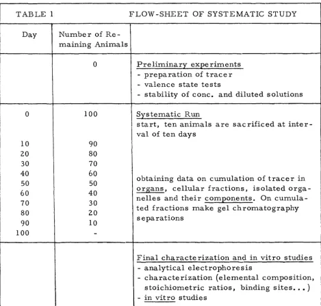
Long-term experiments will be carried out on groups of 100 rats,
sacri-ficed in groups of 10 animals at various intervals of time. A detailed
scheme of the various experimental works which will be carried out for
each metal is given in Table 1. The procedure described is the most
com-plete one. Adaptations will be done as long as the research proceeds to
eliminate the non-essential steps.
Short-term experiments: {1-7 days) the labelled metal will be injected at
acute or subacute levels.
They will be carried out on a small number of animals as preliminary
orientating experiments or to evidentiate differences between chronic and
acute effects.
3. 1 Preparation of the Radiotracers
The systematic study is made possible by the availability of radiotracers,
radiochemical facilities and highly selective measuring techniques, not
usu-ally available to biochemical laboratories and which make it possible to
la-bel in vivo and in vitro very minute amounts of pollutant metal. The
radio-tracers of each element were selected on the basis of:
1) possibility of obtaining very high specific activity,
2) half-life sufficiently long for long-term experiments,
3) easy detection of the emitted radiation.
The preparations are essentially done:
a) by proton irradiations in a cyclotron,
b) by neutron irradiations in a nuclear reactor.
Radioisotopes production in a nuclear reactor by (n, "() reaction and
deve-loped during pr~vious years. An irradiation facility will become avail-able during 197 5 by the installation of a hydraulic irradiation channel
in the ESSOR reactor with a neutron thermal flux of 3. 5 x 1 014 neutrons
-2 -1
em sec • By its use radioisotopes will be prepared both with very
high specific activity and using nuclear reactions other than (n,
o ).
Table 2 shows the nuclear data of the radioisotopes proposed for this
study.
3. 2 Administration of the Radiotracers
The concentrated solutions of the radiotracers obtained in the
preceed-ing step will be stored in a refrigerator. Dilution for long-term
experi-ments will be carried out with well-characterized mineral water.
Exhaus-tive preliminary experiments will be done to make sure that the tracer is
taken by the animal in the desidered chemical form and that uncertainty of
the chemical state, lack of equilibrium of the diluted solution and
absorp-tion effects do not induce misleading experimental artifacts.
3. 3 Sacrifice, Dissection and Counting of Isolated Organs
The animals will be sacrificed by heart puncture after ether anaesthesia.
The following tissues will be separated: brain, heart, kidney, liver, spleen,
muscle, lung, thyroid, testes, bone, blood, stomach, G. I. tract •
. ~he organs will be weighed, washed with buffer solution (a buffer
per-fusion will be done for liver or other organs, if found necessary) and
genized. Homogenization will be done in a refrigerated and shielded
homo-genizer which ensures maximum safety in case of rupture of the organ
con-tainer.
Sacrifice and dissection will be carried out as rapidly as possible. The
organs will be kept refrigerated by ice baths, in order to minimize
bioche-mical reactions inducing migration of metal ions among various
Aliquots of the homogenized organs will be taken for radioactivity
mea-surements while the rest of it will be frozen and stored for further
hand-ling.
3. 4 Separation of Subcellular Fractions by Differential Centrifugation
The separation of subcellular fractions will be carried out on those
or-gans which showed the most significant accumulation of the radiotracers.
Liver and kidney will always be considered. Cellular fractionation will be
carried out in two steps:
1) Nuclei and mitochondria by preparative centrifuge,
2) Lysosomes, microsomes and soluble cytoplasmatic fraction by
prepara-tive ultracentrifuge.
The procedures adopted will be the classical ones, reported in the
lite-rature. Fig. 1 shows the one for rat liver.
Aliquots from the fractions obtained will be taken for radioactivity
eva-luation, the rest will be frozen and stored for further handling.
3. 5 Purification of Subcellular Organelles from Subcellular Fractions
Purification of the subcellular organelles will be carried out in order to
minimize the effects of misleading cross -contaminations. The most recent
procedures reported in the literature will be applied. The flow-sheets of a
few of them (nuclei, mitochondriae, mic rosomes) are shown in Fig. 1.
3.
6
Fractionation of Purified Organelles into their ComponentsThe organelles which will show a significant acGumulation of the tracer
will be submitted to procedures for the isolation of their components. The
most recent procedures reported in the literature will be applied. The
flow-sheets of a few of them (isolation and separation of subnuclear components,
fractionation of mitochondria and Golgi apparatus are shown in Fig. 2.
3. 7 Chromatographic Separations on Organelles Components
The most significant organelles components will be submitted to a
successive fractionation into components of different molecular weight
by gel chromatography. In-line detection systems allow a continuous
mea-surement of radioactivity and U. V. absorption at two different wavelengths
for continuous analysis of metal and protein content. The effluent will be
collected into separate fractions on which tests for specific enzymatic
ac-tivities will eventually be carried out.
The most significant fractions will be pooled desalted by means of
hal-low fibres, lyophilized, and kept for further handling.
The presence of metals other than the ones labelled will be detected and
quantitatively estimated by activation analysis of microsamples of the
lyo-philized product.
3. 8 Analytical and Preparative Electrophoresis
Analytical gel electrophoresis will be carried out on the most significant
protein fractions. U. V. scanning of the gel will allow to isolate
"electro-phoretically pure" proteins. The gel will then be divided into 1-2 mm slices
and the radioactivity profile will be measured. Activation analysis of
indivi-dual gel slices will allow identification and, possibly, quantitative estimate
of metals other than the one labelled.
Preparative electrophoresis will be carried out on the most significant
fractions in order to obtain larger quantities of specific metal-binding
com-ponents for full characterization and successive in vitro studies.
3. 9 Characterization of the Metal-Binding Component and in Vitro Studies
Studies in vitro to characterize the metal- binding component identified
by the systematic study outlined above will also be carried out:
- stoichiometric ratios,
- identification of metal- binding sites,
- saturation levels,
- synergistic and antagonistic effects of other pollutants.
Greatest advantage will be taken of an automatic dialyser being under
con-struction in order to minimize the operators time and to diminish the risk
arising to persons from relatively high radiation levels.
In particular the dialyser will allow the study in vitro of the interaction
of heavy metals with metalloenzymes and nucleic acids, already well known
as potential metal binding sites. It is known that heavy metals such as Hg
and Cd alterate the structure of nucleic acid by interacting with phosphate
groups and/or bases{SZ).
In addition pollutant metals could interfere with the catalytic function by
dis-placing the essential native metal of metalloenzymes{S3).
Because the extent of binding of metals to nucleic acids is extremely low
and since the research on metalloenzymes must necessarily be carried out
on very minute amounts of materials the availability of metals in
radioac-tive form with high specific activity is of very great help in these studies.
3. 10 In Vivo Studies
The following particular studies will be carried out in vivo after
identi-fication of the metal- binding component:
- parameters which affect the accumulation of metals in the body,
- influence of the chemical forms of the metal,
- influence of the metal in the biosynthesis of the metal-binding component,
STATE OF ADVANCEMENT AND PRELIMINARY RESULTS
4. SET UP OF STABULARIUM AND COLD ROOM RADIOCHEMICAL
FACILITIES
Special modular type cages to minimize metal contamination have
been developed. A particular water-supply device was developed in
or-der to minimize radioactive contamination of cages and animals. The
drops of water lost during the drinking process are recovered into a
polyethylene bottle.
Preliminary experiments showed that after administration of 109cd to
rats the radioactive contamination in all the parts of the cage are
insig-nificant. Feces and urines are collected into a polyethylene container
which can be directly counted to obtain data on the excretion of
radio-tracers.
0
A cold room (area at
+
4 C) radiochemical facility has been set upin order to minimize temperature depending artifacts during the
fractio-nation of the purified cellular organelles into their components. The cold
room has been equipped with gel chromatography (flow analyzers for
mo-nitoring UV absorption at 280 and 2 54 nm, fully automatic fraction
collec-tors, recorders, peristaltic pumps and colum~of different size). The cold
area has been designed and especially equipped with high level of
radio-activity as required for some in vitro studies.
Figure 3 shows the stabularium, the various parts of the modular cage
and the cold room radiochemical facility.
5. PREPARATION OF RADIOTRACERS WITH HIGH SPECIFIC ACTNITY
Table 2 gives the nuclear pertinent data of the radioactive isotopes
used for the proposed study. Most of them have been prepared
carrier-free, by proton irradiation at the cyclotron of the Milan University.
The same laboratory has measured the excitation function for each
iso-tope to provide useful information on the possible contaminants from
The knowledge of the excitation function is also essential to determine
whether enriched targets must be used and the degree of enrichment
which is necessary. The separation from interfering radionuclides and
target element, the preparation for their biological use and the analytical
controls have been set up by our laboratory. Figure 4 shows as an exam-48
ple the experimental excitation function for the production of V from
metallic titanium targets.
A complete description of the production methodology will be reported
elsewhere.
6. PRELIMINARY RESULTS ON BIOCHEMICAL EFFECTS OF CADMIUM
Studies on the subcellular distribution of cadmium show that after a
single injection the cellular cadmium was almost entirely present in the
soluble fraction of liver and kidney(l8} bound to a cadmium-binding
pro-tein {Cd-BP}, a low molecular weight propro-tein with a high number of
cyste-ine residues {21 g atoms/mole of protein(54)). For this the incorporation
of 35s-cysteine in the Cd-BP was used to study the biosynthesis of the
protein in Cd-treated rats in respect to normal animals. In addition, we
have investigated in vivo the interaction of heavy metals with Cd-BP:
- by neutron activation analysis of Cd-BP purified by gel chromatography,
- by incorporation of radiotracers in the protein after administration of
labelled metals to Cd-treated rats.
6. 1 Isolation, Purification, Metal c·ontent and "De-Novo" Biosynthesis
Of Rat Ll.ver 109cd and 35S-double L b 11 d C d a e e a m1um-· B" d" 1n 1ng P
ro-tein (Cd-BP)
0 ne group o rats was treate 1. p. w1t f d . . h 1 09c d an d 3 5s -cysteine. T e . h
animals were sacrificed by cervical dislocation after 24
h.
Liver tissueswere homogenized in 0. 25M sucrose. The soluble cytoplasmatic fraction
filtra-tion of soluble fracfiltra-tion was done with 10 mM Tris -HCl buffer, pH=8. 2
as eluant on Sephadex G-7 5 column. UV absorbance of eluate was
moni-tored at 2 54 and 2 80 nrn by an LKB Uvico rd III instrument.
Th e 109cd d. ra Ioact1v1ty 1n t e e uate was measure . . . h 1 db y an autogamma
(Nai(Tl) crystal) while the 35s radioactivity was detected after
separa-tion by ion exchange chromatography of 109 Cd radioactivity.
All ope rations were done in the cold room at 4
°
C.Homogeneity of the Cd-BP was verified by disk electrophoresis, while
neutron activation analysis was used to determine simultaneously various
heavy metals in microsamples of determined protein. The nuclear
tech-nique was also used to the metal content in the Cd-BP from gel
chromato-graphy after further purification by disk electrophoresis. Electrophoretic
separation was performed in duplicate. The first gel was stained for
pro-teins, while the second gel was sliced in small discs, 1 mm thickness,
which were examined for their metal content by neutron activation
analy-sis.
The results are shown in Figure 5:
- a new protein peak (Cd-BP) containing all 109cd radioactivity appears
in UV profiles at 254 nm of Cd-treated rats, absent in untreated
con-trols. No protein peak appears at 280 nm (dotted line in the UV profile)
because Cd-BP does not contain aromatic aminoacids(S4) (Figure SA);
- a much higher incorporation of 35s-cysteine was found in this peak in
respect of controls (Figure SA);
- the presence of the following elements has been demonstrated by
neu-tron activation analysis in the Cd-BP isolated by gel chromatography:
Cd, Zn, Cu, Hg, Ag and traces of Fe, As, Mn, Sb, Sc, Cs and Au.
A typical gamma-ray spectrum of the neutron activated rat liver Cd-BP
is shown in Figure SB;
- Cd, Zn, Hg and Cu of the Cd-BP have similar profiles in the gel after
in terms of molecular weight, seems not to be electrophoretically
homogeneous. This was confirmed by measuring also 35S-cysteine
radioactivity in the gel: the profile after electrophoresis was similar
h f 109cd h . . d" "b . . k
to t at o s owing a maximum Istri ution In two pea s.
6. 2 Identification of Cd-BP in Rat Testicles and Spleen
To establish if Cd-BP could be present in rat testicles and spleen,
one group of 10 rats was treated with 109cd carrier-free. The animals
were sacrificed after 24 hand the soluble fraction isolated and fractioned
6 1 109 d"
as described under section • • The results show that all Cd ra
IOac-tivity was present in fractions corresponding to Cd-BP although the
pro-tein was too low to be detected in the UV profile.
109
However, while in liver {Figure 5A} and testicles (Figure 6A} the Cd
is associated only with Cd-BP of low molecular weight, in the spleen the 109
cd is also present in high molecular weight components (Figure 6B}.
6. 3 Incorporation of Radioactive Metals into Cd-BP
Recently Nordberg observed that Cd-BP could play a more general
role in the metabolism of various heavy metals (l )• For this, to provide
systematic information on the relative incorporation into Cd-BP of rat
liver under in vivo conditions, one group of rats received a single
intra-peritoneal injection of 109cd Cl
2 and radioactive metal, while a second
group,as a check, received only the radioactive metal like the first group 109
but without CdC1
2• After 24 h the Cd-BP was isolated as described in
section 6. 1.
65 197 64 110m 113
From 21 metals tested, only Zn, Hg, Cu, Ag and Sn
were found incorporated into "de novo" biosynthesized Cd-BP.
6. 4. l Effects of Heavy Metals on the Biosynthesis of Cd-BP
In order to prove that the incorporation of cadmium is not affected
b y any ot er meta , one groupo rats receive a Sing e InJection o h l f . d . l . . . f 109 cd
and 35S-labelled cysteine in the presence of other metals, while a second
109 35
group, as a check, received only Cd and S-labelled cysteine without
other metals.
After 24 h the Cd-BP was isolated as described in section 6. 1 and
the 109cd and 35
s
radioactivities measured in the Cd-BP fraction. Theresults show that both incorporation of cadmium and the biosynthesis of
Cd-BP are not influenced by the presence of other metals such as:
2+ 2+
Co 2+ Cu 2+ 2+ 2+ 2+ .2+ 3+ 3+ Cr 3+
Be , Ca ,
'
'
Fe , Hg , Mn , NI , Al , Au , 'G 3+ 3+ 4+ 4+ 4+ + B.3+
Pb
2~
Tl+ 6+ 5+ M 6+a ' La , Ir , Sn , Zr , Ag , I '
'
u
, As , 0 'v
5+ Sb 5+ 4+'
' Se •6. 4. 2 The Biosynthesis of Cd-BP in Short-term Experiments: Normal
Level of Cd-BP in the Rat
Th e simu taneous Incorporation o · 1 · · f 109 C d an d 3 5
s
-cysteine Into·
·
Cd -B Pand its isolation as described under section 6. 1, was used to study the
bio-synthesis of Cd-BP in subacute exposures to cadmium. Some conclusions
seem valid:
- the biosynthesis of Cd-BP is controlled by the cadmium concentration
also in the presence of other heavy metals (section 6. 4. l );
- when rats are intoxicated daily with amounts of cadmium of 0. 8 mg/kg
for 8 days, the biosynthesis is linear with the cadmium concentration
(Figure 8);
- the daily dietary intake for rats is of the order of 5 / g cadmium
sufficient to synthesize the amount of Cd-BP corresponding to the
fraction adsorbed (6o/o from literature data (55)). The Cd-BP should
therefore be normally present in the rat liver. The hypothesis seems
confirmed by the experiments described under section 6. 3 and 7 on
the incorporation of various metals in the Cd-BP. It was found that
a small amount of the different metal ions incorporated in the Cd-BP
65 64 197 113 110m
of Cd-treated rats ( Zn, Cu, Hg, Sn and Ag) is also
pre-sent in normal animals (without Cd-treatment),probably due to minute
amounts of Cd-BP not detectable by UV -measurement.
7. DISTRIBUTION OF HIGH SPECIFIC ACTIVITY RADIOTRACERS IN
THE SUBCELLULAR FRACTIONS FROM RAT LIVER: SYSTEMATIC
STUDY IN THE SOLUBLE FRACTION
A systematic study of the distribution in rats of radiotracers, shows
that liver and kidney are important deposition organs of many heavy
me-tals(56). The knowledge of the subcellular distribution of heavy metals,
which is of great importance to understand the biochemical effects in
rats intoxicated with pollutant elements, is far from being investigated.
We have systematically examined in vivo the distribution of radiotrace rs
in the soluble fractions of rat liver. Animals were injected i. p. with
radioactive metal ions and sacrificed after 24 hours. The soluble
frac-tion of liver homogenate, obtained by centrifugafrac-tion at 105.000 x g for
90 min, was chromatographed on Sephadex G-7 5 column (1 00 x 5 em) and
proteins and radioactivities measured in the collected fractions as
des-c ribed in sedes-ction 6. l. The results are illustrated in Figure 9:
- all radiotrac e rs were always recovered from the supernatant after
Se-phadex G-7 5 chromatography in association with the fraction of high
201 51 7 203 65
molecular weight components, although Tl, C r, Be, Pb, Zn,
197 113
Hg, Sn are also present in the fractions corresponding to low
molecular weight;
109 110m
- 198 Au was also associated with a fraction corresponding to a
molecu-lar weight of 5. 000 -
6.
000;64
Cu is associated with a fraction corresponding to Cd-BP and with
a more specific protein, probably cytocuprein(57) of moleculq.r weight
30.000 - 35.000.
In conclusion, it appears that high molecular weight components of
the soluble fraction of rat liver, are greatly involved in the metabolism
of many heavy metals. The study of the biochemical nature of these
com-ponents will give us informations in view to identify the intracellular
me-tal-binding site(s).
8. PRELIMINARY RESULTS ON BIOCHEMICAL EFFECTS OF LEAD
Di Ferrante and Bordeau ( SS) recently reviewed the distribution of
stable and radioactive lead (210Pb) in different organs of man and
con-cluded that data on the distribution of lead in the different compartments
of the organism are far from being consistent. In addition, very few works
concern the subcellular distribution of lead (37' 38). The purpose of this
study was to investigate the distribution of 203Pb in organs, subcellular
fractions, components of purified subcellular fractions and molecular
components of fractioned subcellular fractions. The study has been
car-ried out by means of 203Pb radioisotope which has favourable nuclear
characteristics for short-term experiments(59). The use of 210Pb in the
study of lead metabolism is limited by the high toxicity of the daughter 210
Po and by the difficulty of measuring accurately the counting rate of
the emitted 0(-radiation.
8 1 . Preparation and Counting o . . f 203Pb
---203
Fig. 10 shows the excitation function for the production of Pb by
203
proton irradiation of thallium target • The purification of Pb was
car-ried out by double coprecipitation with ferric hydroxide and subsequent
was performed by cation exchange resin.
1 f 203 b d" . . d b y
A 1 measurements o P ra 1oact1v1ty were one y fJ -ray
spectros-copy with a Ge(Li) detector at the characteristic line of 279 KeV.
8 2 • Distr1but1on o . . f 2 O 3 Pb . 1n Rat T. 1s sues
18
fg
of stable lead nitrate/rat plus 700fCi
of 203Pb carrier-free/rat were injected i. v. to eight animals. The rats were sacrificed after
24 h. by heart puncture after ether anaesthesia. Blood was collected and
the organs removed and homogenized. Aliquots of homogenates were
di-rectly counted for 203Pb radioactivity. The results are reported in Fig. 11.
203
- Subcellular distribution of Pb in rat liver and kidney: gel
chromato-graphy of soluble fractions from liver, kidney and spleen
The homogenates of liver and kidney were fractioned by differential
. f . Th 2 O 3 Pb d. . . d . 1 .
centr1 ugat1on. e ra 1oact1v1ty was measure 1n nuc ear,
m1to-chondrial, lysosomal, microsomal and soluble fractions. Further
puri-fication of nuclei and mitochondria has been carried out (see
procedu-res in Fig. 1 ). The procedu-results are shown in Fig. 11 and 12. The soluble
fractions from kidney, liver and spleen were chromatographed on
Sepha-dex G-75 column as reported for the isolation of Cd-BP (see section 6. 1).
203
The UV adsorbance and Pb radioactivity profiles from gel
ch.romato-graphy are shown in Fig. 13.
· "b · f 203Pb . h f 1 . d . h d .
- D1str1 ut1on o 1n t e components o nuc e1 an m1toc on r1a
from· liver and kidney
Purified liver and kidney nuclei and mitochondria were respectively
fractioned in membrane and "bulk chromatine" (:q.uclei) and soluble,
in-terface, light and heavy fractions (rnitochondriae) (see procedures in
Fig. 2). The 203Pb radioactivity was measured in these fractions {Fig.
- Chromatographic separation of liver and i:idney nuclear chromatine
and soluble mitochondrial fractions
The nuclear "bulk chromatine" and the mitochondrial soluble
frac-tion from liver and kidney were chromatographed on Sephadex G-7 5
co-lumn and the UV absorbance and 203Pb radioactivity profiles were
mea-sured. The results are shown in Fig. 11 and 12.
The following conclusions can be drawn from the experiments which
were described above:
- Rat kidney and liver are the organs in which most of 203Pb can be
re-covered after 24 h. from a single i. v. injection.
- The subcellular distribution of 203Pb shows that most of the radiolead
was contained in the nuclear and soluble fractions of kidney and liver.
Mitochondria of kidneys bind much more lead than those of liver.
- Gel chromatography of soluble fractions from liver, kidney and spleen
show that 203Pb are associated almost with fractions corresponding to
high molecular components.
-About 65-70% of 203Pb in nuclei is associated with membrane, while
about 30% is present in the chromatine. In this latter case radiolead
is present in one protein peak.
- Different distribution of 203Pb in the mitochondria components of
kid-ney and liver were obtained. However, gel chromatography of mitochon-203
drial soluble fractions shows association of Pb radioactivity with
fractions corresponding to high molecular weights.
ACKNOWLEDGEMENTS
We wish to thank Mr. Amantini, Luciano and Mr. Ubertalli, Livia
REFERENCES
{1) NORDBERG, G. F.; Environ. Physiol. Biochem., ~' 7, 1972
{2) GHAFGHAZI, T., MENNAER, J. H.; Toxicol. Appl.
Pharma-cal., 26, 231, 1973
{3) KAGI, J.H.R., VALLEE, B.L.; J. Bioi. Chern., 235, 3460,
1960
{4) NORDBERG, G. F., NORDBERG, M., PISCATOR, M.,
VESTER-BERG, 0-. ; Bioohem. J. , 126, 491, 1972
{5) NOMOTO, S., Me. NEELY, M.D., SUNDERMAN, F. W.;
Bioche-mistry,
!Q,
1647, 1971(6)
MING-DEAN LUH, BAKER, R. A. , HENLEY, D. E.; The Scienceof the Total Environment,~~ 1, 1973
{7) HULTIN, T., NASLUND, P. H.; Chern. Biol. Interactions, ~' 315,
1974
{8) ERICSSON, Y.; Acta Odont. Scand., 24, 40 5, 1966
{9)
AGNEW, W. F., TA CHENG, J.; Toxicol. Appl. Pharmacal., 20,346, 1971
{10) HURSH, J. B., BROWN, C.; Proc. Soc. Exp. Biol. Med., 131,
116, 1969
{11) HILES, R.A.; Toxicol. Appl. Pharmacal., 27, 366, 1974
{12) FOSSET, M., CHAPPELET-TORDO, D., LAZDUNSKI, M.;
Bio-chemistry,
g,
1783, 1974{13) SANG-HWAN Oh, GANTER, H. E., HOEKSTRA, W. G.;
Bioche-mistry,
g,
182 5, 197 4{14) WOOD, J. M.; Science, 183, 1049, 1974
{15) FISHBEIN, L., FLAMM, W. G.; The Science of Total Environm.,
.!_,
117' 1972{16) SCHROEDER, H. A., NASON, A. P., TIPTON, I. H., BALASSA,
J. J. ; J. C h ron. Dis. , 2 0, 1 7
9,
19 6
7{17) SCHROEDER, H. A., BALASSA, J. J.; J. Chron. Dis., 14, 236,
1961
{18) SHAIKH, Z. A., LUCIS, 0. J.; Arch. Ep.viron. Health, 24, 419,
1972
{19) SQUIBB, K. S., COUSINS, R. J.; Environ. Phys.iol. Biochem., i_,
24, 1974
{20) NORDBERG, G. F., PISCATOR, M., LIND, B.; Acta Pharmacal.,
{21} PISCATOR, M.; Nord. Hyg., 45, 76, 1964
{22} NORDBERG, G. F.; Environ. Physio1. Biochem., ~' 7, 1972
{23} PARIZEK, J., OSTADALOVA, I., KALOUSKOVA, J., BABICKY,
A. , PAULIK, L., BIRR, B.; J. Reprod. Fert. , 2 5, 157, 1971
{24} RODERICH, W., SCHWARTZ, I. L., ROY, J.; Ann. New York
Ac. Sciences, 192, 175, 1972
{2 5} MARTIN, J. L., GERLACH, M. L.; Ann. New York Ac. Sciences,
192, 181, 1972
{26} GANTHER, H.E.; Biochemistry,~' 1089, 1966
{27} NORSETH, T., BRENDEFORT, M.; Bioch. Pharmacal., 20,
1101, 1971
{28} BALTROP, D., SMITH, A.M.; Experientia, 29, 1178, 1973
{29} KATZ, S.; J. Aer. Chern. Soc., 74, 2238, 1952
{30} MATSUDA, M., TAKEUCHI, E.; The J. of Bioch.,
g,
523, 1967{31} MATHUR, R.K., DOISY, R.J.; Proc. Soc. Exp. Biol. Med., 139,
836, 1972
{32} SAMIT Z, M. H.; Acta Derm. - Venereo1., 49, 142, 1969
{33} HOPKINS, L. L., SCHWARZ, K.; Bioc. Bioph. Acta, 90, 484,
1964
{34} WACKER, W.E.C.; VALLEE, B.L.; Fed. Proc., ~' 345, 1959
{35} CASTELLINO, N., ALOJ, S.; Brit. J. Industr. Med., ~' 308,
1964
{ 36) DE BRUIN, A. ; Arch. Environm. Health, 2 3, 249, 1971
{37) CASTELLINO, N., ALOJ, S.; Brit. J. Industr. Med., 26, 139,
1969
{38) BALTROP, D., BARRETT, A. J., DINGLE, J. T.; J. Lab. Clin.
Med., 77, 705, 1971
{39) BALTROP, D., SMITH, A.; Experientia, 28, 76, 1971
{40) VACHER, J., STONER, H.B.; Bioch. Pharmacal.,
.!1..,
93, 1968{41) WITSCHI, H. P.; Biochem. J., 120, 623, 1970
{42) THOMAS, M., ALDRIGE, W.N.; Biochem. J., 98, 94, 1966
{43) REEVES, A. L., DEITCH, D., VORWALD, A. J.; Cancer Res.,
27, 439, 1967
(44) SHAKMAN, R. A.; Arch. Environm. Health, 28, 105, 1974
(46) MARTIN, G. M., BENDITT, E. P., ERIKSEN, N.; Fed. Proc.,
~' 482, 1959
(47) SABBIONI, E., GIRARDI, F., GIULIANI, F.; Report EUR 5060,
1972
(48) JOHNSON, J. L. et al.; Bioc. Biophys. Acta, 56, 4, 1974
(49) WHANGER, P.D.; Tox. Appl. Pharmacal., 25, 323, 1973
(50) SUNDERMAN, F. W., SELIN, C. E.; Tox. Appl. Pharmacal.,
g,
207' 1968(51) SUNDERMAN, F. W., ESFAHANI, M.A., BEACH, D. J., ELSER,
R.C.; Proc. Am. Assoc. Cancer Res., _2, 69, 1968
(52) EICHHORN, G. L. , AESHIN, Y. ; J. Am. Chern. Soc. , 90, 7 32 3,
1968
(53) VALLEE, B. L., WACKER, W. E. C.; "The Protein", ed. H.
Neu-rath, S. N. Y. Academic, 2nd ed. , 1 97 0
(54) KAGI, J.H.R., VALLEE, B.L.; J .. Bioi. Chern., 236,2435,1961
(55) FRIBERG, L .. , PISCATOR, M., NORDBERG, G. F.; Cadmium in
the Environment, CRC Press Div., Chern. Rubber Comp., Cleveland, 1971
(56) DURBIN, P.; Health Physics,~' 225, 1960
(57) MARCEAU, N., ASPIN, N.; Bioch. Biophys. Acta, 293, 338,
1973
(58) DI FERRANTE,
E..,
BORDEAUX, P.; Report EUR 5004, 1973(59) GIRARDI, F., GOETZ, L., SABBIONI, E., MARAFANTE, E.,
MERLIN!, M., ACERB!, E., BARATTARI, C., CASTIGLIONI,
TABLE 1 FLOW -SHEET OF SYSTEMATIC STUDY
Day Number of
Re-maining Animals
0
10 20 30 40 50
60
70 80
90
100
0
100
90
80 70
60
50 40 30 20 10
Preliminary experiments - preparation of tracer - valence state tests
- stability of cone. and diluted solutions
Systematic Run
start, ten animals are sacrificed at inter-val of ten days
obtaining data on cumulation of tracer in organs, cellular fractions, isolated orga-nelles and their components. On cumula-ted fractions make gel chromatography separations
Final characterization and in vitro studies - analytical electrophoresis
[image:30.584.49.518.92.540.2]TABLE 2 Pollutant and type of expe-Isotope T 1/2 riment 115mCd 43 d. Cd 109Cd 450 d. Zn 65 Long-term Zn 245 d. experiments Cr 51 Cr 2 7. 8 d. Hg 203Hg 46. 5 d. Se 75Se 127 d. Pb 203Pb 2. 17 d. Be 7 Be 53. 6 d. Short-term v 48v 16. 2 d. experiments 49v 330 d. Ni 63Ni 80 y. RADIOISOTOPES INVOLVED IN THE SYSTEMATIC STUDY Principal Radiochemical separation and Source
emission (MeV)
( o.2s
M
tuose ,
Tris·HC:I
rH
=72)
Filtrdtion
by
cheese
cloth
roo ~ ~ Resiat~~
..,__ _ _ _ _ _
,. CTude.
Nu~LfiIO mirt
.Su
per"ata"t
9oooK o Residue . .
t---'=d----.Gr..,de MtrocHoNDR•A
10 mit\
Supernata"t
~--~o_.ooo_•...__R_rs_id_ut~Crvdt
LYSOSOHES1"'"'i"
s
ure.rnatar\t
Supnnattnt
._ _ _ _ _ _ _
_:'
So\o~\t c:rroPLIISHAli~fYaction
PUR.IFiCATiON
OFRAT
LiVfR
MiTOCHOND~iA
(G.L.Soltocas~ ,J: "rCell B.ol. ( 13~1). 3Z.415)
Resuspenq
··erode
HiTocHoiiDRiA.. • ...0-2.5'1t suc:rose
24ooo
a'
So~un~t~
..
t
10 min
~'Ott
• 'Su~errutaMt
t - - - •
1).-s,~rf/.2S
""i"
(s~; ... ~'"'' rotor)
Re~ialoe
:PuRifiED HiTocHotiJ>RiAo.4s-M
Suero.se.
o:Oftt
KCI,o.o~ Tr•.s·~CI
pH=
1-.Z
~
Atit/
7
volu~tte.sof
''Je,se
sucrose
so/vt.onu
(2.011 Sucrose ,o.011f KCI. O.Oi Tris-HCJ ~H:7-2)
~
Layer
over
dlar9e
vo/u.,e
of
''o/eHtt. sucro~e-'solut,·on,,
220()()
~ ~ Su~erna~.,t
J1 - - - -.. ..])i.SCarq,
ro
mi,Resusbend ··"
0.311 Sucrose
0.0111
h'CI
a.o~V'r.'If!Ct tJ/!}2
I~ I I I
Adjust
soc.
rose
con
c
e" tra
t;on
to
/.8M,,.&J,
i
•dense
meJ,·c~rn.L.ayer
o~er a~
e'ua/ volutt1f
•f
"~ett~e
.ro/ufi·an.
6oooo •
oSuperna
ta~ 1:.~--=c.J:.-..,_ _ _ _ , . . . IJ i gc
art:L
!Jo
ntinRe.~'due
:
Pu~i
flED NuCLEi.PVRi FiCATiDN
oFRA.T
LiVER
MiCROSOHES
( J: J. M. Ber~Ton. 'J.or
Cell Bioi. (l~l3), 5~. 1~)Resuspewu!
''MicRosoMiALfractiott, ,·.,
I
FRACTioNATION
oFRAT
LI'IER
MiTocHONDRiA
I
Su
1:.rend.
f>Urified.
mitochondria
;.
O.'ls
M
sucrose
sri
ficabon
(
GL. s.t.toCa!>a., T·f
C-ell
f>iol. (l'b~). 3Z,415)Layer
over
a
solution
of
I.IBM
.Sucrose
24.000 ., IBo tt1in($wi~3"'3
.,...__1--=-'~h_t
__
.,.
YO~r )Mitochot\dTid
with
o
h\ore
o'fless
l.t'eJI~re.ser'r'~J.
;.,"er siNcTure butw•thout
t~e ovt~rl.,.e~brdne.r ..
neY ~~ttt brat1e ,system dnd.part
o£
t_},e ~dtri
X cor~td'n' r~~the
bulK
or
tesr"a
lory
~lo\ain eMzym~.s.,Jescrculated.
qerivab~es
of
the
outer
~etttbtane
£t1Z(IIft~:J
thiS
frac.bot1
are 't\otr~~
the
outerntel'MbYa.
.'1aterial
present
be.t4tten
the
~o
tttitDchondr,·al
-...ettebranes.
~rtof
the
...
atriX
MateYial
teleasecL
fro.,
both
eJ,e
iM"etana
oUTer
~ttetttbrantsFRACTIONATION
OFRA1
Ll'JER
GOLGI
APPARATUS
I
FRACTiONATiON
OfNUCLEAR
COMPDNENTS
I
Purifieq
11u~lei(w.w.
frdnKe · f',..ptf.CtJI. ReJ·(1,13),81,3~s-).Susre~d
;,.
o.iM
!>tlcro.se
a11cLTri!>-UCI
. f'H
=
~4
Sonificatiot1
·
AdJ.
•
~13h
salt
exir
acton
me~iu111,,
(
0.3MStJcrose,
USI1
KCI,
0.01ttl;;·s-HC
Stirrerfor
~-L,hour.s
dt
4•c
110.000 .1( ~ I 120.,,rt
t
...---~....
Supernata"
Relidue
:
resuspen~; ..
0.it1
Socro,e,1
o.ol"
I<~'
,o.oi"
Tr•l-H~I
rH·tz
Layer
o'/era
\ine~t .suc.ro.se. sra~iet1tI
(,o.ooo •' 1 ISO"'i"
.t.Juc.lear
Me•~rantba"cleJ
i"rt>3io"
be+-jee~ '·18-1.2.1~t!""-lRe
...
otJeby
a ca .. "ola an~oli
/uite
to o.zM sucrose luo.ooo"3.12.0minr--;::----,.
___
"e.:
NUCLEAR
ltEH.BRAflE
l
I
.Suptrftat~.,t
;
chro~at·"
rrof:eit1S
't>iilysis
a,
aiMst
SM urea.o.oitf
-fi,i
H
t'l
rH=8.3 ( 8\J LK N\J~LE ~ R ) C:HR011Aii N 190.000 ~ ~'2.4
h.
I
Ion
ex
~ht-.~e
c~vomat.o~ta~hy
~AE Ser~~r!exA-S'o
0,,,.
o.25!\,---Re~idue
C:f3
"''"'bra"e.
Res•
due C~ rrwrnbrclt~tSFt
~o~-HisTorlE ~,Nae!l
51\
urea
PROTEiNS ,..._ ___ , ___ ---JRe~idve
CFtmrmbratte
o.otf1
Tris·HCI
pH=83
I
Hi!.TOCold
YOOIII
radiothemical
fatdity
Modular
ca~e
Stabularium
a,ts
ef
mo~u
Jar
c:ase
Yield
10
2
preparation
of
4
Bv
from
Ti
target:
excitation
function
47
~----Sc
t
112
---'
8
v
t
112
'
6
sc
t
112
10
1
~--~--~--~--~--~--~--~--~----~
5
10
15
20
25
30
35
40
45
Mev
Proton
l.P.
INlEC.TiOt-1
Of
109
HOMOGENATE
ol= lt~Z:R. Ar-ID<!EtiTR\Ft.J&ATiON
!Jo
tnht,1osooo
~A
I
GEL
cHI\OMRTOgp.ftPHY
o,.
SEPIIADEX
G-15
1 u.v.
a"d
OF
SOLUBLE
CYTopLASHATiC
FRf\CT\otJ
Rf\DaoJ1Cli'liTi£.S
1 .F'RE
•z
J>Rf
6
tPl'ofiLES
DESALT&N~AriD
E
e
-atJ
or
CA-DHhlt"-BitJDi~4PRoTEiN
(Cd·BP:
~t4E"uTRotl
A~T,'/AT'o
PoLYAcAiLI\Ifi.D£
GEL
ANALYSiS
ofCd-BP
cLECTROP
E
<!,
cpm 10
1 10o:
.... ,
-...
e--uT~P\-O~N--::A~C-r~i V:-:-tiT-i~\) """':"'tJ -:-J17:"NA-:-:L-:-V5-1 1S:
oF
SiN6LE
ELECTRoPHoRE,-iC
8AtJ.D.S tszn+"'sc1~L---L---~~---~~---~ o
500 tOOO 1500 E 't (KeY) 2000
Ccl-evposecl
control
uv
10
~
1\
IU~Cd
1\
I
()
(\
"'
tO
\
IJi~v
~
.
I .
.
0 •• ,. -: \••.
';,/-,-.,
.. 0 . ~----·"'E
--L.£0
u20
0500 1500 2SOO SOO 1SOO
25"00
B
El"f.on
vo"-e
(ml)
....
a
''
.~8
A 0 1---__..;=-....----~""
:::
ll.
~8
~4
l:
ol
~'"""=
·s:
24
.t;
g.
~
.l
Sooo
~
0
u.v.
iOO
2,oo
300
FRACTioN
NtJMBER
E
s:50
~ lO ~
~
~ C)100
""'
E
5ooo
.rt-. \.1 ..._,
~
25'oo
• 0'\ 0
~
0
B:
Spleen
So
100
150
FRACTioN
E
&:. ~ Ln c:--1r-
~
rn
'
Q
J(
f
.c._
u
Cd-BP
•
~
ln
50
C'J~.1
"'
JI-I
'4Cu
_
_
l'oo
~
10
5
~ ~'5!r.
;,c:: ..c::=-=. I ... ~...-;:::;....~~0
~
jUo~nA&
a
4
'o
,(2.
2
L
0
0
0
a
1'
9
!
Hs
...
"'
•
0
-
,
E
L.
u
'-'--d
'-.)en
0-2oo
i.SO
too
6
8
54Mn
sa
co
sszn
132
Te
192lr
197Hg
2o1rt
I I 1
500 1500 2500 500
1500 2500 500
1500 2500
elution volume (ml)
Fi~ure
9. Distribution
of
rad;oifac~rs
in
sohJble
prac.-holll
201
Pb
t112
9.4
200
Pb
tt/2
21.5
~
203
Pb
t
1/2
52.1
202
n
t
112
12.od
10-1
__j______---.---r---.--~----,---r----
5
10
20
30
40
50
60
Proton
energy
(Mev)
Fi~ure
i_O.
Preparation
of
203Pb
fv-om
thallium
tar3et:
y;e/c/
2.01
Pb,
2oopb,
2.03Pb
at~d
202lt
Ns.
pYoEoVl
.c
a.
M 0 N
rt)
•o
-; 5
E
,a_
50
0
II
II c:
c:: .r..J
IU tV
~
E
E
oL.
II .C.
E
CJ_
...
203Pb
'""oL---..JC..te.~----..----r----r---..---t
so
100 15'0 200 25"oFRAc.TiON rJUM BER
20
0
~lfRoH~To~RtlPfiY oF SOLIJ6l.E
fRA~Tioi'J oF HiTOCHotJJ>RIA
u.v.
2.03
Pb
FRACTioN~ Ti ON o~
N tJt.LEl
..c
50
0...
M
0
N 0
Cb c
,
'-.a
E
GJE
Cb c:....,
,
E
0....
.c
(.)~
.
w
+I
.a
a.
("') 0 NBULK CHROMAT•NE ..
FRACTiONATION
50
100 150 200FRACTION NUMBER
,
·c
"0c:
0
" .c.
- I {J
(J 0
::J ~
c:
E
CJ) ~
CJ) 4J c
GJ
E
ro
E o
70
0 Vl c
(I) 0 ....
0 L.. OJ
CJ) u
a.
~ · - ::J
_.~
E
UlFRACTiONATiON
OFM iTO
c.H
ONJ>RiA~40-.0 a_
M
20
0 N
0
0 ---+---~
U'/
uv
f s:.... 50
w
c--1 1-~ '20~ rl) 'o 'II( E A-\J 0CHROM~TO~KHPHY OF
SOLUBLE" FRACTION oF
M iioc.HON DRl~
2o~
Pb
so
100FRACTioN
fi~ure
12.
Subcellu)a'(
d.5tribution
or
2o"3P6
lYlrat Kid.,eys
drld~istribu.t{on
oy
ra.dioleaq
inl'luc.lea.'(
a.n~ ~;rocho~dn'al co~fv~~Vlt5.
(For-
detaJI.s
Q) 0 s:: ttl ,.0 ~ 0 0) ,.0 <: ~ I 0 ,....
><
s Pt
0
s
~1 .o
0.5
0
100
50
0
~
g
0.5(\J (\J
I I
I
0.25
2o~Pb
KIDI!Y
uv
Q) 0 s:: al ,.0 ~ 0 (1) ,.0
o I -<!' -... - --~ .,. ... _______ ~ I
<
100
~
I ~ 50
><
2o~pb
s.
o o I V/((//cZ?f:i ,ztz; I
a
q-1.(\ C\1 ... :. ... 8 s Pt 50 100 3000 20000 1000
SPlEEN
uv
'lo~pb
0 . . Y/(//<(Ci7z 70' '/;. ?:= I
50 100 150 200 250
FRACTION r-;mtBEH