AGGREGATE ON HUMAN PULP TISSUE
- AN IN VIVO STUDYA dissertation submitted
in partial fulfillment of the requirements
for the degree of
MASTER OF DENTAL SURGERY
BRANCH – IV
CONSERVATIVE DENTISTRY AND ENDODONTICS
THE TAMILNADU DR. MGR MEDICAL UNIVERSITY CHENNAI – 600 032
I hereby declare that this dissertation titled “
COMPARATIVEHISTOLOGICAL EVALUATION OF DIRECT PULP CAPPING WITH
BIODENTINE AND MINERAL TRIOXIDE AGGREGATE ON HUMAN PULP
TISSUE- AN IN VIVO STUDY
” is a bonafide and genuine research work
carried out by me under the guidance of
Dr.M.KAVITHA, Professor &
HOD,
Department Of Conservative Dentistry and Endodontics, TamilNadu
Government Dental College and Hospital, Chennai- 600 003.
DR.S.HEMA JANE CHRISTA
This is to certify that
Dr. HEMA JANE CHRISTA
, Post Graduate
student (2012-2015) in the Department of Conservative Dentistry and
Endodontics, Tamil Nadu Government Dental College and Hospital,
Chennai- 600003 has done this dissertation titled “
COMPARATIVEHISTOLOGICAL EVALUATION OF DIRECT PULP CAPPING WITH
BIODENTINE AND MINERAL TRIOXIDE AGGREGATE ON HUMAN PULP
TISSUE- AN IN VIVO STUDY
” under my direct guidance and supervision in
partial fulfillment of the regulations laid down by the TamilNadu Dr.M.G.R
Medical University Chennai -600032, for M.D.S., Conservative Dentistry
and Endodontics (Branch IV) Degree Examination .
DR.M. KAVITHA, M.D.S.
Professor & HOD
Guide
Department of Conservative Dentistry and Endodontics.
TamilNadu Government Dental College and Hospital
This is to certify that the dissertation titled
“COMPARATIVEHISTOLOGICAL EVALUATION OF DIRECT PULP CAPPING WITH
BIODENTINE AND MINERAL TRIOXIDE AGGREGATE ON HUMAN
PULP TISSUE- AN IN VIVO STUDY
’’
is a bonafide research work done by
Dr. S.HEMA JANE CHRISTA
, Post Graduate student (2012-2015) in the
Department of Conservative Dentistry & Endodontics under the guidance of
Dr. M.KAVITHA, M.D.S, HOD and Guide,
Department Of Conservative
Dentistry & Endodontics, TamilNadu Government Dental College and
Hospital, Chennai-600 003.
Dr. M. KAVITHA, M.D.S., Dr.S.PREMKUMAR, M.D.S., Professor & HOD, Principal i/c
Dept of Conservative Dentistry & Endodontics
ACKNOWLEDGEMENT
I wish to place on record my deep sense of gratitude to my guide
DR. M. KAVITHA, M.D.S.,Professor & HOD, Department of Conservative Dentistry and Endodontics, Tamil Nadu Govt. Dental College and Hospital for her keen interest,
inspiration, immense help and expert guidance throughout the course of this study.
I take immense pleasure to extend my heart felt gratitude and sincere thanks to
DR. B. RAMAPRABHA M.D.S.,Professor, Department of Conservative Dentistry and Endodontics, Tamil Nadu Govt. Dental College and Hospital for her timely guidance,
motivation, suggestions and inspiration.
I take this opportunity to convey my sincere thanks and gratitude to
DR. S. PREMKUMAR MDS., Principal i/c, Tamil Nadu Govt. Dental College for permitting me to utilize the available facilities in this institution.
I sincerely thank Dr. K.AMUDHALAKSHMI M.D.S., and Dr. ARUNA RAJ M.D.S.,
Associate professors, for all the support and encouragement throughout this study.
My extended thanks to Dr. G. VINODH M.D.S., Dr. A. NANDHINI M.D.S., Dr. P. SHAKUNTHALA M.D.S., Dr. M. S. SHARMILA M.D.S., Dr.
Department of Veterinary Pathology,Madras Veterinary College, Chennai – 07, for his
guidance,suggestions and allowing me to use the infrastucture facilities in his department.
I also thank my dear co-PGs, seniors and juniors for their timely help and friendship.
TITLE OF DISSERTATION
“
COMPARATIVE HISTOLOGICAL EVALUATION OF DIRECT PULP CAPPING WITH BIODENTINEAND MINERAL TRIOXIDE AGGREGATE ON
HUMAN PULP TISSUE- AN IN VIVO STUDY’’ PLACE OF STUDY Tamil Nadu Government Dental College & Hospital,
Chennai – 03.
DURATION OF THE
COURSE 3 Years
NAME OF THE GUIDE Dr. M. Kavitha
HEAD OF THE DEPARTMENT
Dr. M. Kavitha
I hereby declare that no part of the dissertation will be utilized for gaining financial assistance/ any promotion without obtaining prior permission of the Principal, Tamil Nadu Government Dental College & Hospital, Chennai-600003. In addition, I declare that no part of this work will be published either in print or in electronic media without the guide who has been actively involved in dissertation. The author has the right to reserve for publish of work solely with the prior permission of the Principal, TamilNadu Government Dental College & Hospital, Chennai-600 003.
This agreement herein after the “Agreement” is entered into on this day Dec 2014 between the TamilNadu Government Dental College and Hospital represented by its
Principal having address at TamilNadu Government Dental College and Hospital, Chennai - 600 003, (hereafter referred to as, „the college„)
And
Mrs. Dr. M.KAVITHA, aged 44 years working as Professor and HOD in Department of Conservative Dentistry & Endodontics at the college, having residence address at 69/4,Mettu street, Ayanavaram, chennai-23(herein after referred to as the Principal Investigator„)
And
Mrs. Dr.S.HEMA JANE CHRISTA aged 33 years currently studying as Post Graduate student in Department of Conservtive Dentistry & Endodontics, TamilNadu Government Dental College and Hospital, Chennai 3 (herein after referred to as the PG student and coinvestigator„).
Whereas the PG student as part of her curriculum undertakes to research on
“COMPARATIVE HISTOLOGICAL EVALUATION OF DIRECT PULP CAPPING WITH BIODENTINE AND MINERAL TRIOXIDE AGGREGATE ON HUMAN PULP TISSUE- AN IN VIVO STUDY” for which purpose the Principal Investigator shall act as principal investigator and the college shall provide the requisite infrastructure based on availability and also provide facility to the PG student as to the extent possible as a Co-investigator.
Whereas the parties, by this agreement have mutually agreed to the various issues including in particular the copyright and confidentiality issues that arise in this regard.
Now this agreement witnesseth as follows
1. The parties agree that all the Research material and ownership therein shall become the vested right of the college, including in particular all the copyright in the literature including the study, research and all other related papers.
2. To the extent that the college has legal right to do so, shall grant to license or assign the copyright so vested with it for medical and/or commercial usage of interested persons/entities subject to a reasonable terms/conditions including royalty as deemed by the college.
whatsoever and for any purpose without the express written consent of the college.
6. All expenses pertaining to the research shall be decided upon by the Principal Investigator/ Coinvestigator or borne solely by the PG student. (co-investigator)
7. The college shall provide all infrastructure and access facilities within and in other institutes to the extent possible. This includes patient interactions, introductory letters, recommendation letters and such other acts required in this regard.
8. The Principal Investigator shall suitably guide the Student Research right from selection of the Research Topic and Area till its completion. However the selection and conduct of research, topic an area of research by the student researcher under guidance from the Principal Investigator shall be subject to the prior approval, recommendations and comments of the Ethical Committee of the College constituted for this purpose. 9. It is agreed that as regards other aspects not covered under this agreement, butwhich pertain to the research undertaken by the PG student, under guidance from thePrincipal Investigator, the decision of the college shall be binding and final.
10. If any dispute arises as to the matters related or connected to this agreement herein, it shall be referred to arbitration in accordance with the provisions of the Arbitration and Conciliation Act 1996.
In witness where of the parties herein above mentioned have on this day, month and year herein above mentioned set their hands to this agreement in the presence of the following two witnesses.
College represented by its Principal PG Student
Witnesses Guide
1.
S.No. TITLE PAGENo.
1 INTRODUCTION 1
2 AIM AND OBJECTIVES 5
3 REVIEW OF LITERATURE 6
4 MATERIALS AND METHODS 26
5 RESULTS 37
6 DISCUSSION 49
7 SUMMARY 62
8 CONCLUSION 64
TABLE NO.
TITLE PAGE
NO.
1 Experimental Groups 30
2 Scoring criteria for histopathological evaluation 34
3 Histopathological scores for seven days observation 38
4 Histopathological scores for one month observation 39
5 Histopathological scores for 3 months observation 40
6 Chi-square test for inflammation for all the 3 intervals
42
7 Chi-square test for dentin bridge for all the 3 intervals
43
8 Chi-square test for thickness of dentin bridge for all the 3 intervals
SL. NO. GRAPH PAGE NO.
1 Bar Diagram Showing Comparison of Inflammation in MTA Vs BD Groups at Time Intervals of 7 Days, One Month, 3 Months
45
2 Bar Diagram Showing Comparison of Dentin Bridge Formation in MTA Vs BD Groups at Time intervals of 7 Days, One Month, 3 Months
45
3 Bar Diagram Showing Comparison of Thickness of Dentin Bridge in MTA Vs BD Groups at Time Intervals of 7 Days, One Month, 3 Months
46
4 Bar Diagram Showing Comparison of all the 3 Parameters at Time Intervals of 7 Days, One Month, 3 Months
MTA Mineral Trioxide Aggregate
BD Biodentine
CH Calcium Hydroxide
CSMs Calciumsilicate–Based Materials
ALP Alkaline Phosphatase
hDPCs Human Dental Pulp Cells
OFMSCs Orofacial Bone Mesenchymal Stem Cells
OCN Osteocalcin
DSPP Dentin Sialophosprotein
DMP1 Dentin Matrix Protein 1
BSP Bone Sialo-Protein
BMP-2 Bone Morphogenic Protein-2
CEM Calcium Enriched Mixture
MIP-2 Macrophage Inflammatory Protein
HO-1 Heme Oxygenase-1
LB4 . Leukotrine B4
IL-1 β Interleukin-1 Beta
EMD Emdogain
AIM:
The aim of the study is to compare the pulpal response of human premolars to direct pulp
capping with MTA and BIODENTINE by light microscopic histological evaluation.
MATERIALS AND METHODS:
Direct pulp capping procedure was carried out on human premolars scheduled for orthodontic
extractions under local anaesthesia. Class I cavities were prepared and MTA or BIODENTINE
was placed over the exposed pulp,followed by composite restoration.A total of 42 teeth were
subjected to direct pulp capping. After the experimental periods of 7, 30, and 90 days the teeth
were extracted and histological processing was carried out. The test materials were evaluated
histologically for the degree of inflammation, dentin bridge formation and thickness of dentin
bridge at all the three different observation periods.
RESULTS:
At 7 days observation all the samples in group I and II showed mild inflammatory response. At
30 days thin or partial dentin bridge was evident in all the samples in both groups. At 90 days all
the samples in both groups showed evidence of complete dentin bridge formation. There was no
statistically significant difference between the two groups in all the three criteria.
CONCLUSION:
Biodentine was effective in inducing a pulpal reaction with minimal inflammation and dentin
bridge formation, comparable to MTA .
1
Introduction:
Dental pulp is a highly vascularized tissue of the tooth and has immense healing potential. It performs many functions throughout the life of a tooth, so every effort should be made to maintain its integrity and vitality. The aim of conservative pulp therapy is to maintain the coronal and radicular pulp tissue in a viable condition.
Direct pulp capping is the treatment of an exposed vital pulp by sealing the pulpal wound
with a dental material placed directly on a mechanical or traumatic exposure to facilitate
the formation of reparative dentin and maintenance of the vital pulp26.
Direct pulp capping is indicated whenever the remaining pulp exhibits reversible pulpitis
and can be selectively induced to produce a reparative barrier that protects the tissue from
microbial challenges.
If successful, this procedure eliminates the need for more invasive, more extensive and
more expensive treatment .The intention is to postpone more aggressive therapies that
could eventually lower the long term prognosis for tooth retention and function. Since the
vital pulp is capable of demonstrating competent immune defense mechanisms, it is
desirable to preserve the vitality of an exposed pulp.
A number of factors have been shown to have an impact on the success of direct pulp
2
dentin formation, not affect pulpal tissue vitality in the healing process, not unleash any
adverse effects when used and have good sealing ability.69
A wide array of materials have been used for pulp capping. One such time tested material
is calcium hydroxide. It acts as a stimulating agent for pulpal tissue repair because of its
ability to induce hard tissue formation. Calcium hydroxide’s ability to stimulate
reparative dentine formation with high rates (more than 80% of the treated teeth) of
pulpal survival after capping treatment have been reported in a long term follow up
study33.
But subsequent to pulp capping with this conventional alkaline agent,the adjacent pulp
tissue to calcium hydroxide is usually completely deranged and distorted, forming a zone
of obliteration. The non-adhesive nature of the cement and its dissolution over time may
lead to micro leakage and entry of bacteria to the exposure site. Bacterial contamination
may also occur through imperfections in the calcium hydroxide induced dentin bridges,
the so-called ‘tunnel defects’, which may provide passage for bacteria from the exposure
site to the pulp15.
Impaction of particles of the capping agent, necrotizing action of Calcium hydroxide up
to depths of 1.5 mm compromising vascularity of the pulp and interference by blood clot
are among other factors held responsible for the failure of pulp capping with Calcium
hydroxide. These may cause secondary inflammation of the pulp tissue and are thought to
3
Because of the above mentioned disadvantages of calcium hydroxide, a variety of
materials have been developed, tried and tested as candidates for direct pulp capping,
namely Resin-modified glass ionomers, Emdogain, Bioglass, Cyanoacrylate,
Hydroxyapatite, Calcium phosphate ceramics, Antibiotic and Growth factor
combinations, Dentin bonding agents and Mineral Trioxide Aggregate (MTA).
Use of calciumsilicate–based materials (CSMs) in dentistry became popularized with the
advent of mineral trioxide aggregate (MTA) in 1993 as a root-end filling material.
Mineral Trioxide Aggregate (MTA) has generated considerable interest as a direct pulp
capping agent in recent years. Many of the advantages of MTA include its antibacterial
properties, biocompatibility, high pH, radiopacity, lower solubility, improved mechanical
strength, better marginal adaptation, better sealing ability and homogenous dentin bridge
formation.65
Further MTA has been shown to induce reparative dentin formation by up regulation of
various growth factors and signaling molecules61,75.However, the major drawbacks of MTA are its handling properties, long setting time, and discoloration of the remaining
tooth structure.
In recent years, bioactive tricalcium silicate cements have been introduced, that claim to
overcome these limitations. One such material is Biodentine (BD;
4
technology. The manufacturer claims that Biodentine promotes pulp healing and
remineralization by the production of reactionary dentin and dentin bridges.
Biodentine can be used as a dentin substitute on crowns and roots similar to MTA but
with several added advantages like, cost effectiveness, better handling properties and
reduced setting time. It has a positive effect on vital pulp cells and stimulates tertiary
dentin formation. In direct contact with vital pulp tissue, it also promotes formation of
reparative dentin.16,75
However there are limited in vivo studies using Biodentine. Hence in this study an
attempt has been made to compare and evaluate the effects of Biodentine and MTA as
5
AIM AND OBJECTIVES:
AIM:
The aim of this in vivo study is to compare the outcome of direct pulp
capping with Mineral Trioxide Aggregate [MTA-angelus] and Biodentine
[septodont] cement through histological evaluation.
OBJECTIVES:
To evaluate the following criteria histologically after direct pulp capping with
MTA/Biodentine; at one week ,one month and 3 month intervals
Presence or absence of inflammation
Formation of dentin bridge beneath the pulp capping material.
6
REVIEW OF LITERATURE:
The first description of pulpal treatment in the literature was given by Philip Pfaff in 1756 , dentist to the King of Prussia in his famous German text on the teeth. He seems to have been the first to describe a procedure for applying a cap of lead foil to the exposed
pulp hence the term ‘capping’.
Leonard Kocker in 1826 advocated cauterization of the exposed pulp with a red hot iron wire, after which the wound was covered with a piece of lead foil. Later during the 19th
century , many others also reported incidences of pulp capping treatment like, Rogers (1857) in England, Taft (1859) in the United States and Zur Nedden (1861) in Germany. They replaced the lead cap by prefabricated sheets of tin or thin pieces of ivory. These
were followed by filling materials given over the capping material and these included,
Hills paste (chloropercha) introduced in 1848, zinc oxychloride patented by Sorel in
1856 and zinc oxysulphate (artificial dentine for exposed pulps) made by Fletcher (1874)
in England.
Early attempts to pulpal healing utilized the placeament of metal foil against the exposed
pulp to promote healing .Later a range of medicaments like asbestos, cork, beeswax,
pulverized glass, a variety of calcium compounds, eugenol based compounds came into
7
Shroff stated that an exposed pulp has inherent capacity of healing, by the production of a calcified collagenous barrier, beneath which regeneration of normal dentin may occur
and it is not material specific.
DIRECT PULP CAPPING:
Barthel et al [2000] 8studied the significance of time of placement of the final restoration after pulp capping and reported that, there was a tendency for a higher failure rate in
teeth with temporary restorations, compared with definitive restorations, such as
amalgam, composite, or gold cast restorations during 5 year follow up . A significantly
higher success rate was noticed in teeth with immediate placement of the final restoration
(within 1 or 2 days versus a longer time period ) at the 5 year follow up following direct
pulp capping.
Murray et al [2002]49 conducted a study to collect quantitative information about the dentin bridge and secretory activity and number of odontoblast - like cells following pulp
exposure. The cell density of subjacent reorganizing tissue was found to be strongly
associated with that of odontoblast-like cells. Bacterial microleakage was found to
impede dentin bridge secretion by odontoblast- like cells.
They concluded that pulp reparative activity occurs naturally beneath capping materials
in the absence of bacterial microleakage. The outcome of pulp-capping treatments could
be beneficially influenced by concentrating attention on limiting the width of pulp
exposure, minimizing pulp injury by limiting the creation of operative debris and placing
8
A recent systemic review by Aguilar and Linsuwanont [2011],3 reported the outcome of vital pulp therapy, namely direct pulp capping, partial pulpotomy, and full pulpotomy, in
vital permanent teeth with cariously exposed pulp .It revealed that the overall success rate
was in the range of 72.9%– 99.4%. The fluctuation of the success rate of direct pulp
capping was observed as >6 months–1 year, 87.5%; >1– 2years, 95.4%; >2–3 years,
87.7%; and >3 years, 72.9%.
Cenkhan Bal et al [2011]11 studied the effects of antiseptics on pulpal healing under calcium hydroxide pulp capping to evaluate the healing processes histopathologically and
suggested that the antiseptic materials used, created an environment which may affect
clinical and histological success in a positive way as there were significant differences in
inflammatory response and tissue organization.
Mineral Trioxide Aggregate (MTA):
Use of calcium silicate–based materials (CSMs) in dentistry became popularized with
the advent of mineral trioxide aggregate (MTA) in 1993 as a root-end filling material.
MTA has generated considerable interest as a direct pulp capping agent in recent years.
Studies on MTA reveal that it not only exhibits good sealing ability, excellent long term
prognosis, good biocompatibility but favors tissue regeneration as well.
9
materials. They observed a thick dentin bridge in all pulps capped with MTA. The bridge
was continuous with the adjacent dentin, and dentinal tubules were observed in the
bridge. Based on these results, it appears that MTA has the potential to be used as a pulp
capping material during vital pulp therapy.
Torabinejad et al [1999]65 described the clinical procedures for application of MTA in capping of pulps with reversible pulpitis, apexification, repair of root perforations
nonsurgically and surgically, as well as its use as a root-end filling material.
Faraco et al [2001]22 observed the response of dogs’ dental pulp to mineral trioxide aggregate (MTA) and calcium hydroxide cement when used as pulp capping materials.
The results showed a healing process with complete tubular dentin bridge formation and
no inflammation in any of the pulps capped with MTA. Concluding that MTA exhibited
better results than the calcium hydroxide cement for the capping of the pulp in dogs.
Aeinehchi et al [2003]2 presented a preliminary report comparing MTA and calcium hydroxide as pulp capping agents on human teeth through histologic evaluation after
2,3,4 and 6 months. Their study demonstrated that MTA produced less inflammation,
hyperemia and necrosis. MTA was also seen to promote thick dentinal bridge formation
and odontoblastic layer formation than calcium hydroxide.
Holland et al [2004]32 observed the response of dogs’ dental pulp to white mineral trioxide aggregate (MTA) when used as pulp capping material. The pulp capped with
10
samples. The pulp tissue showed vitality in all cases. This study also demonstrated the
absence of bateria, which confirmed the sealing ability of MTA preventing microleakage.
Menezes et al [2004]45 investigated the pulpal response of dogs’ teeth after pulpotomy and direct pulp protection with MTA Angelus, ProRoot MTA, Portland cement and white
Portland cement. All the materials demonstrated similar results when used as
pulp-capping materials. Pulp vitality was maintained in all specimens and the pulp had healed
with a hard tissue bridge.
They concluded that MTA, Portland cement, and white Portland cement represent
biocompatible substrates to which formative cells can attach and produce new soft or
hard tissue, preserving pulp vitality. The tissue reaction patterns and cell reactions were
identical for ProRoot, MTA Angelus, Portland cement, and white Portland cement.
Queiroz et al [2005]56 evaluated histopathologically the response of the pulp tissue and the periapical region when applying MTA directly over the exposed pulp in dog's teeth,
using calcium hydroxide plus distilled water as a negative control. They concluded that
Mineral trioxide aggregate presented similar response to calcium hydroxide in the pulp
tissue and periapical region when used for direct pulp capping.
Sarkar et al [2005]60 showed that MTA materials were a mixture of a refined portland cement and Bismuth oxide as radiopacifier and trace amounts of SiO 2, CaO, MgO,
11
dicalcium silicate, tricalcium silicate, tricalcium aluminate, gypsum and tetracalcium
aluminoferrite.
In a long term clinical study by Farsi et al [2006]23 to evaluate the effectiveness of Mineral trioxide aggregate as a direct pulp capping material in caries exposed young
permanent teeth it was observed that none of the cases reported spontaneous pain at the
six months follow up and the pulp showed signs of vitality and absence of periapical
changes. At 2 years, the clinical and radiographic success rate was 93% with evidence of
continued root growth.
Tuna et al [2008]67 in a long term clinical stuy to evaluate the effectiveness of mineral trioxide aggregate (MTA) in comparison with calcium hydroxide when used as a pulp
capping material in primary teeth observed that none of the MTA and calcium hydroxide
groups exhibited clinical or radiographic failure after a follow up period of 12 months.
The author concluded that Mineral trioxide aggregate was found to be as successful as
calcium hydroxide when used for direct pulp capping in primary teeth.
Bogen et al [2008]9 presented an observational study on direct pulp capping with MTA in carious teeth diagnosed with reversible pulpitis. Over an observation period of nine
years, the authors followed 49 of 53 teeth and found that 97.96 percent had favorable
outcomes on the basis of radiographic appearance, subjective symptoms and cold testing.
They concluded that MTA can be a reliable pulp capping material on direct carious
12
pulp therapy using MTA is a treatment option for teeth diagnosed with a condition no
more severe than reversible pulpitis.
Accorinte et al [2008]1evaluated the histomorphologic response of human dental pulps capped with mineral trioxide aggregate (MTA) and Calcium hydroxide cement (CH). All
groups performed well in terms of hard tissue bridge formation and inflammatory
response. However the pulp healing with calcium hydroxide was slower than that of
MTA in the one month samples , but both materials were successful for pulp capping in
human teeth. Despite the similarity in the responses of MTA and calcium hydroxide,a
faster hard tissue bridge formation occurred when MTA was used.
Nair et al [2008]50 have reported a randomized control trial investigating the response of healthy human pulps to experimental capping with MTA through histological,
ultrastructural and quantitative evaluations in comparison with calcium hydroxide. The
results showed that iatrogenic pulpal wounds treated with MTA were mostly free from
inflammation after one week and became covered with a compact, hard tissue barrier of
steadily increasing length and thickness within 3 months of pulp capping. The control
teeth capped with Dycal showed numerous tunnel defects and inflammation even at the
end of the 3 month period. They concluded that MTA should be the choice for direct pulp
capping procedures instead of calcium hydroxide.
13
pulpal responses to MTA capping involve proliferation and migration of progenitors
followed by their differentiation into odontoblast- like cells, a mechanism basically
similar to calcium hydroxide.
Yasuda et al [2008]72 investigated the effect of mineral trioxide aggregate (MTA) on cell viability and mineralization ability of rat dental pulp cells. MTA exhibited no
cytotoxicity, whereas almost all cells had died after 72 hours of culture with Dycal.
Furthermore, MTA increased BMP-2 protein production by about 40%, whereas Dycal
significantly reduced it. Their results suggest that BMP-2 may play an important role in
mineralization stimulated by MTA.
Min et al [2008]48 conducted a study to evaluate the pulpal response to direct pulp capping with either mineral trioxide aggregate (MTA) or calcium hydroxide (CH), with a
focus on dentin bridge formation and dentin sialoprotein (DSP) and heme oxygenase-1
(HO-1) expression. The mean thickness of the dentin bridges observed in the MTA group
was statistically greater than that of CH group. In addition, DSP and HO-1 were
expressed in the odontoblast-like cells and pulp fibroblasts beneath the dentin bridge.
Collectively, their results indicate that MTA is superior to CH in terms of inducing the
dentinogenic process in human pulp capping.
In a systematic review on direct pulp capping by Hilton TJ [2009]30, it is suggested that on the basis of the literature to date, it would appear that MTA’s success is due to the
14
at the site of the pulp exposure. The author has concluded in the light of the previous
studies that MTA is a promising material.
Tabarsi et al [2010]64 examined the in vivo response of dental pulps in dogs to three pulp-capping agents: calcium hydroxide (CH), mineral trioxide aggregate (MTA) and a
new endodontic calcium enriched mixture (CEM) cement. Mineral trioxide aggregate and
CEM cement were associated with a similar favourable biological response to pulpotomy
treatment and demonstrated a more effective induction of dentinal bridge formation
compared to CH.
Gomes et al [2010]27 conducted a study to investigate the role of macrophages and mast cells in mineral trioxide aggregate (MTA) - induced release of neutrophil chemotactic
factor. They found that MTA-induces the release of neutrophil chemotactic factor
substances from macrophages and mast cells with participation of IL-1ᵝ,MIP-2 and LB4 .
Zarrabi et al [2010]74 conducted a study to compare human pulp response to mineral trioxide aggregate (MTA) and a novel endodontic cement (NEC) when used as pulp
capping materials after a time period of 2 and 8 weeks. They concluded that MTA and
NEC showed similar biocompatibility and favorable response to pulp capping by
inducing the formation of the dentinal bridge.
15
and the surface morphology were analyzed. MTA promoted cell survival and
proliferation, which was significantly different from the controls in human DPSCs. MTA
up-regulated the expression of transcription factors like Runx2 and genes like
osteocalcin, alkaline phosphatase, and dentin sialoprotein, which are important odonto-
blastic genes, thereby showing the ability to promote differentiation of the pulpal cells
into odontoblast-like cells, which, in turn, are responsible for dentin bridge formation.
They concluded that MTA promoted cell survival and proliferation, which was
significantly different from the controls.
Mente et al [2010]46 in a retrospective study investigated the outcome of 108 patients with 122 pulp exposures, of which 69 teeth were directly capped with MTA (ProRoot
MTA) and 53 with a non-setting Ca(OH)2 paste (Hy- pocal SN).They concluded that
MTA appeared to be more effective than calcium hydroxide for maintaining long-term
pulp vitality after direct pulp capping.
Kim et al [2010]34 investigated changes in gene expressions related to mineralization when mineral trioxide aggregate (MTA) is applied in vitro to human dental pulp cells
(HDPCs). They found that MTA plays a positive effect on a series of genetic changes in
the pulp cells. It appears to influence mineralization greatly and induces slight
16
Parirokh et al [2011]54 compared a combination of MTA with/ without CaCl2 as pulp-capping agents in dogs’ teeth. Pulpal response to the two pulp-capping materials was evaluated
in terms of necrosis, inflammation, and formation of a calcified bridge after two months.
Histological results showed a higher percentage of inflammation and necrosis and a lower
percentage of calcified bridge formation in MTA/CaCl2 samples compared with MTA.
According to this in vivo study, the addition of CaCl2 to MTA as a pulp-capping
biomaterial has a deteriorating effect on calcified bridge formation, inflammation, and
necrosis.
Eskandarizadeh et al [2011]21 investigated the human pulpal response to white and grey mineral trioxide aggregate (WMTA, GMTA) and Dycal (CH) as pulp capping
agents. The results showed that the calcified bridge in teeth that were capped with GMTA
was significantly thicker than Dycal at 30 and 60 days; whereas WMTA showed
significantly thicker calcified bridge than Dycal at 90 days. In addition, GMTA
specimens showed significantly less inflammation compared to Dycal samples at 90 days
interval. Both types of MTA can be suggested as the materials of choice for direct pulp
capping procedure instead of Dycal.
Al-Hezaimi et al [2011]4 evaluated the formation of reparative hard tissues in baboon pulps after Emdogain (EMD) application in conjunction with 3 pulp-capping materials
namely calcium hydroxide, ProRoot White MTA(white Portland cement) and the control
group (no pulp-capping material). They found that MTA produced a better quality
reparative hard tissue response along with Emdogain, when compared with the use of
17
Paranjpe et al [2011]52 cultured human dental pulp cells on gray MTA, either in direct contact or away from the cells on a cell culture insert, and the levels of gene expression,
secretion of vascular endothelial growth factor (VEGF), and the rates of cell proliferation
were analyzed. MTA when placed in direct contact with the cells promoted up-regulated
expression of important odontoblastic genes like osteocalcin and dentin sialoprotein,
thereby showing that direct contact of the cells with the MTA is necessary to promote
differentiation of the pulp cells into odontoblast-like cells, which in turn are responsible
for dentin bridge formation.
Dreger et al [2012]18 studied the interaction of MTA and white Portland cement with dentin in vivo. Human dentin tubules filled with/without the experimental cements were
implanted subcutaneously in rats. In the periods of 30 and 60 days, the mineral deposition
in the material- dentin interface (interfacial layer) and in the interior of dentinal tubules
was detected. MTA was found to be more effective in promoting the biomineralization
process than Portland cements, mainly after 30 and 60 days.
Hirschman et al [2012]31 compared the cytotoxicity of white mineral trioxide aggregate cement (MTA-Angelus), Brasseler En-dosequence Root Repair Putty (ERRM), Dycal,
and Ultra-blend Plus (UBP) by using human dermal fibroblasts and MTT assay. They
found that after exposure to the 8-day elutes, the percentage of cell survivability was 91%
(Brasseler), 88% (MTA-Angelus), 76% (Ultra-blend Plus), and 37% (Dycal)
18
Seo et al[2013]61 conducted a study to identify the early genetic changes related to odontogenic differentiation when mineral trioxide aggregate (MTA) is applied to dental
pulp stem cells (DPSCs). In this study they found that MTA significantly up-regulated
genes associated with cell migration not only in the uninduced DPSCs but also in the
odontogenic differentiated cells. Their results suggest that MTA can stimulate the
migration both of DPSCs and odontoblast-like cells at the injured site.
Even with such numerous advantages and utilities in sphere of application for MTA,
there are many shortcomings of MTA such as difficult handling characteristics, long
setting time, high cost and potential of discoloration. Leading to the development of new
CSMs such as Biodentine {Septodont}.
BIODENTINE:
Biodentine is a new calcium silicate – based restorative cement with dentin-like
mechanical properties. It can be used as a dentin substitute on crowns and roots similar to
MTA . It has a positive effect on vital pulp cells and stimulates tertiary dentin formation.
It also promotes formation of reparative dentin when in direct contact with vital pulp
tissue.
Laurent et al [2008]40 assessed the ability of a recently developed tricalcium silicate-based cement (Biodentine) to induce reparative dentine synthesis and to investigate its
capacity to modulate pulp cells’ TGF-β1 secretion and found that Biodentine induced
19
odontoblasts. Biodentine significantly increased TGF-β1 secretion from pulp cells
independently of the contact surface increase. Histologically, the bioactive tricalcium
silicate demonstrated the ability to induce odontoblast differentiation. The resulting
mineralized matrix had the molecular characteristics of dentin.
Till Dammaschke et al [2010]16 reported a case in which the pulp of tooth 14 (iatrogenic exposure during caries excavation) of a 43 year old male patient was directly
capped with Biodentine. The cement was used as a base to replace the dentin layer and a
composite restoration was placed to replace the enamel layer. At the follow‐up visit after
six months the tooth was clinically normal and tested positive for sensitivity and negative
for percussion. The IOPAR showed the apical region without any pathological findings.
Due to its improved material properties, Biodentine can be used as an alternative to
conventional calcium hydroxide‐based materials.
According to Gandolfi et al [2011]25, calciumsilicate cements have the ability to release calcium and hydroxyl ions and to form hydroxyapatite crystals on the surface after
contact with phosphate containing liquids like body fluid. They tested the hypothesis that
material extracts of calcium-releasing calcium-silicate cements support biomimetic
microenvironment for survival and differentiation of human orofacial bone mesenchymal
stem cells (OFMSCs).
Study results revealed that, extracts of calciumsilicate cements sustained OFMSC
survival and maintained steady state levels of vascular cell adhesion molecule-1, bone
20
concluding that, ion-releasing calciumsilicate cements support a biomimetic
microenvironment conducive to survival and differentiation of OFMSCs .
Peng et al [2011]55 conducted an in vitro study to investigate the effects of tricalcium silicate (Ca3SiO5) on proliferation and odontogenic differentiation of human dental pulp
cells (hDPCs). The MTT assay showed that hDPCs cultured with Ca3SiO5 extract
proliferated more significantly as compared with Ca(OH)2 extract. Ca3SiO5 enhanced the
expression of odontogenic marker genes. Moreover, the extract of Ca3SiO5 stimulated
mineralization and increased ALP and DSP production conspicuously. These results
revealed that Ca3SiO5 can induce the proliferation and odontogenic differentiation of
hDPCs in vitro and might be a potential candidate for preparation of a new type of
Ca3SiO5 based cement as a pulp-capping agent.
Tran et al [2012]66 evaluated the capacity of a new calcium-silicate-based restorative cement to induce pulp healing in a rat pulp injury model. For this, cavities with
mechanical pulp exposure were prepared on maxillary first molars of 27 six week-old
male rats, and the damaged pulps were capped with either Biodentine, MTA, or calcium
hydroxide. Cavities were then sealed with glass-ionomer cement, and the repair process
was assessed. At day 7, the results showed that both the evaluated cement and MTA
induced cell proliferation and formation of mineralization foci.These foci were strongly
positive for osteopontin.
At longer time-points, they observed the formation of a homogeneous dentin bridge at
the injury site. The dentin bridge was secreted by cells displaying an odontoblastic
21
organization, suggesting a different reparative process from those induced by calcium
silicate cements. Thus concluding that the evaluated cement can be used for direct
pulp-capping.
Koubi et al [2012]37 performed a quantitative evaluation by glucose diffusion of microleakage in aged Calcium Silicate based open-sandwich restorations and found that
the calcium silicate-based material performs as well as the resin-modified glass ionomer
cement in open-sandwich restorations.
This was supported by, Raskin A et al [2012]58 who, in an in vitro study evaluated the microleakage of Biodentine as a dentin substitute compared to Fuji II LC in cervical
lining restorations. The study results concluded that, Biodentine in cervical lining
restorations or as a restorative material in approximal cavities when the cervical extent is
below the CEJ seemed to perform well without any conditioning treatment.
But, Leiendecker AP et al [2012]41examined whether prolonged contact of mineralized dentin with recently commercialized versions of these materials adversely affects dentin
collagen matrix integrity and suggested that, extended contact of mineralized dentin with
CSMs has an adverse effect on the integrity of the dentin collagen matrix.
These study results were further supported by, Zanini M et al in 201273 who conducted a study to evaluate the biological effect of Biodentine on immortalized murine pulp cells
(OD-21). OD-21 cells were cultured with or without Biodentine. Cell proliferation was
22
The expression of several biomolecular markers were also analyzed. The results suggest
that Biodentine is bioactive because it increased OD-21 cell proliferation and
biomineralization in comparison with controls. Because of its bioactivity, Biodentine can
be considered as a suitable material for clinical indications of dentin-pulp complex
regeneration, e.g. direct pulp capping.
Du R et al [2013]19 conducted an invitro study to investigate the role of the extracellular signal-regulated kinase ½ (ERK1/2) pathway in regulating tricalcium silicate(C3S) –
driven proliferation and biomineralization of human dental pulp cells (hDPCs). He
concluded that, C3S stimulated the proliferation and biomineralization of hDPCs in vitro.
ERK1/2 pathway was also found to play a key role in the regulation of these effects.
Zhou et al [2013]77 in his study examined the effect of a new bioactive dentin substitute material (Biodentine) on the viability of human gingival fibroblasts . Cells exposed to
extracts from Biodentine and MTA showed the highest viabilities at all extract
concentrations, whereas cells exposed to glass ionomer cement extracts displayed the
lowest viabilities. Biodentine caused gingival fibroblast reaction similar to that by MTA.
Both materials were less cytotoxic than glass ionomer cement. Hence they concluded that
Biodentine maintains human gingival fibroblast viabililty on cell culture similar to that by
MTA.
Han L et al [2013]29 compared white ProRoot MTA (WMTA), EndoSequence BC sealer (BC sealer) and Biodentine, for their ability to produce apatites and cause Ca- and
phosphate-23
buffered saline (PBS). All materials produced surface precipitates of acicular or lath-like
morphology with Ca/P ratio similar to that of tooth .All three materials formed tag-like
structures that were frequently composed of Ca- and P-rich and Si-poor materials within
dentinal tubules, suggesting intratubular precipitation. Ca- and Si- incorporation depths
were highest with Biodentine . The concentration of released Ca was also found to be
highest with Biodentine.
Nowicka et al [2013]51 conducted a study to compare the response of the pulp-dentin complex in human teeth after direct capping with this new tricalcium silicate–based
cement with that of MTA. Here, pulps in 28 caries-free maxillary and mandibular
permanent intact human premolars scheduled for extraction for orthodontic reasons were
mechanically exposed and assigned to each group. After 6 weeks, the teeth were
extracted, stained and categorized by using a histologic scoring system.The majority of
specimens showed complete dentinal bridge formation and an absence of inflammatory
pulp response.
Layers of well-arranged odontoblast and odontoblast-like cells were found to form
tubular dentin under the osteodentin. During the observation period, statistical analysis
showed no significant differences between the Biodentine and MTA experimental
groups. Thus concluding that, Biodentine had a similar efficacy in the clinical setting and
may be considered an interesting alternative to MTA in pulp-capping treatment during
24
De Rossi et al [2014]16 evaluated the pulpal and peri-apical responses of dogs’ teeth after pulpotomy and pulp capping with tricalcium silicate–based Biodentine when compared
with mineral trioxide aggregate (MTA) by radiographic, histomicrobiological, and
histopathologic analyses.They concluded that Biodentine presented tissue compatibility
and allowed for mineralized tissue bridge formation after pulpotomy in all specimens
with similar morphology and integrity to those formed with use of MTA.
Zhirong Luo et al [2014]76 investigated the response of human dental pulp stem cells (hDPSCs) to biodentine. Their results showed that Biodentine significantly increased
alkaline phosphatase activity and formation of mineralized nodule and the expression of
osteocalcin (OCN), dentin sialophosprotein (DSPP), dentin matrix protein 1 (DMP1), and
bone sialo-protein (BSP).
Corral Nu᷈nez et al [2014]14Conducted a study to assess whether Biodentine caused any changes in cell viability and cytokine expression in fibroblast cell culture [mouse
embryonic fibroblast cells ] compared with MTA. BD and MTA were placed in direct
contact with fibroblast-rich tissues such as dental pulp and periodontal ligament. Cell
viability was assessed with the nontoxic, nonradioactive, water-soluble compound
AlamarBlue. This assay revealed that cells exposed to BD and MTA behaved, in terms of
viability, similarly to each other. They concluded that BD and MTA showed similar
effects in mouse embryonic fibroblast cells at both the cytotoxicity and cytokine
25
Chang et al [2014]12 reported that biocompatibility, inflammatory response, and odontoblastic differentiation of Biodentine were similar to that of Ortho-MTA (OMTA;
BioMTA, Seoul, Korea) and Angelus-MTA (AMTA; Angelus, Londrina, Brazil) in
HDPCs. The results of this study showed that Biodentine, OMTA, and AMTA exhibited
equally favorable cell viabilities that were superior to IRM. They concluded that
26
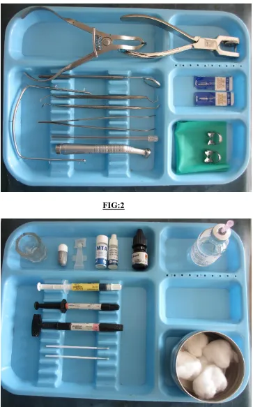
MATERIALS AND METHODS: Materials used:[Fig:2]:
2.2% Lignocaine HCl with 1:80,000 Adrenaline (D.J Laboratories Pvt ltd,
India)
MTA- Angelus[ Angelus,Londrina,PR,Brazil]
Biodentine[Septodont,France]
Chlorhexidine 2%[Asep-RC,Anabond-Stedman,Chennai,India]
Materials for COMPOSITE RESTORATION:
o Tetric N- ceram composite [Ivoclar Vivadent,Schann,Liechtenstein]
o Tetric N –Flow [Flowable comoposite]
o Etchant [35% phosphoric acid]
o Tetric N- bond [ bonding agent]
o Visible light curing unit [Heraeus Kulzer-Hulix Model-200]
o Super Snap Mini Kit for Polishing [Shofu Inc.,Japan]
ARMAMENTARIUM USED FOR THE CLINICAL PROCEDURE:
INSTRUMENTS:[ Fig: 1]:
Mouth mirror,Dental explorer,Tweezer
Sterile cotton and gauze
Suction tip
Dappen dish,Mixing spatula,Plastic instrument
Electric pulp tester [Dental Pulp Tester,Mident Industries,Henan]
27 Airotor handpiece (NSK, Japan)
Straight diamond abrasive [SF-11,Mani,Japan]
No. 2 [1 mm] round tungsten carbide bur (FG-2, SS White, USA)
Suction tips
HISTOLOGICAL PROCESSING:
Diamond disc[#7020,KG,Sorensen Ind,Barueri-SP,Brazil]
10% formalin
Aqueous ethanol
5% Nitric acid
Paraffin wax
28
MATERIALS AND METHODS:
This study was conducted on human subjects, with approval from The Institutional
Ethical Committee- TamilNadu Government Dental College, Chennai.
SUBJECTS AND SPECIMENS:
Young adult patients [age group: 18 to 24 years] from the Department of Orthodontics,
who were scheduled for extraction of premolars for orthodontic reasons, were selected
for the study.
The subjects signed consent forms after they had received a thorough explanation
regarding the experimental rationale, clinical procedure and possible risks. All the
patients were recruited by a single operator. As this study was not based on a population
basis, no randomization was used for patient selection.
Forty five first or second human premolar teeth from fourteen patients were selected.
Seven patients contributed all four premolars each, four other patients contributed three
teeth each,two of them gave consent for two teeth and one patient contributed one tooth.
All teeth were examined clinically and radiographically to ensure absence of caries and
29
EXCLUSION CRITERIA:
The teeth with the following criteria were excluded from the study:
Decayed or fractured teeth
Previous restorative history
Symptomatic teeth
Teeth with any other pathology
Teeth not conducive for rubber dam isolation
EXPERIMENTAL GROUPS: [Table-1]
GROUP I –MTA—[n=21]
GROUP II –BIODENTINE-- [n=21]
GROUP III --CONTROL-NORMAL TEETH—[n=3]
EXPERIMENTAL PROTOCOL:
PHASE I – CAVITY PREPARATION AND PLACEMENT OF MATERIAL PHASE II – ATRAUMATIC EXTRACTION OF THE TEETH AFTER
EXPERIMENTAL PERIODS
30
TABLE 1:
GROUP DPC
MATERIAL
PERIOD OF EXTRACTION
CODE
1 MTA 1 WEEK SM
2 BD 1 WEEK SB
3 MTA 1 MONTH OM
4 BD 1 MONTH OB
5 MTA 3 MONTHS TM
6 BD 3 MONTHS TB
7 NONE --- C
n=7 for Group 1-6 , n=3 for Group 7
DPC : Direct Pulp Capping Material Used
31
CLINICAL PROCEDURES:
Electrical pulp testing was performed on all teeth before preforming the experimental
procedure. Calculus and debris were removed from the tooth surfaces. After
administering local anaesthesia , with 2% lignocaine and rubberdam isolation, the teeth
were cleaned with a rubber cup and prophylactic paste at low speed. The site was then
washed with 2% chlorhexidine.
CAVITY PREPARATION:
Occlusal cavities of dimensions approximately 2mm long , 2 mm wide and of variable
depth till pulp exposure, about 3.5mm deep were prepared using a sterile straight fissure
diamond [SF-11,Mani,Japan] at high speed under water spray coolant [fig- 4 ].The cavity
walls were irrigated with normal saline.Then a pulp exposure was made using a no-2,
round tungsten carbide bur [1mm dia],at high speed without penetrating the pulp
space[fig- 5 ]. New sterilized burs were used for each procedure.
DIRECT PULP CAPPING:
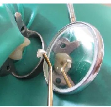
Hemorrhage was controlled by a sterile cotton pellet soaked in saline. Then, the area of
pulp exposure was capped with MTA/ Biodentine [fig- 6,7 ]. The materials were mixed
according to the manufacturers’ instructions.
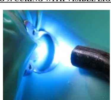
A layer of non-bonded flowable composite resin was then placed over the capping
material and light cured for 40 seconds [fig- 8,9 ] .The walls of the cavity were then
etched,rinsed and bonded with light cured nanofilled composite resin.The immediate final
32
SPECIMEN PREPARATION AND HISTOLOGIC EVALUATION:
All the experimental teeth were carefully followed up and assessed before extraction.The
assessment included history taking and a clinical examination for recording any signs or
symptoms and response to electric pulp testing.The extractions were scheduled after a
period of 1 week, 1 month and 3 months.The teeth were grouped according to the timing
of extraction and the pulp capping material used.
Immediately after the atraumatic extraction procedure,the roots were sectioned midway
between the cemento-enamel junction and the root apex, to facilitate diffusion of the
fixing solution. After fixation with 10% formalin for 96 hours, the specimens were
decalcified in 5% nitric acid, dehydrated in increasing concentrations of aqueous ethanol
and embedded in paraffin.
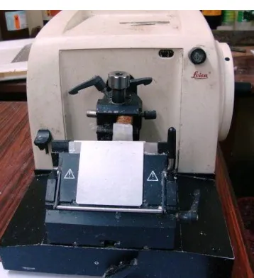
Then serial bucco-lingual sections of four micron thickness were cut longitudinally
through the center of the exposure site, with Leica Microtome [ fig-11 ].The sections
mounted on glass slides were then stained with hematoxylin and eosin in the Leica
semi-automatic stainer [ fig-12 ].
All histological processing procedures were carried out in the Department of Veterinary
Pathology, Madras Veterinary College, Chennai. The sections were then blindly
evaluated by an experienced pathologist and calibrated according to the following
criteria [Table 2].
33
TABLE 2:Scoring criteria for histopathological evaluation: [1, 74, 42,57]
GRADING CHARACTERIZATION
Score Inflammatory cell response
1 None or few scattered inflammatory cells present in the site of pulp exposure, characteristic of normal tissue
2 Mild inflammatory cell infiltration with PMN or mononuclear leukocytes[< 10 cells per high power field]
3 Moderate inflammatory cell infiltration involving coronal pulp[10-25 cells]
4 Severe inflammatory cell infiltration[>25 cells] involving coronal pulp or abcess present
Score Hard tissue formation
1 Absent
2 Lateral deposition of hard tissue on the walls of the cavity of pulp exposure
3 Partial hard tissue bridge-little communication of the capping material with pulp
4 Complete hard tissue bridge-closure of exposure area
Score Thickness of dentinal bridge
1 0mm
2 <0.1 mm
3 0.1–0.25 mm
34
EXPERIMENTAL DESIGN:
ORTHODONTIC PREMOLAR EXTRACTION CASES
[Age 18-24 years] [total number of
samples=45]
INFORMED CONSENT AND CLINICAL ASSESSMENT
RANDOMLY ASSIGNED FOR PULP CAPPING
CONTROL[n=3]
1 MONTH
EXTRACTION AFTER
HISTOLOGICAL EVALUATION
1 WEEK 3
MONTHS BD[n=21]
35
EXPERIMENTAL PROTOCOL
PROCEDURAL-FLOWCHART FOR DIRECT PULP CAPPING:
Local anaesthesia administration
Pre-operative 2% chlorhexidine rinse
Hemorrhage control with sterile cotton pellets soaked in saline
Rubberdam isolation
Occlusal cavity preparation
36
MTA/Biodentine placed on the exposed pulp
Cavity dried with sterile cotton pellets
A layer of non bonded flowable light cure composite resin liner placed and cured
Cavity disinfection with 2% chlorhexidine rinse
Final composite restoration done by incremental placement technique
Remaining cavity walls etched and rinsed
FIG 4: INITIAL CAVITY PREPARATION BEFORE PULP EXPOSURE
[image:56.612.236.450.515.700.2]FIG 7 :TOOTH AFTER THE PLACEMENT OF THE PULP CAPPING MATERIAL
[image:57.612.232.410.314.494.2] [image:57.612.235.414.555.720.2]FIG 9: CURING WITH VISIBLE LIGHT
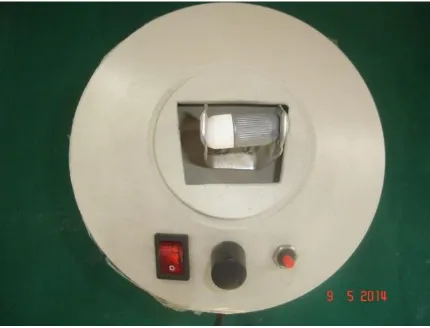
[image:58.612.235.414.423.574.2]FIG 12:LEICA SEMI-AUTOMATIC STAINER
[image:59.612.127.526.331.465.2] [image:59.612.239.410.531.731.2]37
Results:
No apparent adverse events were observed during the experimental periods. All the
individual scores for the two experimental groups in all the observation periods were
38
TABLE 3:Histopathological scores for seven days observation
Si.No Group
Code
Material Sub-Group
Code Age/ Sex scores Inflam- mation Dentin Bridge Thickness of Dentin Bridge
1 G1 MTA G1 SM-01 21/F 1 1 1
2 G1 MTA G1 SM-02 21/F 1 1 1
3 G1 MTA G1 SM-03 21/F 1 1 1
4 G1 MTA G1 SM-04 21/F 2 1 1
5 G1 MTA G1 SM-05 19/M 2 1 1
6 G1 MTA G1 SM-06 19/M 1 1 1
7 G1 MTA G1 SM-07 19/M 2 1 1
8 G2 BD G1 SB-01 18/M 1 1 1
9 G2 BD G1 SB-02 18/M 2 1 1
10 G2 BD G1 SB-03 18/M 2 1 1
11 G2 BD G1 SB-04 18/M 1 1 1
12 G2 BD G1 SB-05 24/M 2 1 1
13 G2 BD G1 SB-06 24/M 2 1 1
39
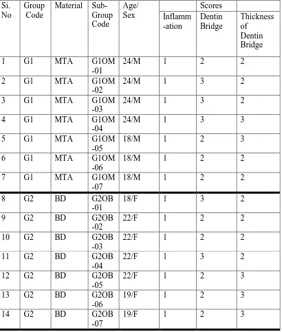
TABLE 4: Histopathological scores for one month observation:
Si. No
Group Code
Material Sub-Group Code Age/ Sex Scores Inflamm -ation Dentin Bridge Thickness of Dentin Bridge
1 G1 MTA G1OM
-01
24/M 1 2 2
2 G1 MTA G1OM
-02
24/M 1 3 2
3 G1 MTA G1OM
-03
24/M 1 3 2
4 G1 MTA G1OM
-04
24/M 1 3 3
5 G1 MTA G1OM
-05
18/M 1 2 3
6 G1 MTA G1OM
-06
18/M 1 2 2
7 G1 MTA G1OM
-07
18/M 1 2 2
8 G2 BD G2OB
-01
18/F 1 3 2
9 G2 BD G2OB
-02
22/F 1 2 2
10 G2 BD G2OB
-03
22/F 1 2 2
11 G2 BD G2OB
-04
22/F 1 3 2
12 G2 BD G2OB
-05
22/F 1 2 3
13 G2 BD G2OB
-06
19/F 1 2 3
14 G2 BD G2OB
-07
40
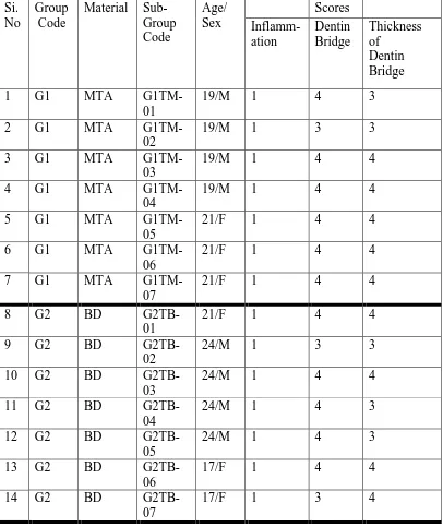
TABLE 5: Histopathological scores for three months observation:
Si. No
Group Code
Material Sub-Group Code Age/ Sex Scores Inflamm-ation Dentin Bridge Thickness of Dentin Bridge
1 G1 MTA
G1TM-01
19/M 1 4 3
2 G1 MTA
G1TM-02
19/M 1 3 3
3 G1 MTA
G1TM-03
19/M 1 4 4
4 G1 MTA
G1TM-04
19/M 1 4 4
5 G1 MTA
G1TM-05
21/F 1 4 4
6 G1 MTA
G1TM-06
21/F 1 4 4
7 G1 MTA
G1TM-07
21/F 1 4 4
8 G2 BD
G2TB-01
21/F 1 4 4
9 G2 BD
G2TB-02
24/M 1 3 3
10 G2 BD
G2TB-03
24/M 1 4 4
11 G2 BD
G2TB-04
24/M 1 4 3
12 G2 BD
G2TB-05
24/M 1 4 3
13 G2 BD
G2TB-06
17/F 1 4 4
14 G2 BD
G2TB-07
41
Statistical Analysis:
The scores attributed to each group were recorded [Table 3,4 and 5]. The results of the
histopathologic evaluation were statistically analyzed by using the Chi-Square Test using
SPSS 16 (statistical package for social sciences version 16) software. A p value <.05 was
42
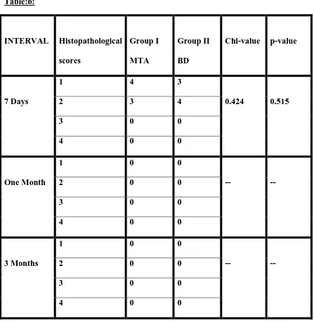
[image:66.612.102.549.131.590.2]INFLAMMATION: Chi-square test for inflammation for all the 3 intervals: Table:6:
INTERVAL Histopathological scores
Group I MTA
Group II BD
Chi-value p-value
7 Days
1 4 3
0.424 0.515
2 3 4
3 0 0
4 0 0
One Month
1 0 0
-- --
2 0 0
3 0 0
4 0 0
3 Months
1 0 0
-- --
2 0 0
3 0 0
43
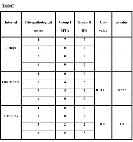
[image:67.612.101.548.114.592.2]DENTIN BRIDGE: Chi-square test for dentin bridge for all the 3 intervals: Table:7
Interval Histopathological scores Group I MTA Group II BD Chi-value p-value 7 Days
1 7 7
-- --
2 0 0
3 0 0
4 0 0
One Month
1 0 0
0.311 0.577
2 4 5
3 3 2
4 0 0
3 Months
1 0 0
0.00 1.0
2 0 0
3 2 2
44
THICKNESS OF DENTIN BRIDGE:
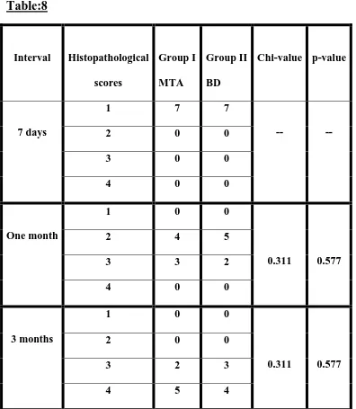
[image:68.612.101.497.133.588.2]Chi-square test for thickness of dentin bridge for all the 3 intervals:
Table:8
Interval Histopathological scores
Group I MTA
Group II BD
Chi-value p-value
7 days
1 7 7
-- --
2 0 0
3 0 0
4 0 0
One month
1 0 0
0.311 0.577
2 4 5
3 3 2
4 0 0
3 months
1 0 0
0.311 0.577
2 0 0
3 2 3
45
Graph-1:
Comparison of Inflammation in MTA Vs BD Groups at Time Intervals of 7 Days, One Month, 3 Months46
47 G rap h -4 : Co m p a rison of all t h
e 3 P
ar a m ete rs at T im e In te rvals o
f 7 Days, On
e M
on
th, 3 M
on
48
Interpretation of results: Seven days observation:
In the 7 days samples groups 1 and 2 showed mild or no inflammatory response.The