Copyright © 1998, American Society for Microbiology
Coreceptor Utilization by Human Immunodeficiency Virus Type 1
Is Not a Primary Determinant of Neutralization Sensitivity
RACHEL A. LACASSE,1KATHRYN E. FOLLIS,1TARSEM MOUDGIL,1MEG TRAHEY,1
JAMES M. BINLEY,2VICENTE PLANELLES,3SUSAN ZOLLA-PAZNER,4,5
ANDJACK H. NUNBERG1*
Montana Biotechnology Center, The University of Montana, Missoula, Montana 598121; Aaron Diamond AIDS
Research Center and The Rockefeller University, New York, New York 100162; University of Rochester Cancer
Center, Rochester, New York 146423; and Veterans Affairs4and New York University5Medical Centers,
New York, New York 10010
Received 24 September 1997/Accepted 4 December 1997
We have examined the relationship between coreceptor utilization and sensitivity to neutralization in a primary isolate of human immunodeficiency virus type 1 and its T-cell line-adapted (TCLA) derivative. We determined that adaptation of the primary-isolate (PI) virus 168P results in the loss of the unique capacity of PI viruses to utilize the CCR5 coreceptor and in the acquisition by the TCLA 168C virus of sensitivity to neutralization by V3-directed monoclonal antibodies (MAbs). In experiments wherein infection by 168P is directed via either the CCR5 or the CXCR4 pathway, we demonstrate that the virus, as well as pseudotyped virions bearing a molecularly cloned 168P envelope protein, remains refractory to neutralization by MAbs 257-D, 268-D, and 50.1 regardless of the coreceptor utilized. This study suggests that coreceptor utilization is not a primary determinant of differential neutralization sensitivity in PI and TCLA viruses.
Although CD4 had long been recognized as the cellular receptor to which the human immunodeficiency virus type 1 (HIV) envelope protein binds (9, 21, 22), it had also been recognized that expression of CD4 alone is insufficient to ren-der nonhuman cells susceptible to HIV infection (4, 5, 22). Similarly, different HIV isolates display different abilities to infect CD4-positive human macrophages, T lymphocytes, and established T-cell lines (31, 32, 35), suggesting that additional molecules may be responsible for cell tropism specificity. Dur-ing the past year, cellular molecules that act in conjunction with CD4 have been identified as required cofactors for HIV envelope protein-mediated binding and entry (1, 6, 10–12, 14). These HIV coreceptors are members of the superfamily of seven-transmembrane segment G-protein-coupled receptors and act primarily as cellular receptors for chemokines.
The discovery of cellular coreceptors for HIV has provided new perspectives for understanding these early events in HIV infection (see review in reference 2). Thus, phenotypically dis-tinct isolates of HIV utilize as coreceptors different chemokine receptor molecules. Although all primary isolates of HIV in-fect primary T lymphocytes, some also inin-fect cells of the mac-rophage lineage (31, 32). These monocyteropic isolates utilize the CCR5 chemokine receptor, whose natural ligands include the chemokines RANTES, MIP-1a, and MIP-1b(1, 6, 10–12). Monocytropic isolates do not induce syncytia in primary lym-phocyte culture and do not infect established T-cell lines (31). During the late course of HIV infection, syncytium-inducing (SI) primary viruses often arise from the population of mono-cytropic viruses (31, 32). These SI primary isolates no longer infect macrophages, and they utilize both CCR5 and another chemokine receptor, CXCR4 (7, 33, 38). CXCR4, whose nat-ural chemokine ligand is SDF-1 (3, 27), was originally identi-fied by Feng et al. as the cofactor used by laboratory-adapted
viruses (14). In fact, the common laboratory viruses (IIIb/LAI, LAV, and RF) are unable to utilize CCR5 coreceptor (1, 6, 10–12), presumably reflecting the lack of CCR5 expression in most established T-cell lines (1, 13). Although some primary isolates utilize additional chemokine receptor molecules, no-tably CCR3 and CCR2b (6, 11, 18), the relationship between these coreceptors and viral phenotypes is less clear. The ability to utilize CCR5 coreceptor, however, is unique to primary-isolate (PI) viruses.
Paralleling these differences in coreceptor utilization and cell tropism are differences in sensitivity to virus neutralization. Although laboratory-adapted isolates of HIV can be potently neutralized by sera elicited by recombinant gp120 (rgp120) protein, primary isolates are largely refractory to neutraliza-tion by rgp120 vaccine sera (23, 37). Similarly, PI viruses are significantly more resistant than T-cell line-adapted (TCLA) viruses to neutralization by gp120-directed monoclonal anti-bodies (MAbs) (25, 37) and to inhibition by soluble forms of CD4 (8). We and others have demonstrated that neutralization sensitivity develops concomitantly with adaptation of primary isolates to persistent growth in established T-cell lines (24, 37). By studying pedigreed PI and TCLA viruses (168P and 168C, respectively), we have shown that adaptation renders the TCLA virus sensitive not only to rgp120 vaccine sera and CD4 immunoadhesin but also to MAbs directed to the V3 loop of gp120 (37). However, the basis for this increase in neutraliza-tion sensitivity remains unclear.
In this report, we explore the relationship between neutral-ization sensitivity and coreceptor utilneutral-ization, especially with regard to changes that accompany adaptation. We examined neutralization sensitivity of the well-characterized SI primary isolate 168P under experimental conditions where infection can be directed via either the CXCR4 or the CCR5 pathway. The pedigreed TCLA derivative 168C utilizes only CXCR4 and was sensitive to neutralization by the panel of V3-directed MAbs used in these assays. However, the primary isolate 168P remained refractory to neutralization regardless of coreceptor pathway taken. Our findings suggest that envelope protein * Corresponding author. Mailing address: Montana Biotechnology
Center, The University of Montana, Missoula, MT 59812. Phone: (406) 243-6421. Fax: (406) 243-6425. E-mail: [email protected] .edu.
2491
on November 9, 2019 by guest
http://jvi.asm.org/
structure, and not coreceptor utilization, is the primary deter-minant of differential neutralization sensitivity in PI and TCLA viruses.
Coreceptor utilization by pedigreed PI and TCLA viruses.
Cross-sectional surveys of coreceptor use have shown that pri-mary SI isolates generally utilize CXCR4 and CCR5 corecep-tors, whereas unrelated laboratory-adapted isolates utilize only CXCR4 (1, 6, 7, 10–12, 14, 33, 38). We wished to confirm this trend in a longitudinal study of adaptation. We previously described the adaptation of the SI primary isolate 168P to persistent growth in the FDA/H9 T-cell line and the concom-itant development of neutralization sensitivity in the resulting TCLA virus 168C (37). In the present study, the ability of these pedigreed viruses to utilize specific coreceptors was tested by infection of U87 human glioma cell lines expressing CD4 (U87-CD4) and the specific coreceptor (19).
For this assay, virus stocks were prepared from cell culture supernatants of phytohemagglutinin (PHA)-stimulated periph-eral blood lymphocytes (PBLs) (168P) or FDA/H9 cells (168C) and standardized to yield a submaximal number of foci of infection on U87-CD4-CXCR4 cells (approximately 100 to 200
gands. Thus, 168P virus infection of CCR5-expressing cells was blocked by the CCR5-specific ligands RANTES, MIP-1a, and MIP-1b(1, 6, 10–12) (Fig. 1). Similarly, infection of CXCR4-expressing U87-CD4 cells by either virus could be blocked by the CXCR4-specific chemokine ligand SDF-1 (3, 27) (data not presented).
Coreceptor pathway and neutralization sensitivity.In pre-vious work, we demonstrated that the PI 168P virus is refrac-tory to neutralization by HIV MN gp120 vaccine sera and by several well-characterized V3-directed murine MAbs which strongly neutralize infectivity of the TCLA 168C virus (37). In the present study, we extended the panel of MAbs to include two V3-directed human MAbs, 257-D and 268-D (17). These well-characterized human MAbs recognize core epitopes at the crown of the V3 loop of gp120 (KRIHI and HIGPGR, respec-tively), linear sequences known to be present in both 168P and 168C envelope proteins (37). These epitope predictions were confirmed by gp120 capture enzyme-linked immunosorbent assay (ELISA) (26) which demonstrated equal binding to en-velope protein in detergent-solubilized 168P and 168C virions (data not presented). Sensitivity to neutralization by these hu-man MAbs was determined in a standard assay using PHA-activated PBLs (37). MAbs 257-D and 268-D were found to potently neutralize 168C but fail to neutralize 168P (Fig. 2). This pattern of neutralization sensitivity is similar to that pre-viously described for the V3-directed murine MAb 50.1 (30, 36, 37).
To examine whether sensitivity to neutralization was
af-FIG. 1. Coreceptor utilization by pedigreed PI and TCLA 168 viruses. U87-CD4 cell lines expressing CXCR4 (■) or CCR5 (u) were used to define the ability of 168P and 168C viruses to utilize the respective coreceptor. CCR5 utilization was further tested by the addition to U87-CD4-CCR5 cells of CCR5-specific chemokines (RANTES, MIP-1a, and MIP-1b; R&D Systems) (M). For details, see text.p, no foci were observed.
FIG. 2. Neutralization sensitivity of 168 viruses in PBL culture. Virus neutralization assays in PHA-stimulated PBL culture were performed as previously described (37). 168P (E,F) and 168C (M,■) virus stocks were standardized to yield submaximal extents of virus spread during the 5-day infection. CCR5-specific chemokines (F,■) were added as described for Fig. 1. The V3-directed MAbs are indicated. p24 antigen was determined by p24 antigen capture ELISA (SAIC Frederick) and was normalized to infected cell control values (168P, 190 ng/ml [170 ng/ml with chemokines]; 168C, 36 ng/ml [33 ng/ml with chemokines]).
on November 9, 2019 by guest
http://jvi.asm.org/
fected by the coreceptor pathway utilized in infection of PBLs, we used inhibitory concentrations of CCR5-specific chemokine ligands RANTES, MIP-1a, and MIP-1b in order to restrict infection to the CXCR4 pathway. Addition of these chemo-kines to the PBL cultures did not affect virus growth, nor did it affect sensitivity to neutralization by the V3-directed human MAbs (Fig. 2). To the extent that CCR5 blockade was com-plete, these results suggest that the simple availability of the CCR5 pathway is not a factor in the resistance of PI viruses to neutralization.
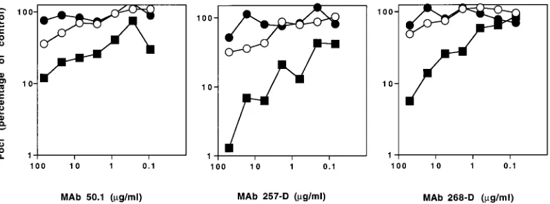
To strengthen this conclusion, we examined neutralization sensitivity in human U87-CD4 cell lines expressing only CXCR4 or CCR5. Using this method, we confirmed that the SI 168P virus remained refractory to neutralization by human MAbs 257-D and 268-D as well as by the murine MAb 50.1, regardless of whether infection occurred via CXCR4 or CCR5 (Fig. 3). These results suggest that availability of the CCR5 pathway is not a primary determinant for the resistance of PI viruses to neutralization. The TCLA 168C virus utilized CXCR4 only and was sensitive to neutralization.
Molecularly cloned PI and TCLA envelope genes.To under-stand better the changes that accompany adaptation and those that determine coreceptor utilization and neutralization sensi-tivity, we molecularly cloned the envelope genes of the 168P and 168C viruses. High-fidelity XL PCR (rTth and Vent DNA polymerases; PE Applied Biosystems) and primers envA and envN (15) were used to amplify a 3.1-kb region of proviral DNA encoding the rev and envelope genes. PCR products were isolated by unidirectional T/A cloning in the eucaryotic expression vector pCR3.1-Uni (Invitrogen). Expression in pCR3.1-Uni is driven by the cytomegalovirus immediate-early promoter. Multiple clones were isolated from each virus, and transient transfection studies in COS-7 cells confirmed the surface expression and fusion competence of all clones tested (data not presented).
DNA sequence analysis demonstrated that all 168C molec-ular clones analyzed encoded the three adaptation-associated amino acid changes previously identified by PCR sequencing of the 168C virus population (V2, I166R; C2, I282N; and V3, G318R) (37). Two molecular clones of each 168P and 168C envelope were subjected to complete DNA sequence analysis (GenBank accession no. AF035532 to AF035534). Molecular clones 168C23 and 168C60 were identical throughout the en-velope gene. Molecular clones 168P5 and 168P23 differed from each other and from the previously determined sequence at four to five positions distinct from those associated with adap-tation. These scattered changes within the primary virus
quasi-species are considered inconsequential at the present level of analysis; the significance of the three adaptation-associated changes is under separate investigation.
Functional analysis of these molecularly cloned envelope genes was performed by incorporation of the molecularly cloned envelope protein into pseudotyped HIV virions. We used an envelope-defective provirus derived from the molec-ularly cloned NL4-3 provirus (kindly provided by I. S. Y. Chen, University of California, Los Angeles). The pNLthyDBgl
pro-virus (28) contains a BglII-BglII deletion within the envelope gene and a substitution of the viral nef gene with a cDNA encoding the murine Thy1.2 cell surface protein. The simian virus 40 ori was subsequently introduced into the plasmid to generate pSVNLthyDBgl (27a). Cotransfection of COS-7 cells
(16, 20) with pSVNLthyDBgl provirus and the envelope
expres-sion plasmid resulted in the production of pseudotyped HIV virions. Culture supernatants were harvested 3 days posttrans-fection, filtered, and used to infect U87-CD4 cell lines express-ing coreceptor. Cells infected by virions bearexpress-ing the comple-menting envelope protein were identified by immunostaining for murine Thy1.2 or HIV proteins.
As anticipated, the molecularly cloned envelope proteins recapitulated the coreceptor specificity of the parental virus population (see the legend to Fig. 4). Pseudotyped virions containing 168C60 were able to infect only U87-CD4 cells expressing CXCR4, while virions containing 168P23 envelope were able to infect U87-CD4 cells expressing either CCR5 or CXCR4. Thus, the viral envelope protein appears to be the major, if not sole, determinant of viral coreceptor use. These findings also indicate that dual coreceptor use is a direct prop-erty of the envelope protein complex and not a result of a mixture of distinct envelope proteins in the SI virus population. This conclusion is corroborated by the failure of CCR5-specific chemokine ligands to diminish 168P virus infection in PBL culture (Fig. 2).
[image:3.612.101.500.68.216.2]Finally, we wished to determine the neutralization sensitivity of pseudotyped virions containing the molecularly cloned 168P23 and 168C60 envelope proteins and to confirm that coreceptor pathway is not a primary determinant of neutral-ization sensitivity. We found that infection of U87-CD4-CXCR4 cells by pseudotyped virions containing 168C60 enve-lope protein was sensitive to neutralization by MAbs 257-D, 268-D, and 50.1 at concentrations comparable to those deter-mined in assays using 168C virus (Fig. 4). Pseudotyped virions containing 168P23 envelope protein remained refractory to neutralization by all three V3-directed MAbs, regardless of the coreceptor expressed by the U87-CD4 cell line.
FIG. 3. Neutralization sensitivity of 168 viruses in U87-CD4 cell lines expressing CCR5 or CXCR4 coreceptor. 168P (E,F) and 168C (■) viruses were used to infect U87-CD4 cell lines expressing CXCR4 (F,■) or CCR5 (E) as described for Fig. 1. The V3-directed MAbs were incubated with virus for 1 h prior to infection.
on November 9, 2019 by guest
http://jvi.asm.org/
In summary, we examined the relationship between corecep-tor utilization and sensitivity to neutralization by V3-directed MAbs. The observed dichotomy in the sensitivity to neutral-ization of PI and TCLA viruses had suggested a discrete dif-ference between these viruses, and we tested one hypothesis: that PI viruses are refractory to neutralization as a result of their unique ability to utilize the CCR5 coreceptor. We exam-ined neutralization sensitivity of a well-characterized SI pri-mary isolate under experimental conditions wherein the virus was forced to utilize either CCR5 or CXCR4 for infection. We showed that coreceptor pathway is not a direct determinant of neutralization sensitivity. The primary virus envelope protein remained refractory to neutralization by V3-directed MAbs regardless of the coreceptor pathway utilized. Similarly, core-ceptor utilization did not affect neutralization sensitivity by soluble CD4 (34) or HIVIG (data not presented).
In discarding the otherwise attractive hypothesis that PI viruses escape neutralization through their unique ability to utilize CCR5, we are left to consider the as yet undefined structural differences between the envelope protein complex of PI and TCLA viruses. Several studies have suggested that critical determinants in the envelope protein of PI viruses are less accessible than those of TCLA viruses and that it is this differential access that determines neutralization sensitivity (reviewed in reference 25). By contrast, our studies have indi-cated similar binding of V3-directed MAbs to PBLs infected with resistant isolate 168P or neutralization-sensitive isolate 168C (37). Thus, the basis for the differential neutralization sensitivity of PI and TCLA viruses remains un-resolved.
Our present studies also do not address whether changes in coreceptor utilization and/or neutralization sensitivity are nec-essarily linked as a consequence of adaptation. The analysis of independently derived PI and TCLA viruses may allow further separation of these viral phenotypes. Subsequent dissection of the amino acid changes that distinguish pedigreed PI and TCLA envelope proteins will help to define the structural bases underlying the changes that accompany adaptation.
This work was supported by NIH AREA grant AI41165 to J.H.N. and by NIH grants AI32424 and AI36085 to S.Z.-P. Additional funds were provided by The University of Montana, the M. J. Murdock Charitable Trust, and the Department of Veterans Affairs.
We thank Ed Berger (NIH) and Ned Landau (Aaron Diamond AIDS Research Center) for useful discussions during the course of these studies. We are also grateful to numerous colleagues who
con-tributed reagents for this work: Dan Littman (HHMI, NYU Medical Center), Thera Mulvania and Jim Mullins (University of Washington) for U87-CD4 cell lines, Irvin Chen (UCLA School of Medicine) for pNLthyDBgl provirus, Terri Wrin (Genentech, Inc.) for 168P and 168C
viruses, Ian Clark-Lewis (University of British Columbia) for SDF-1, Steve Chamow (Genentech, Inc.) for recombinant CD4-Ig, and Fred Prince (New York Blood Center) for HIVIG. Oligonucleotides and DNA sequence analysis were provided by Joan Strange at The Uni-versity of Montana Murdock Molecular Biology Laboratory. We thank John Moore (Aaron Diamond AIDS Research Center) for assistance in the ELISA determination of MAb binding. The NIH AIDS Re-search and Reference Reagent Program provided initial samples of CCR5-specific chemokines, HIVIG, and MAbs 50.1, 257-D, and 268-D, as well as several reagents which were not expressly represented in these studies but which were nonetheless important.
REFERENCES
1. Alkhatib, G., C. Combadiere, C. C. Broder, Y. Feng, P. E. Kennedy, P. M. Murphy, and E. A. Berger.1996. CC CKR5: a RANTES, MIP-1a, MIP-1b
receptor as a fusion cofactor for macrophage-tropic HIV-1. Science 272: 1955–1958.
2. Berger, E. A. 1997. HIV entry and tropism: the chemokine receptor connec-tion. AIDS 11(Suppl. A):S3–S16.
3. Bleul, C. C., M. Farzan, H. Choe, C. Parolin, I. Clark-Lewis, J. Sodroski, and T. A. Springer.1996. The lymphocyte chemoattractant SDF-1 is a ligand for LESTR/fusin and blocks HIV-1 entry. Nature 382:829–833.
4. Broder, C. C., D. S. Dimitrov, R. Blumenthal, and E. A. Berger. 1993. The block to HIV-1 envelope glycoprotein-mediated membrane fusion in animal cells expressing human CD4 can be overcome by a human cell component. Virology 193:483–491.
5. Chesebro, B., R. Buller, J. Portis, and K. Wehrly. 1990. Failure of human immunodeficiency virus entry and infection in CD4-positive human brain and skin cells. J. Virol. 64:215–221.
6. Choe, H., M. Farzan, Y. Sun, N. Sullivan, B. Rollins, P. D. Ponath, L. Wu, C. R. Mackay, G. LaRosa, W. Newman, N. Gerard, C. Gerard, and J. Sodroski. 1996. Theb-chemokine receptors CCR3 and CCR5 facilitate infection by primary HIV-1 isolates. Cell 85:1135–1148.
7. Connor, R. I., K. E. Sheridan, D. Ceradini, S. Choe, and N. L. Landau. 1997. Changes in coreceptor use correlates with disease progression in HIV-1-infected individuals. J. Exp. Med. 185:621–628.
8. Daar, E. S., X. L. Li, T. Moudgil, and D. D. Ho. 1990. High concentrations of recombinant soluble CD4 are required to neutralize primary human immunodeficiency virus type 1 isolates. Proc. Natl. Acad. Sci. USA 87:6574– 6578.
9. Dalgleish, A. G., P. C. L. Beverly, P. R. Clapham, D. H. Crawford, M. F. Greaves, and R. A. Weiss. 1984. The CD4 (T4) antigen is an essential component of the receptor for the AIDS retrovirus. Nature 312:763–766. 10. Deng, H., R. Liu, W. Ellmeier, S. Choe, D. Unutmaz, M. Burkhart, P.
DiMarzio, S. Marmon, R. E. Sutton, C. M. Hill, C. B. Davis, S. C. Peiper, T. J. Schall, D. R. Littman, and N. R. Landau.1996. Identification of a major co-receptor for primary isolates of HIV-1. Nature 381:661–666.
11. Doranz, B. J., J. Rucker, Y. Yi, R. J. Smyth, M. Samson, S. C. Peiper, M. Parmentier, R. G. Collman, and R. W. Doms.1996. A dual-tropic primary HIV-1 isolate that uses fusin and theb-chemokine receptors CKR-5, CKR-3, and CKR-2b as fusion cofactors. Cell 85:1149–1158.
FIG. 4. Neutralization sensitivity of pseudotyped virions in U87-CD4 cell lines expressing CCR5 or CXCR4 coreceptor. Pseudotyped virions were derived by cotransfection of COS-7 cells with pSVNLthyDBgl provirus and plasmid expressing 168P23 (E,F) or 168C60 (■) envelope protein. Virion preparations were incubated with U87-CD4 cell lines expressing CXCR4 (F,■) or CCR5 (E) as described for Fig. 1; V3-directed MAbs were added as indicated. The number of foci was normalized to control values (60 to 100 foci/well for U87-CD4-CXCR4 cells; 10 foci/well for U87-CD4-CCR5 cells).p, no foci were observed.
on November 9, 2019 by guest
http://jvi.asm.org/
12. Dragic, T., V. Litwin, G. P. Allaway, S. R. Martin, Y. Huang, K. A. Na-gashima, C. Cayanan, P. J. Maddon, R. A. Koup, J. P. Moore, and W. A. Paxton.1996. HIV-1 entry into CD41cells is mediated by the chemokine receptor CC-CKR-5. Nature 381:667–673.
13. Endres, M. J., P. R. Clapham, M. Marsh, M. Ahuja, J. D. Turner, A. McKnight, J. F. Thomas, B. Stoebenau-Haggarty, S. Choe, P. J. Vance, T. N. C. Wells, C. A. Power, S. S. Sutterwala, R. W. Doms, N. R. Landau, and J. A. Hoxie.1996. CD4-independent infection by HIV-2 is mediated by fusion/CXCR4. Cell 87:745–756.
14. Feng, Y., C. C. Broder, P. E. Kennedy, and E. A. Berger. 1996. HIV-1 entry cofactor: functional cDNA cloning of a 7-transmembrane G protein-coupled receptor. Science 272:872–877.
15. Gao, F., S. G. Morrison, D. L. Robertson, C. L. Thornton, S. Craig, G. Karlsson, J. Sodroski, M. Morgado, B. Galvao-Castro, H. von Briesen, S. Beddows, J. Weber, P. M. Sharp, G. M. Shaw, B. H. Hahn, and WHO and NIAID Networks for HIV Isolation and Characterization.1996. Molecular cloning and analysis of functional envelope genes from human immunode-ficiency virus type 1 sequence subtypes A through G. J. Virol. 70:1651–1667. 16. Gluzman, Y. 1981. SV40-transformed simian cells support the replication of
early SV40 mutants. Cell 23:175–182.
17. Gorny, M. K., J. Y. Xu, S. Karwowska, A. Buchbinder, and S. Zolla-Pazner. 1993. Repertoire of neutralizing human monoclonal antibodies specific for the V3 domain of HIV-1 gp120. J. Immunol. 150:653–643.
18. He, J., Y. Chen, M. Farzan, H. Choe, A. Ohagen, S. Gartner, J. Busciglio, X. Yang, W. Hofmann, W. Newman, C. R. Mackay, J. Sodroski, and D. Gabuzda.1997. CCR3 and CCR5 are co-receptors for HIV-1 infection of microglia. Nature 385:645–649.
19. Hill, C. M., H. Deng, D. Unutmaz, V. N. Kewalramani, L. Bastiani, M. K. Gorny, S. Zolla-Pazner, and D. R. Littman.1997. Envelope glycoproteins from human immunodeficiency virus types 1 and 2 and simian immunode-ficiency virus can use human CCR5 as a coreceptor for viral entry and make direct CD4-dependent interactions with this chemokine receptor. J. Virol. 71:6296–6304.
20. Jordan, M., A. Schallhorn, and F. M. Wurm. 1996. Transfecting mammalian cells: optimization of critical parameters affecting calcium-phosphate precip-itate formation. Nucleic Acids Res. 24:596–601.
21. Klatzman, D., E. Champagne, S. Chamaret, J. Gruest, D. Guetard, T. Her-cend, J. C. Gluckman, and L. Montagnier.1984. T-lymphocyte T4 molecule behaves as the receptor for human retrovirus LAV. Nature 312:767–770. 22. Maddon, P. J., A. G. Dalgleish, J. S. McDougal, P. R. Clapham, R. A. Weiss,
and R. Axel.1986. The T4 gene encodes the AIDS virus receptor and is expressed in the immune system and the brain. Cell 47:333–348. 23. Mascola, J. R., S. W. Snyder, O. S. Weislow, S. M. Belay, R. B. Belshe, D. H.
Schwartz, M. L. Clements, R. Dolin, B. S. Graham, G. J. Gorse, M. Keefer, M. McElrath, M. Walker, K. Wagner, J. McNeil, F. McCutchan, D. Burke, and NIAID AVEG.1996. Immunization with envelope subunit vaccine prod-ucts elicits neutralizing antibodies against laboratory-adapted but not pri-mary isolates of human immunodeficiency virus type 1. J. Infect. Dis. 173: 340–348.
24. Moore, J. P., L. C. Burkly, R. I. Connor, Y. Cao, R. Tizard, D. D. Ho, and R. A. Fisher.1993. Adaptation of two primary human immunodeficiency virus type 1 isolates to growth in transformed T cell lines correlates with alterations in the response of their envelope glycoproteins to soluble CD4. AIDS Res. Hum. Retroviruses 9:529–539.
25. Moore, J. P., and D. D. Ho. 1995. HIV-1 neutralization: the consequences of viral adaptation to growth on transformed T cells. AIDS 9(Suppl. A):S117– S136.
26. Moore, J. P., J. A. McKeating, I. M. Jones, P. E. Stephens, G. Clements, S. Thomson, and R. A. Weiss.1990. Characterization of recombinant gp120 and gp160 from HIV-1: binding to monoclonal antibodies and sCD4. AIDS 4:307–315.
27. Oberlin, E., A. Amara, F. Bachelerie, C. Bessia, J.-L. Virelizier, F. Arenzana-Seisdedos, O. Schwartz, J.-M. Heard, I. Clark-Lewis, D. F. Legler, M. Loetscher, M. Baggiolini, and B. Moser.1996. The CXC chemokine SDF-1 is the ligand for LESTR/fusin and prevents infection by T-cell-line-adapted HIV-1. Nature 382:833–835.
27a.Planelles, V. Unpublished data.
28. Poon, B., J. B. M. Jowett, S. A. Stewart, R. W. Armstrong, G. M. Rishton, and I. S. Y. Chen.1997. Human immunodeficiency virus type 1 vpr gene induces phenotypic effects similar to those of the DNA alkylating agent, nitrogen mustard. J. Virol. 71:3961–3971.
29. Prince, A. M., H. Reesink, D. Pascual, B. Horowitz, I. Hewlett, K. K. Murthy, K. E. Cobb, and J. W. Eichberg.1991. Prevention of HIV infection by passive immunization with HIV immunoglobulin. AIDS Res. Hum. Retroviruses 7:971–973.
30. Rini, J. M., R. L. Stanfield, E. A. Stura, P. A. Salinas, A. T. Profy, and I. A. Wilson.1993. Crystal structure of a human immunodeficiency virus type 1 neutralizing antibody, 50.1, in complex with its V3 loop peptide antigen. Proc. Natl. Acad. Sci. USA 90:6325–6329.
31. Schuitemaker, H., M. Koot, N. A. Kootstra, M. W. Dergksen, R. E. Y. de Goede, R. P. van Steenwijk, J. M. A. Lange, J. K. M. Eeftink Schattenkerk, F. Miedema, and M. Tersmette.1992. Biological phenotype of human im-munodeficiency virus type 1 clones at different stages of infection: progres-sion of disease is associated with a shift from monocytropic to T-cell-tropic virus populations. J. Virol. 66:1354–1360.
32. Schuitemaker, H., N. A. Kootstra, R. E. Y. de Goede, F. de Wolf, F. Miedema, and M. Tersmette.1991. Monocytropic human immunodeficiency virus type 1 (HIV-1) variants detectable in all stages of HIV-1 infection lack T-cell line tropism and syncytium-inducing ability in primary T-cell culture. J. Virol. 65:356–363.
33. Simmons, G., D. Wilkinson, J. D. Reeves, M. T. Dittmar, S. Beddows, J. Weber, G. Carnegie, U. Desselberger, P. W. Gray, R. A. Weiss, and P. R. Clapham.1996. Primary, syncytium-inducing human immunodeficiency virus type 1 isolates are dual-tropic and most can use either Lestr or CCR5 as coreceptors for virus entry. J. Virol. 70:8355–8360.
34. Smith, D. H., R. A. Byrn, S. A. Marsters, T. Gregory, J. E. Groopman, and D. J. Capon.1987. Blocking of HIV-1 infectivity by a soluble, secreted form of the CD4 antigen. Science 238:1704–1707.
35. Tersmette, M., R. A. Gruters, F. deWolf, R. E. Y. deGoede, J. M. A. Lange, P. T. A. Schellekens, J. Goudsmit, H. G. Huisman, and F. Miedema.1989. Evidence for a role of virulent human immunodeficiency virus (HIV) vari-ants in the pathogenesis of acquired immunodeficiency syndrome: studies on sequential HIV isolates. J. Virol. 63:2118–2125.
36. White-Scharf, M. E., B. J. Potts, L. M. Smith, K. A. Sokolowski, J. R. Rusche, and S. Silver.1993. Broadly neutralizing monoclonal antibodies to the V3 region of HIV-1 can be elicited by peptide immunization. Virology 192:197– 206.
37. Wrin, T., T. P. Loh, J. Charron-Vennari, H. Schuitemaker, and J. H. Nun-berg.1995. Adaptation to persistent growth in the H9 cell line renders a primary isolate of human immunodeficiency virus type 1 sensitive to neu-tralization by vaccine sera. J. Virol. 69:39–48.
38. Zhang, L., Y. Huang, T. He, Y. Cao, and D. D. Ho. 1996. HIV-1 subtype and second receptor use. Nature 383:768.