EVALUATION OF ULTRASONOGRAPHY
IN ASSESSMENT OF FIRST TRIMESTER
BLEEDING
DISSERTATION SUBMITTED FOR
M.D. DEGREE BRANCH II (OBSTETRICS AND
GYNAECOLOGY)
MARCH 2007
MADURAI MEDICAL COLLEGE, MADURAI
THE TAMILNADU Dr. M.G.R. MEDICAL UNIVERSITY
CERTIFICATE
This is to certify that the dissertation entitled “EVALUATION OF ULTRASONAGRAPHY IN ASSESSMENT OF FIRST TRIMESTER
BLEEDING” submitted by Dr. S. SUJATHA to the Faculty of Obstetrics and Gynaecology, The Tamilnadu Dr. M.G.R. Medical university, Chennai in partial fulfillment of the requirement for the award of M.D. Degree Branch II (Obstetrics and Gynaecology) is a bonafide research work carried out by her under our direct supervision and guidance.
Dean, Prof. Dr.S. Raja Rajeswari, M.D.D.G.O.,
Govt. Rajaji Hospital, Professor and Head,
Madurai Medical College, Dept., of Obst. and Gynaecology,
Madurai Govt. Rajaji Hospital,
ACKNOWLEDGEMENT
My sincere and thankful gratitude to Prof. Dr. RajaRajeswari,
M.D.D.G.O., Professor and Head of the Department of Obstetrics and Gynaecology who has been of great help, from the beginning of the study and for her expert guidance through out the study.
I am extremely thankful to the Dean, Madurai Medical College,
for granting me permission to undertake the study.
I am very grateful to Dr. Revathi Janakiram
M.D.D.G.O.,M.N.A.M.S., as a guide in the study.
I am also grateful to Dr. Muthulakshmi M.D.D.G.O., Dr. Dilshath
M.D.D.G.O., and Dr. Parvathavarthini M.D.D.G.O., Additional Professors, Department of Obstetrics and Gynaecology, for their valuable suggestions in preparing this dissertation.
My grateful thanks to the Assistant Professors of Department of Obstetrics and Gynaecology, for their help during this study.
Thanks to my fellow post graduates who had assisted me throughout the study.
CONTENTS
S.No.
Contents
Page
No.
1.
INTRODUCTION
1
2.
AIM OF STUDY
3
3.
BASIC EFFECTS AND BIOLOGICAL EFFECTS
OF ULTRASOUND
4
4.
SONOGRAPHIC APPEARANCE OF NORMAL
FIRST TRIMESTER IMPLANTATION
7
5.
EARLY PREGNANCY WASTAGE
14
6.
REVIEW OF LITERATURE
25
7.
MATERIALS AND METHODS
29
8.
ANALYSIS
32
9.
DISCUSSION
54
10.
SUMMARY
61
11.
CONCLUSION
64
BIBLIOGRAPHY
PROFORMA
INTRODUCTION
Vaginal Bleeding occurring in an early pregnancy pose diagnostic challenges for both obstetrician and sonologist. It is a symptom that frequently interrupts the normal development of early gestation. Despite the latest technological development and laboratory diagnosis the desired goal of early recognition is often not-achieved.
Vaginal bleeding during first trimester has been estimated to occur in 16% of all pregnant women while frequency of spontaneous abortion is estimated at 10-20%.
Prior to the era of ultra sonogram diagnosing the cause of bleeding in First Trimester has been based on history and clinical findings and often confirmed by positive or negative pregnancy test. These are neither specific in indicating the cause of bleeding nor they aid in decision-making.
A spectrum of causes for first trimester bleeding had been identified ranging from threatened abortion, missed abortion, gestational trophoblastic disease, ectopic gestation, complete abortion and incomplete abortion.
It is specially important to the clinician to diagnose each condition in this spectrum specifically because management is radically different. Eg. threatened abortion and vesicular mole.
In the area of fetal medicine Donald’s vision of sonographic diagnosis of handicaps even before the child passes through the valley of shadow of birth is yet to be fulfilled.
The diagnosis of multiple pregnancy and low lying placenta which may migrate upward are virtually impossible by clinical methods. Unnecessary curettage can be avoided in complete abortion and delayed periods with a consequent reduction in patient’s morbidity and mortality.
AIM OF THE STUDY
1) To evaluate the usefulness of ultra sonogram in differential diagnosis of vaginal bleeding in first trimester.
2) To compare the effectiveness of clinical diagnosis and Urine β HcG estimation with ultrasonogram in first trimester bleeding and to evaluate the prognostic value of ultra sonogram in Threatened abortion diagnosed.
BASIC PHYSICS AND
BASIC PHYSICS AND BIOLOGICAL
EFFECTS OF ULTRASOUND
A sound wave is a mechanical disturbance propagating through a liquid, solid, gas medium.
The frequency of a sound wave is determined by the number of oscillations per second made by the vibrating source. The limit for frequency is ‘Hertz’. Sound waves whose frequencies are greater than 20 KHz are referred to as ultrasonic waves.
Pulses versus continuous wave Beams:
When the ultrasound transducer is driven continuously we refer it as continuous wave forms. However pulsed mode is commonly used. Here the transducer vibrates for only a few cycles each time it is activated.
Display of echoes:
The oldest system of display of pulse echo information is the amplitude mode i.e., A-mode.
The second method of display is the brightness mode i.e., B-mode. In this technique the strength of the echoes determines the brightness of dots on the oscilloscope screen.
M-Mode or motion mode is used to correlate position and time for objects in motion.
Ultrasonic Transducers
An ultrasonic transducer is the link between an ultrasound imaging instrument and the patient being examined. It is both the source and detector of ultrasound signals and is a major factor in the overall performance of an instrument.
Real-time ultrasonography:
Real time scanners are smaller, freely mobile, easy to use and readily permit evaluation of motion.
Linear-array transducer:
Linear array transducer gives a wide coverage of area scanned, the rectangular area beneath the transducer and is good for scanning the abdomen for obstetric indications.
Sector Scanning:
SONOGRAPHIC
APPEARANCE OF NORMAL
FIRST TRIMESTER
SONOGRAPHIC APPEARANCE OF
NORMAL FIRST TRIMESTER
IMPLANTATION
It is recognized that assessment of dates from LMP is fraught with errors in 20-40% of gravid women. Some of the reasons for this uncertainty are menstrual irregularities, ovulation and implantation bleeding, pregnancies following the use of contraceptives and conception during lactational period.
Presently it appears that the most effective way to date pregnancies is by use of ultrasonogram. In first trimester, fetal Crown rump length measurement is the most important technique employed, although the gestational sac is the first structure seen in USG examination at around 5 weeks of gestation. As with clinical dating, the first day of LMP is used as a point from which the duration of pregnancy is measured for all method of USG pregnancy dating. It is accepted that the earlier the pregnancy, verification of gestational age is more accurate and precise. That estimate will be for the gestational age.
Rajan et al has located the sac as early as 33rd day of LMP in subjects with 28-29 days cycle and the sac diameter varied from 6 – 8mm. The sac at this stage has no internal echoes since the yolk sac and neural plate are below the resolving power of the equipment. Before the fetal pole anatomical land marks are well described that is at 7 weeks, gestational sac diameter is the only parameter available.
For estimation of gestational age diameter of sac in cms + 3 weeks will give approximately the menstrual age. The premature placenta is seen as thick echogenic crescent beneath decidua basal is at about 5th or 6th week. It is seen as a uniform thickening of gestational sac by 8th week of menstrual age. Gestational sac viewing and measuring is of practical importance to assist blighted ovum detection. An abnormal configuration of sac and a thinned out trophoblastic layer are pathological.
6th Week:
7th week:
This is the time when fetal heart is located in about 99% of cases. The embryo at this stage is shapeless. In Rajan study, fetal pole was located as early as 42nd day in 30% of subjects. Robinson et al 1972 demonstrated the fetal heart motion by 48th day of amenorrhea. Robinson et al in 1977 was always able to detect the presence of fetal life in over 1000 examination by 7th week.
8-10 Weeks:
The gestational sac fills the entire uterine cavity. By this time, it has a more oval configuration in contrast to the round one of early pregnancy. Yolk sac is identified at 7th week and up to 11 weeks it is visible.
13th Week:
It is less reliable after 12 weeks because the fetus curves in its long axis. After this time Biparietal Diameter measurement is a better indicator of gestational age.
Biological safety of USG:
The known detectable effects of ultrasonic energy consists of 1) Heating.
2) Cavitation.
3) Unequal spatial target response to radiation pressure resulting in shear stresses and steaming phenomenon in case of cavities containing fluids.
4) Direct effect.
Heating was not of great significance but cavitation is more likely to appear with prolonged continuous exposure. The interval afforded by pulse echo techniques may provide enough time for recovery.
Ultra sonographically this condition resembles that of normal pregnancy with variable USG findings correlating well with Gestational age. Thus there is no specific findings to classify this type of vaginal bleeding.
Implantation Haemorrhage:
This occurs if the implantation of sac is low in which case it is not be called as threatened abortion. In Hellman’s sample it is of 21% while in Callen 1983 quotes it as high as 50%.
Chorioamniotic Elevation:
A relatively echo free often triangular structure adjacent to the otherwise normal gestational sac between 5-8 weeks menstrual age. Callen reports a 66% incidence while Hallman reports 24%.
There was usually elevation of a tongue of chorion frondosum giving rise to explanation of implantation bleeding. According to Filly triangular echogenic area is hyperplastic endometrium with some central fluid or blood.
Sankanato et al show that in cases where the embryo is alive there is little relationship between genital bleeding and abortion. Their finding in the above study was that the presence of fetal heart motion was the important factor in prognosis of threatened abortion. This correlation was positive to a high degree of accuracy, especially when gestational age was correct.
Levi et al said that it is possible to make a prognosis regarding endangered pregnancies by measuring quantitative measurement of fetal movements and by recording the rhythm of fetal heart. He contents that the diameter of normal gestational sac and that which is destined to abort show a variation but Sakanato et al are not in favor of it.
Levi et al also emphasises that study of fetal movement & heart beat after 7 weeks is an indisputable criteria for recording viable pregnancy.
Drumm 1981 has stressed the importance of USG in assessing fetal viability.
He said that the fetal life detection was of prognostic value and it was the prominent practical finding in his study.
There are some reports which state that perinatal complications significantly occur with first trimester bleeding but it was later showed that there is only an increase in prematurity establishing the other pathological parameter as a secondary consequence to premature delivery.
Early Pregnancy Wastage
Threatened Miscarriage:
Threatened miscarriage is defined as painless vaginal bleeding before 28 weeks of gestation and is estimated to occur is 16-25% of all pregnancies, according to Br.Med Journal, 1980.
Assessment of the patient with threatened miscarriage includes pelvic examination to determine the status of cervix. If the cervix is dilated and the products of conception are in the cervical os or in the vagina a clinical diagnosis of inevitable abortion or incomplete abortion is easily made and evacuation of uterus performed. If the cervix is closed however the extent and duration of bleeding are unreliable indicators of the state and outcome of those pregnancies. The attending physician turns to noninvasive diagnostic tests such as ultra sonography to detect the following questions.
1. Is the patient pregnant? 2. Is the pregnancy intrauterine?
The technique described by Robinson et al using a stat B scanner with A and M mode displays reliably detected embryonic heart movements by the 7th week of pregnancy.
Levin et al, Filly et al suggested the resolution of most conventional real time abdominal transducer allow the experienced operators to identify the heart action when the embryo has reached a crown rump length of 6-10mm i.e., 6-7 weeks. Using high resolution vaginal transducers Blumenfeld et al
suggested that it is now possible to confirm fetal viability at least a week earlier than this point.
Piironen et al, Chilcote et al, Robinson et al analyzed the embryonic heart rate during early pregnancy and demonstrated that between 5 and 8 weeks the normal embryo has a slower heart rate than later in pregnancy.
Schats et al also studied that the abnormal heart rate patterns are associated with subsequent abortion.
Ultra uterine Haematomas
reported that haematomas of more than 500ml volume resulted in subsequent abortion or pre term delivery.
In Stabile, Campbell, Gradzinskas et al study also, concluded that small haematomas (<16ml) is not indicative of subsequent miscarriage.
Low Lying Placenta:
Varma found that the placenta covered the internal os in 7% of women scanned during first and second trimesters, these women experienced more frequent episodes (31%) of vaginal bleeding than the control group (11%). In addition, twice the number of women (16% versus 8% of control) spontaneously aborted.
Mantoni et al determined that the majority of patients (85%) with a low lying placenta do not bleed, among those only a minority 13% will miscarry.
Low Amniotic sac / Fetus Ratio:
Uterine abnormalities:
The increased risk of pregnancy complications associated with uterine abnormalities, eg. Fibroid and Bicornuate uterus is well known as suggested by Mantoni et al and these include abortion, Antepartum Haemorrhage, Premature rupture of membrane etc.
Embryonic growth delay:
Mantoni and Pederson et al suggested these women with threatened miscarriage and smaller than normal embryos were less likely to have a successful outcome of pregnancy. Similar observation was also made by
Tsclobroutsky et al and Benacerraf.
Specific complications of Early pregnancy
Anembryonic Pregnancy:
The two limitations of the abdominal route are
a) Inadequately filled bladder
b) Patient’s obesity both of which are overcome by vaginal sonography.
The sonographic criteria for the diagnosis of a blighted ovum are
1. The presence of a gestational sac with a volume of 2.5ml or more or sac diameter of 3.00 cms or more in which a fetal pole cannot be identified.
2. If a sac of less than 2.5ml & volume of 3 cm diameter is seen at the first examination, the failure of increase of this gestational sac to increase by atleast 75% over a period of one week.
3. If the gestational age is accurately known, failure to locate the fetal pole by 8 completed weeks from LMP.
4. With accurate menstrual dates, gestational sac is small for dates. The normal growth rate of gestational sac diameter beginning at 8 mm for 5 weeks, is 1-2 mm for a day, 14mm at 6 weeks, 25 mm at 7 weeks, 34 mm at 8 weeks.
6. Presence of fluid, the fluid level in gestational sac and the separation of the sac from the uterine wall (subchorionic Haematoma).
Early Embryonic Demise:
This can be diagnosed ultra sonographically by the absence of embryonic heart action in the presence of fetal parts, as suggested by Mantoni and Robinson, provided the embryo is larger than 5mm (6 weeks gestational age). Then Trans vaginal sonography can be used to diagnose embryonic demise if the cardiac action is absent.
The fetal heart motion can be recorded as early as 42 days and accurately by the time the crown – rump length is 10 mm. The fetal trunk movement can also be recorded as early as 56 days from LMP. The presence of fetus with Crown rump length of 10mm or more without fetal heart motion is diagnostic of missed abortion. If the early conception has been dead for some time the fetus and placenta may not be distinguishable. In this case, a scan will demonstrate an ill designed collection of echoes within the uterine cavity.
Complete and Incomplete Miscarriage:
the finding of a well defined regular endometrial line virtually excludes this diagnosis.
Gestational trophoblastic disease
Hydatidiform mole was first described by Hippocrates. Marchand
first described its origin from Trophoblast. Three conditions are usually considered under the gestational trophoblastic disease.
1. Hydatidiform mole – partial mole - Complete mole 2. Invasive mole
3. Choriocarcinoma
Hydatidiform mole
The most benign form of GTD is hydatidiform mole. It is an aberrant placenta composed of swollen villi, from minute vesicles to bullae as large as 2 cm in diameter. 80% of patients with this diagnosis have a benign course.
Sonographic Appearance
small to be delineated on sonography and the mole appears homogeneously echogenic. During the second trimester the vesicles reach a diameter of 10 mm or more and their characteristic appearance is seen. Areas of haemorrhage may be visible and bilateral ovarian thecaluteal cysts are useful for narrowing the differential diagnosis. The most common conditions that can simulate the sonographic appearance of molar pregnancy are missed abortion, leiomyoma, retained products of conception and certain ovarian tumours. Leiomyoma have a characteristic whorling and lack the crystal appearance of a mole. Ovarian tumours can be diagnosed when a normal uterus is demonstrated.
Sonographic appearance of partial mole
It includes an excessively large placenta containing cystic spaces and the presence of a gestational sac with or without macerated fetus. A live or well formed fetus if present is severely growth retarded. A fetal triploidy syndrome that includes intra uterine growth retardation, hydrocephalus, oligohydramnias and hydropic changes in the placenta.
Sonographic appearance of malignant trophoblastic disease
Sonography is part of the routine recommended investigation for the staging of gestational trophoblastic neoplasia.
Sonographic detection of these theca lutein cysts may be an indication of persistant trophoblastic activity.
Ectopic pregnancy
It is defined as gestation in which the fertilized ovum implants itself at a site other than uterine cavity.
The clinical presentation of women with ectopic pregnancy falls into three fairly distinct groups.
1. There may be a life threatening evidence of haemo peritoneum
2. Group of women with history of ectopic pregnancy. (recurrence rate 10%).
3. Women with a sub acute presentation (Positive pregnancy test and abdominal pain).
increased awareness and early recognition and early referral of these cases and greater availability of diagnostic tests.
Role of ultrasonography
The traditional role of ultrasonography for evaluating women suspected of harboring our ectopic pregnancy has been to exclude this condition by demonstrating a viable ultra uterine pregnancy. It remains the
gold standard for the sonographic diagnosis of ectopic pregnancy, either abdominal or vaginal route. Use of high resolution abdominal ultrasonography as the rule diagnostic test for suspected ectopic pregnancy provides an accurate diagnosis of the presence of the condition is 70 – 92% of the affected women, although Nyberg et al, have reported somewhat better results using Trans vaginal sonography.
Sonographic appearance
A pseudogestational sac is seen in 10 – 20% of cases. The pseudogestational sac represents a collection of blood within the hyperplastic endometrium of pregnancy ie the decidual cast. It is always concentrated in the center of the uterine cavity and thus can be differentiated from our early (6 weeks) gestational sac, which is always eccentric. In addition normal intra uterine pregnancy may have a double sac appearance owing to the concentric decidua capsularis and parietalis.
2. Complex adnexal masses in excess of 10ml in volume, seen separate from the ovary.
3. Significant free fluid in the pouch of douglas.
REVIEW OF THE LITERATURE
According to Chard, complication of early pregnancy leading to miscarriage occurring at least 15% of all clinically recognized pregnancies.
The frequency of spontaneous abortion or miscarriage (once a live fetus is identified) is reported to be 2% according to McKenzie et al.
But this estimate is influenced by altering the denominator used ie the pregnancy rate. Studies have analyzed fetal loss after implantation by Jansen & Whittakar. et al; to be less after documented pregnancies by Warburton and Lerodon et al; fetal loss after reorganization of pregnancies by Tietze
and United Nationals Organizations Department, New York.
Ian Donald identified ultrasound images from the gravid uterus using full bladder technique, thus realizing the potential of diagnostic ultra sonography. The distended bladder displaces bowel from the pelvis, creating an acoustic window and allowing visualization of pelvic structures.
miscarriage in that it now possible to offer immediate evacuation of uterus when a nonviable pregnancy is confirmed by ultrasonography thus shortening the hospital stay.
Using high resolution real time abdominal scanning, the earliest sign of pregnancy is an intra uterine gestational sac seen at 6 weeks from last menstrual period onward and at 4.5 – 5 weeks using vaginal transducers. At this stage, the sac has 2 layers, which represent the decidua capsularis and parietalis. This double echogenic wall appearance may also be used to distinguish an early intra uterine pregnancy from the psuedogestational sac of an ectopic pregnancy although the double decidual sign has been reported in 33% of patients with ectopic pregnancy according to Nyberg DA et al.
Characteristically the early gestational sac is eccentrically located within the uterine cavity and is surrounded by an asymmetric trophoblastic ring. This asymmetry distinguishes normal pregnancy from ectopic gestation and from hormone induced changes associated with normal menstrual cycle.
Robinson et al derived the normo grams for the gestational sac volume measurements during the early weeks of pregnancy.
According to Robinson et al, with the introduction of real time equipment, measurement of the embryonic Crown rump length has become a rapid and reliable means of assessing the gestational age to within + 4.7 Days.
Pederson has shown that 11th week is the most accurate time for taking this measurement as after this time the increased curvature of the fetal spine may increase the variability of measurements.
Rizos and Wexler et al made effort towards assessing the diagnostic value of ultra sound localization of first trimester placenta but it is agreed that in only a few cases does the low lying placenta present as a placenta previa at term.
Mantoni and Pederson et al first described the ultra sound appearance of the yolk sac as a 2-5mm spherical structure situated close to the fetus in 50% of 60 consecutive ultrasound examinations during 7-12 weeks. Once the amniotic sac grows to obliterate the extra embryonic coelom (10weeks) the yolk sac decrease in size and is incorporated into the umbilical cord (Sauerbrei, Cooperberg P.L and Poland (11 weeks) J. Ultrasound examination of the normal fetal yolk sac.)
Crooji et al suggested a possible relation between the size and development of the yolk sac and early embryonic death. However Harris et al
Materials and Methods
Type of the Study : Cross – sectional study
Cases : 150 patients admitted in labor ward with first trimester bleeding are selected for the study.
Patients with history of amenorrhoea upto 3 months complaining of bleeding per vaginum and lower abdominal pain are selected for the study.
Machine: Two – Dimensional trans abdominal ultrasound machine. Sonar is a non-invasive procedure and has been proved to be absolutely safe to the conceptus inspite of repeated exposures at any stage of pregnancy.
Multiple factors can be evaluated like
Whether there is a gestational sac?
Whether it is intrauterine or extrauterine?
If intra uterine – whether it is alive or not
To know the period of gestation
To know the number of pregnancies.
To evaluate causes of any pelvic pain
Period : Period of One year 01.09.2005 to 01.10.2006
Place : Government Rajaji Hospital, Madurai.
Methods of Study:
In these 150 patients with history of 3 months of amenorrhoea with complaints like vaginal bleeding, lower abdominal pain were subjected to ultrasound examination. Urine β HcG examination was also done.
The patients were followed up accordingly.
Patients with ultrasound findings of Threatened abortion were followed up with repeat ultrasound examinations and then outcome were studied.
Patients with features of missed abortion and Blighted ovum in ultrasound examination were subjected to Digital evacuation and curettage.
Patients with features of molar pregnancy in ultrasound examination were subjected to evacuation.
Patients with findings of ectopic pregnancies underwent laparatomy.
Patients with findings of Low lying placenta were followed up and repeat ultrasound examinations were done later.
ANALYSIS
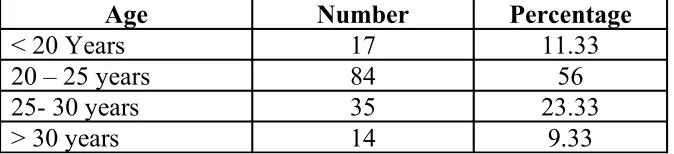
Table 1
Age wise distributions
Age Number Percentage
< 20 Years 17 11.33 20 – 25 years 84 56 25- 30 years 35 23.33 > 30 years 14 9.33
This table analyzed the age wise distribution.
In less than 20 years, there were 17 cases (11.33%).
Between 20-25 years, there were 84 cases (56%).
Between 25-30 years, there were 35 cases (23.33%).
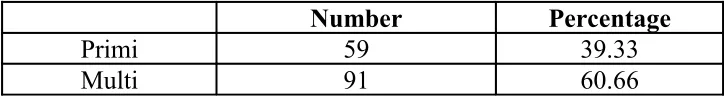
Table 2
Parity Distribution
Number Percentage
Primi 59 39.33
Multi 91 60.66
In this study 59 (39.33%) patients were primi gravidae.
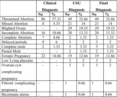
Table 3
Outcome in First Trimester Bleeding
Clinical Diagnosis USG Diagnosis Final Diagnosis
No % No % No %
Threatened Abortion 86 57.33 49 32.66 49 32.66 Missed Abortion 8 5.33 21 14 21 14 Blighted Ovum - - 29 19.33 29 19.33 Incomplete Abortion 16 10.66 20 13.33 20 13.33 Complete Abortion 7 4.66 2 1.33 2 1.33 Delayed periods 9 6 3 2 3 2 Complete mole 2 1.33 5 3.33 5 3.33 Partial Mole - - 2 1.33 2 1.33 Ectopic Pregnancy 22 14.66 19 12.66 19 12.66 Low Lying placenta - - 3 2 3 2 Ovarian cyst
complicating pregnancy
- - 3 2 3 2
Fibroid complicating pregnancy
- - 1 0.66 1 0.66
Bicornuate uterus - - 1 0.66 1 0.66
All the 8 cases (5.33%) diagnosed clinically as missed abortion were confirmed by ultrasonography. In addition 13 cases of threatened abortion were diagnosed as missed abortion by the ultrasonography.
No case of blighted ovum was diagnosed clinically but all the 29 (19.33%) cases were diagnosed by ultrasonography.
Incomplete abortion was diagnosed clinically in 16 cases (10.66%) but with ultrasonography 20 cases (13.33%) turned out to be incomplete abortion
Ectopic pregnancy was diagnosed clinically in 22 cases (14.66%) but confirmed by ultrasonography in 19 cases (12.66%) only. The remaining 3 cases turned out to be that of incomplete abortion by ultrasonography.
Complete abortion was confirmed by ultrasonography only in 2 cases (1.33%) but it was wrongly diagnosed clinically in 7 cases (4.66%).
Partial molar pregnancy was diagnosed by the ultrasonography in 2 cases (1.33%) and none of them were diagnosed clinically.
Low lying placenta and ovarian cyst were diagnosed as associated condition in 2% of cases by ultrasonography.
Fibroid uterus was diagnosed as associated condition in 1 case by ultrasonography.
Bicornuate uterus was diagnosed in 1 case by ultrasonography.
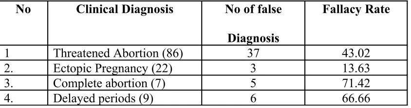
Table 4
Fallacy Rate of Clinical Diagnosis
No Clinical Diagnosis No of false
Diagnosis
Fallacy Rate
1 Threatened Abortion (86) 37 43.02 2. Ectopic Pregnancy (22) 3 13.63 3. Complete abortion (7) 5 71.42 4. Delayed periods (9) 6 66.66
This tabulation gives the fallacy rate of clinical diagnosis. In total there were 51 wrong clinical diagnosis giving a fallacy rate of 48%. Threatened abortion was diagnosed by clinical methods in 86 patients, where 37 cases of first trimester bleeding which was wrongly diagnosed clinically as threatened abortion turned out be pregnancy failures by USG giving a fallacy rate of 43.02%.
Table 5
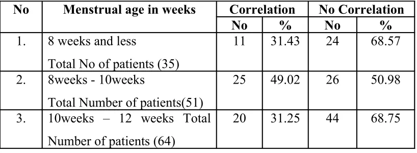
Correlation between menstrual age and clinical assessment
No Menstrual age in weeks Correlation No Correlation
No % No %
1. 8 weeks and less
Total No of patients (35)
11 31.43 24 68.57
2. 8weeks - 10weeks
Total Number of patients(51)
25 49.02 26 50.98
3. 10weeks – 12 weeks Total Number of patients (64)
20 31.25 44 68.75
This table shows the correlation between period of amenorrhoea and clinical assessment of uterine size. In those who presented at 8weeks or less than 8 weeks, there were about 35 patients.
Of these the uterine size correlated with period of gestation in 11 cases giving a percentage of 31.43%. In another 24 cases there was no correlation i.e., the size of the uterus was smaller than the gestational age giving a percentage of 68.57.
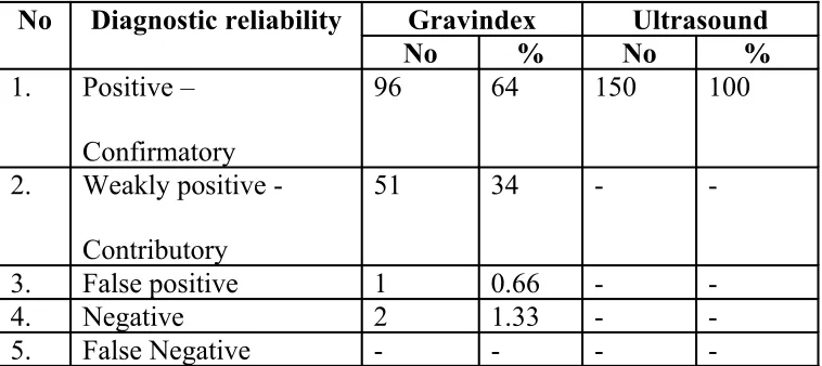
Table 6
Comparison of Reliability of gravindex & ultrasound diagnosis
No Diagnostic reliability Gravindex Ultrasound
No % No %
1. Positive – Confirmatory
96 64 150 100
2. Weakly positive - Contributory
51 34 -
-3. False positive 1 0.66 - -4. Negative 2 1.33 - -5. False Negative - - -
-This table compares the reliability of urine gravindex with ultrasound diagnosis.
The gravindex test was confirmative in 96 cases (64%), while it was contributory in 51 cases (34%).
False positive in 1 case (0.66%).
Negative in 2 cases (1.33%).
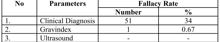
Table 7
Comparison of Fallacy rates of clinical Diagnosis, gravindex
and USG Diagnosis
No Parameters Fallacy Rate
Number %
1. Clinical Diagnosis 51 34 2. Gravindex 1 0.67 3. Ultrasound -
-This table compares the fallacy rate of clinical diagnosis, gravindex and ultrasound examination.
By clinical diagnosis alone, 51 cases were wrongly diagnosed resulting in fallacy rate of 34%.
Gravindex method resulted in 1 false positive diagnosis giving a fallacy rate of 0.67%
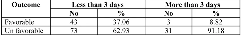
Table 8
Symptoms – Duration of Bleeding and Outcome
Outcome Less than 3 days More than 3 days
No % No %
Favorable 43 37.06 3 8.82 Un favorable 73 62.93 31 91.18
This table compares the duration of vaginal bleeding and pregnancy outcomes.
Out of 150 patients, 116 patients presented with complaints of vaginal bleeding less than 3 days duration. Among these 43 patients continued pregnancy (37.06%) and 73 patients aborted (62.93%).
34 patients had vaginal bleeding more than 3 days duration. Among these, one patient continued pregnancy and 2 patients were diagnosed as delayed periods.
Table 9
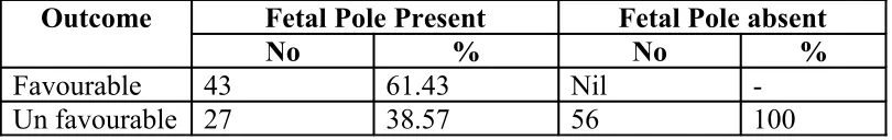
Fetal Pole and Outcome
Outcome Fetal Pole Present Fetal Pole absent
No % No %
Favourable 43 61.43 Nil -Un favourable 27 38.57 56 100
The presence of Fetal pole and pregnancy Outcome is tabulated in this column. Ultrasonographically fetal pole was seen in 70 patients. Of these in 43 patients pregnancy continued inspite of bleeding resulting in a percentage of 61.43.
27 cases (38.57%) aborted.
Table 10
Outcome in ultrasound diagnosis of threatened abortion
Number Outcome Number Percentage
1. Term fetus 36 73.46 2. Preterm Fetus 7 14.29 3. II trimester abortion 3 6.12 4. I trimester abortion 3 6.12
The outcome of patients who were diagnosed by the ultrasonography as threatened abortion is analyzed in this table. In clinically suspected cases of threatened abortion, 49 cases were proved by Ultrasonography as cases of threatened abortion.
Out of 49 patients, 36 patients who went upto term and delivered normal healthy babies resulting in a percentage of 73.46.
In this group, 7 patients (14.29%) went for premature labor.
Table 11
Diagnosis of pregnancy failure by ultrasound
S.No. Type of Pregnancy Failure Number Percentage
1. Blighted Ovum 29 40.27 2. Missed abortion 21 29.16 3. Incomplete abortion 20 27.77 4. Complete abortion 2 2.77
In analyzing the patients with diagnosis of pregnancy failure by ultra sound examination, the type of pregnancy failure recognized by ultrasound examination was either blighted ovum, missed abortion or incomplete abortion or complete abortion.
Blighted ovum was diagnosed in 29 patients (40.27%).
Missed abortion was diagnosed in 21 cases (29.16%).
Incomplete abortion was diagnosed in 20 cases (27.77%)
Table 12
Low Lying placenta in First Trimester & Outcome
S.No Outcome Number Percentage
1. Placenta migration to normal site 2 66.67 2. Spontaneous abortion 1 33.33
Low lying placenta was diagnosed by the ultrasonography in 3 cases.
On follow up of these 3 cases, 2 (66.67%) went to term with placental migration to normal position .
Table 13
Ultrasound Diagnosis of Gestational
Trophoblastic Disease
Outcome Number Percentage
Partial molar degeneration 2 28.57 complete molar degeneration 5 71.43
The incidence of molar pregnancy in cases of first trimester bleeding in our study was 7/150 (4.66%).
Partial molar degeneration was seen in 2 cases (28.57%) and complete molar degeneration in 5 cases (71.43%).
Table 14
Ovarian cyst complicating pregnancy
Diagnosis Clinical Diagnosis Ultrasound
No % No %
1 - - 7 4.66
In our study, associated abdomino pelvic pathology, which were not diagnosed by clinical examination were diagnosed by USG.
In 7 cases (4.66%) ovarian cyst was diagnosed as associated condition along with pregnancy.
Table 15
Duration of hospital stay after diagnosis
Duration of
hospital stay
Number Percentage
2 days 41 27.33 2 – 7 Days 83 55.33 > 7 days 26 17.33
This table analyzed the hospital stay after the diagnosis.
41 (27.33%)patients stayed in the hospital for 2 days.
83(55.33%) patients stayed in the hospital for 2 – 7 Days.
Table 16
Ectopic Pregnancy: Clinical Diagnosis Vs Ultrasound
Outcome Number Percentage
Clinical Diagnosis 22 14.66 Ultrasound Diagnosis 19 12.66
This table analyzed the diagnosis of Ectopic pregnancy by clinical and ultrasound examination.
Clinically 22 cases were diagnosed as ectopic pregnancy resulting in 14.66%. By ultrasonography 19 cases were diagnosed as ectopic pregnancy resulting in 12.66%, which is significantly lesser than clinical diagnosis.
Table 17
Fallacy rate in the Diagnosis of Ectopic Pregnancy
Outcome Number Percentage
Clinical 3 15.78
USG Nil Nil
This table gives the fallacy rate of clinical and ultrasound examination in ectopic pregnancy.
3 cases (15.78%) had a false diagnosis by clinical examination.
Table 18
Outcome of the study
S.No Management Number Percentage
1. Antenatal follow up and repeat scan. 45 30 2. Digital Evacuation & curettage 51 34 3. Suction Evacuation and curettage 27 18 4. Delayed periods and discharged 3 2 5. Laparotomy for Ectopic pregnancy 19 12.66 6. MTP with sterilisation 3 2 7. Readmission for spontaneous abortion 6 4
This table analysed the various outcome in first trimester bleeding.
The cases were followed up during the course of pregnancy with regular antenatal care and ultrasonography till delivery in 30%.
Digital Evacuation & curettage was done in 51 cases (34%) of blighted ovum and missed abortion
Suction Evacuation and Curettage was done in 27 cases (18%) of molar pregnancy and incomplete abortion.
3 multi gravida patients underwent Medical termination of pregnancy with sterilisation.
DISCUSSION
Ultrasound is a non invasive, easily accessible and highly diagnostic tool in the modern era of obstetrics.
Accurate diagnosis and proper intervention is mandatory to save not only the fetus but also the mother. Hence the differential diagnosis must be kept in mind before deciding further management.
Depending upon the correct diagnosis the management may vary from conservative observation to invasive laparotomy.
Ultrasonography nowadays has become a single most effective tool in diagnosing all the causes of first trimester bleeding. The differential diagnosis in our study included
1) Threatened abortion 2) Missed abortion 3) Blighted ovum 4) Complete abortion. 5) Incomplete abortion
It is impossible to diagnose cases clinically like low lying placenta and partial molar degeneration but ultrasound examination confirmed the diagnosis in all cases correctly.
Thus the ultrasound diagnosis helps in the correct line of management thereby, reducing the
a. hospital stay, b. anxiety and
c. in few cases it avoids unnecessary laparotomy.
In our study, 11.33% of the patients were in less than 20years, 56% were between 20-25 years, 23.33% were between 25-30 years 9.33% were above 30 years.
Threatened abortion
Study Incidence Percentage
Rajan et al 324 48
Stabilie & G. Grudzilkas et al 227 48.7 Joupilla et al 236 45
Our study 49 32
Thus in our study, eventhough it is a small cohort study, the incidence of threatened abortion is in par with other studies.
Viability outcome in Threatened abortion
Our study 36 cases 91.40%
In Rajan et al study, of cases of threatened abortion with viable fetus, 93.33% continued for term pregnancy and had healthy term infants. It is in par with our study.
Early diagnosis, timely intervention & necessary treatment initiated immediately has culminated in improving the viability outcome.
Anembryonic pregnancy
Study No. %
How et al - 45
Our study - 40
In our study 40% of cases diagnosed as anembryonic pregnancy. These patients underwent dilatation and curettage.
Irregular sac and pregnancy outcome
Irregular sac in ultrasound almost confirms the non-viability of the conceptus.
Absence of fetal pole and pregnancy outcome
Absence of fetal pole in trans abdominal sonography by 7weeks is indicative of non viable pregnancy.
Scan finding Nyberg et al Our study
Irregular Sac 99% 100% Absence of fetal pole 100% 100%
In Nyberg et al study, absence of yolksac or embryo ended in termination of pregnancy with 100% specificity. It is in par with our study. Absence of fetal echo was diagnosed in 56 cases, all of which ended in termination with 100% specificity.
Incomplete Abortion
Stabile and G.Grudzinkas et al 8.8% Drumm’s et al 28.1% Neelam bharath et al 22% Our study 13.33%
Ultrasound has been proved to be the diagnostic tool in the diagnosis of incomplete abortion. In our study, incomplete abortion was diagnosed by ultrasound examination with 100% specificity in 13.33% of cases. Digital evacuation and curettage was done immediately.
Stabile and G.Grudzinkas et al 0.9%
Our study 2%
Ultrasonography confirms complete abortion (2% of cases) with reliability. It avoids unnecessary Digital evacuation and curettage, an invasive procedure for the patient.
Gestational Trophoblastic Disease
Drumm’s et al 8% Neelam bharath et al 2% Malhotra et al 1% our study 4.36%
Out of 150 cases molar pregnancy was diagnosed in 7 cases by ultrasound. Of these 7 cases, 2 cases were diagnosed clinically. The uterus size was greater than period of amenorrhoea in these 2 cases. Suction evacuation and curettage were done. The specimen sent for histopathological examination. Follow up with quantitative estimation of HcG were done.
Ectopic Pregnancy
Stabile and G.Grudzinkas et al 12.6%
Levi et al 9.2%
Romasufat et al 8%
Our study 12%
many cases were referred late and hence medical / conservative management could not be done. So, all the cases were managed surgically. Of these, one case ended in total abdominal hysterectomy in a multi gravida due to ruptured cornual ectopic pregnancy with profuse bleeding.
Low Lying placenta & their outcome
Mantoni et al 85%
Our study 66%
Mantoni et al determined that the majority of patients (85%) with a low lying placenta had viable pregnancy. In our study, 66% of patients with low lying placenta had live term babies.
Ovarian cysts were diagnosed along with pregnancy as associated condition in 7 cases. Of these 6 were functional cysts. A hemorrhagic cyst was present in left ovary in one case of ectopic pregnancy. Left ovarian cystectomy was done during laparotomy. On the right side, ampullary portion of the tube was the site of ectopic pregnancy and Salphingo Oophrectomy was done on right side.
One case of fibroid uterus was diagnosed along with pregnancy.
SUMMARY
150 patients with period of Amenorrhoea of less than 12 weeks with bleeding per vaginum were included in the study.
Of these 59 cases were primi gravidae and 91 cases were multi gravidae. Their age distributions was as follows
:- 17 patients were less than 20 years.
84 patients were between 20-25 years.
35 patients were between 25-30 years.
14 patients were above 30 years.
Patients presented with bleeding per vagina with duration less than 2 days were 71. 46 patients had bleeding per vagina of 2-7 days duration. 15 patients had bleeding per vagina for more than 7 days. The duration of bleeding per vagina was variable with nearly 80% having duration less than 7 days.
rest of them had non viable pregnancy. These cases of threatened abortion were monitored and pregnancy was allowed to continue.
73.46% went for term pregnancy and delivered term healthy babies.
14.29 % went for preterm labour and delivered preterm babies.
6.12% had second trimester abortion spontaneously.
6.12% had spontaneous first trimester abortion.
Missed abortion was diagnosed in 5.33% cases clinically and in 14% cases by ultrasonography. Digital evacuation and curettage were done for them. Ultrasonography diagnosed more cases of missed abortion than clinical examination.
Blighted ovum was diagnosed in 19.33% cases by USG and no case was detected clinically. Digital evacuation and curettage was done.
12.66% cases were diagnosed as ectopic pregnancy by ultrasonography and 14.66% cases were diagnosed clinically. Laparotomy was done immediately after the diagnosis.
1.33% cases had partial molar pregnancy and 3.33% cases were diagnosed to have complete molar pregnancy. Suction evacuation and curettage were done for all of these cases. Specimen were sent for HPE and were confirmed as molar pregnancy.
CONCLUSION
Ultra Sonography has been proved as an important diagnostic modality in obstetrics. It is an easy available, diagnostic modality and it helps in the earlier diagnosis of complications of first trimester bleeding. In the above study it was demonstrated that it played an important part in diagnosis of first trimester bleeding.
This diagnosis helped in prompt treatment of first trimester pregnancy in a better manner before any complication could develop.
If relied upon the history and clinical findings, it can lead to delay in diagnosis and it may lead to various complications and may increase the morbidity of the patients.
By earlier diagnosis not only the mortality and morbidity are reduced but also the earlier management reduced the hospital stay of the patients.
BIBLIOGRAPHY
1. Blumenfeld . Z. et al . Transvaginal Sonographic assessment of early embryological development. In T. Tritsch and S. Rolten (eds) Trans vaginal Sonography London. Heinemann 1988 Pp 87-108.
2. Benacerraf B.R. Intra uterine growth retardation in the first trimester associated with triploidy J.Ultrasound Med 7:153,1988.
3. Clucote . W. S. and Asokan . S. Evaluation of first trimester pregnancy by ultrasound. clin. Obstet. Gynaecol 20:253, 1977.
4. Chard, T Frequency of Implantation and Early pregnancy was in natural cycles. In Ballieres Clinical Obstetrics and Gynecology (Vol 5 (1) P.179, 199)
5. Campbell,S.Ultrasound in Obstetrics.Br.J.Hosp.Med 8:541,1972
6. Crooij M. J. et al Ultrasonographic measurement of the yolksae Br. J. obstet gynaecol 89:931, 1982.
7. Donald, I. The use of ultrasounds in diagnosis of abdominal swelling.Br.Med.J.2:1154,1963.
9. Hellman, L.M. et al, Growth and Development of the human fetus prior to the twentieth week of gestatation Am. J. obstet Gynecol 103:789, 1969.
10. Harris R.D. Vincent L.M. and Askin F.B. yolksae calcification. A sonographic finding associated with intra uterine embryonic demise in the first trimester. Radiology 166:109, 1988.
11. Jansen R.P.S. Spontaneous abortion incidence in the treatment of Infertility. Am.J.Obstet.lynecol 143: 151,1982 .
12. Jouppila, P.Ultrasound in the diagnosis of early pregnancy and its complications. Acta obstet .Gynecol Scand 15 (sup) 50:7, 1971.
13. Kohorn, E.I and Kaufman, M.Sonar in the first trimester of pregnancy Obstet. Gynecol 44:473, 1974.
14. Leridon. H. Farts and artifacts in the study of intra uterine mortality; a reconsideration from pregnancy histories Popul. stud 30:319, 1976
15. 15. Mckensie, W.E. Holmes. D.S and Newton J.R. Spontaneous abortion rate in ultra sonographically viable pregnancies. Obstet. Gynecol 71:81, 1988.
16. Mantoni M. and Pederson J. F. ultrasound visualization of the human yolksac. J. Clin ultrasound 7:459,1979.
18. Mantoni.M. and Pederson J.F. Intra uterine haematoma an ultrasound study of threatened abortion Br.J. Obstet. gynaecol 88:47, 1981.
19. MuramD.Gillieson M. and Walters J.H.Myomas of the uterus in pregnancy ultrasonographic follow up Am J.Obstet Gynecol 138:16,1980.
20. Nyberg D.A. et al, value of the yolksac in evaluating early pregnancies J. Ultrasound Med 7:129, 1988.
21. Piironen. O. Studies in diagnostic ultrasound. Acta obstret Gynecol. Scand {Suppl} 46:5, 1975.
22. Pederson J.F. Fetal crown rump length measurement by ultrasound in normal pregnancy Br. J. Obstet gynaecol 89:926, 1982.
23. Robinson, H.P.Detection of fetal heart Movement in the first trimester of pregnancy using pulsed ultrasound. Br.Med. J4:466, 1972.
24. Robinson H.P.Gestational Sal Volumes as determined by sonar in the first trimester of pregnancy Br.J.Obstet. Gynecol 82:100,1975.
25. Robinson H.P Sonar measurement of crown rump Length as a means of assessing maturity in first trimester of pregnancy Br.Med. J 4:28, 1973.
27. Rizos . N. etal Natural History of placenta praevia ascertained by diagnostic ultrasound. Am.J. Obstet gynaecol 133:287, 1979.
28. Robinson H.P. and Shaw D. J. Fetal Heart rates as determined by sonar in early pregnancy J. Obstet. Gynaecol . Br. Common w. 80:805, 1973.
29. Robinson H.P. The diagnosis of early pregnancy failure by Sonar Br.J.Obstet. Gynaecol 82:849, 1975.
30. Reuter K.Michdewitz H. and Kahn.P. Early appearance of Hydatidiform mole by ultrasound, a case report A.J.R.134:588,1980.
31. Schats. R. Jansan . C.A.M. and Wladimiroff . J.W.Embryonic heart activity. Appearance and development in early human pregnancy. Br.J.Obstet.Gynaecol :97:989, 1990.
32. Stabile I etal . The diagnostic of early pregnancy failure. In E.S. Teoh. S.S. Rathnam and P.C. Wong (ed) Advances in Fertility and sterility (Vol.1) Parthenon, 1987 Pp 153-160.
33. Seruerbrei E. Cooperberg P.L. and Poland B.J. Ultrasound demonstration of the normal fetil yolk sac .J. Clin ultrasound 8:217, 1980.
34. Tietze, C. Introduction to the statistics of abortion In E. Engle, pregnancy wastage, spring field IL Charles C Thomas, 1953 p.153.
36. United Nations Organisation Department of Social Affairs Fetal, Infant and childhood Mortality. I. The Statistics. Newyork. United Nations, 1954.
37. Varma T.R. The implications of a low implantation of the placenta detected by Ultrasonography in the early pregnancy. Acta.O.scand. 60:265,1981.
38. Whittaker.P.G.Taylor.A and Lind, T.Unsuspected pregnancy loss in healthy women. Lancet 1:1126, 1983.
Proforma
Name Date Age Unit ADD IP No: Parity : LMP : EDD :
Duration of Bleeding
Any other symptom
H/o passing products Past History :
Menstrual History
Married since
Obstetrics Table G P L A
Uterus size Sac measurement Fetal Echo
FH CRL Placenta Adnexa
Outcome in I Trimester Bleeding
86 8 0 16 7 9 20 22 00 00 49 21 29 20 23 52 19 33 11 49 21 29 20 23 52 19 33 11 0 20 40 60 80 100 120 140 160Clinical Diagnosis USG Diagnosis Final Diagnosis Threatened abortion Missedabortion
Blighted abortion Incomplete abortion Complete abortion Delayed periods Complete mole partial mole Ectopic Pregnancy Low lying placenta
Fallacy Rate of Clinical Diagnosis
37 3
5
6
Outcome in ultrasound diagnosis of threatened abortion
36 7
3
3
Diagnosis of pregnancy failure by ultrasound
29
21 20
2
Ectopic Pregnancy Clinical Vs Ultrasound
22 19
Low lying placenta in First trimester and outcome
2 1
Ultrasound diagnosis of gestational trophoblastic disease
2
5
Correlation between menstrual age and clinical assessment
11 25 20 24 26 44 0 5 10 15 20 25 30 35 40 45Age wise distribution
17
84
35
14
0 10 20 30 40 50 60 70 80 90
Number
Outcome
45 51 27 3 19 3 6 0 10 20 30 40 50 60 NumberAntenatal followup and repeat scan Digital Evaluation and curettage Suction evacuation Delayed periods and discharged Laparatomy MTP with sterisation
Symptoms – Duration of Bleeding and Outcome
43 73
3 31
0% 10% 20% 30% 40% 50% 60% 70% 80% 90% 100%
Less than 3 days More than 3 days
Fetal Pole and Outcome
43
0 27
56
0 10 20 30 40 50 60