Biological Systems
Thesis by Jason M. Belitsky
In Partial Fulfillment of the Requirements for the Degree of
Doctor of Philosophy
California Institute of Technology Pasadena, California
2002
To my friend and lab-mate Brock SafronofJ
Acknowledgments
First and foremost I want to thank my family for all their support. I'm particularly glad that my brother Darren moved out here to experience LA with me these last two years.
The strength and togetherness that my parents have shown in overcoming their health problems in recent years will always be an inspiration to me. I love them very much and am
extremely grateful that we were able to reach this milestone together in good health.
Next, I want to thank my advisor, Peter Dervan, for his enthusiasm, encouragement, and for providing me with good research projects and even better co-workers. I also want to
thank the members of my committee, Rich Roberts, Doug Rees, and Bob Grubbs, for all their support. I am grateful to the professors whose classes I had the opportunity to attend, especially to Andy Myers, Barbara Imperiali, and Rich Roberts, and to the professors that I
had the opportunity to TA for, particularly Peter Dervan, Bob Grubbs, Dennis Dougherty,
and Ken Williamson. I would also like to thank Harry Gray for the weekly Organo-metallics seminar, where I able to see a great deal of interesting chemistry. The student-run
Organic Chemistry Journal Club was also a great learning experience for me, for which I especially have to thank J.P. Morgan, Isaac Carrico, John Stellwagon, Amab ChatteIjee, and Tae-Lim Choi. I would never have been in a position to learn anything at Caltech if not for the great education I received from the Amherst College chemistry department, particularly
from my undergraduate thesis advisor David Hansen.
project; growing cells, fighting mold, imaging, and planning our next step. This project
would never have gotten off the ground at Caltech without the help of many people (listed in the Acknowledgements in Chapters 4A and 4B), particularly Inder Nangiani of the Caltech
Protein Expression Facility.
I'm especially thankful for all the great friends I've made at Caltech, especially Doan Nguyen, Isaac and Liz Carrico, Niki Zacharias, J.P. Morgan, Amab Chatterjee, Walter Goldberger, Catherine Baker, Girish Aakalu, Mike Farwell, Soojin Kwon, Carlos Basques, Peter Hackley, and Nick Wurtz. "Renaldo" thanks the entire Grubbs group for their friendship, and the Athenaeum for foozball. I want to thank my current house-mates and friends Niki Zacharias, Gabe Brandt, and Sarah Monahan for putting up with me while I was writing my proposals and thesis. I'm grateful to David Malmud, Rob Leibowitz, Scott Mellender, and Steve Lasher for their long and continuing friendship.
Finally I want to thank the entire Dervan group for their friendship, advice, collaboration, and commiseration. I've been surrounded by great scientists in my class-mates Doan Nguyen, Nick Wurtz, and Adam Urbach. I'm particularly grateful to have been
lab-mates with Doan, Anna Mapp, Paul Floreancig, David Herman, John Trauger, Adam Kerstein, Meredith Howard, Philipp Weyermann, Christoph Briehn, Tim Best, Shira Jacobsen, and John Chevillet. Getting to know and working with Doan, especially, has been a highlight of my Caltech experience. I've also benefited very much from the wisdom and friendship of Ryan Bremer, Clay Wang, Aileen Chang, Christian Melander, Tom Minehan, Ulf Ellervik, and Nick W. I've enjoyed the Amherst connection with Meredith, Adam K., Scott Carter, and Sue Swalley. I've had many interesting conversations with Victor Rucker, and enjoyed getting to know the BI contingent, Bogdan Olenyuk, Pierre Potier, Inger Kers, Konstanze Gottewald, and Simon Friedman. Last but definitely not least, I want to thank three postdocs who helped me out tremendously when I first joined the lab and even after they had left it: Anna Mapp, Paul Floreancig, and Roland Burli. I couldn't ask for better
Abstract
Small molecules that bind selectively to a DNA sequence in the human genome are
potentially useful tools for molecular biology and human medicine. Polyamides containing N-methylimidazole (1m) and N-methylpyrrole (Py) are small molecules that bind DNA
according to a set of "pairing rules" with affinities and specificities that rival natural
transcription factors. By directly competing with a given transcription factor or other DNA
binding protein for its binding site, polyamide can cause inhibition of diverse biological
processes, such as retroviral integration and gene transcription. Polyamides are presented which inhibit the in vitro integration activities for two retroviruses, M-MuLV and HIV-l.
Polyamides are described that inhibit TBP binding to the HER2 promoter, a gene implicated in human breast cancer. Failure to achieve inhibition of HER2 transcription in cell culture
led to the surprising discovery that polyamide-fluorescent dye conjugates are cell-permeable, but that nuclear localization does not occur in many cell lines. Efforts toward
modified polyamides with enhanced nuclear localization properties are presented. In order
Table of Contents
page
Acknowledgments... ... ... ... ... ... ... iv Abstract... ... ... ... ... .... ... ... ... Vi
Table of Contents.... ... ... .... ... .... ... ... vii List of Figures and Tables... IX
CHAPTER ONE:
CHAPTER TWO:
Chapter2A:
Chapter2B:
CHAPTER THREE:
CHAPTER FOUR:
Chapter4A:
Chapter 4B:
Introduction... 1
Inhibition of Retroviral Integration by DNA Binding
Polyamides ... ... ... ... ... 19 Inhibition of Moloney Murine Leukemia Virus Integration Using Polyamides Targetiing the Long-Terminal Repeat (L TR) Sequences ... ... ... ... ... 21 Inhibition ofHIV-l Integration by Hairpin Polyamides... 49
DNA Binding Polyamides for the Inhibition ofHER2
Transcription... 62
Cellular Uptake and Localization of DNA Binding
Polyamide-Fluorescent Dye Conjugates ... 89 Cellular Uptake of
N-MethylpyrroleIN-Methyl-imidazole Polyamide-Dye Conjugates ... 91 Enhanced Nuclear Localization of Polyamide-Dye
CHAPTER FIVE: Synthesis and Investigation of DNA Binding
Polyamides with New C-Terminal Tails ... 140
Chapter SA: Solid-Phase Synthesis of DNA Binding
Polyamides on Oxime Resin ... 142
Chapter SB: From an Aminooxy ~-Alanine Analog to Novel
List of Figures and Tables
CHAPTER ONE
pageFigure 1.1 Structure of double-helical B-fonn DNA ... 2
Figure 1.2 DNA base pairs and schematic representation of the minor goove ... 3
Figure 1.3 Representative X-ray crystal structures of DNA binding proteins... 3
Figure 1.4 Structures of compounds which bind in the minor groove .. ... 4
Figure 1.5 Schematic representation of the polyamide pairing rules... 5
Figure 1.6 X-ray crystal structure polyamide homodimer... 6
Figure 1.7 Inhibition of gene transcription by hairpin polyamides... 8
Figure 1.8 Examples of gene promoters tareted by hairpin polyamides ... 9
Figure 1.9 X-ray crystal structure of the nucleosome core particle ... 12
CHAPTER TWO Figure 2.1 Figure 2.2 Figure 2.3 Figure 2.4 Figure 2.5 Figure 2.6 Figure 2.7 Figure 2.8 Polyamides designed to bind the M-MuLV LTR ... 26
DNase I footprinting experiments... 30
Schematic illustration of in vitro integration assays... ... 31
Inhibition of 3' processing reactions ... 32
Inhibition of strand transfer reactions ... 33
Strand transfer into an exogenous substrate ... 35
ICso of polyamides against M-MuL V integrase... 36
DNA melting experiments ... 42
Figure 2.9 Polyamides designed to bind the HIV-l U5 LTR... 50
Figure 2.10 Binding sites for polyamides in the HIV-1 U5 LTR... 51
Figure 2.11 DNase I footprinting experiment ... 53
Figure 2.12 ICso of polyamides against HIV -1 integrase... 54
Table 2.1 Equilibriwn association constants for M-MuL V L TR ... 29
Table 2.2 DNA melting with internal and terminal polyamide binding sites... 43
Table 2.3 Equilibriwn association constants for HIV -1 U5 LTR ... 53
CHAPTER THREE Figure 3.1 HER2 signaling pathway... 65
Figure 3.2 Sequence ofthe HER2 promoter... 67
Figure 3.3 Overview of poly ami des targeted to the HER2 promoter... 69
Figure3.4 TATABindingProtein ... 70
Figure 3.5 Polyamides targeted to the HER2 promoter in this study... 72
Figure 3.6 DNase I footprinting experiments... 75
Figure 3.7 Comparison oftwo different binding sites... 77
Figure 3.8 TBP electrophoretic mobility shift assays ... 80
Table 3.1 Equilibriwn association constants for HER2 promoter ... 74
Table 3.2 Equilibriwn association constants for M-MuL V L TR ... 76
CHAPTER FOUR Figure 4.1 Structures ofpolyamide-Bodipy FL conjugates... 93
Figure 4.2 Conjugate 1 in live SKBR-3 cells and after methanol addition ... 96
Figure 4.3 Conjugate 1 and the dead cell stain Sytox Orange in Sf9 cells... 96
Figure 4.4 Conjugate 2 in live Sf-9 cells and after methanol addition... 97
Figure 4.5 Conjugates 1 and 3 in live CEM cells... 98
Figure 4.6 Synthesis of polyamide-Bodipy conjugates ... 106
Figure 4.7 Fluorescence of Bodipy conjugate 4 ... 107
Figure 4.8 DNase I footprinting experiments ... 108
Figure 4.9 Conjugate 3 in live Hi-5 cells and conjugate 1 in live LnCap cells ... 112
Figure 4.10 Synthesis of Bodipy-cyclic polyamide conjugate 11 ... 113
Figure 4.11 Tandem dimer and one-to-one binding Bodipy conjugates ... 114
Figure 4.13 Structures of CHL-polyamide-Bodipy conjugates ... 117
Figure 4.14 CHL conjugates 18-19 in live NB4 and primary human T-cells ... 118
Figure 4.15 Conjugate 23 in live LnCap and PC3 cells; small molecule conjugates .... 120
Figure 4.16 Structures of carrier peptide conjugates ... 122
Figure 417 Carrier peptide conjugates 24,26,28 in live Sf9 and Kc cells ... 123
Figure 4.18 Structures offootprinting compounds ... 126
Figure 4.19 DNase I footprinting experiments ... 127
Figure 4.20 DNase I footprinting experiments ... 128
Figure 4.21 Structure of disulfide linked R9 conjugate ... 129
Figure 4.22 Structures of Bodipy-cationic polyamide conjugates ... 131
Figure 4.23 Conjugate 41 in live CEM and Kc cells ... 132
Figure 4.24 PD-4 conjugates 47-48 in live Kc and Sf9 cells ... 133
Table 4.1 Cellular localization of polyarnide-Bodipy conjugates ... 95
Table 4.2 Equilibrium association constants for Bodipy conjugates ... 110
Table 4.3 Screening of standard hairpin-Bodipy conjugates ... III Table 4.4 Confocal microscopy of different polyamide motifs ... 114
Table 4.5 Screening ofpolyamide-chlorambucil-Bodipy conjugates ... 119
Table 4.6 Screening ofpolyamide-Bodipy-small molecule conjugates ... 121
Table 4.7 Screening ofpolyamide-Bodipy-carrier peptide conjugates ... 124
Table 4.8 Equilibrium association constants for peptide conjugates ... 128
Table 4.9 Screening of cationic polyamide-Bodipy conjugates ... 132
CHAPTER FIVE Figure 5.1 Schematic of polyamide binding ... 143
Figure 5.2 Structures of poly ami des with different C-terminal tails ... 145
Figure 5.3 Polyamide synthesis on oxime resin ... 146
Figure 5.4 DNase I footprinting experiments ... 149
Figure 5.5 Model of aminooxy acid unit in a polyamide... ... ... 159
Figure 5.7 Solid-phase synthesis of primary amides from aminooxy linker. ... 162
Figure 5.8 Aminooxy linker route to C-terminal cyclic tertiary amides ... 163
Figure 5.9 Products from reaction with Dp or reductive conditions ... 164
Figure 5.10 DNase I footprinting experiments ... 165
Table 5.1 Table 5.2 Equilibrium association constants for truncated tails... .... 151
Background
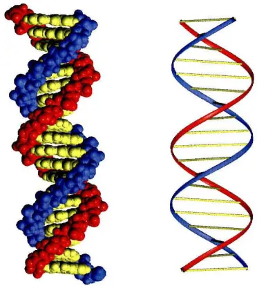
DNA is the storehouse of genetic information, containing all the information required for the growth and development of every organism. I DNA is composed of
deoxyribose-phosphate polymers that display four heterocycles bases, adenine (A), thymidine (T), guanine (G), and cytosine (C). DNA is self-organized as a double-helical duplex of antiparallel strands, which are held together by Watson-Crick hydrogen bonding of A,T and G,C base pairs.2 The recent sequencing of the human genome refers to deciphering the linear order of billions of these base pairs which code for 30,000-40,000 genesY Understanding the complex interrelationships between these genes and their protein products is the central question in molecular biology and human medicine. Aberrant regulation of gene expression, though mutations or the intervention of pathogens, is responsible for a variety of disease states.5 Synthetic ligands which can
affect gene expression by binding to specific predetermined DNA sequences will be powerful tools for our growing understanding of the human genome, and potentially will
[image:14.521.156.342.426.633.2]be valuable therapeutic resources.6
Figure 1.1
Figure 1.2
Major Groove
,H
'\.--Ito' ..
'H-N Nf(
N~:N-H""
'N~l
~N RA 0 0 \) ()
Minor Groove
G~C
C~G
Akr-e-elT
T~A
Figure 1.2. The edges of the base pairs present different hydrogen bond donor and acceptor combinations to the major and minor grooves in the DNA double helix. The top of the bases, as drawn projects into the major groove and the bottom projects into the minor groove. Below the base pairs, the minoT groove is shown in schematic representation, where circles with dots represent lone pairs of N(3) of purines (A,G) and 0(2) of pyrimidines (C,T), and circles containing an H represent the 2-amino group of G.
DNA Recognition
The common B-form of DNA is characterized by a wide (12
A)
and shallow major groove and a narrow (4-6A)
and deep minor groove (Figure 1.1).7 In addition, sequence-dependent structural variations, conformational properties, and solvent and counterion organization can distinguish local DNA structures.7 Individual sequences are distinguished by the pattern of hydrogen bond donors and acceptors present at the edges of the base pairs (Figure 1.2). Proteins which recognize DNA take advantage of specific hydrogen bonding or van der Waals contacts with functional groups in the grooves, Coulombic attraction to the negatively charged phosphodiester backbone or to theFigure 1.3
GCN4 TBP Zif268
Figure 1.3. Representative X-ray crystal structures of DNA binding proteins.8
electrostatic potential in the grooves, and/or intercalation of aromatic functional groups between the DNA bases.8 DNA binding proteins adopt a variety of structural motifs for
sequence-specific recognition including zinc finger9 and leucine zipper!! motifs (Figure
1.3). Specificity for target sites is achieved through specific noncovalent interactions
between the protein side chains and the nucleobases and phosphates of the DNA. However, no single motif exists that represents a general amino acid-base pair code for all DNA sequences.!2 Recognition by some proteins, such as the ubiquitous transcription factor TBP,1O involves large distortions of the target DNA from its common B-form structure, including bending and unwinding of the helix.
Figure 1.4
C~O=;~UN
'"
H-NV H I
Had A OH
Hoechst 33258 Calicheamicin oligosaccharide
H +
H N NH2
r
In
'~"'Y~
n .
~NH
2
H HNI 0 J..N~N~
\ 0 \ 0
Ecteinascidin 743
H,N
J!
"" /\",o OHHN2i>"" ;. '>- I OM.
~
"NHo A N N
~I'" 0 o N & OH
H OM. CC-1065 Distamycin
Figure 1.4. Structures of four natural products and a synthetic compound (Hoechst 33258) which bind in
the minor groove of DNA. Note the similar crescent shape of distamycin, CC-1065 and Hoechst 33258.
0Iiogsaaccharide,18,19 CC_1065,2o,21 and Hoeschst 33258,22,23 possess modular structures that have allowed chemists to synthesize numerous derivatives in order to understand and modify their sequence specificity and binding affinity. Distamycin,24 which binds five base pair A,T rich sequences, is particularly well suited to modification, owing to its relatively simple structure composed of N-methylpyrrole carboxamides.22,25.28
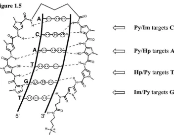
Pyllm targets C· G
PylHp targets
A·T
Hp/Py targets T·
A
[image:17.522.51.403.200.474.2]Im/Py targets
G· C
Figure 1.5. A schematic representation of the polyamide pairing rules.
DNA Recognition by Polyamides
A twenty year research effort in the laboratories of Prof. Peter B. Dervan have led to the development of a class of minor-groove binding small molecules which can recognize a large number of predetermined DNA sequences with affinities that rival natural transcription factors.6,29,3o DNA binding polyamides contain N-methylpyrrole (Py)
side-by-side amino acid pairings in the minor groove that stack the aromatic rings against each other and the walls of the groove allowing backbone amide hydrogens and the substituents at the 3-position of the Py, 1m, and Hp residues to make specific contacts with the edges of the DNA base pairs. Each pair of polyamide residues is selective for a specific DNA base pair based on steric factors and the matching of hydrogen bond donor and acceptor functionalities (Figure 1.5). A pairing of 1m opposite Py (lmJPy) targets a Goe base pair, while Py/lm targets CoG. A PylPy pairing is degenerate, targeting both AoT and ToA base pairs. An Hp opposite a Py (Hp/Py) discriminates ToA from AoT, while Py/Hp targets AoT in preference to ToA and both of these from Goe and CoG.
Footprinting, NMR, and X-ray structure studies validate these pairing rules for DNA minor groove recognition (Figure 1.6).29.31
Figure 1.6
While many different recognition motifs based on pyrrole-imidazole polyamides
are possible, the design known as the hairpin (Figure 1.5) has been widely used for
biological applications. 32 The N- and C-terminal strands of Pyllm aromatic amino acids
are connected by an alkyl amino acid--either y-aminobutyric acid (y), or the chiral,
amine-functionalized derivative (R)-2,4,-diaminobutyric acid «R)H2Ny) __ in an antiparallel
orientation relative to each other and, generally, a 5'-3' N-C orientation relative to DNA.
The linker between Py/Im strands is considered the "tum" of the hairpin, and alkyl units
f3-alanine (13) and N,N-dimethylaminopropylamine (Dp) are typically used to form a
C-terminal "tail." All of these alkyl units are specific for both A,T base pairs. f3-alanine
(13) can also be used internally to form 13/13, f3/Py, and f3lIm pairs, which has extended the
binding site size amenable to recognition by the hairpin polyamides and in certain cases results in increased affinity and specificity.33 Tandem hairpin dimers have been produced
which bind with high affinity and specificity.34.35 The hairpin motif is also a versatile
scaffold for the production of conjugates with diverse functions including
sequence-specific DNA-alkylation36.37 and recruitment of Topoisomerase
e
sGene Regulation
Hairpin polyamides have shown the ability to block transcription factors from
binding to their DNA target sites and thereby inhibiting transcription (Figures 1.7 and
1.8).29,32 In the first demonstration of this approach, an eight-ring hairpin polyamide was
targeted to the binding site of the zinc finger transcription factor TFIIIA in the Xenopus
5S RNA gene promoter, at a sequence which coincides with a zinc finger-minor groove
interaction (Figure 1.8).6 Transcription by RNA polymerase III was suppressed in in
vitro transcription assays and following treatment of whole Xenopus kidney-derived
fibroblast cells with the match polyamide. Very recently it has been demonstrated that
allosteric mechanism.39 Allosteric interactions between the major and minor grooves
were recently shown to be critical for inhibition of the major-groove binding protein UL9
by a minor-groove binding oligopyrrole small molecule (GLX, an indole bridged bis-netropsin).40 However, minor-groove binding ligands including hairpin polyamides can
also co-occupy DNA while certain proteins (such as GCN4, Figure 1.3) occupy the major
groove.41 GCN4 binding has been inhibited by hairpin polyamides containing "positive
patches" targeting protein-phosphate contacts,42.43 and by polyamide-intercalator conjugates.44
Figure 1.7
i
.
::!::J
i
.
::
:
::)
i
.
::::>
,.
m:::>
Figure 1.7. Inhibition of gene transcription by hairpin polyamides. A pair of hairpin polyamides targeted to the DNA sequences adjacent to the binding sites for Ets-I, LEF-l, and TBP inhibit assembly of the transcriptional machinery and transcription of the HIV -I genome.45 Polyamides are shown in ball-and-stick
format. Red and blue circles represent imidazole and pyrrole residues, respectively. ~-Alanine and the y-turn are depicted as green diamonds and curved lines, respectively. The plus sign represents the dimethylaminopropylamide tail.
Minor -groove binding transcription factors, such as TBP (Figure 1.3), LEF-l, and TAX, have been excellent targets for polyamide inhibition, resulting in inhibition of RNA polymerase II transcription of targeted genes in vitro such as HIV -1 and HTL V-I (Figure 1.8):5.47 Interestingly, in the absence of TAX the polyamides shown on the HTL V-I
promoter or another polyamide targeted to the center of the CREB site, do not inhibit the association of major-groove binding CREB with DNA.47 However, in the presence of
Figure 1.8
TFIIIA
I 3-1
58 RNA Promoter 70
----
805'-G C C T G G T TAG T ACT T G GAT G G G-3'
~
3'-C G G A C C A AT CAT G A A C C T A C C C-5'
HlV-l Promoter TBP
5' -A GAT G C T G CAT A T A A G
~
3'-T G T A C G A CEts-1 LEF-1
HIV-I Promoter
5' -G A G T A C A A A G A C T-3'
" " "
CGACGTAGGC
TAX CREB TAX
H1LV-l Promoter
5' -C T C C C T C A C-3'
G-5'
HER2 Promoter Ets (ESX)
5' -G G C T G C T T GAG G A A G TAT A A G A A-3'
+
3'-C C G A C G A A C T C C CAT A T T C T T-5'
CMVMIE Promoter lESS
5'-G C T C G T T TAG T G A A C C G T C A G-3'
B+.~
3'-C G A G C A A A T C ACT T G G C A G T C-5'
Figure 1.8. Examples of gene promoters where transcription factor binding sites that have been targeted
binding. A similar result was obtained on the HIV -1 promoter with a polyamide
targeting Ets-l (Figure 1.8, polyamide on the left), which was able to inhibit binding of
NF-KB to an adjacent DNA site by blocking the formation of a cooperative Ets-loNF-KBo
DNA ternary complex.49 These results show that polyamides are able to inhibit proteins
that they do not directly contact, and may provide a general method for inhibition of the
association of major-groove binding transcription factors with their target DNA.
Proteins that have a majority of major-groove contacts and few or even one
minor-groove contact have been efficiently inhibited by hairpin polyamides. The binding
of two Ets transcription factors, Ets-l and ESX, to the HIV-l and HER2 promoters,
respectively, has been efficiently inhibited by hairpin polyamides (Figure 1.8).45.46.49 Ets
proteins have a winged-helix-turn-helix motif and bind primarily in the major groove
with additional phosphate contacts across the adjacent minor groove(s). Polyamide
inhibition of the binding of NF-KB, which has one such contact across the minor groove,
is likely due, at least in part, to allosteric interactions between the grooves.50 However, in
another case of polyamides inhibiting transcription factor binding with only one
phosphate contact outside the major groove, synthetic modification of the bHLH domain
of the Drosophila Deadpan showed that the inhibition depended on the specific lysine
residue responsible for the phosphate contact.51 Inhibition by two polyamides with
distinct binding sites was then used to demonstrate unusual asymmetric binding of this
homodimeric bHLH protein. This highlights the use of polyamide not only for inhibition
but also for the study of transcription factor-DNA complexes.
Recently, in vitro transcription inhibition has been demonstrated for a hairpin
polyamide binding to sites that do not involve sequence-specific protein-DNA
interactions. A "promoter scanning" experiment52 was devised to probe the portion of the
HIV-1 promoter around the TATA box, using a series of DNA constructs in which a
hairpin polyamide binding site was placed at various distances from the TATA box.53 As
binding and transcription by RNA polymerase II were inhibited. However, repression of
transcription was also observed for sites removed from the TAT A box, which did not
show inhibition of isolated TBP binding. As seen in Figure 1.7, the RNA polymerase II
transcriptional machinery is a huge complex, which occupies at least 40 base pairs of DNA up stream of the transcription start site (the TATA box is at -28 of the HIV-I
promoter). Inhibition was traced to polyarnides blocking the TFIID-TFIIA-DNA ternary
complex, which is not affected by the DNA sequence, based on prior mutagenesis results.
This study suggests that the range of potential target sites for transcription inhibition by polyarnides may be considerably larger than was previously thought.
In addition to inhibition, polyamides have also been shown to activate
transcription. This was accomplished with standard hairpin polyarnides by inhibiting the
binding of a transcriptional repressor protein, IE86, to its cognate site in the viral CMV major intermediate early promoter (Figure 1.8).48 In vitro transcriptional activation has
also been accomplished by hairpin polyamides linked to known activation peptides.54
.55 A peptidic dimerization domain in the original design has been replaced by successively shorter linkers, down to eight atoms, without a significant loss of activation.
Promoter-specific activation over 30-fold above basal levels has been observed.55 Since
polyamides can target large variety of sequences with high affinity, this approach has
great potential for gene-specific activation by small molecules.
In order to activate or inhibit transcription in vivo, polyamides will have to interact with DNA that is not free in solution but organized in the cell nucleus in chromatin. The fundamental repeat of chromatin is the nucleosome, which consists the
nucleosome core particle (NCP) and 20-80 base pairs of linker DNA. The NCP consists of two tight superhelical turns of DNA (147 base pairs) wrapped around a disc-shaped
core composed of eight histones (Figure 1.9).56 The histones are not simply structural
Figure 1.9
Figure 1.9. Representation of the X-ray crystal structure of the NCr used for studying the binding of hairpin polyamides to nuc1eosomal DNA.'6 Due to their flexibility, only one of the histone tails showed up
in the crystal strucure.
specific sequences. In particular, reversible acetylation of the histone tails plays a major
role in transcription.57 Surprisingly, most of the DNA on an isolated NCP was found to be accessible to polyamide binding, including sites where the minor groove faces into the
histone octamer, although affinity was reduced at those sites.58 The only sites which were
found to be inaccessible to polyamide recognition were blocked by the passage of the
histone tails through the minor groove. The nucleosomes remained fully folded upon
polyamide binding and the hairpins did not interfere with reconstitution of the NCPs from
free DNA and histones. Recently similar results on the accessibility of minor groove
sites both facing away from and into the histone core without disruption of the NCP were
obtained for the smaller minor-groove binder Hoechst 33258 (Figure 1.4).59 These
studies suggest that the vast majority of DNA sites in nucleosomes will be available for
recognition by hairpin polyamides. However, it should be noted that nucleosomes are
Polyamides have been shown to regulate gene expression in vivo. The two most powerful demonstrations are the inhibition of HIV -1 replication in human cells by polyamides targeted to the TBP, Ets-I, and Lef-I sites on the HIV-J promoter by hairpin polyamides (Figures 1.7 and 1.8),45 and the activation and repression of selected genes in living Drosophila melanogaster by one-to-one binding polyamides targeted to highly repeated satellite DNA sequences.60•61 In the latter study Laemrnli and co-workers
demonstrated that monomeric and dimeric one-to-one binding polyamides showed specificity for satellite repeat sequences in DNase I footprinting assays, as well as in isolated chromosomes and nuclei as measured by epifluorescence microscopy visualization of fluorescently tagged polyamides. They also showed that monomeric compounds mediated chromatin opening of heterochromatic satellite DNA containing the target repeat. 60 Next, the monomeric polyamides were fed to developing flies, which
proceeded to show specific gain- and loss-of-function phenotypes based on the chromatin opening of the targeted DNA satellites.61 Laemrnli and co-workers have followed up on these studies by developing fJuorescently tagged tandem hairpin dimers targeted to insect and vertebrate telomere repeat sequences.62 These were shown to be excellent probes for
staining telomeres and rapidly estimating telomere length.
Scope of this Work
This thesis describes work examining DNA binding polyamides in biological
systems. Chapters 2 and 3 are both conceptually related to the HIV -1 study with hairpin
polyamides. Chapter 2 describes the development of polyamides as integrase inhibitors, which represents a complimentary attack on the retrovirallife-cycle. Potent inhibitors of M-MuLV (a retrovirus related to HIV-l) integration in cell-free assays are discussed in Chapter 2A. Chapter 2B presents a series of hairpin polyamides designed to inhibit HIV-1 integration. Following on the inhibition of a viral gene in human cells, the Dervan laboratory sought to inhibit the transcription of an endogenous human gene in human cells. HER2, a gene overexpressed in human breast cancer, was chosen as a target.
Chapter 3 describes a series of polyamides targeting the TAT A box of the HER2 promoter. Chapter 4 describes confocal microscopy studies of polyamide-fluorescent dye conjugates which followed from failure to achieve HER2 inhibition in vivo. Chapter 4A suggests that hairpin polyamides are indeed cell-permeable, but that nuclear localization
does not occur in many cell lines. Efforts toward modified poly ami des with enhanced nuclear localization properties are described in Chapter 4B. Chapter 5 describes efforts
to increase the number of sequences amenable to high-affinity recognition by hairpin polyamides by alteration of the C-terminal tail. Chapter 5A presents the development of
References
(1) Lewin, B. Genes V; Oxford University Press: New York, 1994.
(2) Dickerson, R. E.; Drew, H. R.; Conner, B. N.; Wing, M.; Fratini, A. V.; Kopka,
M. L. Science 1982,216,475.
(3) Consortium, I. H. G. S. Nature 2001, 409, 860.
(4) Venter, 1. C. et aI. Science 2001, 291, 1304.
(5) Tjian, R. Sci. Am. 1995,2,54.
(6) Gottesfeld, J. M.; Neely, L.; Trauger, J. W.; Baird, E. E.; Dervan, P. B. Nature
1997,387,202-205.
(7) Saenger, W. Principles of Nucleic Acid Structure; Springer-Verlag: New York,
1984.
(8) Pabo, C. 0.; Sauer, R. T. Annu. Rev. Biochem. 1992,61, 1053-1095.
(9) Pavletich, N. P.; Pabo, C. O. Science 1991, 252, 809.
(10) Kim, Y.; Geiger, J. H.; Hahn, S.; Sigler, P. B. Nature 1993, 365, 512-520.
(11) Ellenberger, T. E.; Brandl, C. J.; Struhl, K.; Harrison, S. C. Cell 1992, 71, 1223.
(12) Kissinger, C. R.; Liu, B.; Martin-Blancco, E.; Kornberg, T. B.; Pabo, C. O. Cell
1990,63,579.
(13) Gao, X. L.; Mirau, P.; Patel, D. J. 1. Mol. Biol. 1992,223, 259-279.
(14) Karnitori, S.; Takusagawa, F. 1. Mol. Biol. 1992,225,445-456.
(15) Guan, Y.; Sakai, R.; Rinehart, K. L.; Wang, A. H.-L. 1. Biomol. Struct. Dyn.
1993,10,793-818.
(16) Wemmer, D. E.; Dervan, P. B. Current Opinion in Structural Biology 1997, 7,
355-361.
(17) Geierstanger, B. H.; Wemmer, D. E. Annu. Rev. Biophys. Biomol. Struct. 1995,
(18) Liu, C.; Smith, B. M.; Ajito, K.; Komatsu, H.; GomezPaloma, L.; Li, T. H.; Theodorakis, E. A; Nicolaou, K.
c.;
Vogt, P. K. Proc. Natl. Acad. Sci. USA 1996, 93,940-944.(19) Kalben, A.; Pal, S.; Andreotti, A H.; Walker, S.; Gange, D.; Biswas, K.; Kahne, D. J. Arn. Chern. Soc. 2000, 122, 8403-8412.
(20) Boger, D. L.; Schmitt, H. W.; B.E., F.; Hendrick, M. P. J. Org. Chern. 2001, 66, 6654-6661.
(21) Boger, D. L.; Stauffer, F.; Hendrick, M. P. Bioorg. Med. Chern. Lett. 2001, 11, 2021-2024.
(22) Satz, A L.; Bruice, T. C. Acc. Chern. Res, 2002,35,86-95.
(23) Minehan, T. G.; Gottwald, K.; Dervan, P. B. Helv. Chirn. Acta.
2000,83,2197-2213.
(24) Arcamone, F.; Penco, S.; Prezzi, P. G.; Nicolella, V.; Pirelli, A Nature 1964, 203, 1064.
(25) Bailly,
c.;
Chaires, J. B. Bioconjugate Chern. 1998,9,513-538.(26) Boger, D. L.; Dechantsreiter, M. A.; Ishii, T.; Fink, B. E.; Hendrick, M. P. Bioorg. Med. Chern. 2000,8,2049-2057.
(27) O'Hare, C.
c.;
Mack, D.; Tandon, M.; Sharma, S. K.; Lown, J. W.; Kopka, M. L.;Dickerson, R. E.; Hartley, J. A Proc. Natl. Acad. Sci. USA 2002, 99, 72-77.
(28) Dyatkina, N. B.; Roberts, C. D.; Keischer, J. D.; Dai, Y.; Nadherny, J. P.; Zhang,
W.; Schmitz, D.; Kongpachith, A; Fung, K.; Novikov, A A; Lou, L.; Velligan, M.; Khorlin, A A ; Chen, M. S. J. Med. Chern. 2002,45,805-817.
(29) Dervan, P. B. Bioorg. Med. Chern. 2001,9,2215-2235.
(30) Dervan, P. B.; Btirli, R. W. Curro Opin. Chern. Biol. 1999,3,688-693.
(31) Kielkopf, C. L.; White, S.; Szewczyk, J. W.; Turner, J. M.; Baird, E. E.; Dervan, P. B.; Rees, D. C. Science 1998,282, 111-115.
(33) Turner, J. M.; Swalley, S. E.; Baird, E. E.; Dervan, P. B. J. Am. Chern. Soc. 1998,
120,6219-6226.
(34) Herman, D. M.; Baird, E. E.; Dervan, P. B. Chem.-Eur. J. 1999,5,975-983.
(35) Maeshima, K; Janssen, S.; Laemmli, U. K EMBO J. 2001,20,3218-3228.
(36) Chang, A. Y.; Dervan, P. B. J. Am. Chern. Soc. 2000, 122,4856-4864. (37) Wurtz, N. R.; Dervan, P. B. Chern. Bioi. 2000, 7, 153-161.
(38) Wang, C. C. C.; Dervan, P. B. J. Am. Chern. Soc. 2001,123,8657-8661. (39) Nguyen, D.H.; Dervan, P.B.; Pabo, C.O. Unpublished Results.
(40) Kwok, Y.; Zhang, W.; Schroth, G. P.; Liang, C. H.; Alexi, N.; Bruice, T. W. Biochemistry 2001, 40, 12628-12638.
(41) Oakley, M. G.; Mrksich, M.; Dervan, P. B. Biochemistry 1992, 31, 10969-10975. (42) Bremer, R. E.; Baird, E. E.; Dervan, P. B. Chern. Bioi. 1998,5, 119-133.
(43) Bremer, R. E.; Wurtz, N. R.; Szewczyk, J. W.; Dervan, P. B. Bioorg. Med. Chern. 2001,9,2093-2103.
(44) Fechter, E.; Dervan, P.B. Unpublished results.
(45) Dickinson, L. A.; Gulizia, R. J.; Trauger, J. W.; Baird, E. E.; Mosier, D. E.; Gottesfeld, J. M.; Dervan, P. B. Proc. Nati. Acad. Sci. USA 1998,95, 12890-12895.
(46) Chiang, S. Y.; Burli, R. W.; Benz, C.
c.; Gawron, L.; Scott, G.
K; Dervan, P. B.; Beerman, T. A. J. Bioi. Chern. 2000,275,24246-24254.(47) Lenzmeier, B. A.; Baird, E. E.; Dervan, P. B.; Nyborg, 1. K J. Mol. BioI. 1999, 291,731-744.
(48) Dickinson, L. A.; Trauger, 1. W.; Baird, E. E.; Ghazal, P.; Dervan, P. B.;
Gottesfeld, J. M. Biochemistry 1999, 39,10801-10807.
(49) Dickinson, L. A.; Trauger, J. W.; Baird, E. E.; Dervan, P. B.; Graves, B. J.;
Gottesfeld, J. M. J. Bioi. Chern. 1999,274, 12765-12773.
(51) Winston, R. L.; Ehley, J. A.; Baird, E. E.; Dervan, P. B.; Gottesfeld, J. M. Biochemistry 2000, 39, 9092-9098.
(52) McBryant, S. J.; Baird, E. E.; Trauger, J. W.; Dervan, P. B.; Gottesfeld, J. M. J. Mol. Bio!. 1999,286,973-981.
(53) Ehley, J. A; Melander,
c.;
Herman, D.; Baird, E. E.; Ferguson, H. A; Goodrich, J. A; Dervan, P. B.; Gottesfeld, J. M. Mo!. Cell. Bio!. 2002,22, 1723-1733.(54) Mapp, A K; Ansari, A Z.; Ptashne, M.; Dervan, P. B. Proc. Natl. Acad. Sci. USA 2000, 97, 3930.
(55) Ansari, A. Z.; Mapp, A. K.; Nguyen, D. H.; Dervan, P. B.; Ptashne, M. Chem. BioI. 2001, 2001, 583-592.
(56) Luger, K ; Mader, A. W.; Richmond, R. K; Sargent, D. F.; Richmond, T. J. Nature 1997, 389, 251-260.
(57) Narliker, G. J.; Fan, H.-y'; Kingston, R. E. Cell 2002, 108,475-487.
(58) Gottesfeld, J. M.; Melander,
c.;
Suto, R. K ; Raviol, H.; Luger, K ; Dervan, P. B. J. Mol. BioI. 2001,309,615-629.(59) Leslie, K D.; Fox, K R. Biochemistry 2002, 41,3484-3497.
(60) Janssen, S.; Durussel, T.; Laemmli, U. K Mol. Cell 2000, 6, 999-1011.
(61) Janssen, S.; Cuvier, 0.; Muller, M.; Laemmli, U. K. Mol. Cell 2000,6, 1013-1024.
Inhibition of Retroviral Integration by
DNA Binding Poly ami des
The text of this chapter was taken in part from a manuscript submitted for publication
coauthored with Professor Peter Dervan (Caltech) and Fan Yang, Rodrigo Villanueva,
and Profressor Monica Roth (Robert Wood Johnson Medical School).
(Yang, F.; Belitsky, 1.M.; Villanueva, R.M.; Dervan, P.B.; Roth, M.l. "Inhibition of
Moloney murine leukemia virus integration using polyamides targeting the long-terminal
Abstract
The retroviral integrase (IN), which carries out the integration of the viral DNA into the host genome, is a potential target for antiviral therapy. Both IN and the DNA sequences at the viral long terminal repeat (L TR) are required for the integration function. Chapter 2A describes the investigation of a series of minor groove binding hairpin polyamides targeting sequences within terminal inverted repeats of the Moloney murine leukemia virus (M-MuL V) LTR which were synthesized as potential inhibitors of integration. Using cell-free
in vitro
integration assays, polyamides targeting the conserved CA dinucleotide with cognate sites closest to the terminal base pairs were effective at blocking 3' processing but not strand transfer. Polyamides which efficiently inhibited 3' processing and strand transfer targeted the LTR sequences through position 9. Polyamides that inhibited integration were effective at nanomolar concentrations and showed subnanomolar affinity for their cognate LTR sites. These studies develop the use of polyamides for antiretroviral therapy.Chapter 2A:
Inhibition of Moloney murine leukemia virus integration using polyamides
targeting the long-terminal repeat (LTR) sequences
Introduction
Emergence of drug-resistant strains of human immunodeficiency virus (HIV)
calls for the identification of new antiviral targets. The retroviral IN is a rational target for antiviral therapy both because it is essential for the viral replication and because it has
no cellular counterpart. The integrase catalyzes the integration of viral DNA into the host
genome. In addition to the integrase, the DNA sequences present in the long terminal
repeat (LTR) at the ends of the linear viral DNA are also required for integrationY The
integration process can be divided into two steps. First, IN cleaves two nucleotides from the 3' ends of both LTR termini, exposing the conserved a 5'-CA-3' dinucleotide.
Second, the 3' processed-ends of the viral DNA are joined to the host DNA in a
coordinated fashion. In vitro assays have been developed to recapitulate both steps of
integration by using purified integrase protein and short oligonucleotide duplexes
mimicking the viral LTR.3.s In addition, purified integrase is able to carry out an in vitro concerted two-end integration reaction using the LTR oligonucleotide as donor and
plasmid DNA as target.6 ,7
Multiple potential IN inhibitors have recently been identified and classified into
four groups based on the proposed sites of action. Inhibitors that target the metal binding
site include salicylhydrazidesB and styrylquinolines.9 The proposed mode of action of
these compounds is through chelating the divalent metal ions required by IN. A second
class targets a potential nucleotide binding site and includes 5-N3-AZTMP,1O
plsopdApdC,JI and guanosine quartets, 12·15 These inhibitors are proposed to compete
with the viral DNA binding site. Alternative nucleic acid binding sites act as potential
found to bind to the active site of the integrase by structural studies. Compounds containing diketo acids are recently reported to specifically block strand transfer by competing for the target binding site. 18.19
The viral DNA is also a target for inhibitors. Inhibitors in this class include DNA intercalators20 as well as DNA minor-groove binders such as distamycin dirners.21.24 Of
particular relevance to this study, the minor groove binder netropsin has been shown to inhibit Moloney murine leukemia virus (M-MuLV) IN at micromolar concentrations.25 N etropsin binds to A
+
T rich sequences of 4-7 base pairs in length. The terminal seven nucleotides of the M-MuL V LTR (5' -AATGAAA) are A+
T rich, and contain sequences especially favorable for netropsin binding.25Studies with modified L TRs containing nucleotide analogues indicate that the IN does contact the LTR minor groove.26 However, the molecular details of integration remain elusive. Integration is a complex and dynamic process, which despite intense efforts,27.29 has resisted direct structural characterization. There is evidence that different IN-LTR interactions are important for the individual steps of integration. 18.30 In the absence of a crystal structure, DNA binding molecules may shed light on the molecular interactions of integrase with the viral L TR. In this regard, it would be especially useful to place minor groove DNA binders at precise but incrementally different positions within the 13 base pair IN recognition sequence, 5' -AATGAAAGACCCC-3'.
promoter have been shown to inhibit viral replication in human cells?3 If polyamides can effectively target the LTR termini, they should act as integrase inhibitors in analogy to
netropsin, which would represent a complimentary attack on the retroviral life cycle. Furthermore, a series of polyamides that recognize distinct L TR sites with high affinity would be molecular probes for understanding the mechanism of integration.
In this study, the ability of a series of polyarnides (1-8), including two mismatch controls, to inhibit the in vitro integration activities of M-MuLV IN was determined. Inhibitors effective at nanomolar concentrations fell into two classes, based on the position of their cognate sites within the M-MuLV LTR. The characterization of these first generation polyamide inhibitors is presented.
Experimental Procedures
Materials. Crude (y_32p) ATP (7,000 Ciimmol) was purchased from ICN. T4
polynucleotide kinase, T4 DNA ligase, DNase I and restriction enzymes were obtained from New England Biolabs. Nj2+-nitrilotriacetic acid agarose was purchased from Qiagen. Target plasmid pGEM-3Zf(+) was purchased from Promega.
Polyamides. All polyamides were synthesized by solid-phase methods as
previously described/6
.38 and characterized by analytical HPLC and MALDI-TOF mass
spectrometry. ImPyPyPylm-y-PyPyPyPyPy-~-Dp (1) [M+H) 1466.99, 1466.57 calculated for [M+H]. ImPy-/3-Pylm-(R)-H2Ny-PyPyPyPyPy-/3-Dp (2) [M+H] 1430.94, 1430.67 calculated for [M+H). ImPy-~-Pylm-(R)-H2Ny-PyPy-~-PyPy-/3-PyPy-/3-Dp (3) [M+H) 1695.08, 1694.79 calculated for [M+H). IrnPy_~_PyIm_(R)_H2Ny_pyImPyPyPy_~_
Dp (4) [M+H) 1431.48, 1431.66 calculated for [M+H). BZlrnPyPy_(R)_H2Ny_pypyPyPy_
~-Dp (5) [M+H) 1233.6, 1233.57 calculated for [M+H). BZPyPyPy_(R)_H2Ny_PylrnPyPy_
PyPylmPy-~-Dp (7) [M+HJ 1233.6, 1233.57 calculated for [M+H]. ImPyPylm_(R)_H2Ny_
PyPylmPy-~-Dp (8) [M+HJ 1238.73, 1238.58 calculated for [M+H].
DNase I footprinting. DNase I footprinting reactions were performed as previously described/9 using a 183 base pair 5'- 32P-Iabeled DNA fragment containing
one copy of the M-MuLV LTR terminus. The labeled fragment was generated by the
PCR method34,40 using primers 5'-AGACAGGATATCAGTGGTCCA and
5'-CATGCCTTGCAAAATGGCGTT, corresponding to positions 10968-10988 and 43-63, respectively, of the M-MuLV linear provirus-containing plasmid, NCA-C,41 which was
used as the template. Primers were prepared and purified by the Caltech Biopolymer Synthesis Facility. The sequence of the labeled fragment is listed here with the terminal 13 base pairs of the M-MuLV LTR shown in bold: 5,_32p CATGCCTTGCAAAATGG
CGTTACTTAAGCTAGCTAGCTTGCCAAACCTACAGGTGGGGTCTTTCATTCC CCCCTTTTTCTGGAGACTAAATAAAATCTTTTATTTTATCTATGGCTCGTACTA
TAGGCTTCAGCTGGTGATATTGTTGAGTCAAAATAGAGCCTGGACCACTGAT ATCCTGTCT.
Oligonucleotides. DNA oligonucleotides used as substrates were prepared by the University of Medicine and Dentistry of New Jersey Biochemistry Department DNA Synthesis Facility and purified by electrophoresis on 20% denaturing polyacrylamide gels. Oligonucleotides used in this study are referred to by their synthesis numbers and were labeled with (y_32p) ATP by kinase reaction as previously described.42 Oligonucleotides 2783 (5' -GTCAGCGGGGGTCTTTCA TT) and its complementary
strand 2785 (5'-AATGAAAGACCCCCGCTGAC) were used for 3' processing assay.
Oligonucleotides 2784 (5'-GTCAGCGGGGGTCTTTCA) and its complementary strand
Purificatiou of M-MuL V integrase. Recombinant M-MuL V integrase (WT IN)
containing a hexahistidine tag were expressed in Escherichia coli BL21(DE3) (Novagen)
and purified by Ni2+-nitrilotriacetate agarose chromatography (Qiagen) as previously
described.42
In vitro assays. Strand transfer and 3' processing reactions were performed as
previously described.42 The reaction buffer contains 20 roM morpholine-ethanesulfonic
acid (MES, pH 6.2), 10 roM DTT, 10 roM MnCl2 , 10 roM KCl and 10% glycerol. The
condition for integration into an exogenous target was the same as that of the strand
transfer reactions except that 200 roM KCl and 10% DMSO (dimethylsulfuroxide) were
added. The LTR oligonucleotide (2783 or 2784) was 5' labeled by T4 polynucleotide
kinase and mixed with complementary strand at a ratio of 1 :2. The oligonucleotides were
annealed by heating for three minutes at 95° C and then cooling to room temperature.
Typically, one reaction mixture (30 Ill) contains 1 pmol of labeled LTR, 0.61lg of target
plasmid DNA and 20 pmol of IN protein. After the pre-incubation of the LTR and the
polyamides, the IN protein was added and incubated on ice for 5 minutes and 37" C for 5
minutes. The target DNA and KCl were then added. The reactions were incubated at 37"
C for two hours and stopped by addition of 10 roM EDTA, pH 8.0, 0.5% SDS and 100
Ilg/ml proteinase K and incubated at 37°C for one hour. 10 III of the reaction was subject
to electrophoresis on a 1 % agarose gel. After gel electrophoresis, the gel was dried and
exposed to Kodak X-Omat Blue XB-l film. In all reactions, the polyamides were mixed
with the LTR substrate first and incubated at room temperature for 18 hours before
adding the IN protein, unless indicated otherwise. Films were scanned and quantified
Results
Polyamide design. The termini of the MuL V L TRs including the conserved
CA::GT dinucleotide, consist of a 13 base pair (bp) inverted repeat sequence
(5'-AATGAAGACCCC). The MuLV U5 and U3 termini sequences are therefore identical.
This degree of conservation of the L TR termini is not found in HIV -1 and Rous sarcoma
virus (RSV) genomes. This facilitates the generation of sequence directed polyamide
inhibitors which would simultaneously recognize both ends of the viral DNA. A series of
polyamides which target the MuLV LTR were generated (Figure 2.1). The pairing rules
are outlined below for hairpin polyamide binding to DNA.3! A pyrrole opposite an
imidazole (Pyllm pairing) targets a CoG bp whereas an ImlPy pair binds a GoC bp. A
PylPy pair recognizes both AoT and ToA pairs. Internal f3-alanines
(13)
may be introducedto improve both flexibility and specificity of the polyamides.
13/13,
f3/Py, and Py/f3 pairsrecognize both AoT and ToA pairs. The "turn" and "tail" residues, y-aminobutyric acid
(y), (R)-2,4,-diaminobutyric acid ((R)H2Ny), unpaired f3-alanine, and dimethyl
amino-propylamine (Dp), are also AoT and ToA specific. Recently a benzoyl group (Bz) has
been shown to be a high affinity AoT and ToA specific N-terminal residue when paired
with py.38
Figure 2.1
B)
1 3 5 7 9 11 13 5'-A A T G A A A G A C C C C-3'
1
~
2 3 4 5 6 7 8
3'-T T ACT T T C T G G G G-5'
5'-A AT G A A A G A C C C C-3'
+~
3'-T T ACT T T C T G G G G-5'
5'-A A T G A A A G A C C C C-3'
~
3'-T T ACT T T C T G G G G-5'5' -A A T G A A r,i\IG A C C C C-3 '
+~
3 '-T T ACT T
L
'l;
.
ic
T G G G G-S'5'-~GAC C C C-3'
+ +
3'-T T A CT T T C T G G G G-5'
5' -A ~~ Ace C C-3'
3'-T
T~T
C T G G G G-5'5'~+GACCCC-3'
3'
-
T~T
T C T G G G G-5'5'-AfA1T Giii.-!A A G A C C C C-3'
~
!; ++ I '
I I
I
3'-TL!JA CLTJT T C T G G G G-S'
Figure 2.1. (A) Chemical structures of selected polyamides. (B) Ball-and-stick representations of the
polyamides and their binding sites within the M-MuLV LTR. Numbering of the LTR is shown above the top entry. Mismatches are highlighted. The symbols for polyamides are: open, filled and slashed circles,
1m, Py and Bz rings, respectively; curved lines, y-aminobutyric acid; diamonds, l3-alanine; dimethylaminopropylamine tail and amino group of (R)-2,4-diaminobutyric acid, plus signs.
The most conserved feature of all retroviral termini is the CA/GT dinucleotide
pair, invariably found precisely at the site of joining to the host DNA. Sequences internal
to the CA, extending up to 15 bp from the termini, have a more limited effect on IN
activity.430
of the surrounded sequences to which they bind (Fig. lB). Polyamides 1-3 were designed
to bind a core sequence from positions 2-9 of the LTR. The central PylPy pair of 1 was
varied to a ~lPy (2) or ~/~ pair (3). Polyamide 1 contains the standard y-turn, whereas
hairpins 2 and 3 contain the charged chiral analog, (R)H2Ny. These latter compounds are doubly charged. Polyamide 3, which contains a C-terminal extension of ~-Py-Py, is
designed to bind a larger site, placing a Py ring at position 1 of the L TR. Polyamide 4
represents a single base pair mismatch for the 2-9 region. Another control compound,
polyamide 8, is a double base pair mismatch for the LTR sequence. The recent
development the AoT and ToA selective N-terminal Bz residue allowed for the targeting
of additional sequences, which place the core of the hairpins closer to the terminus of the
LTR (5-7). Polyamides 5 and 7 cover positions 1-7, and polyamide 6 covers positions
2-8. While polyamides 1-4 place a IrnlPy pair and un-paired ~-alanine against the CA
dinucleotide, polyamides 5-7 place two central ring pairings against the CA dinucleotide,
which may serve as a greater steric impediment to interaction with the integrase.
High-affinity binding of the polyamides to the viral L TR sequence.
Quantitative DNase I footprinting titration experiments40 were performed to determine
the equilibrium association constant (Ka) of the polyamides with the M-MuL V LTR
sequence (Table 2.1, see also Chapter 3). Individual polyamides were incubated with a
32p labeled DNA fragment containing an internal copy of the LTR, followed by DNase I
digestion. Note that this procedure requires an excess of polyamide, and the amount of
DNA used in this procedure is on the order of 5 pM.40 Representative DNase I
footprinting gels for polyamides 2 and 6 are shown in Figure 2.2. Polyamides 5-7, which
are constitutional isomers, varying only by the position of a single nitrogen atom within
the molecule, are readily differentiated by the LTR sequence (Table 2.1). Among the
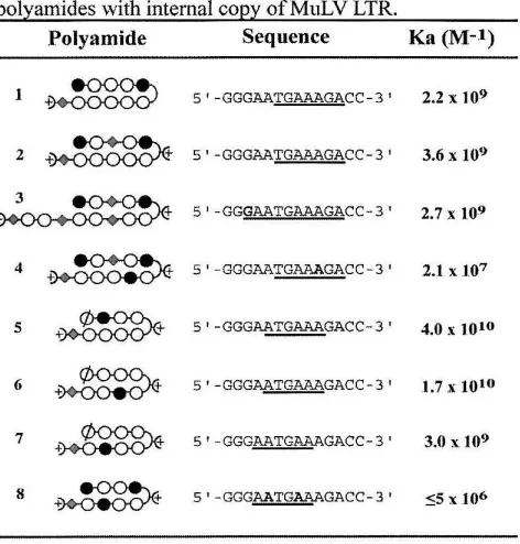
Table 1. Equillibriwn association constants for polyamides with internal coPY ofMuLV LTR.
Polyamide Sequence Ka (M-l)
J ~ 5' -GGGAATGAAAGACC-3' 2.2 x 10⁹
2 ~ 5' -GGGAATGAAAGACC- 3' 3.6 X 10⁹
~ 5' -GGGAATGAAAGACC-3' 2.7 x 10⁹
4
~
S'-GGGAATGAAAGACC-3' 2.1 X 1075
+~
5 t -GGGAA~GACC-3 ' 4.0 x 10¹⁰(;
~
5'-GGG~GACC-31 1.7 X 10'"7
+~
5'-GGGAATGAAAGACC-3' 3.0 x 1098
~
S'-GGG~GACC-3' ~5 x .106Table 2.1. Values reported are the mean values obtained from three DNase I footprint titration experiments. The binding site for each polyamide is underlined. Mismatches are shown in bold. The assays were carried out at 22° C, 10 mM Tris-HCI (pH 7.0), 10 mM KCI, 10 rnM MgCI20 and 5 mM CaCI2•
(Ka
=
4 X WID M-I), while the double base pair mismatch 8 has the lowest binding constant (Ka ::; 5 X 10⁶ M-I).The similar affinity (Ka
=
1.9 - 3.6 x 10⁹M-I) of polyamides 1-3 is of interest.
Polyamide 1 appears to be particularly well matched for the LTR sequence, given its relatively high affinity, and observations that substitution with a central WP pair and/or the positively charged (R)H2N,y-turn failed to increase its affinity (data not shown). Additionally, substitution with a central PlPy pair yielded a drop in affinity, which was recovered by substitution with (R)H2Ny (polyamide 2). With the internal copy of the LTR sequence used for these experiments, the tail of the extended polyamide 3 is placed against G·e base pairs, which represents a mismatch that is not present in the L TR itself.
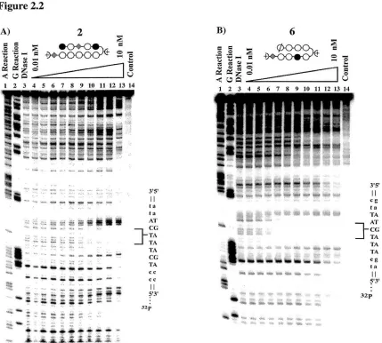
[image:41.518.126.362.86.333.2]Figure 2.2
I 2 3 4 5 6 7 8 9 10 11 12 13 14
-'~----~ --
---
"., -
-
..
-
.,.
-~... =.
":"'l "':'~ ~ '";;: :';'1i't!'!' . .,..:'~;;;; ;'; .: =.:;:;:
~'- ; ::.* =::=<-. ~,.
.
?'
..
~ :~:::...
~:: -~: "-~~-,.,.,..- "'"
~ i-~"~;-,~_.~A.,~;~:::
3'5' II
,,,
la Kl' ] -CG TA TA 1',\ CG TA cc cc II?'3'
32p
B)
....
6
~
~
+ 0000-,<;.=
=
~:=:'0
-
..
~~~
4 5 6 7 8 9 10 11 12 13 14
~ .. , -" .. ,; •. :0--- ... , .... .
""" -to~,_'-... _
3'S' II cg: I . TA AT
] -CG TA 11\ TA cg ta II ?'3' 32p
Figure 2,2. (A) Quantitative DNase I footprint titration experiment with polyamide 2 on a 5' 3Zp labeled 183 base pair DNA fragment containing an internal copy of the M-MuLV LTR: lane I, A reaction; lane 2, G reaction; lane 3, DNase I standard; lanes 4-13, 10 pM, 20 pM, SO pM, 100 pM, 200 pM, 500 pM, 1 nM,
2 nM, 5 nM, and 10 nM polyamide; lane 14, intact DNA. All reactions contained 10 mM Tris-HCI (pH 7.0), 10 mM KCl, 10 mM MgCI2, and 5 mM CaCI2 , and were performed at 22° C. (B) Quantitative DNase
I footprint titration experiment with polyamide 6 on a 5' 32p labeled 183 base pair DNA fragment containing an internal copy of the M-MuLV LTR: lane 1, A reaction; lane 2, G reaction; lane 3, DNase I
standard; lanes 4-13, 10 pM, 20 pM, SO pM, 100 pM, 200 pM, 500 pM, 1 nM, 2 nM, 5 nM, and 10 nM polyamide; lane 14, intact DNA. All reactions contained 10 mM Tris-HCI (pH 7.0), 10 mM KCI, 10 mM MgCI2, and 5 mM CaClz, and were performed at 22° C.
binding to the M-MuLV LTR termini. The binding of polyamides 5-7 was of particular
interest, given the proximity of their cognate sites to the absolute end of the LTR. DNA
performed to address this issue.47 Comparisons of melting curves with and without
polyamides 5-7 showed that the Tm shifts induced by these polyamides were similar for
both sets of oligonucleotides, although slightly lower in each case for the terminal
cognate site (see Appendix). In all cases, the Tm shifts for both sets of oligonucleotides
with polyamides 5-7 were considerably greater than the Tm shifts induced by a mismatch
polyamide. These results indicate that polyamides can bind with high affinity to the
absolute ends of DNA fragments. However, since the Tm shift for the terminal cognate
site was slightly lower than the internal cognate site for polyamides 5-7, the affinity
constants determined by DNase I footprinting for these compounds (Table 2.1) should be
considered as upper limits for their affinity to the M-MuLV LTR termini.
Figure 2.3
L TR·L TR product
5'*---<:ATT LTR 3·
IN
13 ..
process,ng'---<:A
S ' * C A L
-3·~--AC--*
two-end product (linear)
one-end product (circular)
Figure 2.3. Schematic illustration of in vitro integration assays. IN first removes the "IT" dinucleotide from the 5' '2p labeled blunt LTR duplex, leaving a single-strand tail. Then IN integrates the cleaved strand into the target: either the L TR itself in the strand transfer assay, or the plasmid DNA as an exogenous substrate. The major products of integration are as listed.
Inhibition of the 3' processing. The ability of the polyamides to inhibit
integration was tested in in vitro integration assays. Figure 2.3 schematically outlines the
in vitro assays utilized in these studies. In the 3' processing assay, a 20 base-pair (bp)
Figure 2.4
Polyamide:
substrate~
-2product~
Figure 2.4. Inhibition of 3' processing reactions by the polyamides. 3' processing reactions were
performed as described in the Materials and Methods. The position of the -2 3 'processing product is indicated at the left. Lane 1, no protein control; lane 2, M-MuLV IN alone. Lanes 3-9, IN plus polyamide
1; lanes 10-16, IN plus polyamide 2; lanes 17-23, IN plus polyamide 3; lanes 24-30, IN plus polyamide 4; lanes 31-37, IN plus polyamide 5; and lanes 38-44, IN plus polyamide 6. The concentrations of the polyamide used are (left to right): 0.4, 16,40, 100,256,640 and 1600 nM.
(Figure 2.3, top line). The strand containing the conserved CA dinucleotide was 5' 32p
labeled. IN dependent 3' processing of the dinucleotide yields an 18 base product, which
is monitored on a denaturing acrylarnide gel. It is of note that the concentration of DNA
substrate in these and the following integration assays is significantly greater (33 nM)
than the concentration of DNA in the DNase I footprinting titrations. Figure 2.7
summarizes the effects of the polyarnides on the 3' processing reaction. The results of an
exemplary 3' processing assay with selective polyamides is shown in Figure 2.4. The
eight polyamides could be divided into three classes, according their inhibition
efficiencies (Figure 7). The first class includes polyarnides 1,3, 5, and 6 all having an
ICso under 250 nM. The second class includes polyarnides 2 and 7 with an ICso between
450 and 650 nM. Polyarnides 4 and 8 belong to the third class, which has an ICso at or
above 1400 nM. The polyamides are designed to bind with sequence specificity. The
inhibitory effect of the polyarnides correlates with the ability to efficiently bind the LTR
sequences and is therefore not due to nonspecific interactions with the IN protein. A
single mismatch within the polyamide was found to decrease the Ka value by lOa-fold
exemplified by polyamide 4 (Figure 2.4 and data not shown), with a Ka of 2.1 x 10⁷ M·I ,
and an IC50 value at or greater than 1600 nM.
Figure 2.5
Polyamide:
strand
transfer
product
2
3
4
5
6
~~~
*
rl'" ... 10"'1'-0001 0 1"'1 N , . , . . 11'110 r-.oo CI'I 1;1 .... Nt<> ... 10 "'f'-ID<JI 0 .... NM ... Il'lIOr- 0 0 " ' 0 .... N.., ... rl rll"'l .... rl.-4 . . . .-l <'4 N N N N N N N N N . . . ..,I"'I ... M ... 1'1 . . . ... Figure 2.5. Inhibition of strand transfer reactions by the polyamides. Strand transfer assays, based on
oligonucleotides substrates were performed as described in the Materials and Methods. Strand transfer
yields 32P-labeled products larger than the input substrate, indicated at left of panel. Lane 1, no protein
control; lane 2, M-MuLV IN alone. Lanes 3-9, IN plus polyamide 1; lanes 10-16, IN plus polyamide 2;
lanes 17-23, IN plus polyamide 3; lanes 24-30, IN plus polyamide 4; lanes 31-37, IN plus polyamide 5; and
lanes 38-44, IN plus polyamide 6. The concentrations of the polyamide used are (left to right): 004, 16, 40,
100,256,640, and 1600 nM. The asterick indicates the formation of the enhanced integration product.
Inhibition of strand transfer. The polyamides were tested for inhibition of
strand transfer. In this assay, a precleaved substrate (Figure 2.3, second row) was used
instead of the blunt-end substrate, with integration occurring into a second
double-stranded oligonucleotide (Figure 2.3, left arrow). Strand transfer is highly efficient using
a precleaved substrate,48 and allows for the separation of the two steps in the analysis. Similar to 3' processing, the polyarnides can be separated into three classes based on their IC50 for the strand transfer reactions (Figure 2.7). An exemplary strand transfer assay
using selective polyamides is shown in Figure 2.5. The first class consists of 1 and 3,
with the IC50 under 250 nM. Moderate inhibition is found with the second class, including 2 and 5 with IC50 at 450 and 1100 nM, respectively. The third class includes 4
Although polyamides 5 and 6 were among the most effective inhibitors of 3'
processing, they showed little effect on strand transfer. Poly ami des 1 and 3, though, are
highly effective at inhibition of both the
3'
processing and strand transfer reactions. This raises the interesting possibility that distinct regions of the viral termini are required for3' processing and strand transfer. Additionally, the absence of the TT dinucleotide in the
strand transfer substrate will affect the binding of polyamides, particularly 5-7, whose cognate sites are closest to the end of the viral DNA. Each of these polyarnides was less
effective at inhibiting strand transfer than 3'-processing, whereas polyamides 1-3 showed the same efficacy in both assays (Figure 2.7). This further emphasizes the importance of
cognate site recognition in integrase inhibition.
In the oligonucleotide based assay, the L TR substrate functions both as the viral and target DNA. Previous analysis of binding site selection indicated that the major groove was the main determinant within the target DNA for IN docking.49•s3 Therefore it
was believed that binding of the polyamides to the target DNA should not influence target site selection. Experimentally, variations in target site selection were observed. In the presence of high concentration of polyamide 2 (640 and 1600 nM, Figure 2.5, lanes 15 and 16), loss of integration at the predominant sites was paralleled with enhanced production of a single product of slow electrophorectic mobility (marked by the asterisk).
This product was also observed at saturating levels of polyamide 3 (Figure 2.5, lanes
21-23). Formation of this large product is a result of integration at one end of the target DNA. Target site selection using polyamide 5 was also altered, with preference for the faster mobility products at the highest polyamide concentrations (Figure 2.5, lanes
36-37).
Inhibition of strand-transfer into an exogenous target. To separate the effects