Patterns of genome evolution that have accompanied
host adaptation in
Salmonella
Gemma C. Langridgea,1, Maria Fookesa, Thomas R. Connora,2, Theresa Feltwella, Nicholas Feaseya, Bryony N. Parsonsb,c, Helena M. B. Seth-Smitha,3,4, Lars Barquista, Anna Stedmana, Tom Humphreyd, Paul Wigleyb,e, Sarah E. Petersf, Duncan J. Maskellf, Jukka Coranderg, Jose A. Chabalgoityh, Paul Barrowi, Julian Parkhilla, Gordon Dougana, and Nicholas R. Thomsona
aPathogen Genomics, The Wellcome Trust Sanger Institute, Hinxton, Cambridge CB10 1SA, United Kingdom;bInstitute of Infection and Global Health,
University of Liverpool, Liverpool L69 7BE, United Kingdom;cInstitute of Translational Medicine, University of Liverpool, Liverpool L69 3GE, United Kingdom; dCollege of Medicine, Swansea University, Swansea SA2 8PP, United Kingdom;eSchool of Veterinary Science, University of Liverpool, Liverpool L69 3GB, United
Kingdom;fDepartment of Veterinary Medicine, University of Cambridge, Cambridge CB3 0ES, United Kingdom;gDepartment of Mathematics and Statistics,
University of Helsinki, FI-00014 Helsinki, Finland;hDepartamento de Desarrollo Biotecnológico, Instituto de Higiene, Facultad de Medicina, Universidad de la
República, Montevideo, CP 11600, Uruguay; andiSchool of Veterinary Medicine and Science, University of Nottingham, Nottingham LE12 5RD, United Kingdom
Edited by Ralph R. Isberg, Howard Hughes Medical Institute, Tufts University School of Medicine, Boston, MA, and approved November 26, 2014 (received for review August 29, 2014)
Many bacterial pathogens are specialized, infecting one or few hosts, and this is often associated with more acute disease presentation. Specific genomes show markers of this specialization, which often reflect a balance between gene acquisition and functional gene loss. WithinSalmonella entericasubspeciesenterica, a single line-age exists that includes human and animal pathogens adapted to cause infection in different hosts, includingS. enterica serovar Enteritidis (multiple hosts),S.Gallinarum (birds), andS.Dublin (cattle). This provides an excellent evolutionary context in which differences between these pathogen genomes can be related to host range. Genome sequences were obtained from∼60 isolates selected to represent the known diversity of this lineage. Exami-nation and comparison of the clades within the phylogeny of this lineage revealed signs of host restriction as well as evolutionary events that mark a path to host generalism. We have identified the nature and order of events for both evolutionary trajectories. The impact of functional gene loss was predicted based upon po-sition within metabolic pathways and confirmed with phenotyp-ing assays. The structure ofS.Enteritidis is more complex than previously known, as a second clade ofS.Enteritidis was revealed that is distinct from those commonly seen to cause disease in humans or animals, and that is more closely related toS.Gallinarum. Isolates from this second clade were tested in a chick model of infection and exhibited a reduced colonization phenotype, which we postulate rep-resents an intermediate stage in pathogen–host adaptation.
Salmonella
|
host adaptation|
pseudogene|
metabolismT
he central importance of horizontal acquisition of mobile genetic elements in the development of virulence in bacteria has been well described. It has frequently been observed that, as pathogens acquire virulence determinants, they become increasingly adapted to a specific host (1, 2). Exquisitely host-restricted pathogens also often exhibit extensive genome decay, through insertion sequence element proliferation, genomic rear-rangement, and/or pseudogene formation (1, 3, 4). Investigating mechanisms involved in host adaptation is key to an understanding of pathogen evolution and has directly translatable relevance to the epidemiology and potentially the control of human and zoo-notic infectious disease.By concentrating upon individual pathogenic clades, insights have been obtained into specific adaptations relating to specific hosts; however, comparative analysis is relatively rare. By broadening this approach to examine multiple human and animal pathogens, de-rived from a single closely related lineage but with differing host specializations, there is an opportunity to understand the funda-mental evolutionary processes involved in host adaptation. Lineage-specific changes that have become fixed can then be distinguished from those stochastic changes that differentiate individual isolates.
A single lineage within Salmonella entericapresents such an opportunity. S. entericais a leading cause of foodborne gastro-enteritis, globally responsible for 80 million cases annually (5). Differentiation of S. entericais largely based upon somatic (O) and flagellar (H) antigens, but it is increasingly being typed by
Significance
Common features have been observed in the genome sequences of bacterial pathogens that infect few hosts. These“host adapta-tions”include the acquisition of pathogenicity islands of multiple genes involved in disease, losses of whole genes, and even single mutations that affect gene function. Within Salmonella entericais a natural model system of four pathogens that are each other’s closest relatives, including a host-generalist, two host-specialists, and one with strong host associations. With whole-genome sequences, we aimed to improve our un-derstanding of the number, nature, and order of these host adaptation events, shedding light on how human and animal pathogens arose in the past, and potentially allowing us to predict how emerging pathogens will evolve in the future.
Author contributions: G.C.L., T.H., P.W., D.J.M., J.A.C., P.B., J.P., G.D., and N.R.T. designed research; G.C.L., T.F., B.N.P., A.S., and S.E.P. performed research; J.C. contributed new reagents/analytic tools; G.C.L., M.F., T.R.C., N.F., B.N.P., H.M.B.S.-S., L.B., T.H., P.W., S.E.P., D.J.M., J.C., and N.R.T. analyzed data; and G.C.L., M.F., T.R.C., B.N.P., L.B., P.W., J.C., and N.R.T. wrote the paper.
The authors declare no conflict of interest. This article is a PNAS Direct Submission.
Freely available online through the PNAS open access option.
Data deposition: The sequences reported in this paper have been deposited in the European Nucleotide Archive (ENA),www.ebi.ac.uk/ena(accession nos.ERS003157–
ERS003161,ERS004906,ERS004907,ERS004909–ERS004912,ERS004914,ERS007756,
ERS022673–ERS022687,ERS024552,ERS217197,ERS217199,ERS217201,ERS217204,
ERS217211,ERS217214,ERS217216,ERS217218,ERS217222,ERS217225,ERS217233,
ERS217235,ERS217238,ERS217239,ERS217241,ERS217242,ERS217252,ERS217264,
ERS400244,ERS400251,ERS400254,ERS400256–ERS400259,ERS400262,ERS400264,
ERS429283,ERS429284,ERS430067,LK931482, andLK9315020), and annotated
plas-mid assemblies are available from the European Molecular Biology Laboratory (EMBL) European Bioinformatics Institute,www.ebi.ac.uk(accessions nos.HG970000
andHG970001).
See Commentary on page 647.
1To whom correspondence should be addressed. Email: [email protected].
2Present address: Cardiff School of Biosciences, Cardiff University, Cardiff CF10 3AX,
United Kingdom.
3Present address: Functional Genomics Center Zürich, Universität Zürich, CH-8057 Zurich,
Switzerland.
4Present address: Institute for Veterinary Pathology, Vetsuisse Faculty, Universität Zürich,
CH-8057 Zurich, Switzerland.
This article contains supporting information online atwww.pnas.org/lookup/suppl/doi:10.
1073/pnas.1416707112/-/DCSupplemental.
www.pnas.org/cgi/doi/10.1073/pnas.1416707112 PNAS | January 20, 2015 | vol. 112 | no. 3 | 863–868
MICRO
BIOLOGY
SEE
COM
genomic methods, such as multilocus sequence typing (MLST). Somatic serogrouping and MLST have identified a single lineage with closely related members that exhibit a range of different host specializations (6–9). These include two of the most im-portantSalmonellapathogens:S.Enteritidis andS.Gallinarum. In addition to the contribution of S.Enteritidis to human dis-ease, in many countries both of these pathogens are notifiable diseases in poultry farming and egg production. Despite their close phylogenetic relatedness, they exhibit strikingly different host ranges, withS.Enteritidis capable of infecting multiple host species, whereasS.Gallinarum is restricted to infection in gal-liforme birds. This lineage also includes S. Gallinarum biovar Pullorum (hereafterS.Pullorum), also restricted to galliformes, and S. Dublin, which is strongly associated with infection of cattle and more rarely that of humans (10). The Salmonella pathogens adapted to particular hosts are associated with a much more invasive disease than generalists likeS.Enteritidis, which tend to cause enteritis.
Fifty-nine isolates of thisSalmonellalineage were selected to capture the diversity of sequence types, phage types, and geo-graphical and temporal spread available at the time. Through genome sequencing, we generated a phylogeny to act as a framework with which to reconstruct the evolutionary history of the lineage, onto which observed gene loss and acquisition could be plotted.
This dataset provides a compelling record of the degradation of common metabolic pathways during host specialization to date. We have documented the order of events during the evo-lution of an entireSalmonellalineage. To link this specifically to host adaptation, we have tested isolates occupying key positions in the phylogeny in their cognate host to assess the effects of gene degradation on the manner and severity of disease caused.
Results and Discussion
ClassicalS.Enteritidis Is Not the Ancestor of theS.Gallinarum Biovars.
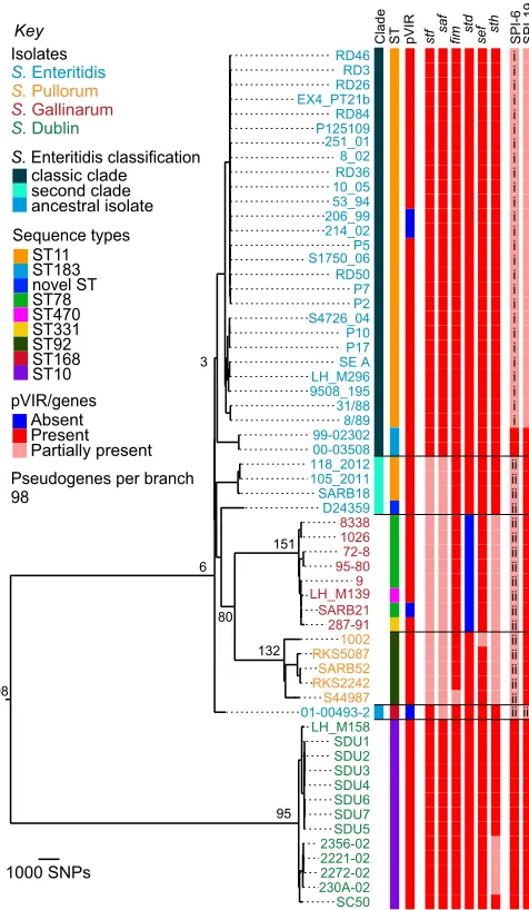
To establish an accurate phylogeny, we selected isolates of S.Enteritidis,S.Gallinarum,S.Pullorum, andS.Dublin repre-senting different MLSTs, phage types (PTs), and geographical and temporal spread to ensure that we sampled a broad diversity in this lineage. In total, genomic DNA from 59 isolates was used to generate whole genomes by Illumina multiplex sequencing (Table S1). Single-nucleotide polymorphisms (SNPs) were de-tected by mapping sequences against a pan-chromosome and virulence plasmid pseudomolecule (SI Methods). The maximum-likelihood phylogenetic tree shown in Fig. 1 was constructed based upon the core regions of the reference S. Enteritidis strain P125109 genome.
The phylogeny of these serotypes is largely consistent with previous studies showing that S. Dublin represents an in-dependent clade (9, 11). Our data show that there are∼23,000 SNPs differentiating S. Dublin from the most recent common ancestor of S. Enteritidis and the avian-adapted salmonellae, S.Gallinarum andS.Pullorum. However, rather than being a sin-gle clade itself,S.Enteritidis appears to be more structurally com-plex: isolate 01-00493-2 occupies a position on the tree basal to all other S.Enteritidis and S.Gallinarum/S.Pullorum complex isolates, and a second clade ofS.Enteritidis appears within the S.Gallinarum/S.Pullorum complex (Fig. 1).
Statistical clustering supports the two clades ofS.Enteritidis depicted in Fig. 1; a Bayesian analysis of population structure (BAPS) was used to identify significant differences in poly-morphism sharing across strains (12). This initially indicated that members of clade 2 clustered separately from clade 1. A sub-sequent level of clustering then separated the S. Enteritidis 01-00493-2 isolate labeled“ancestral”in Fig. 1 from the rest of clade 2 (Dataset S1). No further substructure inS. Enter-itidis was found to be significant according to the Bayesian statistical model.
We refer to members of the first clade as“classic”S. Enter-itidis because this clade includes the most commonly isolated MLST, ST11, and PTs affecting humans and animals. It is of note that both PT11 isolates (99-02302 and 00-03508) occupy a deep branch within the classic clade. PT11s are unusual in that they are rare in human infections and have been reported to be associated with hedgehogs (Erinaceus europaeus) (13).
The second clade (Fig. 1, second clade) includes isolates SARB18, 118-2012K-186, 105-2011K-1654, and D24359, which share the same O and H antigen designations as classicS. Enter-itidis and are closely related at the sequence level. It is this second clade ofS.Enteritidis from which the ancestors of the S.Gallinarum/S.Pullorum complex emerged. This phylogeny suggests thatS.Gallinarum andS.Pullorum did not directly
1000 SNPs RD46 RD3 RD26 EX4_PT21b RD84 P125109 251_01 8_02 RD36 10_05 53_94 206_99 214_02 P5 S1750_06 RD50 P7 P2 S4726_04 P10 P17 SE A LH_M296 9508_195 31/88 8/89 99-02302 00-03508 118_2012 105_2011 SARB18 D24359 8338 1026 72-8 95-80 9 LH_M139 SARB21 287-91 1002 RKS5087 SARB52 RKS2242 S44987 01-00493-2 LH_M158 SDU1 SDU2 SDU3 SDU4 SDU6 SDU7 SDU5 2356-02 2221-02 2272-02 230A-02 SC50 98 95 Absent Present Partially present
S. Enteritidis
S. Pullorum
S. Gallinarum
S. Dublin
Key Clade ST pVIR stf saf fim std sef sth SPI-6 SPI-19
classic clade second clade ancestral isolate 132 151 80 6 3 ST11 ST183 novel ST ST78 ST470 ST331 ST92 ST168 ST10 Sequence types Isolates
S. Enteritidis classification
pVIR/genes
[image:2.585.305.543.52.461.2]Pseudogenes per branch 98 i i i i i i i i i i i i i i i i i i i i i i i i i i ii ii ii ii ii ii ii ii ii ii ii ii ii ii ii ii ii iii ii
Fig. 1. Chromosome-based phylogenetic relationships. Midpoint-rooted maximum-likelihood phylogenetic tree based upon the chromosome. Branch lengths are determined by number of SNPs. Numbers on the tree indicate how many pseudogenes were identified on that branch. Colored strain names show serotype:S. Enteritidis, blue;S. Pullorum, gold;S. Gallinarum, red;S. Dublin, green. Metadata columns: Clade, phylogenetic grouping of S. Enteritidis isolates; ST, sequence types; pVIR, virulence plasmid;stf–sth, fimbriae; SPI-6 and -19,Salmonellapathogenicity islands; i, ii, SPI-6 partial hits represent two distinct versions at this locus; iii, SPI-19 partial presence is different from classicS. Enteritidis in this strain. Partial presence indicates one or more genes have been lost.
evolve from within the most commonly isolated (classic) clade ofS.Enteritidis, as was previously postulated (8), and that there is much more substructure in the phylogeny of these important human and animal pathogens than was previously known.
Chromosomal Macroevolution.Evolution of pathogenicity by Salmo-nellais strongly associated with the acquisition of mobile genetic elements calledSalmonellapathogenicity islands (SPIs). Many of these SPIs were acquired very early in the evolution ofS.enterica (14), and so, perhaps unsurprisingly, their complement was found to be conserved across this entire lineage, with the ex-ception of SPI-6 and SPI-19. Among other functions, these SPIs both encode type VI secretion systems (T6SSs), which are known to promote colonization in the avian gut (15). As previously reported for S. Enteritidis PT4 P125109, the majority of the classicS.Enteritidis isolates sequenced here harbor degenerate versions of both these SPIs. In these isolates, little more than the saffimbrial operon is maintained on SPI-6, whereas on SPI-19 only 13 out of 33 genes, mainly encoding exported proteins, re-main intact. The borders of these deletions are the same for all isolates. We also observed that two isolates of the classic S.Enteritidis clade, both PT11, carry intact versions of both SPIs. This indicates that the loss of SPI-6 and SPI-19 is re-stricted and suggests that it occurred following the divergence of the main classicS.Enteritidis group from these strains. That the common ancestor of the entire lineage contained both loci is also supported by their (intact) presence inS.Dublin.
SPI-19 is intact in all other isolates sequenced in this study (Fig. 1), and forS.Gallinarum andS.Pullorum, the retention of a functional SPI-19 is consistent with the T6SS it encodes being important forS. Gallinarum survival in chicken macrophages (16). However, the degradation of SPI-6 and loss of the T6SS, seen within the second clade ofS.Enteritidis and theS.Gallinarum/S.Pullorum complex, may have only occurred once. Examining the recombination events across the entire lineage revealed that the region surrounding SPI-6 appears to have been independently donated from theS. Gallina-rum lineage toS. Pullorum and isolates in the second clade of S.Enteritidis (Fig. 1, SPI-6 version ii;Dataset S1).
Within the chromosome, other clade-specific whole gene losses were related to fimbriae. InSalmonella, the loss or inactivation of fimbriae is associated with host adaptation (8, 17).S.Enteritidis harbors 13 fimbrial operons, and the variable presence of 6 of these occurred in patterns that matched the phylogeny of isolates in this lineage (Fig. 1). The majority of these differences result from partial- or whole-gene losses in host-restrictedS.Gallinarum andS.Pullorum, but partial losses of thessfandsafoperons were also observed in the second clade ofS.Enteritidis.
Extrachromosomal Macroevolution.Only four isolates in this study do not carry a version of the Salmonella virulence plasmid (pVIR) encoding the virulence-associated spv operon (Fig. 1). Using the alignment produced by mapping to a pseudomolecule representing the pan-chromosome combined with the pVIR, we extracted the subset of sequences aligning to the pVIR of S.Gallinarum 287-91, which is the largest and most complete virulence plasmid in this lineage. The SNPs identified in this alignment were used to construct a pVIR phylogenetic tree (Fig. S1). The plasmid phylogeny mirrors that of the chromo-some for all isolates except for the classic Enteritidis clade, which appears to harbor a pVIR more distantly related to the plasmids carried within this lineage (∼1,000 SNPs) than the outgroup, pSLT (∼300 SNPs).
The smaller size of the classic S.Enteritidis pVIR (∼60 kb) relative to that ofS.Gallinarum (∼90 kb) can be largely attrib-uted to major deletions in thetraregion responsible for conjugal transfer (18). Other differences that contribute to the phyloge-netic distance between pVIRs include the presence of genes for a partial K88-like fimbria (19), which was found in all of the
S.Dublin, S.Gallinarum, and S.Pullorum pVIRs. ApefA-like remnant upstream of SG_p0053 was also conserved across these plasmids, which we believe to be a scar of thepef fimbrial op-eron. This remnant has not been annotated in any published K88-like–containing pVIRs to date but has been randomly mu-tated inS.Gallinarum by transposon mutagenesis. In contrast to the parental S.Gallinarum, no morbidity or mortality was ob-served in chickens inoculated with the resultant mutants (19). This suggests that the pefAremnant retains some functionality, although it is not clear what interaction this may have with the K88-like replacement. Thepef fimbrial operon itself is present only in pVIR-carrying strains ofS.Enteritidis from the classic clade. Alongside the pVIR phylogeny, this suggests that the ancestral pVIR plasmid of the entire lineage was replaced by a different plasmid before the expansion of the classicS.Enteritidis clade. In further support of this, we observe that the second clade and an-cestralS.Enteritidis all have a pVIR more closely related to those carried byS.Gallinarum andS.Pullorum than to those found in the classicS.Enteritidis clade.
Pseudogene Formation Largely Occurred After Serovar Diversification.
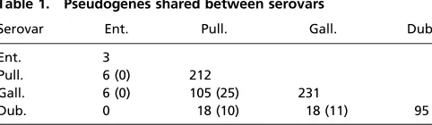
Manual, whole-genome comparisons were used to define the ex-tent to which gene degradation has occurred along the branches of the chromosomal phylogenetic tree. In this context, we defined a pseudogene as a gene harboring a mutation (i.e., premature stop codon, frame shift, truncation, or syntenic deletion) relative to an intact version of that gene. Table 1 summarizes the number of pseudogenes present in each of the respective serovars, and how many shared pseudogenes are due to identical or nonidentical mutations. Full pseudogene sets are listed inDataset S2. In total, 98 ancestral pseudogenes are common to the entire set of serovars. These are typically fragments of nonfunctional genes and are likely due to stochastic loss. However, pseudogenes that are found in particular regions of the tree are more likely to have resulted from a primary event interrupting metabolic or other pathways, poten-tially leading to further degradation of related genes.
S.Enteritidis is generally regarded as a promiscuous serovar, and our analysis found only one nonancestral pseudogene shared by all classic isolates (Fig. 1). When considered alongside the maintenance of intact fimbriae, replacement of the ancestral plasmid, and loss of both T6SSs, it is apparent that these are markers of adaptation to host generalism in classicS.Enteritidis.
Fig. S2 depicts the differing routes toward host generalism and adaptation seen across the entire lineage, from a hypothetical shared ancestor.
[image:3.585.299.540.54.124.2]Previous studies have indicated that the degree of host spec-ificity displayed by particularSalmonellaserovars loosely corre-lates with the level of gene degradation (8, 17, 20, 21). In keeping with this observation, host-restrictedS.Gallinarum andS.Pullorum harbor 231 and 212 pseudogenes, respectively, whereasS.Dublin, which is associated with but not restricted to cattle, harbors 95 (Table 1).
Significantly, given their shared host restriction, analysis of pseudogene positioning across the tree indicated that over 60% Table 1. Pseudogenes shared between serovars
Serovar Ent. Pull. Gall. Dub.
Ent. 3
Pull. 6 (0) 212
Gall. 6 (0) 105 (25) 231
Dub. 0 18 (10) 18 (11) 95
Dub., Dublin; Ent., Enteritidis; Gall., Gallinarum; Pull., Pullorum. Total number of pseudogenes shared between serovars. Pseudogenes that have different mutations between serovars are given in parentheses. The 98 ancestral pseudogenes common to all are not included. Only pseudogenes present in all sequenced strains of the serovar are shown here.
Langridge et al. PNAS | January 20, 2015 | vol. 112 | no. 3 | 865
MICRO
BIOLOGY
SEE
COM
of the genome degradation found inS.Gallinarum (151 of 231) and S.Pullorum (132 of 212) occurred after the two diverged. Our recombination analysis revealed that there has been one large recombination event between them, representing a region of around 180 kb (Dataset S1), but this accounts for only six shared pseudogenes, indicating that recombination has not been a significant cause of shared gene loss. However, this re-gion encompasses 165 genes, including four fimbrial operons (saf,sti, stf, andstb) and SPI-6, suggesting that recombination has played some role in these two pathogens converging on the same host niche.
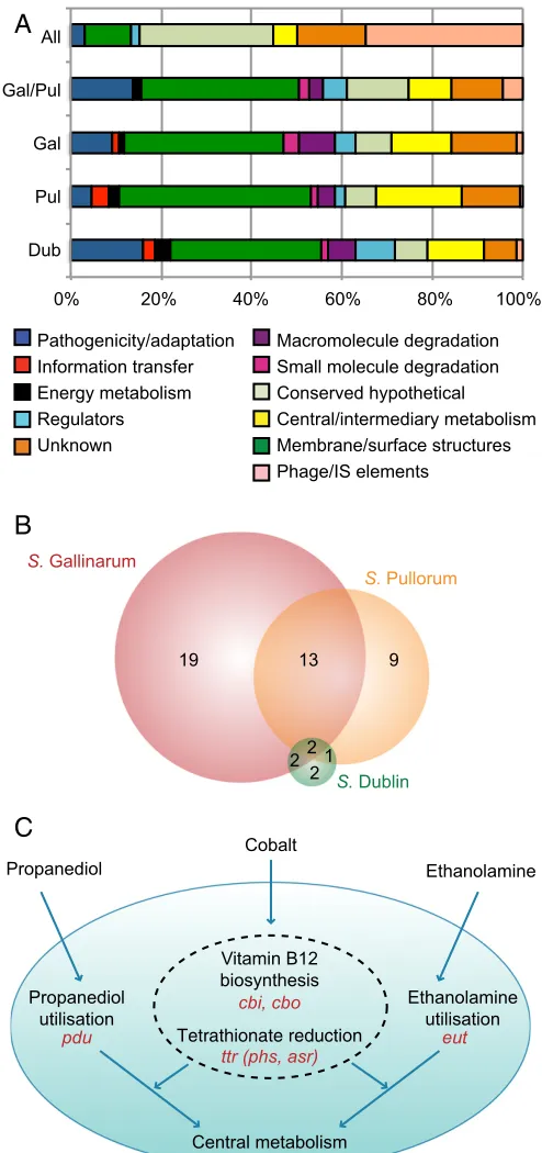
Loss of Metabolic Capacity.Functionally, the 98 identified ances-tral pseudogenes consist mainly of phage, insertion sequences, and genes of unknown function. This is in stark contrast to the functional categories represented by pseudogenes found toward the tips of the tree (Figs. 1 and 2A), where membrane/surface structure and central/intermediary metabolism genes are more commonly inactivated. This latter pattern is broadly consistent forS.Gallinarum andS.Pullorum, both individually and in their shared 78 pseudogenes, and also in theS.Dublin pseudogenes, suggesting that loss of related functions is associated with ad-aptation to a host organism.
We established that∼15–20% of pseudogenes from each sero-var had functional locations according to a database of metabolic pathways predicted to be present in S.Enteritidis (Dataset S3). This allowed us to establish whether any metabolic functions were commonly affected (i.e., by the same or different pseudogenes in the same pathway) between serovars. The overlap of the affected pathways is shown in Fig. 2Band indicates that many specific pathways or transport reactions are affected in more than one of the adapted serovars in our set, and indeed in other host-adapted salmonellae (see below).
One sodium/galactoside transporter has been lost in all of the host-adapted serovars within our set, but degradation of the carbon sourceD-glucarate is the only full pathway that has been affected (by different mutations) in them all. Interestingly, the human-restricted serovarsS.Typhi andS.Paratyphi A also har-bor pseudogenes related to the transport ofD-glucarate into the cell, suggesting that the utilization of this carbon source may not be advantageous in causing invasive infection. Two isoenzymes catalyze a key reaction in this pathway, one of which is mutated in S.Dublin, whereas both are inactivated inS.Gallinarum.
The differing impact of these pseudogenes was supported by a phenotyping screen (Dataset S4), becauseS.Dublin has only suf-fered a loss of redundancy and therefore remains capable of using D-glucarate, whereasS.Gallinarum has lost the function entirely.
There are 13 metabolic pathways and two transport reactions commonly affected in S.Gallinarum andS.Pullorum (Dataset S3). The majority of these (12 of 15) are due to identical pseu-dogenes that occurred before their divergence. One of these is in the biosynthesis of the siderophore enterobactin, used to scav-enge iron from the environment (22). Iron acquisition in the host is key to many bacterial infections (23), and other iron uptake and transport systems remain intact. This is in contrast to pre-vious findings that human-restrictedS.Typhi is dependent upon iron uptake through enterobactin via the fep genes (24) and therefore suggests that enterobactin is not required in the avian host. However, another of the pathways, putrescine biosynthesis, is also affected in S. Typhi, and the same enzyme, ornithine decarboxylase, is mutated in all three. Because the alternative putrescine pathway remains intact, loss of redundancy may have an important role alongside loss of function in host adaptation. This is further supported by the phenotyping screen performed inS.Dublin. In accordance with the lower absolute number of pseudogenes, S.Dublin has only three pathways affected, and four transport reactions (Dataset S3). Of the four relevant sub-strates present in the phenotyping screen,S.Dublin cannot use
one, but remains capable of using three others, indicating that reduction in redundancy is more common than loss of function in this host-adapted serovar.
All
Gal/Pul
Gal
Pul
Dub
Macromolecule degradation Small molecule degradation Conserved hypothetical Central/intermediary metabolism Membrane/surface structures Phage/IS elements
Pathogenicity/adaptation Information transfer Energy metabolism Regulators Unknown
0% 20% 40% 60% 80% 100%
A
B
1 22
2
13 9
19
S. Dublin
S. Pullorum
S. Gallinarum
Tetrathionate reduction
Vitamin B12 biosynthesis
Cobalt
Propanediol Ethanolamine
Central metabolism Propanediol
utilisation
Ethanolamine utilisation
ttr (phs, asr) cbi, cbo
pdu eut
[image:4.585.301.548.50.575.2]C
Fig. 2. Functions lost through pseudogene formation. (A) Functional clas-sification of pseudogene sets. All, pseudogenes shared by all strains in the phylogeny (98); Gal/Pul, shared byS. Gallinarum andS. Pullorum (80); Gal, Pul, Dub, remaining pseudogenes present in all strains ofS. Gallinarum (151), S. Pullorum (132), andS. Dublin (95), respectively. (B) Venn diagram showing the distribution of metabolic pathway and transport loss betweenS. Gallinarum, S. Pullorum, andS. Dublin, irrespective of causative pseudogenes. (C) Sche-matic depicting interconnectivity of pseudogene-affected pathways and transport systems. Processes inside the dotted line only occur anaerobically. Operons involved are shown in red.
Three metabolic pathways are affected inS.Gallinarum andS. Pullorum due to different pseudogenes that occurred after the two diverged. Two of these functions, allantoin degradation and adenosylcobalamin (vitamin B12) biosynthesis, are also mutated inS.Typhi andS.Paratyphi A. Given the pseudogene accumu-lation in these pathways in host-restricted serovars, they emerge as strong contenders for markers of a switch to invasive rather than enteric disease.
In birds, allantoin can be found in the serum and is used as a carbon source duringS.Enteritidis infection of chickens (25). Inactivation of the genes encoding the regulator allS and the degradative enzymeallDinS.Gallinarum andS.Pullorum, re-spectively, means that neither can use allantoin. Although the relevance of allantoin in mammalian hosts is unknown, pseu-dogenes relating to allantoin utilization have also been identified in a strain ofS.Typhimurium belonging to ST313, which causes invasive nontyphoidal salmonellosis in humans (26).
Vitamin B12 is required for the anaerobic degradation of 1,2-propanediol, which uses tetrathionate as an electron acceptor (27). Tetrathionate plays an important role in enteric infection: its production is triggered by the inflammatory response to S.Typhimurium in the gut and provides a niche for respiration, in competition with other gut microbes (28). A recent report by Nuccio and Bäumler (21) identified loss of function in central anaerobic metabolism as a key indicator of invasive versus gas-trointestinal salmonellae. From plotting pseudogenes onto the chromosomal phylogeny, we know that S. Gallinarum and S.Pullorum lost the ability to reduce tetrathionate before their divergence, and that their subsequent loss of vitamin B12 syn-thetic ability occurred independently. The knock-on effect of those initial pseudogenes in tetrathionate reduction is illustrated in Fig. 2C, as further pseudogenes have also arisen in other re-lated functions, consistent with Nuccio and Bäumler’s findings.
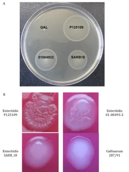
Second Clade and Ancestral S. Enteritidis Display Intermediate Characteristics.Of the five S. Enteritidis isolates outside the classic clade, one (ancestral) was basal to allS. Enteritidis and S. Gallinarum, whereas the remainder (second clade) were basal to bothS. Gallinarum andS.Pullorum (Fig. 1). To assess whether their position on the phylogenetic tree had any phenotypic con-sequences, we initially looked at colony morphology and motility of the ancestral isolate (01-00493-2) and a representative isolate from the second clade (SARB18). These indicated that both have in-termediate characters between S. Enteritidis andS. Gallinarum (Fig. S3). One explanation for this could be recombination; mul-tiple separate events have recombined∼175 kb fromS.Gallinarum orS.Pullorum into the second clade isolates, but not into any from the classicS.Enteritidis clade (Dataset S1). Approximately 160 kb originating from S. Gallinarum recombined into all four of the second clade isolates, around the SPI-6 locus, resulting in the presence of a different degenerate version of this pathogenicity island compared with that carried by the classic clade isolates. A separate event was responsible for the recombination of∼85 kb into the ancestral isolate 01-00493-2 fromS. Gallinarum, also in-cluding SPI-6. Unlike classicS.Enteritidis, SPI-19 remains intact in the ancestral and second clade isolates ofS.Enteritidis, as it does inS.Gallinarum andS.Pullorum (Fig. 1).
Our pseudogene analysis was dependent upon a single refer-ence strain per serovar, and therefore new pseudogenes could not be confirmed in theS.Enteritidis isolates outside the classic clade. However, metabolic phenotyping evidence suggests nu-merous differences are present between the SARB18 second clade and 01-004-93-2 ancestral isolate, with each able to use six or eight different carbon sources, respectively, that could not be used by the other isolate (Dataset S4).
Given the genotypic and phenotypic differences observed, we tested the pathogenesis of these two isolates in comparison with the reference strains ofS.Enteritidis andS.Gallinarum in the
natural avian host. The S. Gallinarum-infected group showed signs of systemic salmonellosis and reached the humane end-point of this experimental protocol at 5 d postinfection. The remaining groups showed no signs of ill health and the experiment continued for the full 7 d. At postmortem, the S. Gallinarum-infected birds displayed considerable hepatosplenomegaly, white spot lesions on the spleen and discoloration of the liver accom-panied by “bronzing” on exposure to air, consistent with fowl typhoid. In contrast, the groups infected withS.Enteritidis strains showed mild hepatosplenomegaly consistent with infection by this Salmonellaserovar. Also as expected,S. Gallinarum was the most invasive, indicated by significantly higher colony counts in both the spleen and liver on day 5 (P<0.01) (Fig. 3AandB). However, the classic S. Enteritidis strain P125109 showed significantly higher levels of colonization (bacteria present in the caeca) compared with both the nonclassicS. Enteritidis strains andS. Gallinarum (P< 0.05) (Fig. 3C). This was also borne out by histopathological scoring of the tissue (Table S2). This reduced colonization phenotype from the ancestral and second clade S. Enteritidis isolates further suggests that they represent intermediate stages in the evolution of host adaptation.
In conclusion, whole-genome comparisons across an entire Salmonellalineage have enabled us to establish the progression of pseudogene formation that has resulted in differently host-adapted pathogens. The strongest signal of metabolic loss was seen inS.Gallinarum andS.Pullorum, the fully host-restricted
A
B
C
6 5 4 3 2 17 6 5 4 3 2 1 7 6 5 4 3 2 1
Liver
Spleen
Caeca
log CFU/g
10
log CFU/g
10
log CFU/g
10
*
**
**
**
**
[image:5.585.305.535.53.387.2]Gal Ent outlier Ent second Ent classic
Fig. 3. Infection of the avian host by nonclassicS. Enteritidis isolates. In-vasion ofSalmonellastrains into the (A) spleen, (B) liver, and (C) colonization of chick ceca 7 d (exceptS. Gallinarum 287/91, 5 d, separated by dashed line) postinfection (n=5). Solid lines represent means. *P<0.05, **P<0.01. classic, classic clade isolate; Ent,S. Enteritidis; GAL,S. Gallinarum; second, second clade isolate.
Langridge et al. PNAS | January 20, 2015 | vol. 112 | no. 3 | 867
MICRO
BIOLOGY
SEE
COM
members of the lineage, consistent with patterns seen in other host-restricted salmonellae. It is therefore plausible that pseu-dogene formation progresses in a predictable fashion, given a par-ticular host. We also observed the presence ofS.Enteritidis isolates that fall outside the classic host generalist clade, instead occupying positions basal to the avian-restricted strains. Their metabolic, ge-nomic, and infection phenotypes all suggest that these represent intermediate, but extant, stages in the process of pathogen–host adaptation, demonstrating an unexpected diversity in this serovar.
Methods
Sequencing and Phylogenetic Analysis.All isolates were sequenced on the Illumina platform, with additional 454 sequencing where required. A non-redundant pseudomolecule was used a reference for mapping and to pro-duce genome alignments. The genetic structure of the population was estimated with the software BAPS, version 6.0 (12, 29), and recombination detected using the BratNextGen method (30). Pseudogene identification was performed by manual genome comparison, and metabolic pathway reconstruction was based upon the annotated genome of S.Enteritidis,
using Pathway Tools software (SRI International). High-throughput pheno-type screening was performed using the Biolog system (Biolog). Details are described inSI Methods.
Chick Infection Models.All experiments were conducted in accordance with United Kingdom legislation governing experimental animals under project licenses PPL 40/3063 and PPL 40/3652 and were approved by the University of Liverpool ethical review process before the award of the license. Four or five chicks were inoculated per experimental group and were killed at 5 or 7 d postinfection. Colony-forming units were quantified from the liver, spleen, and cecal contents, and pathology scoring on hematoxylin-and-eosin–stained sections was performed. Full details are described inSI Methods.
ACKNOWLEDGMENTS. We thank Mark Achtman and Mark Stevens for kindly providing strains. M.F., T.R.C., H.M.B.S.-S., J.P., and N.R.T. were funded by Wellcome Trust Grant 098051. The chicken infection work by B.N.P., T.H., and P.W. was funded by the Houghton Trust. G.C.L. was funded by Microme, a European Union Framework 7 Programme Collaborative Project, Grant Agreement 222886-2. J.C. was funded by European Research Council Grant 239784 and Academy of Finland Grant 251170.
1. Parkhill J, et al. (2003) Comparative analysis of the genome sequences ofBordetella
pertussis,Bordetella parapertussisandBordetella bronchiseptica.Nat Genet35(1):
32–40.
2. Rohmer L, et al. (2007) Comparison ofFrancisella tularensisgenomes reveals evolu-tionary events associated with the emergence of human pathogenic strains.Genome Biol8(6):R102.
3. Cole ST, et al. (2001) Massive gene decay in the leprosy bacillus.Nature409(6823): 1007–1011.
4. Moran NA, Plague GR (2004) Genomic changes following host restriction in bacteria.
Curr Opin Genet Dev14(6):627–633.
5. Majowicz SE, et al.; International Collaboration on Enteric Disease“Burden of Illness”
Studies (2010) The global burden of nontyphoidalSalmonella gastroenteritis.Clin
Infect Dis50(6):882–889.
6. Boyd EF, et al. (1993)Salmonellareference collection B (SARB): Strains of 37 serovars of subspecies I.J Gen Microbiol139(Pt 6):1125–1132.
7. Porwollik S, et al. (2005) Differences in gene content betweenSalmonella enterica
serovar Enteritidis isolates and comparison to closely related serovars Gallinarum and Dublin.J Bacteriol187(18):6545–6555.
8. Thomson NR, et al. (2008) Comparative genome analysis ofSalmonellaEnteritidis PT4
andSalmonellaGallinarum 287/91 provides insights into evolutionary and host
ad-aptation pathways.Genome Res18(10):1624–1637.
9. Achtman M, et al.;S.Enterica MLST Study Group (2012) Multilocus sequence typing as a replacement for serotyping inSalmonella enterica.PLoS Pathog8(6):e1002776. 10. Selander RK, et al. (1992) Molecular evolutionary genetics of the cattle-adapted
se-rovarSalmonelladublin.J Bacteriol174(11):3587–3592.
11. Olsen JE, Skov M (1994) Genomic lineage ofSalmonella entericaserovar Dublin.Vet
Microbiol40(3-4):271–282.
12. Corander J, Marttinen P, Sirén J, Tang J (2008) Enhanced Bayesian modelling in BAPS software for learning genetic structures of populations.BMC Bioinformatics9(1):539. 13. Nauerby B, Pedersen K, Dietz HH, Madsen M (2000) Comparison of Danish isolates of
Salmonella entericaserovar enteritidis PT9a and PT11 from hedgehogs (Erinaceus
europaeus) and humans by plasmid profiling and pulsed-field gel electrophoresis.
J Clin Microbiol38(10):3631–3635.
14. Fookes M, et al. (2011) Salmonella bongori provides insights into the evolution of the Salmonellae.PLoS Pathog7(8):e1002191.
15. Blondel CJ, et al. (2010) Contribution of the type VI secretion system encoded in SPI-19 to chicken colonization bySalmonella enterica serotypes Gallinarum and Enteritidis.
PLoS One5(7):e11724.
16. Blondel CJ, et al. (2013) The type VI secretion system encoded inSalmonella patho-genicity island 19 is required forSalmonella entericaserotype Gallinarum survival within infected macrophages.Infect Immun81(4):1207–1220.
17. Parkhill J, et al. (2001) Complete genome sequence of a multiple drug resistant
Sal-monella entericaserovar Typhi CT18.Nature413(6858):848–852.
18. Rodríguez-Peña JM, Buisan M, Ibáñez M, Rotger R (1997) Genetic map of the viru-lence plasmid ofSalmonella enteritidisand nucleotide sequence of its replicons.Gene
188(1):53–61.
19. Rychlik I, Lovell MA, Barrow PA (1998) The presence of genes homologous to the K88 genes faeH and faeI on the virulence plasmid ofSalmonella gallinarum.FEMS
Microbiol Lett159(2):255–260.
20. Chiu C-H, et al. (2005) The genome sequence ofSalmonella entericaserovar Choler-aesuis, a highly invasive and resistant zoonotic pathogen.Nucleic Acids Res33(5): 1690–1698.
21. Nuccio S-P, Bäumler AJ (2014) Comparative analysis ofSalmonellagenomes identifies a metabolic network for escalating growth in the inflamed gut.MBio5(2):e00929-14. 22. Pollack JR, Ames BN, Neilands JB (1970) Iron transport inSalmonella typhimurium:
Mutants blocked in the biosynthesis of enterobactin.J Bacteriol104(2):635–639. 23. Skaar EP (2010) The battle for iron between bacterial pathogens and their vertebrate
hosts.PLoS Pathog6(8):e1000949.
24. Barquist L, et al. (2013) A comparison of dense transposon insertion libraries in the
Salmonellaserovars Typhi and Typhimurium.Nucleic Acids Res41(8):4549–4564.
25. Dhawi AA, et al. (2011) Adaptation to the chicken intestine inSalmonellaEnteritidis PT4 studied by transcriptional analysis.Vet Microbiol153(1-2):198–204.
26. Kingsley RA, et al. (2009) Epidemic multiple drug resistantSalmonellaTyphimurium causing invasive disease in sub-Saharan Africa have a distinct genotype.Genome Res
19(12):2279–2287.
27. Price-Carter M, Tingey J, Bobik TA, Roth JR (2001) The alternative electron acceptor tetrathionate supports B12-dependent anaerobic growth ofSalmonella enterica se-rovar typhimurium on ethanolamine or 1,2-propanediol.J Bacteriol183(8):2463–2475. 28. Winter SE, et al. (2010) Gut inflammation provides a respiratory electron acceptor for
Salmonella.Nature467(7314):426–429.
29. Tang J, Hanage WP, Fraser C, Corander J (2009) Identifying currents in the gene pool for bacterial populations using an integrative approach.PLOS Comput Biol5(8): e1000455.
30. Marttinen P, et al. (2012) Detection of recombination events in bacterial genomes from large population samples.Nucleic Acids Res40(1):e6.
Supporting Information
Langridge et al. 10.1073/pnas.1416707112
SI MethodsSequencing and Phylogenetic Analysis.S.Pullorum isolate S44987 andS.Dublin SC50 were sequenced as 37-bp paired-end runs on the Illumina platform, and 3-kb insert libraries for these isolates were also sequenced as FLX2 paired-end run on the 454 py-rosequencer (Roche) to provide reference sequences for other isolates of the same serotype. The Illumina data were assembled using Velvet (1), with a kmer of 27 for S44987 and 31 for SC50. The 454 data were assembled and combined with the Illumina data with Newbler, version 2.1 (Roche). Joins in the sequences were made manually in Gap4 (2) based upon contig overlaps and 454 read-pair information, and the resulting assembly was cor-rected using iCORN (3).
All other isolates were sequenced as 50- to 76-bp paired-end runs on the Illumina platform. Two published reference genomes were also included: S. Gallinarum 287/91 and S. Enteritidis P125109 (4). All accession numbers are listed in Table S1.
Pan-Chromosome and pVIR Pseudomolecule Preparation.A BLAST-based iterative procedure was used to obtain a nonredundant pseudomolecule, which we used as a reference for mapping (5). The entire S.Enteritidis P125109 chromosomal sequence was concatenated to the unique regions of S. Gallinarum 287-91, S.Gallinarum var. Pullorum S44987,S.Enteritidis SARB18 and 01-00493-2, andS.Dublin SC50, using draft assemblies where appropriate. In addition, theS.Gallinarum 287/91 pVIR sequence [European Molecular Biology Laboratory (EMBL): HG970001] and the unique regions of the pVIR counterparts of P125109 (EMBL: HG970000), S44987 (EMBL: LK931482), SC50 (EMBL: LK931502), and S. Typhimurium LT2 (EMBL: AE006471) were also added, creating a final pan-chromosome and pVIR pseudomolecule.
Sequence Mapping and Phylogenetic Analysis.All raw sequence data were mapped to the pan-chromosome and pVIR pseudomolecule using SMALT (www.sanger.ac.uk/resources/software/smalt). The maximum-likelihood whole-genome phylogeny was generated using 28,089 variant sites, corresponding to the chromosome-based alignment ofS.Enteritidis P125109, after removing high-density SNP regions (6).
Per isolate, Velvet assemblies (1) were checked to ascertain the presence of a pVIR. For the purposes of creating a pVIR-based phylogeny, a subset alignment corresponding to the pVIR of S. Gallinarum 287/91 was extracted for all pVIR-containing isolates. This alignment was used as input to produce a maximum-likelihood phylogenetic tree using all sites (RAxML). Finally, SNPs were reconstructed with parsimony (7).
Population Genetic Analysis.The genetic structure of the pop-ulation was estimated with the software BAPS, version 6.0 (8, 9), in particular its module hierBAPS (10). BAPS is widely used for the analysis of bacterial genome data for establishing lineages, and in particular the nested clustering identifies significant dif-ferences in polymorphism sharing across strains in different groups. When a lineage lacks significant substructure according to the Bayesian statistical model, it will not be split further in the higher levels of the analysis. hierBAPS fits a nested clustering to the genome data using a Bayesian statistical model and sto-chastic optimization algorithm to estimate the optimal clusters. Two nested levels of molecular variation were fitted to the data using 10 independent runs of the stochastic optimization algo-rithm with the a priori upper bound of the number of clusters
varying over the interval 20–40 across the runs. The estimated mode of the posterior distribution had 4 and 12 clusters at the levels 1 and 2 of the hierarchy, respectively. All clusters in the mode were significantly supported when compared against al-ternative partitions (posterior probability of any cluster at least 100-fold higher than for the alternative).
Analysis of Recombination. The BratNextGen method (11) was used to detect recombination events using the pan-chromosome and pVIR sequence alignment obtained above. The estimation of recombination was performed with the hyperparameterα=1 using 20 iterations of the estimation algorithm, which was as-sessed to be sufficient because changes in the hidden Markov model parameters were already negligible over the last 60% of the iterations. Levels of significance for estimated recombina-tions were determined as in ref. 11 using a permutation test with 100 permutations executed in parallel on a cluster com-puter with the threshold of 5% to conclude significance for each region.
Basic Phenotyping Methods.Four isolates were streaked out onto Brilliant Green Agar (Oxoid) and incubated overnight at 37 °C and for a further 7 d at 25 °C. Colony morphology was examined after these 7 d. Each isolate was inoculated from a single colony into a soft agar plate of LB with 0.3% Noble agar (Oxoid). The diameter of the swim zone was measured after a 5-h incubation at 37 °C. See Fig. S3.
Chick Infection Models. Four isolates were used in the animal models: S.Enteritidis P125109, SARB18, and 01-00493-2, and S.Gallinarum 287/91.
Unvaccinated commercial female egg-layer Lohmann Brown chicks were obtained from a commercial hatchery and housed in four groups in secure floor pens at a temperature of 25 °C. Chicks were given ad libitum access to water and to a laboratory grade vegetable protein-based pellet diet (SDS). Five chicks per strain were inoculated by gavage at 10 d of age and received a dose of 108Salmonellain a volume of 0.2 mL. Subsequently, four to five birds from each group were killed 5 or 7 d postinfection (p.i.). Birds inoculated withS.Gallinarum 287/91 were killed at 5 d p.i. as they became ill and had reached the humane endpoint for this experimental protocol.
At postmortem, the liver, spleen, and cecal contents were removed aseptically from each bird and diluted 1:10 (wt/vol) in sterile PBS. Tissue and cecal content samples were then homogenized in a Colworth 80 BioMaster microstomacher (A. J. Seward & Co. Ltd.). Samples were serially diluted and dispensed onto Brilliant Green agar (Oxoid) to quantify Sal-monellanumbers as described previously (12). Statistical analysis was performed using SPSS, version 20 (IBM). Comparison of bacterial load between infected groups was tested using Kruskal– Wallis, an equivalent nonparametric test to ANOVA, because the data were not distributed normally.
Pathology scoring on hematoxylin-and-eosin–stained sections at days 3 and 7 (5 forS.Gallinarum-infected birds) following the methods described previously (13). Statistical comparison be-tween groups was performed using the Mann–WhitneyUtest.
Pseudogene Curation.The presence of pseudogenes was assessed in two stages. First, a four-way genome comparison was gener-ated by Blast and visualized with the Artemis Comparison Tool (ACT) (14), using a reference sequence for each of the fol-lowing:S.Gallinarum 287/91,S.Pullorum S44987,S.Enteritidis
P125109, andS.Dublin SC50. A pseudogene predicted in any reference was manually checked in all references, generating lists of pseudogenes present in each reference sequence that specified whether pseudogene-causing mutations were identical or different between references (Dataset S2). Mutations shared between ref-erences allowed placement on shared branches of the chromosomal phylogeny. Mutations that only appeared in the reference sequence were assessed in the second stage to establish they were present in all sequenced isolates of that serovar. Per reference, pseudogenes were BLASTed against each isolate, which confirmed whether or not the gene was present. If present, sequences across the hit region were extracted and aligned per pseudogene. The alignments were visualized in SeaView (15), then translated and compared against the reference in Artemis (16), looking for evidence of the original pseudogene-causing mutation. If that mutation was not present in all of the strains, the pseudogene was classed as“variable”and not counted toward the final tally of pseudogenes per serovar.
Metabolic Analysis.Functional classification of pseudogenes was performed based upon the functional classes assigned in the genome annotations ofS.Enteritidis P125109 andS.Gallinarum 287/91 (4). Where a functional category of “pseudogene” was indicated in both these genomes, manual assessment of the po-tential gene product and/or comparisons were made with other serovars to establish which category was suitable for assignment. To determine which metabolic functions may be affected by pseudogene formation, a metabolic database, SenCyc, was gen-erated in Pathway Tools version 15.0 (SRI International) using the genome sequence and annotation ofS.Enteritidis P125109. The database was manually curated according to the steps in the user manual, and the Pathway Hole Filler algorithm results
manually checked and verified against otherSalmonellaserovars, EcoCyc (www.ecocyc.org), and the literature. This resulted in a database containing 195 predicted metabolic pathways and 137 predicted transport reactions. Pseudogene sets were loaded into the software and highlighted on the cellular overview to establish which pathways/transport reactions were affected.
Metabolic Phenotyping.Strains were prepared from agar plates as per the manufacturer’s instructions (Biolog). The carbon source panels PM1 and PM2A were used with dye A, and additives were provided at a final concentration of 0.5 μM to the following strains: nicotinic acid and thiamine for 287/91, nicotinic acid only for SC50; none were required for P125109, SARB18, and 01-004-93-2. Duplicate panels for each strain were incubated for 48 h in the Omnilog (Biolog). Each serovar was tested at the appropriate host temperature: 42 °C for 287/91, 37 °C for SC50, and both temperatures for P125109, SARB18, and 01-004-93-2. Data were exported from the Biolog File Manager, and further analysis was conducted in R. The 48-h time series for each well was used to compute a signal value, described in ref. 17. Log-transformed signal values displayed a bimodal distribution, with modes corresponding to background dye reduction and active respiration, respectively. We fitted normal distributions to the densities surrounding each mode using the MASS package, and calculated a log2-odds ratio for each replicate of each well.
Wells were classified as actively respiring if both replicates had a log2-odds ratio of at least 2 (four times more likely to occur
under the fitted“active respiration”distribution), ambiguous if both replicates had a log-odds ratio of at least −2, and not respiring otherwise.
1. Zerbino DR, Birney E (2008) Velvet: Algorithms for de novo short read assembly using de Bruijn graphs.Genome Res18(5):821–829.
2. Bonfield JK, Smith Kf, Staden R (1995) A new DNA sequence assembly program.
Nucleic Acids Res23(24):4992–4999.
3. Otto TD, Sanders M, Berriman M, Newbold C (2010) Iterative Correction of Reference Nucleotides (iCORN) using second generation sequencing technology.Bioinformatics
26(14):1704–1707.
4. Thomson NR, et al. (2008) Comparative genome analysis ofSalmonellaEnteritidis PT4
andSalmonellaGallinarum 287/91 provides insights into evolutionary and host
ad-aptation pathways.Genome Res18(10):1624–1637.
5. Bronowski C, et al. (2013) Genomic characterisation of invasive non-typhoidal
Sal-monella entericasubspeciesentericaserovar Bovismorbificans isolates from Malawi.
PLoS Negl Trop Dis7(11):e2557.
6. Croucher NJ, et al. (2011) Rapid pneumococcal evolution in response to clinical in-terventions.Science331(6016):430–434.
7. Harris SR, et al. (2010) Evolution of MRSA during hospital transmission and in-tercontinental spread.Science327(5964):469–474.
8. Corander J, Marttinen P, Sirén J, Tang J (2008) Enhanced Bayesian modelling in BAPS software for learning genetic structures of populations.BMC Bioinformatics9(1):539.
9. Tang J, Hanage WP, Fraser C, Corander J (2009) Identifying currents in the gene pool for bacterial populations using an integrative approach.PLoS Comput Biol5(8):e1000455. 10. Cheng L, Connor TR, Sirén J, Aanensen DM, Corander J (2013) Hierarchical and spatially
explicit clustering of DNA sequences with BAPS software.Mol Biol Evol30(5):1224–1228. 11. Marttinen P, et al. (2012) Detection of recombination events in bacterial genomes
from large population samples.Nucleic Acids Res40(1):e6.
12. Salisbury AM, Bronowski C, Wigley P (2011)SalmonellaVirchow isolates from human and avian origins in England—molecular characterization and infection of epithelial cells and poultry.J Appl Microbiol111(6):1505–1514.
13. Parsons BN, et al. (2013) Invasive non-typhoidalSalmonella typhimuriumST313 are not host-restricted and have an invasive phenotype in experimentally infected chickens.PLoS Negl Trop Dis7(10):e2487.
14. Carver TJ, et al. (2005) ACT: The Artemis comparison tool.Bioinformatics21(16):3422–3423. 15. Galtier N, Gouy M, Gautier C (1996) SEAVIEW and PHYLO_WIN: Two graphic tools for sequence alignment and molecular phylogeny.Comput Appl Biosci12(6):543–548. 16. Rutherford K, et al. (2000) Artemis: Sequence visualization and annotation.
Bio-informatics16(10):944–945.
17. Homann OR, Cai H, Becker JM, Lindquist SL (2005) Harnessing natural diversity to probe metabolic pathways.PLoS Genet1(6):e80.
pSLT
pPullorum
pGallinarum
pSARB18
pDublin
p118_2012 p105_2011
70 SNPs
pClassicClade
p99-02302p00-03508
[image:8.585.133.462.552.675.2]pD23459
Fig. S1. Plasmid-based phylogenetic relationships. Unrooted maximum-likelihood phylogenetic tree based upon the virulence plasmid (pVIR). p denotes plasmid of the named isolate or group. Branches labeled by serotype or clade indicate that all pVIRs from that group are found on that branch. pSLT is the virulence plasmid derived fromS. Typhimurium LT2. Four strains (oneS. Gallinarum and threeS. Enteritidis) lacked evidence of a pVIR.
metabolic pathways impaired
T6SS SPI-19 K88-like
fimbria
pVIR
Host adaptation
no T6SS
loss of fimbriae metabolic
pathways intact
pef fimbria imported
pVIR
fimbriae intact
Host generalism
fimbriae intact K88-like
fimbria
T6SS SPI-19
T6SS SPI-6
pVIR Hypothetical ancestor
metabolic pathways
[image:9.585.174.422.51.420.2]intact
Fig. S2. Evolution of host adaptation and generalism. Schematic depicting the hypothetical common ancestor of the lineage, and the features that are found in host-adapted and host-generalist clades.
Fig. S3. Basic phenotyping tests. (A) Comparative 140-mm swim plate showing the intermediate phenotype of two nonclassicS. Enteritidis isolates. (B) Single-colony morphology after growth on Brilliant Green Agar. GAL,S. Gallinarum 287/91; P125109, classicS. Enteritidis; 01004932 and SARB18, ancestral and second cladeS. Enteritidis isolates.
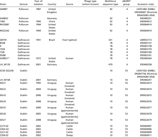
Table S1. List of isolates used in this study
Strain Serovar
Year of
isolation Country Source
Phage type (where known)
Multilocus sequence type
eBURST
group Accession no(s).
P125109 Enteritidis United Kingdom
Human (gastroenteritis)
4 11 4 AM933172
SARB18 Enteritidis 1988 United States
4 11 4 ERS400262
31/88 Enteritidis 1988 Uruguay Human (gastroenteritis)
11 4 ERS022680
8/89 Enteritidis 1989 Uruguay Human (invasive)
11 4 ERS022681
53/94 Enteritidis 1994 Uruguay Food 11 4 ERS022682
9508_195 Enteritidis 1994 United States
Egg 23 11 4 ERS217239
SE A Enteritidis 1994 United States
Egg 13a 11 4 ERS217235
EX4_PT21b Enteritidis 1996 United Kingdom
Human (gastroenteritis)
21b 11 4 ERS217238
D24359 Enteritidis 1998 Malawi Human (invasive)
Novel ERS024552
99-02302 Enteritidis 1999 Germany Hedgehog 11 183 4 ERS400254
206/99* Enteritidis 1999 Uruguay Food 11 4 ERS022683
00-03508 Enteritidis 2000 Germany Hedgehog 11 183 4 ERS400256
LH_M296 Enteritidis 2001 United States
11 4 ERS400259
251/01 Enteritidis 2001 Uruguay Egg 11 4 ERS022684
01-00493-2* Enteritidis 2001 Germany Reptile 168 4 ERS400244
8/02 Enteritidis 2002 Uruguay Human (gastroenteritis)
11 4 ERS022685
214/02* Enteritidis 2002 Uruguay Human (invasive)
11 4 ERS022686
10/05 Enteritidis 2005 Uruguay Human (gastroenteritis)
11 4 ERS022687
RD50 Enteritidis 2005 United Kingdom
Egg farm 6 11 4 ERS217225
RD36 Enteritidis 2005 United Kingdom
Egg farm 7 11 4 ERS217218
RD3 Enteritidis 2005 United Kingdom
Egg farm 24 11 4 ERS217214
RD26 Enteritidis 2005 United Kingdom
Egg farm 35 11 4 ERS217216
RD46 Enteritidis 2005 United Kingdom
Egg farm 36 11 4 ERS217222
RD84 Enteritidis 2005 United Kingdom
Egg farm 45 11 4 ERS217233
S1750_06 Enteritidis 2006 United Kingdom
Human (gastroenteritis)
1 11 4 ERS217242
S4726_04 Enteritidis 2006 United Kingdom
Human (gastroenteritis)
14b 11 4 ERS217241
P17 Enteritidis 2007 Human
(gastroenteritis)
2 11 4 ERS217211
P5 Enteritidis 2007 Human
(gastroenteritis)
3 11 4 ERS217199
P2 Enteritidis 2007 Human
(gastroenteritis)
5 11 4 ERS217197
P7 Enteritidis 2007 Human
(gastroenteritis)
58 11 4 ERS217201
P10 Enteritidis 2007 Spain Human
(gastroenteritis)
14b 11 4 ERS217204
105_2011K Enteritidis 2011 United States
Human (gastroenteritis)
366 4 ERS217252
118_2012K Enteritidis 2012 United States
Human (gastroenteritis)
366 4 ERS217264
Table S1. Cont.
Strain Serovar
Year of
isolation Country Source
Phage type (where known)
Multilocus sequence type
eBURST
group Accession no(s).
S44987 Pullorum 1987 United Kingdom
92 4 LK931482 (EMBL)
ERS430067 (Illumina) ERS429283 (454)
SARB52 Pullorum Germany 92 4 ERS400251
1002 Pullorum 1995 China 92 4 ERS004912
RKS5087 Pullorum 1994 United States
92 4 ERS004914
RKS2242 Pullorum 1994 United States
92 4 ERS004910
287/91 Gallinarum 1991 Brazil Fowl typhoid 331 4 AM933173
8338 Gallinarum 78 4 ERS003157
9 Gallinarum 78 4 ERS003158
1026 Gallinarum 78 4 ERS003159
72/8 Gallinarum 78 4 ERS003160
95/80 Gallinarum 78 4 ERS003161
SARB21* Gallinarum 1972 United States
Human 78 4 ERS400264
LH_M139 Gallinarum 2001 Germany 470 4 ERS400258
SC50 (S2229) Dublin 10 53 LK931502 (EMBL)
ERS007756 (Illumina) ERS429284 (454)
LH_M158 Dublin 2001 Germany 10 53 ERS400257
SDU1 Dublin 1995 Uruguay Human
(invasive)
10 53 ERS022673
SDU2 Dublin 2004 Uruguay Human
(invasive)
10 53 ERS022674
SDU3 Dublin 2006 Uruguay Human
(invasive)
10 53 ERS022675
SDU4 Dublin 2008 Uruguay Human
(invasive)
10 53 ERS022676
SDU5 Dublin 2000 Uruguay Human
(gastroenteritis)
10 53 ERS022677
SDU6 Dublin 2005 Uruguay Human
(gastroenteritis)
10 53 ERS022678
SDU7 Dublin 2008 Uruguay Human
(gastroenteritis)
10 53 ERS022679
2272-02 Dublin 2002 Cattle 10 53 ERS004907
230A-02 Dublin 2002 Cattle 10 53 ERS004906
2221-02 Dublin 2002 Cattle 10 53 ERS004909
2356-02 Dublin 2002 Sheep 10 53 ERS004911
Includes accession numbers.
*These isolates do not harbor a virulence plasmid.
Table S2. Pathology scores in chickens following experimental infection
Isolate
3 d p.i. 7 d p.i.*
Spleen Liver Ileum Spleen Liver Ileum
Mean
Median (range) Mean
Median (range) Mean
Median (range) Mean
Median (range) Mean
Median (range) Mean
Median (range)
S. Gallinarum 287/91 2.8 3.0 (2–4) 3.0 3.0 (2–4) 1.0 1 (1–1) 4.5 4.5 (4–5) 3.7 3.5 (3–4) 1.4 1.0 (0–2) S.Enteritidis P125109 1.4 1.0 (1–2) 2.0 2.0 (1–3) 3.0 3.0 (2–4) 2.0 2.0 (1–3) 2.0 2.0 (1–3) 3.6 3.5 (3–4) S. enteritidis01-00493-2 1.4 1.0 (1–2) 2.0 2.0 (1–3) 2.2 2.0 (2–3) 2.0 2.0 (2–2) 2.0 2.0 (1–3) 2.8 3.0 (3–4) S. Enteritidis SARB18 1.6 1.5 (1–2) 1.8 2.0 (1–3) 3.0 3.0 (2–4) 2.2 2.0 (1–3) 2.0 2.0 (1–3) 1.8 2.0 (1–3)
Statistical comparison between groups using the Mann–WhitneyUtest shows increased systemic pathology in theS.Gallinarum-infected group at 5 d p.i. over that of theS.Enteritidis-infected birds at 7 d p.i. (P<0.015 for spleen;P<0.03 for liver). Little difference was seen in systemic pathology between S.Enteritidis strains. At 3 d p.i., all threeS. Enteritidis isolates showed significantly greater inflammation and damage in the ileum thanS.Gallinarum (P<0.013) and little difference between each other. However, at 7 d p.i. damage and inflammation was lower thanS. Enteritidis P125109, but not at statistical significance, in 01-00493-2 and significantly lower in SARB18 (P=0.014). The reduced intestinal pathology is likely to be a reflection of reduced intestinal load ofSalmonellain these isolates. Intestinal pathology remained lower in theS. Gallinarum-infected group, although was not significantly lower than that seen with SARB18 (P=0.40).
*S. Gallinarum data at 3 and 5 d p.i. as birds had reached humane endpoint at 5 d p.i.
Dataset S1. BAPS clusters and recombination events
Dataset S1
Dataset S2. Pseudogenes across the lineage, listed with gene IDs and pseudogene designations
Dataset S2
Dataset S3. List of metabolic pathways and transport reactions affected by pseudogenes
Dataset S3
Includes an interpretation of pseudogene impact using Biolog phenotyping results.
Dataset S4. Biolog phenotyping results forS. Dublin,S. Gallinarum, and threeS. Enteritidis isolates
Dataset S4