Virulence and Genome Integrity by Compensating for Low Cellular
dUTPase Activity in the Central Nervous System
Akihisa Kato,a,bJun Arii,a,bYoshio Koyanagi,cYasushi Kawaguchia,b
Division of Molecular Virology, Department of Microbiology and Immunology, The Institute of Medical Science, The University of Tokyo, Tokyo, Japana
; Department of Infectious Disease Control, International Research Center for Infectious Diseases, The Institute of Medical Science, The University of Tokyo, Tokyo, Japanb
; Laboratory of Viral Pathogenesis, Institute for Virus Research, Kyoto University, Kyoto, Japanc
ABSTRACT
A mutation in herpes simplex virus 1 dUTPase (vdUTPase), which precluded its phosphorylation at Ser-187, decreased viral neurovirulence and increased mutation frequency in progeny virus genomes in the brains of mice where endogenous cellular dUTPase activity was relatively low, and overexpression of cellular dUTPase restored viral neurovirulence and mutation fre-quency altered by the mutation. Thus, phosphorylation of vdUTPase appeared to regulate viral virulence and genome integrity by compensating for low cellular dUTPase activityin vivo.
IMPORTANCE
Many DNA viruses encode a homolog of host cell dUTPases, which are known to function in accurate replication of cellular DNA genomes. The viral dUTPase activity has long been assumed to play a role in viral replication by preventing mutations in prog-eny virus genomes if cellular dUTPase activity was not sufficient. Here, we showed that a mutation in herpes simplex virus 1 dUTPase, which precluded its phosphorylation at Ser-187 and reduced its activity, decreased viral neurovirulence and increased mutation frequency in progeny virus genomes in the brains of mice where endogenous cellular dUTPase activity was relatively low. In contrast, overexpression of cellular dUTPase restored viral neurovirulence and mutation frequency altered by the muta-tion in the brains of mice. This is the first report, to our knowledge, directly showing that viral dUTPase activity regulates viral genome integrity and pathogenicity by compensating for insufficient cellular dUTPase activityin vivo.
P
reserving the integrity of their genetic information duringge-nome replication is of vital importance for all organisms. Noncanonical nucleotides are constantly generated during nucle-otide metabolism, and their misincorporation into the genome during replication may result in increased mutagenesis and over-load of the DNA excision repair system, leading to multiple DNA
strand breaks and cell death (1,2). The most common
noncanoni-cal nucleoside triphosphate is dUTP, which is continuously pro-duced in the pyrimidine biosynthesis pathway by phosphorylation
of dUDP or deamination of dCTP (1,3). dUTPase catalyzes dUTP
cleavage to dUMP and pyrophosphate, thereby reducing
misin-corporation of dUTP into the genome (4, 5). In addition,
dUTPase also plays a role in providing a substrate for thymidylate synthase, which converts dUMP to TMP, a major biosynthetic
pathway for TTP (6–8). Interestingly, a number of viruses,
includ-ing herpesviruses, poxviruses, adenoviruses, D-type retroviruses, and African swine fever virus (ASFV), encode their own dUTPases, suggesting the importance of dUTPases in the life
cy-cles of these viruses (4,9–11).
In this study, we focused on the dUTPase encoded by herpes simplex virus 1 (HSV-1), one of the best-studied members of the
Herpesviridaefamily, which causes a variety of diseases such as mucocutaneous diseases, keratitis, skin diseases, and encephalitis
(12). HSV-1 dUTPase (vdUTPase) is encoded by the UL50 gene
and is conserved throughout theHerpesviridaefamily (13,14). We
recently reported that Us3, an HSV-1-encoded serine/threonine protein kinase, phosphorylated vdUTPase at serine 187 (Ser-187), which upregulated its enzymatic activity in HSV-1-infected cells
(15). We also showed that this phosphorylation promoted viral
replication in human neuroblastoma SK-N-SH cells and viral rep-lication and virulence in the central nervous system (CNS) of mice
(15,16). Thus, regulation of vdUTPase activity by Us3
phosphor-ylation of vdUTPase Ser-187 appeared to be important for HSV-1 replication and pathogenicity. Although the mechanism by which dUTPases encoded by HSV-1 and other viruses act in viral
repli-cation and pathogenicityin vitroandin vivohas not completely
been elucidated, it has long been assumed that dUTPase activity is critical in the replication of viruses encoding a dUTPase. Such viral dUTPase activity may compensate if there is not sufficient host cell dUTPase activity for efficient viral replication, e.g., in resting and differentiated cells, such as neurons and macrophages,
where cellular dUTPase activity has been suggested to be low (12,
17). In agreement with this hypothesis, replication of
recombi-nant ASFV and D-type retroviruses with mutations in their
vdUTPases was significantly reduced in nondividing cellsin vitro,
Received29 August 2014 Accepted6 October 2014
Accepted manuscript posted online15 October 2014
CitationKato A, Arii J, Koyanagi Y, Kawaguchi Y. 2015. Phosphorylation of herpes simplex virus 1 dUTPase regulates viral virulence and genome integrity by compensating for low cellular dUTPase activity in the central nervous system. J
Virol 89:241–248.doi:10.1128/JVI.02497-14.
Editor:R. M. Sandri-Goldin
Address correspondence to Yasushi Kawaguchi, [email protected]. Copyright © 2015, American Society for Microbiology. All Rights Reserved.
doi:10.1128/JVI.02497-14
on November 7, 2019 by guest
http://jvi.asm.org/
whereas replication in actively dividing cells was only minimally
decreased (17–21).
We recently reported data supporting this hypothesis more
directly (22). First, YK751 (vdUTPaseS187A), an HSV-1 mutant
with an alanine substitution for vdUTPase Ser-187 (S187A) (Fig.
1), which blocked Us3 phosphorylation of vdUTPase Ser-187 and
reduced vdUTPase activity in HSV-1-infected cells (15),
repli-cated less efficiently than wild-type virus in SK-N-SK cells that had lower endogenous cellular dTUPase activity than in human carci-noma HEp-2 cells, in which YK751 (vdUTPaseS187A) replicated
as well as did wild-type virus (22). Second, knockdown of
cel-lular dUTPase in HEp-2 cells reduced replication of YK751
(vdUTPaseS187A) but not of wild-type virus (22). Third,
overex-pression of cellular dUTPase in SK-N-SH cells and in cellular dUTPase knockdown HEp-2 cells restored the replication of an HSV-1 mutant virus with the vdUTPase S187A mutation to that of
the wild-type virus in both cell types (22). These results suggested
that Us3 phosphorylation of vdUTPase Ser-187 upregulated its enzymatic activity and that this upregulation compensated for low cellular dUTPase activity for efficient HSV-1 replication, at least in
cell cultures. This also appeared to be the casein vivo, based on our
previous report that Us3 phosphorylation of vdUTPase Ser-187 promoted viral replication and virulence in the CNS of mice, in which most cells are not dividing, but played no obvious role in viral replication and pathogenicity in the eyes and vaginas of mice,
in which the HSV-1 target epithelial cells are actively dividing (16,
23, 24). However, experimental data to directly prove that the
dUTPase of HSV-1 and other viruses is needed to compensate for insufficient cellular dUTPase activity for viral replication and
pathogenicity in some cellsin vivohave not been reported thus far.
Furthermore, there is a lack of information on the effect of regu-lation of vdUTPase activity on viral genome integrity during
HSV-1 replicationin vitroandin vivo.
In the present study, we compared the levels of cellular dUTPase activity in mouse brains, eyes, and vaginas and examined whether overexpression of cellular dUTPase restored HSV-1 rep-lication and virulence impaired by the vdUTPase S187A mutation in the CNS of mice, to address questions about the function of vdUTPase in HSV-1 infections. We also investigated the effect of the vdUTPase S187A mutation on the mutation frequency in progeny HSV-1 genomes in cellular dUTPase knockdown and control HEp-2 cells and in the CNS of mice. In addition, we stud-ied the effect of overexpression of cellular dUTPase in the CNS of mice on the mutation frequency in progeny virus genomes of an HSV-1 strain with the vdUTPase S187A mutation.
MATERIALS AND METHODS
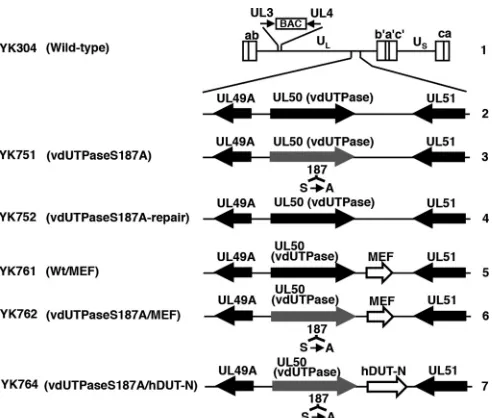
Cells and viruses.The simian kidney epithelial Vero line was described previously (25). sh-hDUT-HEp-2 and sh-Luc-HEp-2 cells were a cellular dUTPase knockdown HEp-2 cell line stably expressing short hairpin RNA (shRNA) against the 3=untranscribed region (UTR) in cellular dUTPase mRNA and a control cell line expressing shRNA against the open reading frame (ORF) in firefly luciferase mRNA, respectively, and were described previously (22). HSV-1 wild-type strain HSV-1(F); recombinant virus YK751 with a vdUTPase S187A mutation (vdUTPaseS187A); recombi-nant virus YK752, in which the vdUTPase S187A mutation in YK751 was repaired (vdUTPaseS187A-repair); recombinant virus YK761 carrying an expression cassette consisting of the Egr-1 promoter, an MEF tag consist-ing of Myc and Flag epitopes, and a tobacco etch virus (TEV) protease cleavage site and bidirectional polyadenylation signals of HSV-1 UL21 and UL22 genes (EGRp-MEF-polyA) in the intergenic region between UL50 and UL51 genes (Wt/MEF); recombinant virus YK762 with an S187A mutation in vdUTPase and the EGRp-MEF-polyA expression cas-sette (vdUTPaseS187A/MEF); and recombinant virus YK764 with an S187A mutation in vdUTPase and an expression cassette consisting of the Egr-1 promoter, the human hDUT-N ORF encoding the dominant nu-clear isoform of human dUTPase, and bidirectional polyadenylation sig-nals of HSV-1 UL21 and UL22 genes (vdUTPaseS187A/hDUT-N) were described previously (15,22,26) (Fig. 1).
Animal studies.Female ICR mice were purchased from Charles River. For all animal studies, after choosing the mean weight⫾4 g of mice, mice were randomly allocated into each group. For intracranial infection, 3-week-old mice were infected intracranially with 102PFU of each of the
indicated viruses as described previously (25,27). Mice were monitored daily, and mortality from 1 to 21 days postinfection was attributed to the inoculated virus. To determine viral titers in the brains, mice were inoc-ulated intracranially with 102PFU of the indicated virus as described
previously (27,28). Briefly, at 3 days postinfection, the mice were sacri-ficed, and the whole brain was removed, sonicated in 500l of 199 me-dium containing 1% fetal calf serum and antibiotics, and frozen at⫺80°C. Frozen samples were later thawed, and viral titers in the supernatants obtained after centrifugation of the samples were determined by standard plaque assays on Vero cells. All animal experiments were carried out in accordance with the Guidelines for Proper Conduct of Animal Experi-ments, Science Council of Japan. The protocol was approved by the Insti-tutional Animal Care and Use Committee (IACUC) of the Institute of Medical Science, The University of Tokyo (IACUC protocol approval number 19-26).
dUTPase enzyme assay.Brains, eyes, or vaginas were removed from 3-week-old female mice, 5-week-old female mice, or 5-week-old female mice pretreated with 1.67 mg of medroxyprogesterone (Depo-M; Vesco),
FIG 1Schematic diagrams of the genome structure of the wild-type and re-combinant HSV-1 viruses used in this study. Line 1, wild-type HSV-1 (YK304) genome carrying a bacmid (BAC) in the intergenic region between UL3 and UL4. Line 2, domain with the UL49A, UL50 (vdUTPase), and UL51 open reading frames. Line 3, recombinant virus YK751 with an S187A mutation in the UL50 (vdUTPase) gene. Line 4, recombinant virus YK752 with the re-paired vdUTPase S187A mutation. Line 5, recombinant virus YK761 carrying the MEF foreign gene expression cassette (an MEF tag with Myc and Flag epitopes and a TEV protease cleavage site) inserted into the intergenic region between UL50 (vdUTPase) and UL51. Line 6, recombinant virus YK762 with the vdUTPase S187A mutation and carrying MEF. Line 7, recombinant virus YK764 with the vdUTPase S187A mutation and carrying the hDUT-N foreign gene expression cassette (the nuclear isoform of human dUTPase) inserted
into the intergenic region between UL50 (vdUTPase) and UL51.
on November 7, 2019 by guest
http://jvi.asm.org/
[image:2.585.41.287.64.273.2]respectively; washed with 10 ml phosphate-buffered saline (PBS); and sonicated in 500l Nonidet P-40 (NP-40) buffer (50 mM Tris-HCl [pH 8.0], 150 mM NaCl, 1.0% NP-40). Protein concentrations in the super-natants obtained after a brief centrifugation were determined using a Bio-Rad protein assay kit. A 60-ng sample of each supernatant was then as-sayed for dUTPase enzymatic activity as described previously (22,29,30). Briefly, sample from HSV-1-infected mouse tissues was mixed with 200l reaction buffer (50 mM Tris-HCl [pH 8.0], 2 mM-mercaptoethanol, 1 mM MgCl2, 0.1% bovine serum albumin, 2 mMp-nitrophenylphosphate,
0.24 nM [3H]dUTP [28.8 Ci/mmol; PerkinElmer]). The reaction was
al-lowed to proceed for 30 min at 37°C and then terminated by spotting the reaction mixture onto DE81 circle discs (Whatman). The discs were washed three times for 5 min each with washing solution (1 mM ammo-nium formate, 4 M formic acid), followed by one wash with 95% ethanol for 3 min. The discs were air dried and counted for radioactivity using an LS3801 scintillation counter (Beckman).
HSV mutation frequency in cell cultures and mouse CNS.To mea-sure the mutation frequency in the progeny virus genomes in cell cultures, sh-hDUT-HEp-2 and sh-Luc-HEp-2 cells were infected with the indi-cated virus at a multiplicity of infection (MOI) of 5, harvested 36 h postin-fection, and lysed in 500l lysis buffer (10 mM Tris-HCl [pH 7.5], 150 mM NaCl, 1.5 mM MgCl2, 0.1% Nonidet P-40 [NP-40]). After a brief
centrifugation,-mercaptoethanol and EDTA were added to 400l of each supernatant to final concentrations of 50 mM and 1 mM, respec-tively. DNA was extracted with phenol-chloroform and precipitated with ethanol. HSV-1 UL54 nucleotides 114425 to 115153 were then amplified by PCR from the isolated viral genomes with Tks Gflex DNA polymerase (TaKaRa) and cloned into pBluescript II KS(⫹) (Stratagene). DH5␣cells were transformed with the cloned DNA, and 192 or 96 white colonies containing replicated viral genomes from sh-hDUT-HEp-2 or sh-Luc-HEp-2 cells, respectively, infected with each of the indicated viruses were picked. Sequencing was performed using a BigDye Terminator v3.1 cycle sequencing kit (Applied Biosystems). To measure the mutation frequency in the progeny virus genomes in the CNS of infected mice, a 3-week-old female ICR mouse was infected intracranially with 102PFU of each of the
indicated viruses and sacrificed at 3 days postinfection. To isolate viral DNAs from the brains of infected mice, whole brains were then removed, washed in PBS, frozen at⫺80°C, later thawed, lysed in urea lysis buffer (4.7 M urea, 1.3% [wt/vol] SDS, 0.23 M NaCl, 0.67 mM EDTA [pH 8.0], 6.7 mM Tris-HCl [pH 8.0]), extracted with phenol-chloroform, and eth-anol precipitated as described previously (31). UL54 nucleotides 114425 to 115153 were then amplified, cloned, and sequenced as described above, except using a pCR-Blunt II-TOPO vector (Invitrogen) and XL1-Blue cells.
Statistical analysis.Differences in relative dUTPase activity and virus titers were statistically analyzed using the two-tailed Studentttest. Differ-ences in frequency of the mutated clones were statistically analyzed using the2test. Differences in mortality of infected mice were statistically
analyzed by the log rank test. For the three comparison analyses,Pvalues of⬍0.0167 (0.05/3), 0.025 (0.05/2), or 0.05 (0.05/1) were sequentially considered significant after Holm’s sequentially rejective Bonferroni mul-tiple-comparison adjustment.
RESULTS
Endogenous cellular dUTPase activity in mice.We reported previously that YK751 (vdUTPaseS187A) replication and vir-ulence in the brains of mice following intracranial inoculation were impaired compared to the YK752 (vdUTPaseS187A-repair)
repaired virus (Fig. 1), but YK751 (vdUTPaseS187A) and YK752
(vdUTPaseS187A-repair) replication and pathogenicity in the eyes and vaginas of mice following ocular and vaginal inoculation,
respectively, were almost identical (16). To examine endogenous
cellular dUTPase activity in mouse tissues, the brains, eyes, and vaginas were removed from 3-week-old female mice, 5-week-old female mice, and 5-week-old female mice pretreated with
Depo-M, respectively, which were the same conditions as those in
the mouse experimental models that we used previously (16). As
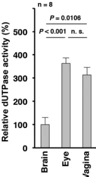
shown inFig. 2, endogenous dUTPase activity in the brains of
3-week-old female mice was significantly lower than in the eyes of 5-week-old female mice or in the vaginas of 5-week-old female mice pretreated with Depo-M. Thus, low endogenous cellular dUTPase activity in viral host tissues may be linked to the lower virulence and replication of YK751 (vdUTPaseS187A) described above, which had reduced vdUTPase activity due to its mutation
(15).
Effect of overexpression of cellular endogenous dUTPase on replication and virulence of HSV-1 with the vdUTPase S187A mutation.To investigate a direct linkage between low cellular dUTPase activity and HSV-1 replication and virulence, we studied the effect of cellular dUTPase overexpression in the CNS of mice on the replication and virulence of HSV-1 with reduced vdUTPase
activity due to the vdUTPase S187A mutation (15). Three
previ-ously reported (22) recombinant viruses were used for these
ex-periments (Fig. 1): (i) YK761 (Wt/MEF), which carried an
expres-sion cassette for the MEF tag in the intergenic region between UL50 and UL51; (ii) YK762 (vdUTPaseS187A/MEF), which had the vdUTPase S187A mutation and carried the MEF tag expres-sion cassette in the intergenic region between UL50 and UL51; and (iii) YK764 (vdUTPaseS187A/hDUT-N), which had the vdUTPase S187A mutation and carried the hDUT-N expression cassette. The MEF tag gene was used as a control foreign gene unrelated to the cellular hDUT-N gene. We previously reported that insertion of foreign genes into the intergenic region between UL50 and UL51 had no effect on viral replication in cell cultures or
on viral virulence in mice following intracranial inoculation (32).
We also reported that (i) YK764 (vdUTPaseS187A/hDUT-N) overexpressed cellular dUTPase in cell cultures, (ii) the growth curves of the three recombinant viruses (YK761, YK762, and YK764) were almost identical to that of wild-type HSV-1(F) in
FIG 2Endogenous cellular dUTPase activity in mouse brains, eyes, and vagi-nas. Using the same conditions as in the mouse experimental models described previously (16), the brains, eyes, and vaginas from eight 3-week-old female mice, 5-week-old female mice, and 5-week-old female mice pretreated with Depo-M for a week, respectively, were solubilized and 60-ng samples were assayed for dUTPase activity. Each value is the mean⫾standard error and is expressed relative to the mean dUTPase activity from brains, which was nor-malized to 100%.Prepresents the statistical significance value according to the two-tailed Studentttest. n.s., not significant.
on November 7, 2019 by guest
http://jvi.asm.org/
[image:3.585.372.468.66.243.2]Vero cells infected at multiplicities of infection (MOIs) of 5 and 0.01, and (iii) insertion of the foreign genes into the intergenic region between UL50 and UL51 in the three recombinant viruses
had no effect on expression of UL50 and UL51 (22).
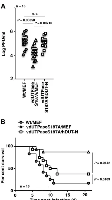
To investigate the effect of overexpression of cellular dUTPase on HSV-1 replication and virulence in the brains of mice,
3-week-old female ICR mice were infected intracranially with 102PFU of
YK761 (Wt/MEF), YK762 (vdUTPaseS187A/MEF), or YK764
(vdUTPaseS187A/hDUT-N) (Fig. 1); virus titers were assayed in
the brains of infected mice at 3 days postinfection; and mouse survival was monitored for 21 days postinfection. In agreement with our previous results that the vdUTPase S187A mutation sig-nificantly reduced HSV-1 replication and virulence in the CNS of
mice following intracranial inoculation (16), there was
signifi-cantly less progeny virus in the brains of mice infected intracrani-ally with YK762 (vdUTPaseS187A/MEF) than in the brains of
mice infected with YK761 (Wt/MEF) (Fig. 3A) and greater
sur-vival in mice infected with YK762 (vdUTPaseS187A/MEF) than in
mice infected with YK761 (Wt/MEF) (Fig. 3B). In contrast, the
survival of mice infected with YK764 (vdUTPaseS187A/hDUT-N) was significantly less than that of mice infected with YK762 (vdUTPaseS187A/MEF), although it was not as low as that of mice
infected with YK761 (Wt/MEF) (Fig. 3B). Furthermore, the level
of replication of YK764 (vdUTPaseS187A/hDUT-N) in the brains
of mice was similar to that of YK761 (Wt/MEF) (Fig. 3A). These
results indicated that overexpression of cellular hDUT-N signifi-cantly compensated for the reduction in HSV-1 replication and virulence due to the vdUTPase S187A mutation in the CNS of mice.
Effect of the vdUTPase S187A mutation on the mutation fre-quency in progeny viral genomes in cell cultures and mouse
CNS. To examine whether Us3 phosphorylation of vdUTPase
Ser-187 was involved in regulation of viral genome integrity in cells with low cellular dUTPase activity, two sets of experi-ments were performed. In the first set of experiexperi-ments, we used
sh-hDUT-HEp-2 and sh-Luc-HEp-2 cells (22). It was also
re-ported previously that cellular dUTPase was barely detectable in sh-hDUT-HEp-2 cells and that endogenous dUTPase activity in sh-hDUT-HEp-2 cells was significantly lower than that in
sh-Luc-HEp-2 cells (22). Therefore, sh-hDUT-HEp-2 and sh-Luc-HEp-2
cells were infected at an MOI of 5 with wild-type HSV-1(F), YK751 (vdUTPaseS187A), or YK752 (vdUTPaseS187A-repair) and harvested 36 h postinfection, and the mutation frequency in the progeny virus genomes in the infected cells was measured. As
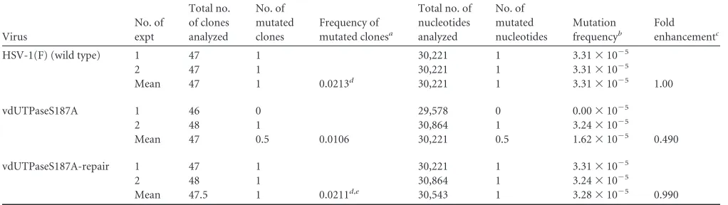
shown inTable 1, in two experiments the relative mutation
fre-quency in progeny virus genomes in sh-hDUT-HEp-2 cells in-fected with YK751 (vdUTPaseS187A) was 5.15-fold higher than that in sh-hDUT-HEp-2 cells infected with wild-type HSV-1(F) and was 5.42-fold higher than that in sh-hDUT-HEp-2 cells in-fected with YK752 (vdUTPaseS187A-repair). In contrast, the rel-ative mutation frequency of progeny virus genomes in sh-Luc-HEp-2 cells infected with YK751 (vdUTPaseS187A) was similar to that in sh-hDUT-HEp-2 cells infected with wild-type HSV-1(F) or
YK752 (vdUTPaseS187A-repair) (Tables 1and 2). The S187A
mutation in vdUTPase significantly elevated the frequency of the mutated clones from progeny virus genomes in sh-hDUT-HEp-2
cells (Table 1), whereas the mutation had no effect on that in
sh-Luc-HEp-2 (Table 2). These results indicated that the level of
endogenous cellular dUTPase activity in HEp-2 cells was sufficient for accurate replication of HSV-1 genomes with the vdUTPase S187A mutation and that wild-type vdUTPase enzymatic activity,
which was downregulated by the vdUTPase S187A mutation (15),
was required for suppression of incorporation of mutations into replicating HSV-1 genomes in cells with low endogenous dUT-Pase activity.
In the second set of experiments, 3-week-old female
ICR mice were infected intracranially with 102 PFU of
YK751 (vdUTPaseS187A), YK752 (vdUTPaseS187A-repair), YK761 (Wt/MEF), YK762 (vdUTPaseS187A/MEF), or YK764 (vdUTPaseS187A/hDUT-N), and the mutation frequency in progeny virus genomes in the brains of the infected mice was
measured at 3 days postinfection. As shown inTable 3, the relative
mutation frequency in progeny virus genomes in mice infected with YK751 (vdUTPaseS187A) was higher (3.79-fold) than that in mice infected with YK752 (vdUTPaseS187A-repair). In agree-ment with this result, the relative mutation frequency in progeny virus genomes in mice infected with YK762 (vdUTPaseS187A/
FIG 3Effect of cellular dUTPase overexpression on HSV-1 replication in the CNS of mice. (A) Fifteen 3-week-old female mice were infected intracranially with the indicated viruses. At 3 days postinfection, the brains of infected mice were harvested, and virus titers were assayed. Each data point is the virus titer in the brain of one mouse. The horizontal bars indicate the mean for each group. (B) Effect of cellular dUTPase overexpression on neurovirulence in the CNS of mice. Sixteen 3-week-old female ICR mice were infected intracranially with the indicated viruses, and survival was monitored for 21 days.P repre-sents the statistical significance value according to the two-tailed Studentttest (A) or the log rank test (B). n.s., not significant.
on November 7, 2019 by guest
http://jvi.asm.org/
[image:4.585.66.263.62.412.2]MEF) was higher (4.61-fold) than that in mice infected with
YK761 (Wt/MEF) (Table 4). The relative mutation frequency
in progeny virus genomes in mice infected with YK764 (vdUTPaseS187A/hDUT-N) was lower than that in mice infected
with YK762 (vdUTPaseS187A/MEF) in the two experiments (
Ta-ble 4). The S187A mutation in vdUTPase significantly elevated the frequency of mutated clones from progeny virus genomes in mice and overexpression of cellular dUTPase in part but significantly restored the wild-type frequency of the mutated clones from
prog-eny virus genomes in mice (Tables 3and4). These results
indi-cated that, in the brains of infected mice, wild-type vdUTPase activity was required for suppression of incorporation of muta-tions into replicating HSV-1 genomes, and overexpression of cel-lular dUTPase (hDUT-N) reduced the increase in mutation fre-quency in progeny virus genomes due to the vdUTPase S187A mutation.
DISCUSSION
In the present study, we showed that the endogenous dUTPase activity in the CNS of mice, in which the HSV-1 vdUTPase S187A
mutation reduced viral replication and pathogenicity (16), was
significantly lower than that in the eyes or vaginas of mice, in which the HSV-1 vdUTPase S187A mutation had no effect on
viral replication and pathogenicity (16). These observations
indi-cated that low endogenous cellular dUTPase activity in viral host
tissuesin vivowas linked to lower replication and pathogenicity of
[image:5.585.39.549.77.223.2]HSV-1 viruses with the vdUTPase S187A mutation. The more striking feature in this study was that overexpression of cellular dUTPase significantly restored the viral replication and virulence that were impaired by the vdUTPase S187A mutation in the CNS of mice. Taken together, the data presented here and in our pre-vious studies suggested that a particular level of dUTPase activity
TABLE 1Sequence analysis of progeny HSV-1 genomes in sh-hDUT-HEp-2 cells
Virus
No. of expt
Total no. of clones analyzed
No. of mutated clones
Frequency of mutated clonesa
Total no. of nucleotides analyzed
No. of mutated nucleotides
Mutation frequencyb
Fold enhancementc
HSV-1(F) (wild type) 1 89 3 57,227 3 5.24⫻10⫺5
2 86 3 55,298 3 5.43⫻10⫺5
Mean 87.5 3 0.0343d 56,263 3 5.33⫻10⫺5 1.00
vdUTPaseS187A 1 88 12 56,584 15 26.5⫻10⫺5
2 93 15 59,799 17 28.4⫻10⫺5
Mean 90.5 13.5 0.149 58,192 16 27.5⫻10⫺5 5.15
vdUTPaseS187A-repair 1 92 2 59,156 2 3.38⫻10⫺5
2 92 4 59,156 4 6.76⫻10⫺5
Mean 92 3 0.0326e,f 59,156 3 5.07⫻10⫺5 0.945
aFrequency of mutated clones was calculated as (mean of number of mutated clones)/(mean of total number of clones analyzed).
b
Mutation frequency was calculated as (number of mutated nucleotides)/(total number of nucleotides analyzed).
cFold enhancement was calculated as [mean of mutation frequency of HSV-1(F) genome], (mean of mutation frequency of vdUTPaseS187A genome), or (mean of mutation
frequency of vdUTPaseS187A-repair genome)/[mean of mutation frequency of HSV-1(F) genome].
dStatistically significant difference from vdUTPaseS187A (P⬍0.001). e
Statistically significant difference from vdUTPaseS187A (P⫽0.00286).
fStatistically nonsignificant difference from HSV-1(F).
TABLE 2Sequence analysis of progeny HSV-1 genomes in sh-Luc-HEp-2 cells
Virus
No. of expt
Total no. of clones analyzed
No. of mutated clones
Frequency of mutated clonesa
Total no. of nucleotides analyzed
No. of mutated nucleotides
Mutation frequencyb
Fold enhancementc
HSV-1(F) (wild type) 1 47 1 30,221 1 3.31⫻10⫺5
2 47 1 30,221 1 3.31⫻10⫺5
Mean 47 1 0.0213d 30,221 1 3.31⫻10⫺5 1.00
vdUTPaseS187A 1 46 0 29,578 0 0.00⫻10⫺5
2 48 1 30,864 1 3.24⫻10⫺5
Mean 47 0.5 0.0106 30,221 0.5 1.62⫻10⫺5 0.490
vdUTPaseS187A-repair 1 47 1 30,221 1 3.31⫻10⫺5
2 48 1 30,864 1 3.24⫻10⫺5
Mean 47.5 1 0.0211d,e 30,543 1 3.28⫻10⫺5 0.990
a
Frequency of mutated clones was calculated as (mean of number of mutated clones)/(mean of total number of clones analyzed).
bMutation frequency was calculated as (number of mutated nucleotides)/(total number of nucleotides analyzed).
c
Fold enhancement was calculated as [mean of mutation frequency of HSV-1(F) genome], (mean of mutation frequency of vdUTPaseS187A genome), or (mean of mutation frequency of vdUTPaseS187A-repair genome)/[mean of mutation frequency of HSV-1(F) genome].
d
Statistically nonsignificant difference from vdUTPaseS187A.
eStatistically nonsignificant difference from HSV-1(F).
on November 7, 2019 by guest
http://jvi.asm.org/
[image:5.585.39.550.526.670.2]was required for efficient HSV-1 replication and virulence in the CNS of mice and that Us3 phosphorylation of vdUTPase Ser-187 upregulated vdUTPase activity to compensate for the low cellular dUTPase activity in the CNS of mice for efficient HSV-1 replica-tion and virulence.
This is the first report, to our knowledge, directly showing that a virus-encoded dUTPase was able to compensate for low cellular dUTPase activity in host cells for efficient viral replication and
pathogenicityin vivo. Since this conclusion is in agreement with
the observations in cell cultures that we reported previously (22),
one might argue that the present study simply confirmed the ear-lier observations in cell cultures. However, it is known that a viral
replication phenotype exhibitedin vitrois not always in agreement
with the phenotype displayedin vivo. In fact, we previously
re-ported that the S147A mutation in Us3 and the T887A mutation in HSV-1 envelope glycoprotein B (gB), which precludes autophos-phorylation of Us3 at Ser-147 and Us3 phosautophos-phorylation of gB at Thr-887, respectively, had no effect on HSV-1 replication in cell cultures, although these mutations significantly reduced viral
rep-lication in mice (33–36). In contrast, we reported that the null
mutation in HSV-1 alkaline nuclease UL12 reduced viral replica-tion in cell cultures approximately 10,000-fold more than the enzyme-dead mutation in UL12 in cell cultures, but the null mutation in UL12 reduced viral neurovirulence in mice only approximately 10-fold more than the enzyme-dead mutation in
UL12 (37). Therefore, confirmation that phosphorylation of
vdUTPase Ser-187 compensated for low cellular dUTPase activity
for efficient viral replication and pathogenicityin vivois
signifi-cant and provides insight into the roles of other virus-encoded
dUTPases in viral replication and pathogenicityin vivoand into
the mechanism of HSV-1 CNS-specific virulence. There has been
an analogous report by Chen et al. (38) that HSV-1 thymidine
kinase (vTK), which is another viral homolog of a host cell enzyme involved in nucleotide metabolism and is required for viral repli-cation in ganglia of mice and for reactivation from the latency
following ganglionic explant (39–42), seemed to compensate for
endogenous cellular TK in gangliain vivo, based on the
[image:6.585.39.544.87.194.2]observa-tion that replacement of vTK with human TK was able to take over
TABLE 3Sequence analysis of progeny HSV-1 genomes in the brains of mice infected with YK751 (vdUTPaseS187A) or YK752 (vdUTPaseS187A-repair)
Virus
No. of expt
Total no. of clones analyzed
No. of mutated clones
Frequency of mutated clonesa
Total no. of nucleotides analyzed
No. of mutated nucleotides
Mutation frequencyb
Fold enhancementc
vdUTPaseS187A 1 74 9 47,582 9 18.9⫻10⫺5
2 93 14 59,799 14 23.4⫻10⫺5
Mean 83.5 11.5 0.138 53,691 11.5 21.2⫻10⫺5 3.79
vdUTPaseS187A-repair 1 88 2 56,584 2 3.54⫻10⫺5
2 74 5 47,582 5 10.5⫻10⫺5
Mean 81 3.5 0.0432d 52,083 3.5 7.02⫻10⫺5 1.00
aFrequency of mutated clones was calculated as (mean of number of mutated clones)/(mean of total number of clones analyzed).
b
Mutation frequency was calculated as (number of mutated nucleotides)/(total number of nucleotides analyzed).
cFold enhancement was calculated as (mean of mutation frequency of vdUTPaseS187A genome) or (mean of mutation frequency of vdUTPaseS187A-repair genome)/(mean of
mutation frequency of vdUTPaseS187A-repair genome).
dStatistically significant difference from vdUTPaseS187A (P⬍0.001).
TABLE 4Sequence analysis of progeny HSV-1 genomes in the brains of mice infected with YK761 (Wt/MEF), YK762 (vdUTPaseS187A/MEF), or YK764 (vdUTPaseS187A/hDUT-N)
Virus
No. of expt
Total no. of clones analyzed
No. of mutated clones
Proportion of mutated clonesa
Total no. of nucleotides analyzed
No. of mutated nucleotides
Mutation frequencyb
Fold enhancementc
Wt/MEF 1 86 2 55,298 2 3.62⫻10⫺5
2 94 5 60,442 6 9.93⫻10⫺5
Mean 90 3.5 0.0389d 57,870 4 6.77⫻10⫺5 1.00
vdUTPaseS187A/MEF 1 78 11 50,154 12 23.9⫻10⫺5
2 78 13 50,154 13 25.9⫻10⫺5
Mean 78 12 0.154 50,154 12.5 24.9⫻10⫺5 4.61
vdUTPaseS187A/hDUT-N 1 87 5 55,941 5 8.94⫻10⫺5
2 81 8 52,083 8 15.4⫻10⫺5
Mean 84 6.5 0.0774e,f 54,012 6.5 12.0⫻10⫺5 2.01
aFrequency of mutated clones was calculated as (mean of number of mutated clones)/(mean of total number of clones analyzed).
b
Mutation frequency was calculated as (number of mutated nucleotides)/(total number of nucleotides analyzed).
cFold enhancement was calculated as (mean of mutation frequency of Wt/MEF), (mean of mutation frequency of vdUTPaseS187A/MEF), or (mean of mutation frequency of
vdUTPaseS187A/hDUT-N genome)/(mean of mutation frequency of Wt/MEF genome).
dStatistically significant difference from vdUTPaseS187A/MEF (P⬍0.001). e
Statistically significant difference from vdUTPaseS187A/MEF (P⫽0.0116).
fStatistically nonsignificant difference from Wt/MEF.
on November 7, 2019 by guest
http://jvi.asm.org/
[image:6.585.39.545.516.661.2]the vTK functions in the ganglia of infected mice (38). These re-sults and those in this paper suggested that enzymes encoded by various viruses, which are homologs of host cell enzymes involved in nucleotide metabolism, may in general compensate for low activity of the homologous cellular enzymes for efficient viral
rep-licationin vivo.
Another striking feature in this study was that the vdUTPase S187A mutation increased the mutation frequency in progeny HSV-1 genomes in cellular dUTPase knockdown HEp-2 cells, whereas the mutation did not increase the mutation frequency in control HEp-2 cells. We also showed that the vdUTPase S187A mutation increased the mutation frequency in progeny HSV-1 genomes in the CNS of mice and that overexpression of cellular dUTPase attenuated the increase in the mutation frequency due to the vdUTPase S187A mutation. These results indicated that wild-type vdUTPase activity regulated by Us3 phosphorylation of vdUTPase Ser-187 compensated for insufficient cellular dUTPase
activity for accurate HSV-1 genome replicationin vitro andin
vivo. Thus, HSV-1 dUTPase functioned as an antimutator. In
agreement with this, Pyles and Thompson previously reported that loss of HSV-1 dUTPase increased the viral mutation
fre-quency in mouse fibroblast NIH 3T3 cells (43). Furthermore,
Le-rner et al. reported that feline immunodeficiency virus dUTPase was required for reduction of the viral mutation frequency in macro-phages in infected cats in which endogenous cellular dUTPase activ-ity was low, but not in lymphocytes with higher endogenous
cel-lular dUTPase activity (19).
We should note, however, that Pyles and Thompson
previ-ously reported that the loss of vdUTPase in the HSV-1 17syn⫹
strain increased the mutation frequency approximately 5-fold in NIH 3T3 cells as described above but had no effect on viral
repli-cation in these cells (43). That report contradicts our observation
that the vdUTPase S187A mutation in wild-type HSV-1(F) in-creased the viral mutation frequency approximately 5-fold and significantly reduced viral replication in cellular dUTPase knock-down HEp-2 cells. This discrepancy may be due to differences in cell types, viral strains, and/or methods of detecting mutations in the two studies. A similar contradiction was also reported for vTK:
Pyles and Thompson reported that vTK of the HSV-1 17syn⫹
strain had mutator activity (43), but Hwang et al. were not able to
detect mutator activity of vTK in the HSV-1 KOS strain using a
different method of detecting mutations (44). Specifically, Pyles
and Thompson used thelacZmutagenesis assay to examine the
mutation frequency of thelacZgene, when it was inserted into the
viral genome, by observing the relative ratio of the number of white/blue plaques in plaque assays after X-Gal
(5-bromo-4-chloro-3-indolyl--D-galactopyranoside) staining (43). Unlike
the method used to detect mutations in the present study, thelacZ
mutagenesis assay does not directly detect mutations in progeny
viral genomes. It has been suggested elsewhere (45) that
identify-ing plaques with mutatedlacZgenes may not be as simple as
ex-pected, since there may be experimental bias using cells infected
with a mixture of viruses with wild-type and mutantlacZgenes in
which insertion of thelacZgene at different locations in the viral
genome of each recombinant virus, including those used to deter-mine the baseline relative mutation frequency, may affect the rep-lication fidelity.
We have also presented data here showing that a high mutation frequency in progeny HSV-1 genomes in cellular dUTPase knock-down HEp-2 cells and in the CNS of mice was linked to lower viral
replication and/or virulence of viruses with the vdUTPase S187A mutation. Therefore, compensation for low cellular dUTPase ac-tivity by Us3 phosphorylation of vdUTPase Ser-187 for accurate viral genome replication during HSV-1 replication may be impor-tant for maintenance of viral replication and pathogenicity.
ACKNOWLEDGMENTS
We thank Tomoko Ando and Shihoko Koyama for excellent technical assistance.
This study was supported by the Funding Program for Next Genera-tion World-Leading Researchers and Grants for Scientific Research from the Japan Society for the Promotion of Science (JSPS); a contract research fund for the Program of Japan Initiative for Global Research Network on Infectious Diseases (J-GRID) and a grant for Scientific Research on Inno-vative Areas from the Ministry of Education, Culture, Science, Sports and Technology (MEXT) of Japan; and grants from the Takeda Science Foun-dation and the Ichiro Kanehara FounFoun-dation.
REFERENCES
1.Kouzminova EA, Kuzminov A.2004. Chromosomal fragmentation in dUTPase-deficient mutants of Escherichia coli and its recombinational repair. Mol Microbiol 51:1279 –1295. http://dx.doi.org/10.1111/j.1365 -2958.2003.03924.x.
2.Galperin MY, Moroz OV, Wilson KS, Murzin AG.2006. House clean-ing, a part of good housekeeping. Mol Microbiol59:5–19.http://dx.doi .org/10.1111/j.1365-2958.2005.04950.x.
3.Mathews CK.2006. DNA precursor metabolism and genomic stability. FASEB J20:1300 –1314.http://dx.doi.org/10.1096/fj.06-5730rev. 4.Vertessy BG, Toth J.2009. Keeping uracil out of DNA: physiological role,
structure and catalytic mechanism of dUTPases. Acc Chem Res42:97– 106.http://dx.doi.org/10.1021/ar800114w.
5.Shlomai J, Kornberg A.1978. Deoxyuridine triphosphatase of Esche-richia coli. Purification, properties, and use as a reagent to reduce uracil incorporation into DNA. J Biol Chem253:3305–3312.
6.Kunz BA, Kohalmi SE.1991. Modulation of mutagenesis by deoxyribo-nucleotide levels. Annu Rev Genet25:339 –359.http://dx.doi.org/10.1146 /annurev.ge.25.120191.002011.
7.Sedwick WD, Brown OE, Glickman BW.1986. Deoxyuridine misin-corporation causes site-specific mutational lesions in the lacI gene of Escherichia coli. Mutat Res162:7–20.http://dx.doi.org/10.1016/0027 -5107(86)90066-7.
8.Bessman MJ, Lehman IR, Adler J, Zimmerman SB, Simms ES, Korn-berg A.1958. Enzymatic synthesis of deoxyribonucleic acid. III. The in-corporation of pyrimidine and purine analogues into deoxyribonucleic acids. Proc Natl Acad Sci U S A44:633– 640.
9.Elder JH, Lerner DL, Hasselkus-Light CS, Fontenot DJ, Hunter E, Luciw PA, Montelaro RC, Phillips TR.1992. Distinct subsets of retrovi-ruses encode dUTPase. J Virol66:1791–1794.
10. Baldo AM, McClure MA.1999. Evolution and horizontal transfer of dUTPase-encoding genes in viruses and their hosts. J Virol73:7710 –7721. 11. McClure MA.2001. Evolution of the DUT gene: horizontal transfer be-tween host and pathogen in all three domains of life. Curr Protein Pept Sci
2:313–324.http://dx.doi.org/10.2174/1389203013381062.
12. Roizman B, Knipe DM, Whitley RJ.2013. Herpes simplex viruses, p 1823-1897.InKnipe DM, Howley PM, Cohen JI, Griffin DE, Lamb RA, Martin MA, Racaniello VR, Roizman B (ed), Fields virology, 6th ed. Lip-pincott-Williams & Wilkins, Philadelphia, PA.
13. McGeehan JE, Depledge NW, McGeoch DJ. 2001. Evolution of the dUTPase gene of mammalian and avian herpesviruses. Curr Protein Pept Sci2:325–333.http://dx.doi.org/10.2174/1389203013380964.
14. Preston VG, Fisher FB.1984. Identification of the herpes simplex virus type 1 gene encoding the dUTPase. Virology138:58 – 68.http://dx.doi.org /10.1016/0042-6822(84)90147-8.
15. Kato A, Tsuda S, Liu Z, Kozuka-Hata H, Oyama M, Kawaguchi Y.2014. Herpes simplex virus 1 protein kinase Us3 phosphorylates viral dUTPase and regulates its catalytic activity in infected cells. J Virol88:655– 666.
http://dx.doi.org/10.1128/JVI.02710-13.
16. Kato A, Shindo K, Maruzuru Y, Kawaguchi Y.2014. Phosphorylation of a herpes simplex virus 1 dUTPase by a viral protein kinase, Us3, dictates
on November 7, 2019 by guest
http://jvi.asm.org/
viral pathogenicity in the central nervous system but not at the periphery. J Virol88:2775–2785.http://dx.doi.org/10.1128/JVI.03300-13. 17. Payne SL, Elder JH.2001. The role of retroviral dUTPases in replication
and virulence. Curr Protein Pept Sci2:381–388.http://dx.doi.org/10.2174 /1389203013381008.
18. Threadgill DS, Steagall WK, Flaherty MT, Fuller FJ, Perry ST, Rushlow KE, Le Grice SF, Payne SL.1993. Characterization of equine infectious anemia virus dUTPase: growth properties of a dUTPase-deficient mutant. J Virol67:2592–2600.
19. Lerner DL, Wagaman PC, Phillips TR, Prospero-Garcia O, Henriksen SJ, Fox HS, Bloom FE, Elder JH.1995. Increased mutation frequency of feline immunodeficiency virus lacking functional deoxyuridine-triphosphatase. Proc Natl Acad Sci U S A92:7480 –7484.http://dx.doi.org /10.1073/pnas.92.16.7480.
20. Ladner RD, McNulty DE, Carr SA, Roberts GD, Caradonna SJ.1996. Characterization of distinct nuclear and mitochondrial forms of human deoxyuridine triphosphate nucleotidohydrolase. J Biol Chem271:7745– 7751.http://dx.doi.org/10.1074/jbc.271.13.7745.
21. Turelli P, Petursson G, Guiguen F, Mornex JF, Vigne R, Querat G.
1996. Replication properties of dUTPase-deficient mutants of caprine and ovine lentiviruses. J Virol70:1213–1217.
22. Kato A, Hirohata Y, Arii J, Kawaguchi Y.2014. Phosphorylation of herpes simplex virus 1 dUTPase upregulated viral dUTPase activity to compensate for low cellular dUTPase activity for efficient viral replication. J Virol88:7776 –7785.http://dx.doi.org/10.1128/JVI.00603-14. 23. Martin-Belmonte F, Perez-Moreno M.2012. Epithelial cell polarity, stem
cells and cancer. Nat Rev Cancer12:23–38.http://dx.doi.org/10.1038 /nrc3169.
24. Jakobsson J, Lundberg C.2006. Lentiviral vectors for use in the central nervous system. Mol Ther13:484 – 493.http://dx.doi.org/10.1016/j.ymthe .2005.11.012.
25. Tanaka M, Kagawa H, Yamanashi Y, Sata T, Kawaguchi Y. 2003. Construction of an excisable bacterial artificial chromosome containing a full-length infectious clone of herpes simplex virus type 1: viruses recon-stituted from the clone exhibit wild-type properties in vitro and in vivo. J Virol77:1382–1391.http://dx.doi.org/10.1128/JVI.77.2.1382-1391.2003. 26. Ejercito PM, Kieff ED, Roizman B.1968. Characterization of herpes simplex virus strains differing in their effects on social behaviour of in-fected cells. J Gen Virol2:357–364.http://dx.doi.org/10.1099/0022-1317 -2-3-357.
27. Tanaka M, Kato A, Satoh Y, Ide T, Sagou K, Kimura K, Hasegawa H, Kawaguchi Y.2012. Herpes simplex virus 1 VP22 regulates translocation of multiple viral and cellular proteins and promotes neurovirulence. J Virol86:5264 –5277.http://dx.doi.org/10.1128/JVI.06913-11.
28. Imai T, Arii J, Minowa A, Kakimoto A, Koyanagi N, Kato A, Kawaguchi Y.2011. Role of the herpes simplex virus 1 Us3 kinase phosphorylation site and endocytosis motifs in the intracellular transport and neurovirulence of envelope glycoprotein B. J Virol85:5003–5015.http://dx.doi.org/10 .1128/JVI.02314-10.
29. Williams MV, Cheng Y.1979. Human deoxyuridine triphosphate nucle-otidohydrolase. Purification and characterization of the deoxyuridine triphosphate nucleotidohydrolase from acute lymphocytic leukemia. J Biol Chem254:2897–2901.
30. Pyles RB, Sawtell NM, Thompson RL.1992. Herpes simplex virus type 1 dUTPase mutants are attenuated for neurovirulence, neuroinvasiveness, and reactivation from latency. J Virol66:6706 – 6713.
31. Nie C, Sato K, Misawa N, Kitayama H, Fujino H, Hiramatsu H, Heike
T, Nakahata T, Tanaka Y, Ito M, Koyanagi Y.2009. Selective infection of CD4⫹effector memory T lymphocytes leads to preferential depletion of memory T lymphocytes in R5 HIV-1-infected humanized NOD/SCID/ IL-2Rgammanull mice. Virology394:64 –72.http://dx.doi.org/10.1016/j .virol.2009.08.011.
32. Morimoto T, Arii J, Akashi H, Kawaguchi Y. 2009. Identification of multiple sites suitable for insertion of foreign genes in herpes simplex virus genomes. Microbiol Immunol 53:155–161. http://dx.doi.org/10.1111/j .1348-0421.2008.00104.x.
33. Imai T, Sagou K, Arii J, Kawaguchi Y.2010. Effects of phosphorylation of herpes simplex virus 1 envelope glycoprotein B by Us3 kinase in vivo and in vitro. J Virol84:153–162.http://dx.doi.org/10.1128/JVI.01447-09. 34. Sagou K, Imai T, Sagara H, Uema M, Kawaguchi Y.2009. Regulation of the catalytic activity of herpes simplex virus 1 protein kinase Us3 by auto-phosphorylation and its role in pathogenesis. J Virol83:5773–5783.http: //dx.doi.org/10.1128/JVI.00103-09.
35. Kato A, Arii J, Shiratori I, Akashi H, Arase H, Kawaguchi Y.2009. Herpes simplex virus 1 protein kinase Us3 phosphorylates viral envelope glycoprotein B and regulates its expression on the cell surface. J Virol
83:250 –261.http://dx.doi.org/10.1128/JVI.01451-08.
36. Kato A, Tanaka M, Yamamoto M, Asai R, Sata T, Nishiyama Y, Kawaguchi Y.2008. Identification of a physiological phosphorylation site of the herpes simplex virus 1-encoded protein kinase Us3 which regulates its optimal catalytic activity in vitro and influences its function in infected cells. J Virol82:6172– 6189.http://dx.doi.org/10.1128/JVI.00044-08. 37. Fujii H, Mugitani M, Koyanagi N, Liu Z, Tsuda S, Arii J, Kato A,
Kawaguchi Y.2014. Role of the nuclease activities encoded by herpes simplex virus 1 UL12 in viral replication and neurovirulence. J Virol88:
2359 –2364.http://dx.doi.org/10.1128/JVI.03621-13.
38. Chen SH, Cook WJ, Grove KL, Coen DM. 1998. Human thymidine kinase can functionally replace herpes simplex virus type 1 thymidine kinase for viral replication in mouse sensory ganglia and reactivation from latency upon explant. J Virol72:6710 – 6715.
39. Coen DM, Kosz-Vnenchak M, Jacobson JG, Leib DA, Bogard CL, Schaffer PA, Tyler KL, Knipe DM. 1989. Thymidine kinase-negative herpes simplex virus mutants establish latency in mouse trigeminal gan-glia but do not reactivate. Proc Natl Acad Sci U S A86:4736 – 4740.http: //dx.doi.org/10.1073/pnas.86.12.4736.
40. Tenser RB, Miller RL, Rapp F.1979. Trigeminal ganglion infection by thymidine kinase-negative mutants of herpes simplex virus. Science205:
915–917.http://dx.doi.org/10.1126/science.224454.
41. Jacobson JG, Ruffner KL, Kosz-Vnenchak M, Hwang CB, Wobbe KK, Knipe DM, Coen DM.1993. Herpes simplex virus thymidine kinase and specific stages of latency in murine trigeminal ganglia. J Virol67:6903– 6908.
42. Gordon YJ, Gilden DM, Becker Y.1983. HSV-1 thymidine kinase pro-motes virulence and latency in the mouse. Invest Ophthalmol Vis Sci
24:599 – 602.
43. Pyles RB, Thompson RL.1994. Mutations in accessory DNA replicating functions alter the relative mutation frequency of herpes simplex virus type 1 strains in cultured murine cells. J Virol68:4514 – 4524.
44. Hwang YT, Wang YA, Lu Q, Hwang CB.2003. Thymidine kinase of herpes simplex virus type 1 strain KOS lacks mutator activity. Virology
305:388 –396.http://dx.doi.org/10.1006/viro.2002.1776.
45. Hwang CB.2011. DNA replication fidelity of herpes simplex virus.In
Jelena KT (ed), DNA replication and related cellular processes. InTech, Rijeka, Croatia.http://dx.doi.org/10.5772/23548.