Hairpin RNAs: Sequence Analysis of Surviving Virus Shows Added
Selective Pressure of Combination Therapy
Anne Dallas,aHeini Ilves,aHan Ma,bDaniel J. Chin,bIan MacLachlan,cKlaus Klumpp,bBrian H. Johnstona SomaGenics, Inc., Santa Cruz, California, USAa
; Hoffmann-La Roche, Nutley, New Jersey, USAb
; Tekmira Pharmaceuticals Corporation, Burnaby, BC, Canadac
ABSTRACT
We have recently shown that a cocktail of two short synthetic hairpin RNAs (sshRNAs), targeting the internal ribosome entry site of hepatitis C virus (HCV) formulated with lipid nanoparticles, was able to suppress viral replication in chimeric mice
in-fected with HCV GT1a by up to 2.5 log10(H. Ma et al., Gastroenterology 146:63– 66.e5,http://dx.doi.org/10.1053/j.gastro.2013.09
.049) Viral load remained about 1 log10below pretreatment levels 21 days after the end of dosing. We have now sequenced the
HCV viral RNA amplified from serum of treated mice after the 21-day follow-up period. Viral RNA from the HCV sshRNA-treated groups was altered in sequences complementary to the sshRNAs and nowhere else in the 500-nucleotide sequenced re-gion, while the viruses from the control group that received an irrelevant sshRNA had no mutations in that region. The ability of
the most commonly selected mutations to confer resistance to the sshRNAs was confirmedin vitroby introducing those
muta-tions into HCV-luciferase reporters. The mutamuta-tions most frequently selected by sshRNA treatment within the sshRNA target sequence occurred at the most polymorphic residues, as identified from an analysis of available clinical isolates. These results demonstrate a direct antiviral activity with effective HCV suppression, demonstrate the added selective pressure of combination therapy, and confirm an RNA interference (RNAi) mechanism of action.
IMPORTANCE
This study presents a detailed analysis of the impact of treating a hepatitis C virus (HCV)-infected animal with synthetic hairpin-shaped RNAs that can degrade the virus’s RNA genome. These RNAs can reduce the viral load in these animals by over 99% after 1 to 2 injections. The study results confirm that the viral rebound that often occurred a few weeks after treatment is due to emer-gence of a virus whose genome is mutated in the sequences targeted by the RNAs. The use of two RNA inhibitors, which is more effective than use of either one by itself, requires that any resistant virus have mutations in the targets sites of both agents, a higher hurdle, if the virus is to retain the ability to replicate efficiently. These results demonstrate a direct antiviral activity with effective HCV suppression, demonstrate the added selective pressure of combination therapy, and confirm an RNAi mechanism of action.
H
epatitis C virus (HCV) infection is the most common cause of chronic liver disease and liver transplantation in the United States and a leading cause of liver cirrhosis and hepatocellular carcinoma worldwide (1,2). HCV has high genetic heterogeneity due to its intrinsically error-prone polymerase, high replication rate, and large number of infected cells in a typical infection. This results in a virus population with a huge number of sequence variations in each infected individual and a high potential for de-veloping resistance to direct-acting antiviral agents (DAA) (3–6). It has been demonstrated that a combination of molecules with nonoverlapping resistance profiles can be successful in treating HCV infection (7–10). A highly potent combination of antiviral agents will be especially important for subpopulations of difficult-to-treat patients, such as interferon nonresponders and patients with high viral load and high quasispecies diversity. RNA interfer-ence (RNAi) has the potential to provide multiple agents with nonoverlapping resistance profiles, in that multiple interfering RNAs targeting different parts of the viral RNA can be adminis-tered together. This is in contrast to current DAA combination therapy, in which multiple small-molecule drugs, each targeting a different viral protein, are combined.HCV has a single positive-strand RNA genome that functions as both a template for replication and message for polyprotein
synthesis. A single RNAi-induced cleavage of the genomic RNA can abolish both replication and translation of the virus. The 5=
end of the genome contains an internal ribosome entry site (IRES), which is the site of translation initiation and interacts directly with the 40S ribosomal subunit (11). Because the IRES is the most conserved region of the virus (12,13), it is an attractive target for RNAi-based therapies that are inherently very sequence specific. RNAi is particularly well suited for hepatitis therapy, be-cause the liver is both the site of HCV replication and the tissue most easily targeted by systemically administered oligonucleo-tides (reviewed in reference14), and a number of clinical trials of
Received30 September 2013Accepted15 January 2014
Published ahead of print29 January 2014
Editor:K. Kirkegaard
Address correspondence to Brian H. Johnston, [email protected].
A.D., H.I., and H.M. contributed equally to this work.
Supplemental material for this article may be found athttp://dx.doi.org/10.1128 /JVI.00105-14.
Copyright © 2014, American Society for Microbiology. All Rights Reserved.
doi:10.1128/JVI.00105-14
on November 7, 2019 by guest
http://jvi.asm.org/
RNAi agents against liver-specific targets are in progress (15). While several other groups have demonstrated RNAi-mediated inhibition of HCV in cell-based reporter gene systems, HCV sub-genomic replicon systems, and cell culture-based infectious HCV systems (16–21), RNAi-mediated inhibition of HCV replication in anin vivomodel system permissive for HCV replication has only recently been described (22).
We have previously characterized a novel class of RNAi agents called short synthetic hairpin RNAs (sshRNAs) that target domain IV of the HCV IRES and are highly potent inhibitors of HCV IRES-dependent luciferase gene expression in cell-based assays (23–27). sshRNAs that are chemically modified with 2=-O-methyl (2=-OMe) residues to abrogate immune-stimulatory effects and enhance stability in serum (24) show potential as therapeutic agents. We have shown that sshRNAs induce cleavage of target RNAs by a Dicer-independent RNAi mechanismin vitro(25,28). sshRNAs are predominantly loaded as intact molecules into Ar-gonaute (Ago)-containing complexes without prior processing by Dicer and are activated by Ago2-mediated cleavage of the passen-ger arm of the hairpin (28). When formulated with lipid nanopar-ticles (LNP), HCV-directed sshRNAs can be efficiently delivered to the liver, and they show potent and prolongedin vivoactivity in HCV-luciferase reporter mice with no immune stimulation (29,30).
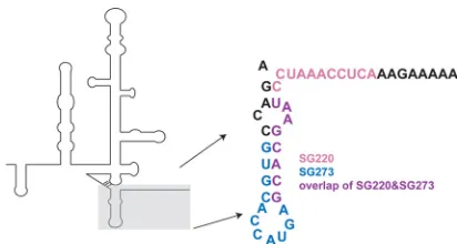
To assess the activity of the HCV sshRNAs in a robustin vivo HCV infection model, chimeric human hepatocyte uPA-SCID mice were stably infected with HCV of genotype 1a (GT1a). In this study, the mice received two intravenous injections, given a week apart, of two different LNP-formulated sshRNAs (SG220 and SG273) with overlapping target sequences in the HCV IRES (Fig. 1). Treatment groups included SG220 alone, SG273 alone, a com-bination of the two, and an irrelevant sshRNA as a control. The results showed substantial and sustained reduction in viral load in the groups treated with the HCV-specific sshRNAs (22). Twenty-one days after the last dose, the viral load of those groups remained about 90% below pretreatment levels. In contrast, there was no reduction of HCV RNA titer in the control sshRNA group during the dosing or follow-up periods. The LNP-formulated sshRNAs were well tolerated, with no treatment-related body weight loss, elevation of liver ALT levels, or reduction in serum human albu-min concentration during the dosing and follow-up periods. These results suggested that the reduction of serum HCV RNA concentration by the HCV-targeting sshRNAs was specific and that there was little or no nonspecific loss of human hepatocytes.
To assess to what extent treatment imposed selective pressure on the sequence makeup of the virus and determine whether re-maining viruses were resistant to the treatment, we undertook a sequence analysis of HCV that could be recovered from the treated mice, which we present here. We find that almost all of the recov-ered viral RNA was mutated in the regions targeted by the respec-tive sshRNAs and that these mutations conferred significant resis-tance to the agents. Use of a combination of SG220 and SG273 selected for mutations that conferred resistance to both sshRNAs, but viral reboundin vivotook longer to develop. These findings demonstrate the value of combining sshRNAs targeting different sites on the viral genome in constraining mutational escape, and they provide strong evidence for an RNAi mechanism of action for these agents.
MATERIALS AND METHODS
Reporter plasmids.Forin vitrocell culture experiments, a dual-luciferase expression plasmid (IRES/fLuc) was used in which the sequence encoding the HCV IRES of genotype 1b is placed between the coding sequences for Renillaandfireflyluciferase (fLuc), such that fLuc expression is dependent on the IRES. We introduced single and double point mutations in the HCV IRES target that result in mismatches to SG220 at position 5, 17, or both 5 and 17, as well as single mutations resulting in mismatches to SG273 at positions 6 and 16 using the QuikChange mutagenesis kit (Strat-agene/Agilent, Santa Clara, CA) according to the manufacturer’s instruc-tions. The presence of these mutations was confirmed by sequencing (Ret-rogen, San Diego, CA).
Transfection andin vitroassays.The human kidney cell line 293FT (Invitrogen, Carlsbad, CA) was maintained in Dulbecco’s modified Eagle medium (DMEM; Cambrex, Walkersville, IN) with 10% fetal bovine se-rum (HyClone, Logan, UT), supplemented with 2 mML-glutamine and 1 mM sodium pyruvate. One day prior to transfection, cells were seeded at 23,000 cells/well in a 96-well plate, resulting in⬃80% cell confluence at the time of transfection. Transfections were performed using Lipo-fectamine 2000 (Invitrogen) by following the manufacturer’s instruc-tions. Thirteen nanograms of pIRES/fLuc (an IRES-linked firefly lucifer-ase reporter construct), 20 ng of pSEAP2 control plasmid (BD Biosciences Clontech, San Jose, CA) as a transfection control, and the indicated amounts of sshRNAs were cotransfected into the 293FT cells. Forty-eight hours later, the cells were lysed and luciferase activity was measured in a MicroLumat LB 96P luminometer (Berthold Technologies, Bad Wildbad, Germany). All sshRNA samples were tested in triplicate. Percent silencing was calculated relative to transfections with the reporter plasmid in the absence of sshRNA inhibitors.
Formulation of sshRNA into LNP.Monomeric sshRNAs and control short interfering RNAs (siRNAs) were formulated into LNP by the pro-cess of stepwise ethanol dilution and spontaneous vesicle formation as previously described (31,32). LNP were dialyzed against phosphate-buff-ered saline (PBS) and filter sterilized through a 0.2-m filter before use. Mean particle sizes were 85 to 90 nm with polydispersity values of⬍0.1 by dynamic light scattering, and 92 to 98% of the siRNA was encapsulated within the lipid particles.
Treatment of human liver-uPA-SCID chimeric mice and measure-ments.Male uPA⫹/⫹/SCID mice with human albumin (ⱖ7 mg/ml) and
HCV (genotype 1a) RNA titers between 2.5⫻107and 5.1 ⫻108 copies/ml were chosen for the study. All mice received 2 intravenous injections via tail vein of the LNP-formulated HCV sshRNAs or an irrelevant sshRNA at the indicated doses at day 0 and day 7. At the indicated time points before or after the sshRNA dosing, blood sam-ples were collected via the retro-orbital route under isoflurane anes-thesia, and serum HCV RNA titer, serum total alanine aminotransfer-ase activity, and human albumin were measured as described previously (33). On day 28, all animals were anesthetized with isoflu-rane, and a minimum of 300l of blood was collected via cardiac FIG 1sshRNA target sites in the HCV IRES. Schematic diagram of the HCV
IRES secondary structure with the region targeted by sshRNAs in domain IV shown in detail. Residues targeted only by SG273 are shown in blue, residues targeted only by SG220 in pink, and residues targeted by both SG220 and SG273 in purple.
on November 7, 2019 by guest
http://jvi.asm.org/
[image:2.585.59.265.64.174.2]puncture, after which the animals were sacrificed via exsanguination. Animal husbandry and all animal experimental procedures used in this study have been approved by the animal ethics committee of Phoenix-Bio (Hiroshima, Japan, where the injections were performed) in ac-cord with appropriate Japanese regulatory authorities.
Isolation of HCV RNA from serum samples from treated human liver-uPA-SCID chimeric mice.Serum was isolated from blood collected on day 28 of the study (described above) and stored at⫺80°C until analysis. On the day of analysis, the serum samples were thawed on ice, and 300-l aliquots were transferred to tubes containing 300l of 2⫻denaturing solu-tion (Ambion). RNA was isolated using themirVana PARIS kit (Ambion) by following the manufacturer’s protocol for liquid samples. RNA was eluted with 100l of elution buffer (as supplied by themirVana kit).
Synthesis, amplification, and cloning of HCV cDNA.The following primers were used to synthesize the HCV cDNA: HCVrev2 (5=-GGATA GGATCCCGTCTACCTCGAGGTTGC-3=), HCVrev3 (5=-AAAAAAGG ATCCCCCAAATTGCGTGACCTGC-3=), and HCVrev4 (5=-AAAAAAG GATCCCGACGAGCGGAATGTACC-3=). Underlined sequences are complementary to HCV-gt1a RNA residues 509 to 526, 682 to 700, and 745 to 762, respectively (relative to reference genomeAF009606) (34). Two to fivel of serum RNA was used for cDNA synthesis with Super-script II reverse tranSuper-scriptase by following the manufacturer’s protocol (Invitrogen). Two to 10l of HCV cDNA was subjected to PCR amplifi-cation with primers HCVfw (5=CCTGATAAGCTTGACACTCCACCAT GAATCA-3=; underlined nucleotides [nt] correspond to nt 21 to 39) and HCVrev2, HCVrev3, or HCVrev4 by using the PfuTurbo Hotstart DNA polymerase by following the manufacturer’s protocol (Stratagene). Am-plified products were digested with HindIII and BamHI and ligated to plasmid pBluescript SK⫹previously digested with HindIII and BamHI. The resulting recombinant plasmids (9 to 13/per mouse for a total of 20 mice) were sequenced using a T7 sequencing primer by Retrogen (San Diego, CA). Approximately 10 clones from each mouse serum sample were sequenced to detect variants. All cloned HCV sequences were aligned against the sequence of the HCV genotype 1a inoculum by using the ClustalW2 multiple-sequence alignment algorithm (www.ebi.ac.uk /Tools/msa/clustalw2/).
HCV 5=-untranslated region polymorphism analysis.A total of 609 confirmed and nonredundant genotype 1 (GT1) HCV sequences were ob-tained from the EU HCV database (http://euhcvdb.ibcp.fr/euHCVdb/). The 609 GT1 sequences consist of 245 GT1a and 364 GT1b sequences. The con-firmed HCV sequences are generally either full-length sequences or near-full-length sequences. The mean near-full-length of GT1a sequences is 9,272 nt (standard deviation, 149), and the mean length of GT1b sequences is 9,358 nt (standard deviation, 128). All GT1 sequences contain the complete 30-nt sshRNA target region. The GT1a and GT1b sequences were compared to the 30-nt sequence by blastn (http://www.ncbi.nlm.nih.gov/blast) with a minimal window size of 8. The blastn results were analyzed by a custom script; the nucleotide base variations at each position of the 30-nt reference were counted for GT1a and GT1b sequences separately or in aggregate.
RESULTS
Characterization of drug-resistant HCV variants selected
dur-ing treatment.HCV has a high mutation rate due to its
intrinsi-cally error-prone RNA polymerase and high replication rate (35). The emergence of drug-resistant variants is one of the major causes of the failure of DAA-based treatment regimens to achieve a sustained antiviral response (6,9,36). In our efficacy study of two LNP-formulated sshRNAs targeting overlapping sequences in the HCV IRES (SG220 and SG273) (Fig. 1) in chimeric human hepatocyte uPA-SCID mice infected with HCV genotype 1a, the mice were given two intravenous doses a week apart (details of this study are provided in reference22). We found that after the first injection, all mice receiving HCV-specific sshRNAs showed sub-stantial reduction in serum HCV RNA concentration. Treatment
with 2.5 mg/kg of body weight SG220 or SG273 led to a 1.8 log10or 1.2 log10reduction in HCV RNA, respectively, at 72 h after the first injection (P⬍0.01). A higher dose of SG220, 5 mg/kg, did not lead to further reduction in viral load. However, the combination of 2.5 mg/kg SG220 and 2.5 mg/kg SG273 provided the strongest inhibition of HCV replication, with a 2 log10reduction in HCV RNA observed 72 h after the first treatment and an additional 0.5 log10reduction 7 days after the second injection (P⬍0.01). The LNP-formulated irrelevant control sshRNA administered at 5 mg/kg showed no effect. The mean viral load reduction from base-line in all HCV sshRNA-treated groups remained detectable for an extended period of time. In particular, in the mice treated with either SG220 or with the combination of SG220 and SG273, the HCV serum viral load remained⬎10-fold lower than the pretreat-ment level (P⬍0.01) up to the last study time point 3 weeks after the second and last injections. Interestingly, in this study we ob-served a further reduction in HCV RNA titer after the second dose when a combination of SG220 and SG273 was given, but this was not observed in the case of the individual HCV sshRNAs (22). To determine if this was indicative of the emergence of resistant vari-ants in the case of the individual sshRNAs, we characterized cDNA clones of HCV RNA amplified from the terminal serum (recov-ered on day 28, 3 weeks after the second dose) of all mice in each of the treatment groups (SG220, 2.5 mg/kg; SG220, 5.0 mg/kg; SG273, 2.5 mg/kg; SG220 plus SG273, 2.5 mg/kg each; SG221 [scrambled control], 5.0 mg/kg). We obtained cDNA clones for all mice with the exception of one mouse in the SG220 2.5-mg/kg dosing group that had terminal serum HCV RNA levels 2.6 log10 below pretreatment levels (22). A total of 169 clones were obtained for the five dose groups, corresponding to 9 to 13 sequences per mouse.
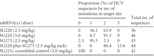
We found that all HCV cDNA clones from mice receiving the HCV-specific SG220 and almost all receiving only SG273 were mutated in the target region of their respective sshRNA and no-where else within the surrounding 500 nt sequenced (Table 1). In contrast, there were no sequence changes in the target regions of the two HCV sshRNAs in any of the 42 HCV clones we examined from the mice treated with the irrelevant sshRNA SG221 (Table 1). Positions in the target sequence that were mutated in each dosing group are summarized inFig. 2, and detailed alignments for each mouse are provided in Fig. S1 in the supplemental mate-rial. The mutations in HCV clones from SG220-treated mice oc-curred within the SG220 target region but not in the region unique to SG273 (Fig. 2AandB) andvice versafor mutations in HCV clones from the SG273-treated mice (Fig. 2C). Importantly, the mutations obtained from the mice treated with a combination of SG220 and SG273 occurred predominantly in the overlapping
re-TABLE 1Proportion of HCV sequences with mutations in the sshRNA target site from serum samples collected 21 days after the last injection
sshRNA(s) (dose)
Proportion (%) of HCV sequences by no. of mutations in target site
Total no. of sequences
0 1 2 3
SG220 (2.5 mg/kg) 0 36.1 63.9 0 36
SG220 (5.0 mg/kg) 0 8.7 91.3 0 46
SG273 (2.5 mg/kg) 2.3 95.3 2.3 0 43
SG220 plus SG273 (2.5 mg/kg each) 0 0 86.4 13.6 44
SG221c scrambled control (5.0 mg/kg) 100 0 0 0 42
on November 7, 2019 by guest
http://jvi.asm.org/
[image:3.585.299.545.89.179.2]gion of the two sshRNAs and within the target region of SG220 (the more potent of the two sshRNAs characterizedin vitro) but not in the region only targeted by SG273 (Fig. 2D). The number of mutations in the target site also tended to increase at higher indi-vidual doses and in the combination treatment group (Table 1). Mice receiving only SG220 at 2.5 mg/kg had 1 (36%) or 2 (64%) mutations per sequence, whereas those at the 5.0-mg/kg dose mostly had 2 mutations (91%). Most of the mice receiving only SG273, which was less potent than SG220 inin vitroscreening assays (24,25) andin vivo(22), had only 1 mutation in the target site (95%). In the sequences obtained from the combination group, all mice had 2 to 3 sites mutated in the target region. The overwhelming majority of mutations were point mutations, but 1 clone had a 6-nucleotide deletion instead (Fig. 2A).
The most common mutations in the SG220-treated group were at position 17 of the target site, corresponding to U362 in genotype 1a (SG220/S17; 35 out of 36 clones in the 2.5-mg/kg group and 45 out of 46 clones in the 5.0-mg/kg group), followed by positions 9 and 14. Of the substitutions at U362, 87% were U362C, but mutations to A and G were also observed (see Fig. S1 in the supplemental material). The most common mutations in the SG273-treated mice were at position 6 (C340) (SG273/S6; 24 out of 43), followed by position 16 (G350) (SG273/S16; 13 out of 43). The most frequent mutations in the combination group were at position 5 of the SG220 target (G350) (SG220/S5, which is the same position as SG273/S16) (Fig. 3A), followed by position 17 in the target of SG220. Substitutions at G350 were overwhelmingly G350A (96%) (see Fig. S1), with mutation to U also observed. C340 is in the translation start codon loop of the IRES, whereas G350 and U362 are nucleotides in the IRES just downstream of the start codon and are translated as the N-terminal residues of the core protein. These results indicate that the HCV-targeting sshRNAs were exerting selection pressure on the virus, effectively inhibiting the replication of
viruses having the wild-type IRES sequence while allowing replica-tion of HCV variants with certain mutareplica-tions in the respective target sites. These results are highly suggestive of a specific RNAi mecha-nism of action, consistent with our previous results obtained in cell culture (25,28).
Since the region targeted by the two sshRNAs contains nucle-otides coding for the core protein N-terminal residues M1S2T3N4P5K6P7, we analyzed the mutated sequences for codon substitutions and resulting amino acid changes (Table 2; also see Table S1 in the supplemental material) and performed protein sequence alignments (see Fig. S2). In the 169 sequences, there were no mutations in the AUG start codon (M1) or in the serine residue (S2). For the most frequently mutated codons, T3and P7, virtually all mutations occurred in the third position (at nucleotides G350 and U362), which did not result in an amino acid change. Similarly, 59 out of 60 mutations to the lysine codon also resulted in no change to the amino acid. The one-codon change resulting in an amino acid change was a conser-vative substitution of a basic residue (K6¡R). The N4and P5 codons were less frequently mutated but did result in some codon substitutions. No mutations were observed from nucle-otides 345 to 349, which make up codons S2and the first 2 nucleotides of T3and correspond to a base-paired stem closing the loop with the initiation codon, consistent with its conser-vation across all genotypes (12, 21). Altogether, 252 codon changes were observed, of which the majority (206 codons) resulted in synonymous substitutions rather than amino acid identity changes (46 codons), suggesting that mutations with minimal impact on viral protein function or ability of the vi-ruses to replicate were preferentially selected.
In vitrocharacterization of sshRNA activity against the most
frequently mutated sequences.To determine whether the
iden-tified HCV sequence variants could confer resistance to inhibition
FIG 2Sequence analysis of cDNA clones of HCV RNA amplified from terminal serum (day 28) of sshRNA-treated mice. The target sites of the 2 HCV sshRNAs with 8-nt overlaps are indicated by colors (blue, SG273; pink, SG220; purple, overlapping sequence). Sequence variants are aligned relative to wild-type genotype 1a virus (gt1a), with each dash representing one wild-type nucleotide of the target region. The positions of mutant variants (X) or deleted nucleotides (⌬) identified for each of the 5 dosing groups are shown aligned with the wild-type sequence (top line). The number of clones obtained out of the total number of cloned sequences is indicated in parentheses beside each sequence variant.
on November 7, 2019 by guest
http://jvi.asm.org/
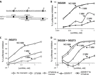
[image:4.585.137.450.65.283.2]by the HCV sshRNAs, we engineered the mutations most fre-quently observed in each of the HCV sshRNA treatment groups (C340U, G350A, U362C, and the double mutation G350A-U362C) into the HCV IRES region of an IRES-directed firefly luciferase (fLuc) reporter plasmid (described in Ge et al. [25]) (Fig. 3A). These mutations correspond to target sites SG273/S6, SG273/S16 (identical to SG220/S5), SG220/S17, and double mu-tation SG273/S16-SG220/S17, respectively (Fig. 3A). HEK-293FT cells were cotransfected with SG220, SG273, or both sshRNAs plus
either the wild-type or the mutant plasmid, and luciferase reporter gene activity was measured to determine the activities of the sshRNAs against the mutated IRES.
SG220/S17, which lies in the seed region (nt 2 to 8 of the guide sequence) of SG220 and is the most frequent mutation to emerge from SG220 treatment, significantly attenuated the gene silencing activity of SG220 (Fig. 3B). SG220/S5, a mutation outside the seed region, did not reduce the silencing activity of SG220, consistent with the fact that this mutation did not emerge in the SG220-only
[image:5.585.103.486.65.365.2]FIG 3Mutations selected by HCV sshRNA treatments reduced the silencing ability of HCV sshRNA. (A) The three most common mutations among the HCV cDNA clones derived from the terminal serum are shown. Note that SG273/S16 is the same as SG220/S5. (B) Effect of mutations at the indicated sites on the gene-silencing activity of SG220. (C) Effect of mutations at the indicated sites on the gene-silencing activity of SG273. (D) Effect of mutations at the indicated sites on the gene-silencing activity of the SG220 and SG273 combination. The values shown are means and standard deviations from triplicate transfections. MM indicates the number of mutations in the target sequence.
TABLE 2Nucleotide and amino acid changes in the core protein residues contained within the sshRNA target sites
Codon type
Sequence change for core protein residuea:
M1 S2 T3 N4 P5 K6 P7 Q8
GT1a AUG AGC ACG AAU CCU AAA CCU CAAc
Substitutionb ACA (T; 52/169) CUC (L; 3/169) UCU (S; 26/169) AAC (K; 58/169) CCC (P; 81/169) AAA(K; 1/169)
ACU (T; 2/169) ACU (T; 10/169) CUU (L; 1/169) AAG (K; 1/169) CCA (P; 9/169) AUU (I; 1/169) AGA (R; 1/169) CCG (P; 3/169) CUC (L; 2/169) CAA (Q; 1/169)
Total no. of substitutions 0/169 0/169 54/169 14/169 27/169 60/169 96/169 1/169
a
Sequences were obtained from viral cDNA isolated on day 28 of the study. Codon changes resulting in amino acid substitutions are shaded gray. Changed codons resulting in a synonymous substitution are not shaded. Results are combined from all dose groups.
b
The amino acid substitution is in parentheses with the frequency listed.
cOnly the first 2 nucleotides of this codon are within the target sequence.
on November 7, 2019 by guest
http://jvi.asm.org/
[image:5.585.38.550.577.687.2]treatment groups. As expected, the double mutation of SG220/S5 and SG220/S17 did attenuate SG220 silencing.
In contrast to SG220, the less potent SG273 was vulnerable to inactivating mutations outside as well as inside its seed region: both SG273/S16 (within the SG273 seed region) and SG273/S6 (outside the seed region) attenuated SG273 silencing activity, al-though the seed mutation S16 had a stronger effect (Fig. 3C). This finding is consistent with the chimeric mouse results, where mu-tations emerged at either position upon SG273 treatment, indicat-ing that each mutation was sufficient to confer resistance to SG273 in vivo.
Treatment of HCV-infected mice with the SG220-SG273 com-bination induced the selection of sequence variants at the SG220 binding site, including the region overlapping the SG273 binding site (where mutations selected by SG220 alone were rare), but not the region unique to SG273 (Fig. 2). Of these variants, the SG220/ S17 mutation affected thein vitrosilencing activity of the sshRNA combination to an extent similar to that of SG220 alone (Fig. 3D). As noted above, the SG220/S5 (equivalent to SG273/S16) muta-tion alone had no measurable impact on the activity of SG220 (Fig. 3B) and did not significantly reduce the silencing effect of the sshRNA combination (Fig. 3D), whereas it did confer resistance to SG273 (Fig. 3C). The SG220/S5/S17 double mutant reduced the silencing activity of the sshRNA combination to an extent similar to that of SG220 alone (compareFig. 3BtoD). Taken together with the clustering of mutations in the overlap region with the combination treatment, these results suggest that the gene-silenc-ing activity of the sshRNA combination was due predominantly to SG220, the more potent of the two sshRNAs, but SG273 helped narrow the possibilities for mutational escape.
Polymorphism analysis of target site revealed potential
mechanism of resistant variant selection.To estimate the relative
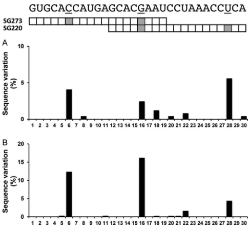
barriers to resistance for the two sshRNAs tested here, we investigated the conservation of the target sequences of the HCV sshRNAs in clinical isolates. A total of 609 nonredundant and confirmed geno-type 1 (GT1a and GT1b combined) HCV sequences were collected from the EU HCV database (http://euhcvdb.ibcp.fr/euHCVdb/). These sequences were analyzed with respect to the 30-nt sequence representing the target sequences of the two sshRNAs used in this study, using a custom script to count the polymorphisms at each position of the 30 nucleotides. The results are shown inFig. 4. Strik-ingly, the three nucleotide positions with the highest frequency of
sequence polymorphism in clinical isolates in the 30-nt region coin-cide with the three most frequent mutations induced by sshRNA treatment: 8.7% of the GT1 sequences in the databases contain vari-ants at the SG273/S6 position (the most common mutation in the SG273-treated group), 4.8% of the sequences contain variants at the SG220/S17 position (the most common mutation in the SG220-treated group), and 10.7% of the sequences contain vari-ants at the SG220/S5 (SG273/S16) position (the most common mutation in the combination treatment group). These three nu-cleotide positions remain the most polymorphic sites when GT1a and GT1b sequences are separately analyzed against the 30-nt se-quence, although the three sites are slightly more conserved in GT1a (Fig. 5A) than GT1b (Fig. 5B). The natural polymorphism at these sites suggests that sequence changes at these sites have less impact on replication fitness than changes at the more conserved sites; therefore, they are preferentially selected for. This, together with the reverse genetic characterization results presented inFig. 3, indicates that the mechanism of selection of HCV sshRNA re-sistancein vivois similar to that of other direct-acting antivirals: mutation of the DAA binding site leads to reduced drug binding affinity or binding effectiveness and, as a result, drug resistance.
DISCUSSION
In recent studies, we investigated thein vivoefficacy of LNP-for-mulated sshRNAs targeting the HCV IRES (22,29). Our results showed that, consistent with their potentin vitro inhibition of HCV IRES activity, these sshRNAs induced potent and rapid in-hibition of HCV IRES-dependent reporter gene expression in the mouse liver (29) and also efficiently inhibited HCV replication in the uPA-SCID human hepatocyte chimeric mouse model (22). Several features of these mice make them a good model for the human disease. First, the uPA-SCID mice used in this study sup-ported high levels of HCV replication. The HCV RNA titer in these mice (serum HCV RNA in the range of 107to 108copies/ml) was comparable to or higher than that typically observed in
pa-FIG 4Natural sequence polymorphism of HCV GT1 clinical isolates within the target region of the overlapping HCV sshRNAs SG220 and SG273. The percentage of HCV sequences deviating from the HCV sshRNA reference se-quence at any particular nucleotide position is shown. The shaded squares indicate the three most frequently mutated sites found in HCV isolated from mice treated with the active sshRNAs.
FIG 5Comparison of natural polymorphism within the sshRNA target region for GT1a (245 confirmed isolates) (A) and GT1b (364 confirmed isolates) (B). Analyses were performed and are displayed as described forFig. 4.
on November 7, 2019 by guest
http://jvi.asm.org/
[image:6.585.299.543.64.284.2] [image:6.585.43.283.64.183.2]tients infected with HCV genotype 1. It has been reported that the human hepatocytes in the uPA-SCID chimeric mouse model have the capacity to produce HCV virions at a rate comparable to that of the hepatocytes in chronically infected patients (33). Second, data generated with direct-acting antivirals in this mouse model have correlated well with the intrinsic clinical antiviral activity of those compounds in HCV-infected patients (33,37). Thein vivo efficacy of the HCV sshRNAs reported previously (22,29), to-gether with the resistance selection profile described here, estab-lishes sshRNAs as a novel class of direct-acting antiviral agents and represents, to the best of our knowledge, the first demonstrations of HCV inhibition by synthetic RNAs acting through an RNAi mechanism in a robust HCV infection model. Importantly, the RNA interference effect of LNP-formulated sshRNA treatment was long lasting, suggesting that such compounds could be effec-tive even with infrequent dosing. In the chimeric mice treated with SG220 (at both 2.5 and 5.0 mg/kg) or with the combination of SG220 and SG273, the serum HCV viral load increased slowly over the 3 weeks of follow-up after the end of dosing. The mean viral load remained about 1 log10lower than the baseline and the con-trol groups 21 days after the second and last treatments (22).
Treatment of the HCV-infected chimeric mice with HCV-tar-geted sshRNA effectively selected for drug-resistant HCV variants. The selected sequence changes were restricted to the target regions of the sshRNAs, demonstrating that the effect was sequence spe-cific. Interestingly, treatment with the less potent molecule SG273 selected mainly HCV variants with single point mutations in the sshRNA binding site (Table 1andFig. 2C), whereas with the more potent SG220 molecule (or the combination of both sshRNAs) the majority of the clones obtained had acquired 2 or even 3 muta-tions (Table 1andFig. 2A,C, andD). In the combination treat-ment group, the mutations clustered to the region of overlap of the target regions of SG220 and SG273 as well as the region unique to SG220. These results are consistent with SG220 having exerted a higher selection pressure on HCV than SG273, as expected based on its greater potency bothin vitroandin vivo(22,24), but with SG273 contributing additional selection pressure in the combina-tion so as to require that escape mutacombina-tions be directed to the over-lap region. Interestingly, mutations in the overover-lap region that were selected by the combination treatment appeared infrequently (1 out of 82 clones) in mice treated with SG220 alone, although they were more common in the mice receiving the less potent SG273 alone (13 out of 43 clones had the G16 position of SG273 mu-tated). This correlates with thein vitroassay showing that a single mutation of G350A in the overlap region had no observable effect on the silencing activity of SG220 but severely attenuated the si-lencing activity of SG273 (Fig. 3BandC).
The target region of SG220 and SG273 lies within the sequence common to both the IRES and the 5=end of the coding region of the core protein. Together with the 5= nontranslated region (NTR), these sequences are among the most conserved regions of HCV (13,38). The HCV core protein forms the viral nucleocap-sid. Its N-terminal domain is highly basic and has been shown to have RNA binding activity (38,39). Whereas most IRES require only the 5=-NTR for translation initiation, several studies have shown that efficient initiation of translation with the HCV IRES is dependent upon the sequence downstream of the initiation AUG codon (40–43). The N-terminal sequence of the core protein has a cluster of basic residues that are important for HCV infectivity (44). While extensive mutagenesis of these residues to alanine
knocks down core protein expression to undetectable levels, ala-nine-scanning mutants limited to amino acids in the sequence targeted by SG220/SG273 had only a slight effect on core expres-sion (44). In addition, the nucleotide sequence that forms the N terminus of the core protein contains an adenosine-rich region, which has been shown to interact directly with an IREStrans -acting factor called NSAP1. In addition to binding to this A-rich region, NSAP1 also interacts with the 40S subunit and is impor-tant for correct positioning of the start codon in the translation initiation complex (45,46). Interestingly, introduction of silent mutations that convert A residues in this residue to G dramatically reduced binding activity of NSAP1 to HCV RNA (45). Since the region targeted by SG220 and SG273 comprises only a part of the A-rich element, the mutations of A residues we observe may not be sufficient to severely reduce the binding affinity of NSAP1. There were not any observed mutations from nucleotides 345 to 349, which make up codons S2and the first 2 nucleotides of T3. The amino acids coded by these residues not only are conserved (38) but also are part of the base-paired stem that closes the loop containing the initiation codon, suggesting that this conserved stem structure is important for translation initiation. Indeed, Honda et al. found in a mutagenesis study that the internal stabil-ity of this stem loop is inversely correlated with efficiency of initi-ation of transliniti-ation and may serve a regulatory function (47). Interestingly, we did not find any mutations in the nucleotides that comprise the other strand of the stem (335 to 338) that are targeted by SG273, so no double compensatory mutations were observed in sequence space targeted by 2 different sshRNAs. Nev-ertheless, most (206/252) of the observed escape mutations occur at the third positions of core codons and do not alter the amino acid sequence. This is also consistent with the previous observa-tion that, across all genotypes, most of the frequently observed changes in the coding region occur at synonymous sites (13).
Our analysis of over 600 sequences of HCV genotype 1 revealed that the resistant variants selected in the chimeric mice coincide with the most highly polymorphic sites within the target region. Other recent studies using the chimeric mouse HCV infection model also generated drug-resistant variants similar to those ob-served in treated patients with chronic HCV infection (37,48,49). Preferential selection of resistance mutations at polymorphic sites suggests that those mutants are associated with reasonable cation fitness, a feature that could be tested in competitive repli-cation assays (6,50). The frequency of sequence polymorphism in regions encoding the targets of direct-acting antiviral agents can have a clear impact on the rate of resistance selection and the clinical efficacy of these drugs.
It has been shown that siRNAs and shRNAs can inhibit gene expression nonspecifically by mechanisms such as activation of innate immunity, which can result in cytotoxicity from induction of interferon and inflammatory cytokines (51–54). Abrogation of such unwanted effects is important for the development of RNAi agents for therapeutic use, so care must be taken to avoid the misinterpretation of gene silencing resulting from nonspecific or off-target effects (reviewed in reference55). The induction of in-nate immunity is influenced by both RNA structure and sequence (52,56). Because sshRNAs have a novel structural design, it is critical to verify that these molecules act through an RNAi mech-anism of action and carefully examine their potential to induce unwanted immune stimulation if they are to be considered for therapeutic applications. We have previously shown that this is
on November 7, 2019 by guest
http://jvi.asm.org/
the case in cultured cells, where chemically modified sshRNAs specifically knock down their intended target genes via an RNAi mechanism without immune stimulation (24). Furthermore, the RNA target was cleaved at the site expected for an RNAi mecha-nism in cells treated with these sshRNAs, and the sshRNAs could be immunoprecipitated in association with Ago2 (28), a key com-ponent of RNA-induced silencing complex (RISC). We next showed that LNP-formulated sshRNAs induce sequence-specific inhibition of HCV with little or no immune stimulatory effectin vivo(22,29). In this study, we provide further, definitive evidence that thisin vivoanti-HCV activity is a specific, RNAi-mediated effect by showing that mutations of drug-resistant HCV variants selected duringin vivotreatment are limited to sequences targeted by the sshRNAs, and that these mutations confer resistance to the sshRNAsin vitro. Together, these results demonstrate convinc-ingly that the sshRNAs used act specifically on the HCV RNA through an RNAi mechanismin vivo.
The ability of viruses with error-prone polymerases to escape inhibition of siRNA and shRNA agentsin vitrois well known (57–65). Strategies for mitigating resistance selection include tar-geting highly conserved target sites, increasing the inhibitory quo-tient, and using combination therapy. The latter approach could combine drugs of different mechanisms, such as small-molecule DAAs and interfering RNA, or employ a mixture of interfering RNAs (66). In the present context, additional sshRNAs could be combined with SG220 and SG273 to target the most common escape mutants revealed in our study or to target independent, conserved sites. Such combinations of siRNAs targeting different conserved regions have been shown to mitigate the risk of resis-tancein vitro(60,67). In the case of morbilliviruses, a combina-tion of three siRNAs targeting conserved regions was found to prevent viral escape, but two were insufficient (67). In that study, the emergence of escape mutants was delayed by 6 to 13 passages when two siRNAs were simultaneously delivered in the cell cul-ture, and the association of three siRNAs prevented any escape over 20 passages in cell culture. A similar result was obtained by Kusov et al. (68), who efficiently prevented the selection of resis-tant HIV-1 variants by using siRNAs targeting various sites in the HIV-1 nonstructural genes.
Because even treatment of HCV-infected chimeric mice with a combination of 2 HCV-targeted sshRNAs effectively selected for drug-resistant HCV variants, a successful sshRNA-based therapy against HCV will probably require a cocktail of more than 2 sshRNAs targeting highly conserved sequences. Oligonucleotide delivery agents are continually being improved, with the latest generation of LNP effective at doses well below 1 mg/kg (15,69). This effectively reduces nonspecific toxicity and allows more agents to be combined while maintaining an effective dose of each component. Whether such cocktails consist only of sshRNAs or sshRNAs combined with small-molecule DAAs, the potent and effective reduction of HCV replication and the favorable tolerability profile we observe with one or two doses of LNP-formulated sshRNAs suggest that sshRNAs have the potential to become effective components of highly active treat-ment regimens for HCV infection.
In summary, we have investigated thein vivoefficacy of LNP-formulated sshRNAs targeting the HCV IRES (22,29) as a novel RNAi-based therapeutic modality. Our results showed that, con-sistent with their potentin vitroinhibition of HCV IRES activity, these sshRNAs induced potent and rapid inhibition of HCV IRES-dependent reporter gene expression in mouse liver (29) and also
efficiently inhibited HCV replication in the uPA-SCID human hepatocyte chimeric mouse model (22). Thein vivoefficacy of the HCV sshRNAs reported in the studies described above (22,29) together with the resistance selection profile described here estab-lish sshRNAs as a novel class of DAA and represent, to the best of our knowledge, the first demonstrations of HCV inhibition by synthetic RNAs acting through an RNAi mechanism in a robust HCV infection model.
ACKNOWLEDGMENTS
This work was supported by NIH grants R43AI056611, R44AI056611, R43AI074256, and R43AI074214 (to B.H.J.).
We thank Yoshio Morikawa and colleagues at PhoenixBio for con-ducting the liver uptake and efficacy studies in the chimeric mice.
REFERENCES
1.Clark PJ, Muir AJ.2012. Overcoming barriers to care for hepatitis C. N. Engl. J. Med.366:2436 –2438.http://dx.doi.org/10.1056/NEJMp1202608. 2.Holmberg SD, Spradling PR, Moorman AC, Denniston MM. 2013. Hepatitis C in the United States. N. Engl. J. Med.368:1859 –1861.http: //dx.doi.org/10.1056/NEJMp1302973.
3.Farci P.2011. New insights into the HCV quasispecies and compartmen-talization. Semin. Liver Dis. 31:356 –374. http://dx.doi.org/10.1055/s -0031-1297925.
4.Martell M, Esteban JI, Quer J, Genesca J, Weiner A, Esteban R, Guardia J, Gomez J.1992. Hepatitis C virus (HCV) circulates as a population of different but closely related genomes: quasispecies nature of HCV genome distribution. J. Virol.66:3225–3229.
5. Powdrill MH, Tchesnokov EP, Kozak RA, Russell RS, Martin R, Svarovskaia ES, Mo H, Kouyos RD, Gotte M.2011. Contribution of a mutational bias in hepatitis C virus replication to the genetic barrier in the development of drug resistance. Proc. Natl. Acad. Sci. U. S. A.108:20509 – 20513.http://dx.doi.org/10.1073/pnas.1105797108.
6.Sun JH, O’Boyle DR, II, Zhang Y, Wang C, Nower P, Valera L, Roberts S, Nettles RE, Fridell RA, Gao M.2012. Impact of a baseline polymor-phism on the emergence of resistance to the hepatitis C virus nonstruc-tural protein 5A replication complex inhibitor, BMS-790052. Hepatology 55:1692–1699.http://dx.doi.org/10.1002/hep.25581.
7.Chayama K, Takahashi S, Toyota J, Karino Y, Ikeda K, Ishikawa H, Watanabe H, McPhee F, Hughes E, Kumada H.2012. Dual therapy with the nonstructural protein 5A inhibitor, daclatasvir, and the nonstructural protein 3 protease inhibitor, asunaprevir, in hepatitis C virus genotype 1b-infected null responders. Hepatology55:742–748.http://dx.doi.org/10 .1002/hep.24724.
8.Gane EJ, Roberts SK, Stedman CA, Angus PW, Ritchie B, Elston R, Ipe D, Morcos PN, Baher L, Najera I, Chu T, Lopatin U, Berrey MM, Bradford W, Laughlin M, Shulman NS, Smith PF.2010. Oral combi-nation therapy with a nucleoside polymerase inhibitor (RG7128) and danoprevir for chronic hepatitis C genotype 1 infection (INFORM-1): a randomised, double-blind, placebo-controlled, dose-escalation trial. Lan-cet376:1467–1475.http://dx.doi.org/10.1016/S0140-6736(10)61384-0. 9.Lok AS, Gardiner DF, Lawitz E, Martorell C, Everson GT, Ghalib R,
Reindollar R, Rustgi V, McPhee F, Wind-Rotolo M, Persson A, Zhu K, Dimitrova DI, Eley T, Guo T, Grasela DM, Pasquinelli C.2012. Pre-liminary study of two antiviral agents for hepatitis C genotype 1. N. Engl. J. Med.366:216 –224.http://dx.doi.org/10.1056/NEJMoa1104430. 10. Zeuzem S, Buggisch P, Agarwal K, Marcellin P, Sereni D, Klinker H,
Moreno C, Zarski JP, Horsmans Y, Mo H, Arterburn S, Knox S, Oldach D, McHutchison JG, Manns MP, Foster GR.2012. The protease inhib-itor, GS-9256, and non-nucleoside polymerase inhibitor tegobuvir alone, with ribavirin, or pegylated interferon plus ribavirin in hepatitis C. Hepa-tology55:749 –758.http://dx.doi.org/10.1002/hep.24744.
11. Pestova TV, Shatsky IN, Fletcher SP, Jackson RJ, Hellen CU.1998. A prokaryotic-like mode of cytoplasmic eukaryotic ribosome binding to the initiation codon during internal translation initiation of hepatitis C and classical swine fever virus RNAs. Genes Dev.12:67– 83.http://dx.doi.org /10.1101/gad.12.1.67.
12. Bukh J, Miller RH, Purcell RH.1995. Genetic heterogeneity of hepatitis C virus: quasispecies and genotypes. Semin. Liver Dis.15:41– 63.http://dx .doi.org/10.1055/s-2007-1007262.
on November 7, 2019 by guest
http://jvi.asm.org/
13. Simmonds P.2004. Genetic diversity and evolution of hepatitis C vi-rus–15 years on. J. Gen. Virol.85:3173–3188.http://dx.doi.org/10.1099 /vir.0.80401-0.
14. Barros SA, Gollob JA.2012. Safety profile of RNAi nanomedicines. Adv. Drug Deliv. Rev.64:1730 –1737.http://dx.doi.org/10.1016/j.addr.2012.06 .007.
15. Sehgal A, Vaishnaw A, Fitzgerald K.2013. Liver as a target for oligonu-cleotide therapeutics. J. Hepatol. 59:1354 –1359. http://dx.doi.org/10 .1016/j.jhep.2013.05.045.
16. Wilson JA, Jayasena S, Khvorova A, Sabatinos S, Rodrigue-Gervais IG, Arya S, Sarangi F, Harris-Brandts M, Beaulieu S, Richardson CD.2003. RNA interference blocks gene expression and RNA synthesis from hepa-titis C replicons propagated in human liver cells. Proc. Natl. Acad. Sci. U. S. A.100:2783–2788.http://dx.doi.org/10.1073/pnas.252758799. 17. Yokota T, Sakamoto N, Enomoto N, Tanabe Y, Miyagishi M, Maekawa
S, Yi L, Kurosaki M, Taira K, Watanabe M, Mizusawa H.2003. Inhi-bition of intracellular hepatitis C virus replication by synthetic and vector-derived small interfering RNAs. EMBO Rep.4:602– 608.http://dx.doi.org /10.1038/sj.embor.embor840.
18. Seo MY, Abrignani S, Houghton M, Han JH.2003. Small interfering RNA-mediated inhibition of hepatitis C virus replication in the human hepatoma cell line Huh-7. J. Virol.77:810 – 812.http://dx.doi.org/10.1128 /JVI.77.1.810-812.2003.
19. Kanda T, Steele R, Ray R, Ray RB.2007. Small interfering RNA targeted to hepatitis C virus 5=nontranslated region exerts potent antiviral effect. J. Virol.81:669 – 676.http://dx.doi.org/10.1128/JVI.01496-06.
20. Randall G, Panis M, Cooper JD, Tellinghuisen TL, Sukhodolets KE, Pfeffer S, Landthaler M, Landgraf P, Kan S, Lindenbach BD, Chien M, Weir DB, Russo JJ, Ju J, Brownstein MJ, Sheridan R, Sander C, Zavolan M, Tuschl T, Rice CM.2007. Cellular cofactors affecting hepatitis C virus infection and replication. Proc. Natl. Acad. Sci. U. S. A.104:12884 –12889.
http://dx.doi.org/10.1073/pnas.0704894104.
21. Kronke J, Kittler R, Buchholz F, Windisch MP, Pietschmann T, Barten-schlager R, Frese M.2004. Alternative approaches for efficient inhibition of hepatitis C virus RNA replication by small interfering RNAs. J. Virol.78: 3436 –3446.http://dx.doi.org/10.1128/JVI.78.7.3436-3446.2004.
22. Ma H, Dallas A, Ilves H, Shorenstein J, Maclachlan I, Klumpp K, Johnston BH.2013. Formulated minimal-length synthetic small hairpin RNAs are potent inhibitors of hepatitis C virus in mice with humanized livers. Gastroenterology146:63– 66.e5.http://dx.doi.org/10.1053/j.gastro .2013.09.049.
23. Vlassov AV, Korba B, Farrar K, Mukerjee S, Seyhan AA, Ilves H, Kaspar RL, Leake D, Kazakov SA, Johnston BH.2007. shRNAs targeting hepa-titis C: effects of sequence and structural features, and comparison with siRNA. Oligonucleotides17:223–236.http://dx.doi.org/10.1089/oli.2006 .0069.
24. Ge Q, Dallas A, Ilves H, Shorenstein J, Behlke MA, Johnston BH.2010. Effects of chemical modification on the potency, serum stability, and im-munostimulatory properties of short shRNAs. RNA16:118 –130.http: //dx.doi.org/10.1261/rna.1901810.
25. Ge Q, Ilves H, Dallas A, Kumar P, Shorenstein J, Kazakov SA, Johnston BH.2010. Minimal-length short hairpin RNAs: the relationship of struc-ture and RNAi activity. RNA16:106 –117.http://dx.doi.org/10.1261/rna .1894510.
26. Wang Q, Contag CH, Ilves H, Johnston BH, Kaspar RL.2005. Small hairpin RNAs efficiently inhibit hepatitis C IRES-mediated gene expres-sion in human tissue culture cells and a mouse model. Mol. Ther.12:562– 568.http://dx.doi.org/10.1016/j.ymthe.2005.04.014.
27. Ilves H, Kaspar RL, Wang Q, Seyhan AA, Vlassov AV, Contag CH, Leake D, Johnston BH.2006. Inhibition of hepatitis C IRES-mediated gene expression by small hairpin RNAs in human hepatocytes and mice. Ann. N. Y. Acad. Sci.1082:52–55.http://dx.doi.org/10.1196/annals.1348 .060.
28. Dallas A, Ilves H, Ge Q, Kumar P, Shorenstein J, Kazakov SA, Cuellar TL, McManus MT, Behlke MA, Johnston BH.2012. Right- and left-loop short shRNAs have distinct and unusual mechanisms of gene silencing. Nucleic Acids Res.40:9255–9271.http://dx.doi.org/10.1093/nar/gks662. 29. Dallas A, Ilves H, Shorenstein J, Judge A, Spitler R, Contag C, Wong SP,
Harbottle RP, Maclachlan I, Johnston BH.2013. Minimal-length syn-thetic shRNAs formulated with lipid nanoparticles are potent inhibitors of hepatitis C virus IRES-linked gene expression in mice. Mol. Ther. Nucleic Acids2:e123.http://dx.doi.org/10.1038/mtna.2013.50.
30. Kolli S, Wong SP, Harbottle R, Johnston B, Thanou M, Miller AD.
2013. pH-triggered nanoparticle mediated delivery of siRNA to liver cells in vitro and in vivo. Bioconjug. Chem.24:314 –332.http://dx.doi.org/10 .1021/bc3004099.
31. Jeffs LB, Palmer LR, Ambegia EG, Giesbrecht C, Ewanick S, Ma-cLachlan I.2005. A scalable, extrusion-free method for efficient liposomal encapsulation of plasmid DNA. Pharm. Res.22:362–372.http://dx.doi .org/10.1007/s11095-004-1873-z.
32. Semple SC, Akinc A, Chen J, Sandhu AP, Mui BL, Cho CK, Sah DW, Stebbing D, Crosley EJ, Yaworski E, Hafez IM, Dorkin JR, Qin J, Lam K, Rajeev KG, Wong KF, Jeffs LB, Nechev L, Eisenhardt ML, Jayaraman M, Kazem M, Maier MA, Srinivasulu M, Weinstein MJ, Chen Q, Alvarez R, Barros SA, De S, Klimuk SK, Borland T, Kosovrasti V, Cantley WL, Tam YK, Manoharan M, Ciufolini MA, Tracy MA, de Fougerolles A, MacLachlan I, Cullis PR, Madden TD, Hope MJ.2010. Rational design of cationic lipids for siRNA delivery. Nat. Biotechnol. 28:172–176.http://dx.doi.org/10.1038/nbt.1602.
33. Kamiya N, Iwao E, Hiraga N, Tsuge M, Imamura M, Takahashi S, Miyoshi S, Tateno C, Yoshizato K, Chayama K.2010. Practical evalua-tion of a mouse with chimeric human liver model for hepatitis C virus infection using an NS3-4A protease inhibitor. J. Gen. Virol.91:1668 – 1677.http://dx.doi.org/10.1099/vir.0.019315-0.
34. Kuiken C, Simmonds P.2009. Nomenclature and numbering of the hepatitis C virus. Methods Mol. Biol. 510:33–53.http://dx.doi.org/10 .1007/978-1-59745-394-3_4.
35. Castro C, Arnold JJ, Cameron CE.2005. Incorporation fidelity of the viral RNA-dependent RNA polymerase: a kinetic, thermodynamic and structural perspective. Virus Res.107:141–149.http://dx.doi.org/10.1016 /j.virusres.2004.11.004.
36. Sullivan JC, De Meyer S, Bartels DJ, Dierynck I, Zhang EZ, Spanks J, Tigges AM, Ghys A, Dorrian J, Adda N, Martin EC, Beumont M, Jacobson IM, Sherman KE, Zeuzem S, Picchio G, Kieffer TL. 2013. Evolution of treatment-emergent resistant variants in telaprevir phase 3 clinical trials. Clin. Infect. Dis.57:221–229.http://dx.doi.org/10.1093/cid /cit226.
37. Shi N, Hiraga N, Imamura M, Hayes CN, Zhang Y, Kosaka K, Okazaki A, Murakami E, Tsuge M, Abe H, Aikata H, Takahashi S, Ochi H, Tateno-Mukaidani C, Yoshizato K, Matsui H, Kanai A, Inaba T, McPhee F, Gao M, Chayama K. 2013. Combination therapies with NS5A, NS3 and NS5B inhibitors on different genotypes of hepatitis C virus in human hepatocyte chimeric mice. Gut62:1055–1061.http://dx .doi.org/10.1136/gutjnl-2012-302600.
38. Bukh J, Purcell RH, Miller RH.1994. Sequence analysis of the core gene of 14 hepatitis C virus genotypes. Proc. Natl. Acad. Sci. U. S. A.91:8239 – 8243.http://dx.doi.org/10.1073/pnas.91.17.8239.
39. Santolini E, Migliaccio G, La Monica N.1994. Biosynthesis and bio-chemical properties of the hepatitis C virus core protein. J. Virol.68:3631– 3641.
40. Honda M, Ping LH, Rijnbrand RC, Amphlett E, Clarke B, Rowlands D, Lemon SM.1996. Structural requirements for initiation of translation by internal ribosome entry within genome-length hepatitis C virus RNA. Virology222:31– 42.http://dx.doi.org/10.1006/viro.1996.0395. 41. Lu HH, Wimmer E.1996. Poliovirus chimeras replicating under the
translational control of genetic elements of hepatitis C virus reveal un-usual properties of the internal ribosomal entry site of hepatitis C virus. Proc. Natl. Acad. Sci. U. S. A.93:1412–1417.http://dx.doi.org/10.1073 /pnas.93.4.1412.
42. Reynolds JE, Kaminski A, Kettinen HJ, Grace K, Clarke BE, Carroll AR, Rowlands DJ, Jackson RJ.1995. Unique features of internal initiation of hepatitis C virus RNA translation. EMBO J.14:6010 – 6020.
43. Wang TH, Rijnbrand RC, Lemon SM.2000. Core protein-coding se-quence, but not core protein, modulates the efficiency of cap-independent translation directed by the internal ribosome entry site of hepatitis C virus. J. Virol. 74:11347–11358. http://dx.doi.org/10.1128/JVI.74.23.11347 -11358.2000.
44. Alsaleh K, Delavalle PY, Pillez A, Duverlie G, Descamps V, Rouille Y, Dubuisson J, Wychowski C.2010. Identification of basic amino acids at the N-terminal end of the core protein that are crucial for hepatitis C virus infectivity. J. Virol.84:12515–12528.http://dx.doi.org/10.1128/JVI.01393 -10.
45. Kim JH, Paek KY, Ha SH, Cho S, Choi K, Kim CS, Ryu SH, Jang SK. 2004. A cellular RNA-binding protein enhances internal ribosomal entry site-dependent translation through an interaction downstream of the
on November 7, 2019 by guest
http://jvi.asm.org/
atitis C virus polyprotein initiation codon. Mol. Cell. Biol.24:7878 –7890.
http://dx.doi.org/10.1128/MCB.24.18.7878-7890.2004.
46. Park SM, Paek KY, Hong KY, Jang CJ, Cho S, Park JH, Kim JH, Jan E, Jang SK.2011. Translation-competent 48S complex formation on HCV IRES requires the RNA-binding protein NSAP1. Nucleic Acids Res.39: 7791–7802.http://dx.doi.org/10.1093/nar/gkr509.
47. Honda M, Brown EA, Lemon SM.1996. Stability of a stem-loop involv-ing the initiator AUG controls the efficiency of internal initiation of trans-lation on hepatitis C virus RNA. RNA2:955–968.
48. Bartels DJ, Zhou Y, Zhang EZ, Marcial M, Byrn RA, Pfeiffer T, Tigges AM, Adiwijaya BS, Lin C, Kwong AD, Kieffer TL.2008. Natural prev-alence of hepatitis C virus variants with decreased sensitivity to NS3.4A protease inhibitors in treatment-naive subjects. J. Infect. Dis.198:800 – 807.http://dx.doi.org/10.1086/591141.
49. Le Pogam S, Seshaadri A, Kosaka A, Chiu S, Kang H, Hu S, Rajyaguru S, Symons J, Cammack N, Najera I.2008. Existence of hepatitis C virus NS5B variants naturally resistant to non-nucleoside, but not to nucleo-side, polymerase inhibitors among untreated patients. J. Antimicrob. Che-mother.61:1205–1216.http://dx.doi.org/10.1093/jac/dkn085.
50. Adiwijaya BS, Herrmann E, Hare B, Kieffer T, Lin C, Kwong AD, Garg V, Randle JC, Sarrazin C, Zeuzem S, Caron PR.2010. A multi-variant, viral dynamic model of genotype 1 HCV to assess the in vivo evolution of protease-inhibitor resistant variants. PLoS Comput. Biol. 6:e1000745.
http://dx.doi.org/10.1371/journal.pcbi.1000745.
51. Robbins M, Judge A, Ambegia E, Choi C, Yaworski E, Palmer L, McClintock K, MacLachlan I.2008. Misinterpreting the therapeutic ef-fects of small interfering RNA caused by immune stimulation. Hum. Gene Ther.19:991–999.http://dx.doi.org/10.1089/hum.2008.131.
52. Robbins M, Judge A, MacLachlan I.2009. siRNA and innate immunity. Oligonucleotides19:89 –102.http://dx.doi.org/10.1089/oli.2009.0180. 53. Hornung V, Guenthner-Biller M, Bourquin C, Ablasser A, Schlee M,
Uematsu S, Noronha A, Manoharan M, Akira S, de Fougerolles A, Endres S, Hartmann G.2005. Sequence-specific potent induction of IFN-alpha by short interfering RNA in plasmacytoid dendritic cells through TLR7. Nat. Med.11:263–270.http://dx.doi.org/10.1038/nm1191.
54. Sioud M.2005. Induction of inflammatory cytokines and interferon re-sponses by double-stranded and single-stranded siRNAs is sequence-dependent and requires endosomal localization. J. Mol. Biol.348:1079 – 1090.http://dx.doi.org/10.1016/j.jmb.2005.03.013.
55. Judge A, MacLachlan I.2008. Overcoming the innate immune response to small interfering RNA. Hum. Gene Ther.19:111–124.http://dx.doi.org /10.1089/hum.2007.179.
56. Judge AD, Robbins M, Tavakoli I, Levi J, Hu L, Fronda A, Ambegia E, McClintock K, MacLachlan I. 2009. Confirming the RNAi-mediated mechanism of action of siRNA-based cancer therapeutics in mice. J. Clin. Investig.119:661– 673.http://dx.doi.org/10.1172/JCI37515.
57. Berkhout B, Das AT.2012. HIV-1 escape from rnai antivirals: yet another Houdini action? Mol. Ther. Nucleic Acids 1:e26. http://dx.doi.org/10 .1038/mtna.2012.22.
58. Boden D, Pusch O, Lee F, Tucker L, Ramratnam B. 2003. Human immunodeficiency virus type 1 escape from RNA interference. J. Virol. 77:11531–11535.http://dx.doi.org/10.1128/JVI.77.21.11531-11535.2003. 59. Das AT, Brummelkamp TR, Westerhout EM, Vink M, Madiredjo M, Bernards R, Berkhout B.2004. Human immunodeficiency virus type 1 escapes from RNA interference-mediated inhibition. J. Virol.78:2601– 2605.http://dx.doi.org/10.1128/JVI.78.5.2601-2605.2004.
60. Lavender H, Brady K, Burden F, Delpuech-Adams O, Denise H, Palmer A, Perkins H, Savic B, Scott S, Smith-Burchnell C, Troke P, Wright JF, Suhy D, Corbau R.2012. In vitro characterization of the activity of PF-05095808, a novel biological agent for hepatitis C virus therapy. Anti-microb. Agents Chemother. 56:1364 –1375. http://dx.doi.org/10.1128 /AAC.05357-11.
61. Nevot M, Martrus G, Clotet B, Martinez MA.2011. RNA interference as a tool for exploring HIV-1 robustness. J. Mol. Biol.413:84 –96.http://dx .doi.org/10.1016/j.jmb.2011.08.035.
62. Shah PS, Pham NP, Schaffer DV.2012. HIV develops indirect cross-resistance to combinatorial RNAi targeting two distinct and spatially dis-tant sites. Mol. Ther.20:840 – 848.http://dx.doi.org/10.1038/mt.2012.3. 63. Shah PS, Schaffer DV.2012. Response to “HIV escape from RNAi
anti-virals: yet another Houdini action?” Mol. Ther. Nucleic Acids1:e28.http: //dx.doi.org/10.1038/mtna.2012.23.
64. von Eije KJ, ter Brake O, Berkhout B.2008. Human immunodeficiency virus type 1 escape is restricted when conserved genome sequences are targeted by RNA interference. J. Virol.82:2895–2903.http://dx.doi.org/10 .1128/JVI.02035-07.
65. Wilson JA, Richardson CD.2005. Hepatitis C virus replicons escape RNA interference induced by a short interfering RNA directed against the NS5b coding region. J. Virol.79:7050 –7058.http://dx.doi.org/10.1128/JVI.79 .11.7050-7058.2005.
66. Grimm D, Kay MA.2007. Combinatorial RNAi: a winning strategy for the race against evolving targets? Mol. Ther.15:878 – 888.
67. Gitlin L, Stone JK, Andino R.2005. Poliovirus escape from RNA inter-ference: short interfering RNA-target recognition and implications for therapeutic approaches. J. Virol.79:1027–1035.http://dx.doi.org/10.1128 /JVI.79.2.1027-1035.2005.
68. Kusov Y, Kanda T, Palmenberg A, Sgro JY, Gauss-Muller V. 2006. Silencing of hepatitis A virus infection by small interfering RNAs. J. Virol. 80:5599 –5610.http://dx.doi.org/10.1128/JVI.01773-05.
69. Novobrantseva TI, Borodovsky A, Wong J, Klebanov B, Zafari M, Yucius K, Querbes W, Ge P, Ruda VM, Milstein S, Speciner L, Duncan R, Barros S, Basha G, Cullis P, Akinc A, Donahoe JS, Narayanannair Jayaprakash K, Jayaraman M, Bogorad RL, Love K, Whitehead K, Levins C, Manoharan M, Swirski FK, Weissleder R, Langer R, Anderson DG, de Fougerolles A, Nahrendorf M, Koteliansky V.2012. Systemic RNAi-mediated gene silenc-ing in nonhuman primate and rodent myeloid cells. Mol. Ther. Nucleic Acids 1:e4.http://dx.doi.org/10.1038/mtna.2011.3.