Molecular Evolution of Herpes Simplex
Virus 2 Complete Genomes: Comparison
between Primary and Recurrent
Infections
Miguel A. Minaya,
aTravis L. Jensen,
bJohannes B. Goll,
bMaria Korom,
a*
Sree H. Datla,
aRobert B. Belshe,
cLynda A. Morrison
a,cDepartment of Molecular Microbiology and Immunology, Saint Louis University School of Medicine, St. Louis, Missouri, USAa; The EMMES Corporation, Rockville, Maryland, USAb; Department of Internal Medicine, Saint
Louis University School of Medicine, St. Louis, Missouri, USAc
ABSTRACT
Herpes simplex virus 1 (HSV-1) and HSV-2 are large, double-stranded
DNA viruses that cause lifelong persistent infections characterized by periods of
qui-escence and recurrent disease. How HSV evolves within an infected individual
expe-riencing multiple episodes of recurrent disease over time is not known. We
deter-mined the genome sequences of viruses isolated from two subjects in the Herpevac
Trial for Women who experienced primary HSV-2 genital disease and compared
them with sequences of viruses isolated from the subsequent fifth or sixth episode
of recurrent disease in the same individuals. Each of the HSV-2 genome sequences
was initially obtained using next-generation sequencing and completed with Sanger
sequencing. Polymorphisms over the entire genomes were mapped, and amino acid
variants resulting from nonsynonymous changes were analyzed based on the
sec-ondary and tertiary structures of a previously crystallized protein. A phylogenetic
re-construction was used to assess relationships among the four HSV-2 samples, other
North American sequences, and reference sequences. Little genetic drift was
de-tected in viruses shed by the same subjects following repeated reactivation events,
suggesting strong selective pressure on the viral genome to maintain sequence
fi-delity during reactivations from its latent state within an individual host. Our results
also demonstrate that some primary HSV-2 isolates from North America more closely
resemble the HG52 laboratory strain from Scotland than the low-passage-number
clinical isolate SD90e from South Africa or laboratory strain 333. Thus, one of the
se-quences reported here would be a logical choice as a reference strain for inclusion
in future studies of North American HSV-2 isolates.
IMPORTANCE
The extent to which the HSV-2 genome evolves during multiple
epi-sodes of reactivation from its latent state within an infected individual is not known.
We used next-generation sequencing techniques to determine whole-genome
se-quences of four viral samples from two subjects in the Herpevac Trial. The sequence
of each subject’s well-documented primary isolate was compared with the sequence
of the isolate from their fifth or sixth episode of recurrent disease. Only 19 genetic
polymorphisms unique to the primary or recurrent isolate were identified, 10 in
sub-ject A and 9 in subsub-ject B. These observations indicate remarkable genetic
conserva-tion between primary and recurrent episodes of HSV-2 infecconserva-tion and imply that
strong selection pressures exist to maintain the fidelity of the viral genome during
repeated reactivations from its latent state. The genome conservation observed also
has implications for the potential success of a therapeutic vaccine.
KEYWORDS
complete genome, Herpevac Trial for Women, evolutionary biology,
genetic polymorphisms, herpes simplex virus, next-generation sequencing
Received14 June 2017Accepted12 September 2017
Accepted manuscript posted online20 September 2017
CitationMinaya MA, Jensen TL, Goll JB, Korom M, Datla SH, Belshe RB, Morrison LA. 2017. Molecular evolution of herpes simplex virus 2 complete genomes: comparison between primary and recurrent infections. J Virol 91:e00942-17.https://doi.org/10.1128/JVI .00942-17.
EditorRichard M. Longnecker, Northwestern University
Copyright© 2017 American Society for Microbiology.All Rights Reserved.
Address correspondence to Miguel A. Minaya, [email protected], or Lynda A. Morrison, [email protected].
*Present address: Maria Korom, Department of Microbiology, Immunology and Tropical Diseases, George Washington University School of Medicine, Washington, DC, USA.
crossm
on November 6, 2019 by guest
http://jvi.asm.org/
H
erpes simplex virus 2 (HSV-2) is among the most successful human viruses in terms
of its global distribution, evolutionary coexistence with humans, and persistence in
the individual host (1–4). Current estimates suggest that HSV-2 afflicts more than 400
million people worldwide (5) and one-sixth of the U.S. population (6). HSV-2 primarily
causes genital ulcerative disease (7). Babies are at risk of infection from their mothers
during birth (8, 9). In addition, genital HSV infection increases the likelihood of HIV
infection and transmission (10, 11). HSV-2 enters the body through the anogenital
epithelium and then spreads via sensory nerve axons to the lumbosacral ganglia, where
it establishes latent infection (12). Latent virus periodically reactivates due either to
local stimuli, such as injury to tissues innervated by neurons harboring the virus, or to
systemic stimuli, such as physical or emotional stress or hormonal imbalance (13).
During a reactivation, the viral genome and virion components are synthesized in the
nerve cell body and transported anterogradely within axons to the genital mucosa or
skin at the original site of infection.
Periodic lesions caused by the reactivated virus (recurrent disease) or periods of
asymptomatic shedding are the reservoirs for transmission from person to person (14,
15). Thus, limiting viral reactivation from the latent state is a central concern in
prevention and treatment strategies. Common therapies for herpesvirus infection
typically employ nucleoside analogs, such as acyclovir (ACV) and valacyclovir, which
target the viral DNA polymerase. Although these drugs can be used episodically or
chronically to treat HSV-2 infection (16, 17), they are incompletely effective (18, 19). In
addition, resistance to the agents is an emerging problem for disease management
(20–22). In an effort to protect HSV-naive individuals, several recombinant vaccines to
prevent HSV-2 genital infection have been clinically evaluated (23). Among them, a
vaccine composed of glycoprotein D (gD) in alum and 3-
O
-deacylated monophosphoryl
lipid A adjuvants showed promise in early vaccine trials (24), prompting a large,
multicenter phase III trial in HSV-1/HSV-2-seronegative women, the Herpevac Trial for
Women (25). The vaccine candidate provided 58% protection against HSV-1 disease but
did not protect against HSV-2 infection or disease. Forty-four subjects developed
primary HSV-2 disease, some of whom had multiple episodes of recurrent disease
during the study period.
There is a paucity of information regarding factors that influence HSV-2 reactivation
frequency, including the significance of individual isolates’ genetic makeup. HSV-2
contains a linear, double-stranded DNA (dsDNA) genome of approximately 154,700 bp
that has a GC content of 69% (26–28). At least 84 unique protein-coding open reading
frames (ORFs) have been recognized, along with several RNA transcripts not proven to
encode proteins (12). The HSV-2 genome consists of two covalently linked components,
the unique long (U
L) and unique short (U
S) segments, which are flanked by inverted
repeat long (IR
L) and inverted repeat short (IR
S) regions (29). Five genes are located
within the IR
Land IR
Ssequences and are therefore diploid (30). The large size and high
GC content of the HSV-2 genome have been longstanding obstacles to understanding
its molecular structure and evolution. The advent of next-generation sequencing, with
its high throughput and low cost, has allowed investigation of the complete genomes
of multiple geographically disparate HSV-2 strains (30, 31). Two of the completely
sequenced and best-studied HSV-2 strains are HG52 and SD90e. HSV-2 HG52 is a
high-passage-number laboratory strain from Scotland (27), while SD90e is a
low-passage-number clinical isolate from South Africa (32). Compared to the HG52 strain,
SD90e contains numerous single-nucleotide variants (SNVs), 13 insertions/deletions
(indels), and 9 short compensatory frameshifts (33) that lead to differences in
immu-nological and pathogenic properties (34). Because HG52 is less virulent than SD90e, it
may not be representative of wild-type HSV-2 strains (34–37). It has therefore been
suggested that the HSV-2 SD90e genome rather than that of HG52 should serve as the
HSV-2 reference genome (30, 33).
Most investigations involving HSV genetics on the whole-genome scale have
fo-cused on characterization of drug resistance, geographic variation, phylogenetic
asso-ciations, and recombination frequency (30, 31, 38). No studies to date compare the
December 2017 Volume 91 Issue 23 e00942-17 jvi.asm.org 2
on November 6, 2019 by guest
http://jvi.asm.org/
complete genome sequences of HSV-2 isolates from primary and recurrent episodes of
disease. Understanding such viral genetic variability within an infected individual over
time will facilitate research aimed at vaccine design, diagnosis, and therapeutic
ap-proaches. To begin to address this question, we used whole-genome sequencing to
evaluate the frequencies and locations of nucleotide and amino acid changes in the
HSV-2 genome that occur between primary infection and a recurrent disease episode
in the same subjects.
RESULTS
Sequencing and genomic assembly.
Purified viral DNAs of primary HSV-2 isolates
(samples 8 and 16) from two subjects in the Herpevac Trial and a later recurrent isolate
(fifth or sixth symptomatic episode, samples 14 and 19) from the same subjects were
sequenced using the Illumina platform (Fig. 1). The total number of reads for each viral
isolate ranged from 2,568,857 to 29,742,520 (Table 1), the latter of which contained a
large number of
de novo
-assembled contigs mapping to the African green monkey
genome (contaminating cellular DNA). The viral genomic sequence coverage of the
de
novo
-assembled contigs compared with the established HSV-2 strain HG52 genome
ranged from 97.4% to 98% (Table 1). Per-base position qualities showed that, for all
samples, the lower quartile of the quality score was
⬎
30 for any base position (an
incorrect base call probability of 1 in 1,000). While sample 8 had the lowest per-base
position and average read quality compared to the other three samples, all Illumina
reads passed the FastQC (
http://www.bioinformatics.babraham.ac.uk/projects/fastqc/
)
default per-base and per-read sequence quality score checks. For all samples, the
majority of reads were 45 bp or longer, with the primary and recurrent samples from
subject A (samples 8 and 14, respectively) having a small percentage of reads around
25 bp in length (data not shown).
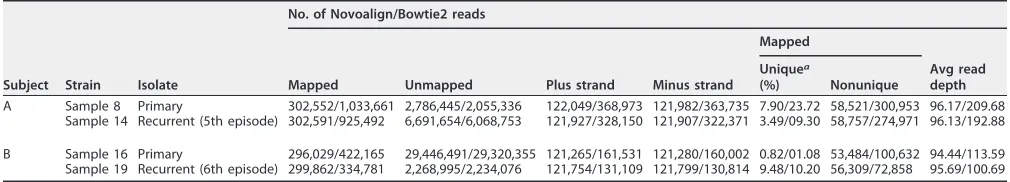
Reads were independently reference aligned using HG52 as the reference sequence.
The number of mapped reads contributing to Novoalign (Novocraft) alignments
(
⬃
300,000) and the number of plus- and minus-strand reads in Novoalign (
⬃
121,000)
were similar across the four samples and lower than the number of mapped reads
contributing to Bowtie2 (83) alignments. However, the Novoalign alignments resulted
in up to a 3-fold-lower percentage of unique mapped reads than Bowtie2 (Table 2).
Regions with low read coverage (read depth,
⬍
25) were filled using Sanger sequencing
(Fig. 1 and 2). The combination of the Novoalign sequence consensus and Sanger
sequences yielded complete coverage of the HG52 HSV-2 genome for all four samples
(Fig. 2), and they were uploaded to GenBank as a single contiguous sequence for each
sample (Table 3).
Variant alleles detected in
ⱖ
10% of the read depth were tabulated with the ultimate
goal of discerning genomic regions under evolutionary pressure. With HG52 used as
the reference genome, the total number of SNVs in coding and noncoding positions
within the U
Sand U
Lregions of all four samples combined was higher in the Bowtie2
alignments (560) than in the Novoalign alignments (485) (Table 4). Nonetheless, the
percentages of total SNVs that fell within the ORFs were similar when the Novoalign
(80.6%) and Bowtie2 (72.3%) alignments were compared, as were the percentages of
nonsynonymous SNVs (41.4% and 39.8%, respectively). The total numbers of indels
appearing in
ⱖ
10% of mapped reads in all four samples were also similar between the
programmatic alignments (37 in Novoalign and 33 in Bowtie2), suggesting that neither
of the programmatic alignments skewed the data set toward SNVs or indels (Table 4).
One SNV resulting in a stop codon was found in a Novoalign alignment (U
L13 gene;
14% of mapped reads; sample 16), and one more was found in the Bowtie2 alignments
(U
L46 gene; 11% of mapped reads; samples 8 and 14) (Tables 4 and 5). Based on this
comparison, we consider the Novoalign alignment to be the most likely approximation
for two reasons. First, the Novoalign alignments had fewer total SNVs in the four
samples, and a smaller number of changes is the most plausible scenario according to
evolutionary theories (39, 40). Second, the numbers of SNVs unique to the primary- or
recurrent-disease samples in subjects A and B were 4- to 6-fold lower in the Novoalign
on November 6, 2019 by guest
http://jvi.asm.org/
(subject A, 10; subject B, 9) than in the Bowtie2 (45 and 64, respectively) alignment
(Table 4).
Using the consensus sequence of the Novoalign mapped reads plus Sanger
se-quences, the entire HSV-2 genomes obtained from the four samples were manually
adjusted using, consecutively, the HG52 and SD90e reference sequences and then
analyzed for polymorphisms. More SNVs and indels were found in coding and
non-coding positions when we compared all four samples to the low-passage-number
clinical isolate SD90e strain from South Africa (641 SNVs and 210 indels) than when we
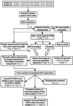
FIG 1Flow chart summarizing the data generation and analysis workflow. (1) HSV-2 was isolated from
primary and recurrent infections in two subjects, and the viruses were expanded a single time in Vero cells. (2) Illumina Hi-Seq sequencing was performed on the whole genome of each sample. (3)De novo
assembly of contigs was carried out using Velvet, and the contigs were aligned with the HG52 reference strain using Abacas/Nucmer to establish draft genome coverage rates. (4) Programmatic Novoalign and Bowtie2 alignments (BAM files), which excluded the IRLand IRSregions and used the HG52 strain as a reference, were compared based on the numbers of SNVs and indels that appeared inⱖ10% of the Illumina read depth. Sanger sequencing was then performed on the HSV-2 regions with low read coverage (read depth,⬍25) based on Novoalign alignments. Sanger plus Novoalign consensus se-quences for each sample (FASTA files) were aligned using nucleic and amino acid sese-quences. The HG52 and SD90e strains were used independently as reference genomes. Using the IUPAC code, SNVs observed in Novoalign alignments (ⱖ10% of the read depth) were incorporated into the manually adjusted alignment of each sample. The BAM and FASTA files were uploaded to GenBank (Table 3). The rectangular and rounded boxes represent, respectively, databases and actions done in the databases.
December 2017 Volume 91 Issue 23 e00942-17 jvi.asm.org 4
on November 6, 2019 by guest
http://jvi.asm.org/
[image:4.585.62.350.70.486.2]compared them to the high-passage-number laboratory HG52 strain from Scotland
(455 and 69, respectively) (Table 4). However, the percentages of SNVs that fell within
ORFs were virtually the same regardless of whether the reference sequence used was
HG52 (77%) or SD90e (65.8%). Nonsynonymous SNVs comprised similar percentages of
the total SNVs whether the subjects’ sequences were manually adjusted against the
HG52 or the SD90e reference sequence (35.6% versus 27.3%, respectively). The stop
codon found in a minority of mapped reads for the U
L13 gene (sample 16) remained
in the alignment whether the reference strain was HG52 or SD90e. Overall, fewer SNVs
and indels appeared in coding and noncoding positions in any of the four samples
when HG52 was used as the reference (Table 4), and the numbers were very similar
between isolates (data not shown).
Using the manually adjusted alignments with the HG52 strain as the reference
genome, we analyzed the sequence diversity of the individual ORFs. Our results
indicate low sequence diversity and generally even SNV distribution across the coding
regions (Fig. 3; see Table S1 in the supplemental material). The genes with the highest
average percentages of SNVs were U
L39, U
L49, and U
S12. U
L39 also had a high average
frequency of nonsynonymous mutations, in addition to U
L3, U
L11, and U
L43. No
nucleotide variation was observed in U
L23, U
L25, U
L33, U
L35, U
L41, U
L45, U
L48, U
L55,
U
S5, U
S9, or U
S11.
Unique polymorphisms within the primary or recurrent isolates.
Having
assem-bled whole-genome sequences of primary and recurrent isolates from the same
subjects provided the opportunity to examine how the HSV-2 genome evolves over
time within an individual. Using the Novoalign alignments, we annotated unique
polymorphisms in coding regions of each genome that represented
ⱖ
10% of the total
reads at a given nucleotide position. A total of 10 nucleotide positions in the primary
or recurrent isolate of subject A were altered in
ⱖ
10% of reads (Table 5). Seven of these
nucleotide substitutions would produce a nonsynonymous change in the amino acid
sequence of the gene. Similarly, 9 nucleotide positions varied in
ⱖ
10% of reads
between the primary and recurrent isolates of subject B. Only five of them would alter
the amino acid sequence of the encoded proteins. The unique polymorphic residue
shared by subjects A and B was a synonymous mutation detected in the U
L14 gene
(nucleotide position 321). In some cases, genetic variability at a nucleotide position
within the primary-infection reads resolved to a single homogeneous nucleotide in the
recurrent sample (e.g., nucleotide position 1039 in U
L21 of subject B); in other cases,
[image:5.585.40.544.84.156.2]more diversity occurred in the recurrent isolate (e.g., nucleotide 1133 in U
L13 of subject
TABLE 1De novopseudomolecule assembly summary constructed using Velvet
Subject Strain No. of reads
Pseudomolecule length (bp)
No. of contigs (>100 bp)
No. of gaps in pseudomolecule
No. of overlapping contig separators in pseudomolecule
Genome sequence coverage (%)
A Sample 8 3,088,997 174,355 577 182 144 97.7
Sample 14 6,994,245 173,075 2,662 186 140 97.4
B Sample 16 29,742,520 164,695 7,138 126 88 97.7
Sample 19 2,568,857 170,115 436 141 108 98.0
TABLE 2Novoalign- and Bowtie2-based read-mapping statistics using HSV-2 strain HG52 as the reference genome
Subject Strain Isolate
No. of Novoalign/Bowtie2 reads
Mapped Unmapped Plus strand Minus strand
Mapped
Avg read depth Uniquea
(%) Nonunique
A Sample 8 Primary 302,552/1,033,661 2,786,445/2,055,336 122,049/368,973 121,982/363,735 7.90/23.72 58,521/300,953 96.17/209.68 Sample 14 Recurrent (5th episode) 302,591/925,492 6,691,654/6,068,753 121,927/328,150 121,907/322,371 3.49/09.30 58,757/274,971 96.13/192.88
B Sample 16 Primary 296,029/422,165 29,446,491/29,320,355 121,265/161,531 121,280/160,002 0.82/01.08 53,484/100,632 94.44/113.59 Sample 19 Recurrent (6th episode) 299,862/334,781 2,268,995/2,234,076 121,754/131,109 121,799/130,814 9.48/10.20 56,309/72,858 95.69/100.69
aUniquely mapped reads are defined as having a mapping quality (MQ) ofⱕ30.
on November 6, 2019 by guest
http://jvi.asm.org/
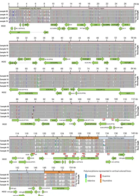
[image:5.585.40.545.640.731.2]FIG 2Features of the complete HSV-2 genomes aligned with the HSV-2 strain HG52 reference sequence. Assembled Illumina mapped reads were aligned with the reference sequence and viewed in the IGV program. The scale at the top represents the genome position in kilobases. The horizontal bars below the genome scale represent the locations of the IRLregions (light brown), ULregion (light pink), IRSregions (orange),
(Continued on next page)
December 2017 Volume 91 Issue 23 e00942-17 jvi.asm.org 6
on November 6, 2019 by guest
http://jvi.asm.org/
[image:6.585.44.490.65.694.2]B). Overall, these data demonstrate that the number of nucleotide positions
undergo-ing change between the primary and a later recurrent isolate from the same individual
was low.
It was of interest to determine whether nonsynonymous variants would produce a
predicted change in protein conformation (Table 6). Among nonsynonymous variants
in subject A, comparison of the predicted secondary and tertiary structures of the
protein with those of a previously crystallized protein was only possible for U
L13 (A190V
and D229Y) and U
L37 (H493P). Four nonsynonymous variants from subject B also could
be examined by comparison to crystal structures: G378V and the minor variant
R288STOP in U
L13, A347T in U
L21, and C951Y in U
L30. A crystal structure of the G
protein-coupled receptor kinase 2 (grk2) (41) (Protein Data Bank [PDB] accession no.
1YM7
; residues 147 to 516) was used with 100% confidence to analyze the locations of
the substitutions in the HSV-2 pUL13 protein kinase. Although residue 229 scored high
for conservation and mutational sensitivity (Table 6), no change in the secondary

-sheet structure was observed (not shown). A crystal structure of the HSV-1 tegument
protein pUL21 C-terminal domain (42) (PDB accession no.
5ED7
; residues 277 to 527)
was used with 100% confidence to evaluate the C-terminal domain of HSV-2 pUL21.
The C-terminal domain of U
L21 is composed of 10
␣
-helices and one
-helix arranged
into a dragonfly fold with the left “wing” formed by
␣
1 to
␣
4 and
1 (43, 44). Residue
347, located in helix
␣
3 and present as threonine instead of alanine in 33% of the reads
from subject B’s initial isolate, was strongly conserved and had a high score for
FIG 2Legend (Continued)
[image:7.585.43.548.83.365.2]and USregion (light blue) in the genome of HSV-2 reference strain HG52. Samples are labeled on the left. BAM file reads were capped at 100 for visualization in IGV, and the read depth at each nucleotide position is represented in dark gray for each sample (obtained from IGV). The vertical colored lines inserted in the gray section represent polymorphisms in the genome using the HG52 strain as a reference. HSV-2 coding regions are shown as green arrows indicating the direction of the ORF. The red arrows represent the locations of forward and reverse primers used in Sanger sequencing (Table 7) to fill the regions of low read depth coverage.
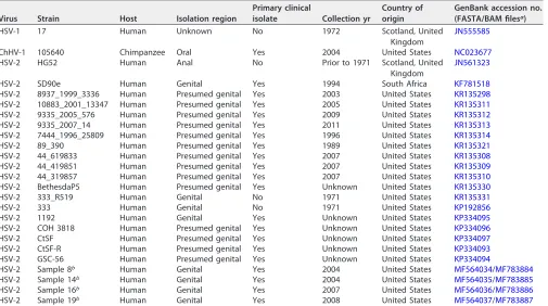
TABLE 3Genomes and accession numbers
Virus Strain Host Isolation region
Primary clinical
isolate Collection yr
Country of origin
GenBank accession no. (FASTA/BAM filesa)
HSV-1 17 Human Unknown No 1972 Scotland, United
Kingdom
JN555585
ChHV-1 105640 Chimpanzee Oral Yes 2004 United States NC023677
HSV-2 HG52 Human Anal No Prior to 1971 Scotland, United
Kingdom
JN561323
HSV-2 SD90e Human Genital Yes 1994 South Africa KF781518
HSV-2 8937_1999_3336 Human Presumed genital Yes 2003 United States KR135298
HSV-2 10883_2001_13347 Human Presumed genital Yes 2005 United States KR135311
HSV-2 9335_2005_576 Human Presumed genital Yes 2009 United States KR135312
HSV-2 9335_2007_14 Human Presumed genital Yes 2011 United States KR135313
HSV-2 7444_1996_25809 Human Presumed genital Yes 1996 United States KR135314
HSV-2 89_390 Human Presumed genital Yes 1989 United States KR135321
HSV-2 44_619833 Human Presumed genital Yes 2007 United States KR135308
HSV-2 44_419851 Human Presumed genital Yes 2007 United States KR135309
HSV-2 44_319857 Human Presumed genital Yes 2007 United States KR135310
HSV-2 BethesdaP5 Human Presumed genital Yes Unknown United States KR135330
HSV-2 333_R519 Human Genital No 1971 United States KR135331
HSV-2 333 Human Genital No 1971 United States KP192856
HSV-2 1192 Human Genital Yes Unknown United States KP334095
HSV-2 COH 3818 Human Presumed genital Yes Unknown United States KP334096
HSV-2 CtSF Human Presumed genital Yes Unknown United States KP334097
HSV-2 CtSF-R Human Presumed genital Yes Unknown United States KP334093
HSV-2 GSC-56 Human Presumed genital Yes Unknown United States KP334094
HSV-2 Sample 8b Human Genital Yes 2004 United States MF564034/MF783884
HSV-2 Sample 14b Human Genital Yes 2004 United States MF564035/MF783885
HSV-2 Sample 16b Human Genital Yes 2007 United States MF564036/MF783886
HSV-2 Sample 19b Human Genital Yes 2008 United States MF564037/MF783887
aBAM files were obtained from the Novoalign mapped and unmapped reads. FASTA files were obtained from the final version of the whole HSV-2 genome (Fig. 1). bStrain newly sequenced in this study.
on November 6, 2019 by guest
http://jvi.asm.org/
mutational sensitivity (Table 6) but did not produce a change in the secondary
␣
-helix
structure (not shown). A crystal structure of the HSV-1 DNA polymerase (45) (PDB
accession no.
2GV9
; residues 61 to 1202) was used with 100% confidence to model the
HSV-2 pUL30 DNA polymerase. Four structural domains comprise pUL30. These
do-mains assemble to form a disk-like shape around a central hole (45). The C951Y
substitution found in 20% of the reads from subject B’s primary isolate (Table 6)
produced an extension of the

-sheet located within the palm subdomain of the
polymerase domain but did not otherwise alter its structure (not shown). The P1133L
substitution in 60% of subject A’s recurrent isolate could not be analyzed because
amino acids (aa) 1100 to 1138 were not included in the protein that had been
crystalized. Although subjects A and B received valacyclovir therapy for their infections,
we noted no decrease in ACV sensitivity of their recurrent isolates (data not shown),
consistent with the genetic sequence information. A crystal structure of the
pseudo-rabiesvirus pUL37 N-terminal half (46) (PDB accession no.
4K70
; residues 24 to 570) was
used with 100% confidence to evaluate the secondary and tertiary structures of HSV-2
pUL37. Domain III of pUL37 is a highly conserved helical bundle, with a central helix
(
␣
19) surrounded by six helices (
␣
16 to
␣
22) (46). Ninety-three percent of the reads
from subject A’s primary isolate specified proline at residue 493 (Table 6), whose
replacement by histidine in the recurrent isolate disrupted helix
␣
21 within domain III
of pUL37 (Fig. 4).
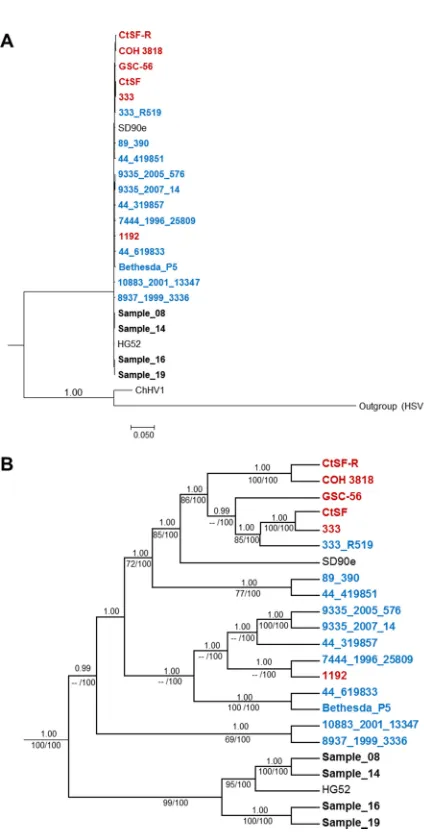
[image:8.585.42.550.83.356.2]Phylogenetic reconstruction.
Phylogenetic associations among the HSV-2
primary-and recurrent-disease samples; previously published unverified (30) or partial (31)
HSV-2 genome sequences from North America; and the HSV-2 reference strains HG52
(Scotland), SD90e (South Africa), and 333 (North America) were evaluated using HSV-1
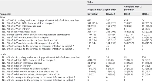
TABLE 4Comparison of SNV and indel events among different alignments and reference genomes
Parameter
Value
Programmatic alignmentsa
Complete HSV-2 genomeb
Novoalign Bowtie2 HG52 SD90e
SNVs
No. of SNVs in coding and noncoding positions (total of all four samples) 485 560 455 641
No. (%) of SNVs in ORFs (total of all four samples) 391 (80.6)c 405 (72.3) 350 (77) 422 (65.8)
No. (%) of SNVs in intergenic regions 94 (19.4) 155 (27.7) 100 (22) 191 (29.8)
No. (%) of SNVs in intronsd NDe ND 5 (1) 28 (4.36)
No. (%) of nonsynonymous SNVs 201 (41.4) 223 (39.8) 162 (35.6) 175 (27.3)
No. of stop codons within an ORF creating possible pseudogenes 1 (UL13) 1 (UL46) 1 (UL13) 1 (UL13)
No. (%) of SNVs common to all four samples 191 (39.4) 216 (38.6) 149 (32.7) 214 (33.4)
No. (%) of SNVs only in subject A (samples 8 and 14) 129 (26.6) 180 (32.1) 146 (32) 164 (25.6)
No. (%) of SNVs only in subject B (samples 16 and 19) 165 (34) 164 (29.2) 160 (35.3) 164 (25.6)
No. of SNVs unique to the primary or recurrent infection in subject A 10 45 0 0
No. of SNVs unique to the primary or recurrent infection in subject B 9 64 0 0
Indels
No. of indels in coding and noncoding positions (total of all four samples) 37 33 69 210
No. (%) of indels in ORFs (total of all four samples) 4 (10.81) 2 (6.06) 33 (47.8) 32 (15.2)
No. (%) of indels in intergenic regions 33 (89.2) 31 (93.9) 33 (47.8) 144 (68.6)
No. (%) of indels in intronsd ND ND 3 (4.4) 34 (16.2)
No. (%) of indels common to all four samples 12 (32.4) 5 (15.1) 28 (40.6) 123 (58.6)
No. (%) of indels only in subject A (samples 8 and 14) 15 (40.5) 15 (45.4) 15 (21.7) 33 (15.7)
No. (%) of indels only in subject B (samples 16 and 19) 10 (27) 13 (39.4) 26 (37.7) 35 (16.6)
No. of indels unique to the primary or recurrent infection in subject A 5 0 0 0
No. of indels unique to the primary or recurrent infection in subject B 3 4 0 0
aThe programmatic alignments, which included the high-quality mapped reads and HG52 strain as the reference sequence, were analyzed using SAMtools (v1.3)
software. SNVs and indels were considered only when they appeared inⱖ10% of the read depth.
bComplete HSV-2 genome obtained for each sample using Illumina and Sanger sequences improved manually using, consecutively, the nucleic acid and amino acid
sequences. Only one copy of the IRLand IRSregions was included to compare the number of SNVs and indels observed when the HG52 or SD90e strain was used as a reference.
cThe numbers in parentheses are the percentages of the total SNVs or indels. dProgrammatic alignments did not distinguish between intergenic regions and introns. eND, not determined.
December 2017 Volume 91 Issue 23 e00942-17 jvi.asm.org 8
on November 6, 2019 by guest
http://jvi.asm.org/
and chimpanzee herpesvirus 1 (ChHV-1) genomes as outliers. The phylogenetic
recon-struction based on the whole HSV-2 genome provides a graphical representation of the
relationships among the samples (Fig. 5). Expansion of the HSV-2-specific nodes (Fig.
5B) indicated a close relationship between the isolates from subject A (samples 8 and
14) and those from subject B (samples 16 and 19). It also suggested a close relationship
between the laboratory HG52 strain from Scotland and the samples from subject A,
while the low-passage-number clinical isolate SD90e from South Africa appeared to be
strongly nested within a group that includes other North American strains published
previously.
DISCUSSION
This research presents for the first time a comparison of whole HSV-2 genome
sequences of primary isolates and isolates from later recurrent-disease episodes from
two infected individuals. Coverage of
⬎
97% was reached based on
de novo
pseudo-molecule assembly contig mapping compared to the HSV-2 HG52 strain, and 100%
coverage was reached when we combined Novoalign sequence alignments and Sanger
sequences (Fig. 2). The two individuals became infected while they participated in the
Herpevac Trial for Women (25). Two of the genome sequences derive from the subjects’
primary isolates and two of them from the well-documented fifth (subject A) or sixth
(subject B) recurrent episodes of genital disease. Although various mechanisms of
natural variation were evident, including SNVs and indels, our analyses demonstrate a
high degree of sequence homology when each subject’s primary and recurrent isolates
were compared, suggesting a low rate of evolution over time in an infected individual.
The striking sequence conservation between the primary isolate and a later recurrent
isolate from the same subjects suggests strong selection pressures on the virus to
maintain genetic fidelity during reactivations from its latent state, but more temporally
distant isolates will be needed to firmly establish the rate of genetic drift or the impact
of immune selection.
Identification of unique polymorphisms in primary infection versus recurrent
epi-sodes of disease throughout the complete HSV-2 genome sequence may help us to
ascertain regions of the genome undergoing molecular evolution during reactivation
events. The strong sequence conservation we observed, whether in a subject with a
high frequency of recurrence (subject A) or less than once per month (subject B), also
augers well for the success of a therapeutic vaccine because it suggests evolution of the
viral genome is constrained by selective pressures, including the immune response in
an infected individual. The apparent rate of mutational drift in HSV is much lower than
in other persistent virus infections where it has been studied, such as HIV and hepatitis
C virus (47). Possible reasons include the proofreading activity of the HSV DNA
polymerase and HSV’s complex life cycle that must accommodate the restrictions
imposed by diverse necessities of existing in a latent state in neurons and rapidly
replicating in mucosal epithelia.
All Illumina reads passed the FastQC default per-base and per-read sequence quality
checks. The Illumina coverage distribution observed in our results was similar to that
previously seen for HSV-1 (48) and HSV-2 (31), with lower coverage in the repeat
regions and higher coverage in the U
Land U
Scoding regions. The ambiguous regions
of Illumina sequence were filled using conventional Sanger sequencing, with special
conditions for GC-rich DNA, as has been previously published for sequencing of other
strains of HSV-2 (33) and HSV-1 (38). The combination of the assembled Illumina reads
and Sanger sequences yielded a full-length version of the HSV-2 genomes for samples
8, 14, 16, and 19 (Fig. 2).
Phylogenetic analyses were performed to further evaluate the association among
the primary and recurrent disease isolates, other North American HSV-2 strains
previ-ously published, and the HG52 and SD90e strains, using HSV-1 strain 17 and ChHV-1
strain genomes as outgroups (Fig. 5). In contrast to previous studies that focused on
selected regions of the genome or excluded intergenic sequences (49, 50), we used the
full genome alignment, including coding and noncoding positions, to assess
on November 6, 2019 by guest
http://jvi.asm.org/
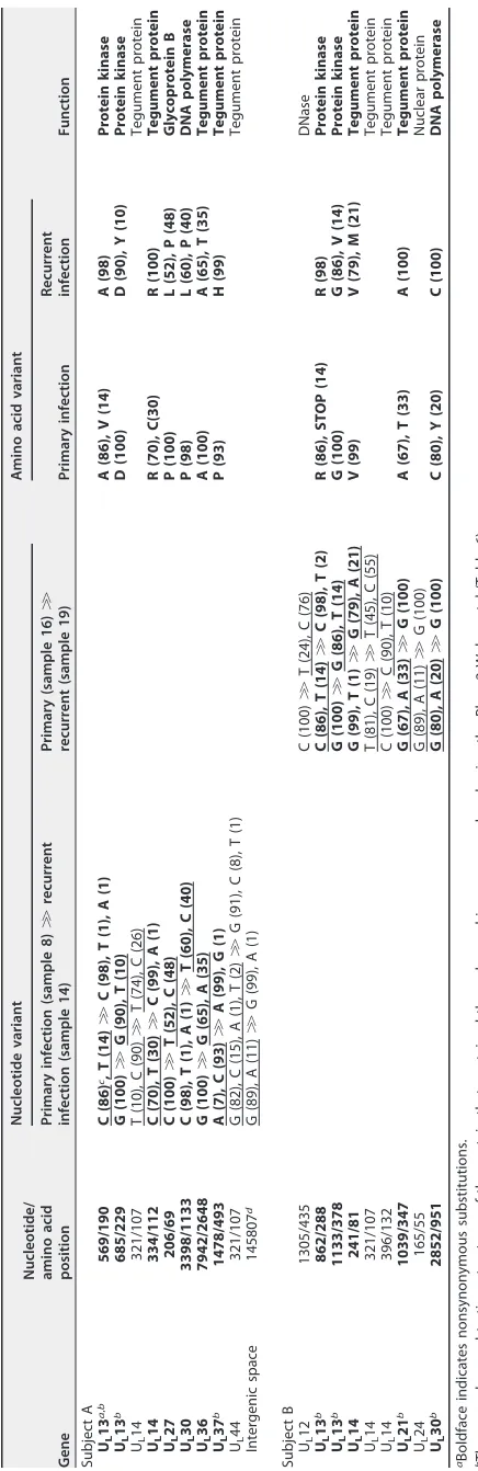
TABLE 5 SNVs unique to the primary or recurrent infection in subject A or B Gene Nucleotide/ amino acid position Nucleotide variant Amino acid variant Function Primary infection (sample 8) ⬎⬎ recurrent infection (sample 14) Primary (sample 16) ⬎⬎ recurrent (sample 19) Primary infection Recurrent infection Subject A UL 13 a , b 569/190 C (86) c, T (14) ⬎⬎ C (98), T (1), A (1) A (86), V (14) A (98) Protein kinase UL 13 b 685/229 G (100) ⬎⬎ G (90), T (10) D (100) D (90), Y (10) Protein kinase UL 14 321/107 T (10), C (90) ⬎⬎ T (74), C (26) Tegument protein UL 14 334/112 C (70), T (30) ⬎⬎ C (99), A (1) R (70), C(30) R (100) Tegument protein UL 27 206/69 C (100) ⬎⬎ T (52), C (48) P (100) L (52), P (48) Glycoprotein B UL 30 3398/1133 C (98), T (1), A (1) ⬎⬎ T (60), C (40) P (98) L (60), P (40) DNA polymerase UL 36 7942/2648 G (100) ⬎⬎ G (65), A (35) A (100) A (65), T (35) Tegument protein UL 37 b 1478/493 A (7), C (93) ⬎⬎ A (99), G (1) P (93) H (99) Tegument protein UL 44 321/107 G (82), C (15), A (1), T (2) ⬎⬎ G (91), C (8), T (1) Tegument protein Intergenic space 145807 d G (89), A (11) ⬎⬎ G (99), A (1) Subject B UL 12 1305/435 C (100) ⬎⬎ T (24), C (76) DNase UL 13 b 862/288 C (86), T (14) ⬎⬎ C (98), T (2) R (86), STOP (14) R (98) Protein kinase UL 13 b 1133/378 G (100) ⬎⬎ G (86), T (14) G (100) G (86), V (14) Protein kinase UL 14 241/81 G (99), T (1) ⬎⬎ G (79), A (21) V (99) V (79), M (21) Tegument protein UL 14 321/107 T (81), C (19) ⬎⬎ T (45), C (55) Tegument protein UL 14 396/132 C (100) ⬎⬎ C (90), T (10) Tegument protein UL 21 b 1039/347 G (67), A (33) ⬎⬎ G (100) A (67), T (33) A (100) Tegument protein UL 24 165/55 G (89), A (11) ⬎⬎ G (100) Nuclear protein UL 30 b 2852/951 G (80), A (20) ⬎⬎ G (100) C (80), Y (20) C (100) DNA polymerase aBoldface indicates nonsynonymous substitutions. bThe secondary and tertiary structures of the protein that contained the polymorphisms were analyzed using the Phyre2 Web portal (Table 6 ). cThe numbers in parentheses are the percentages of Novoalign mapped reads supporting each polymorphism. The reported polymorphisms appeared in ⱖ 10% of mapped reads. Underlining indicates the polymorphic variant allele. dPolymorphism position observed in an intergenic space derived from the Novoalign alignments (BAM files).
December 2017 Volume 91 Issue 23 e00942-17 jvi.asm.org 10
on November 6, 2019 by guest
http://jvi.asm.org/
[image:10.585.51.269.69.740.2]ships among strains. Several salient observations arose. First, our results demonstrate
that the primary isolates (samples 8 and 16) cluster with their respective
recurrent-disease isolates (samples 14 and 19) rather than with each other (Fig. 5B). Therefore,
accumulated SNVs that occur with repeated reactivations of HSV-2 from a latent state
do not necessarily lead to sequence convergence when considering the genome as a
whole. Second, there is a strongly supported cluster between the HG52 strain and the
samples from subject A (samples 8 and 14) and subject B (samples 16 and 19), while the
SD90e and 333 strains appear to be nested within groups that include other North
American strains (30, 31). Thus, comparison of the sequences from subjects A and B
with genomes from other North American HSV-2 isolates indicates that our sequences
represent a distinct group with the greatest homology to HG52 (Fig. 5B). These
phylogenetic observations corroborate our numerical observations: our four HSV-2
genome sequences from North American clinical isolates (subjects A and B) had fewer
SNVs and indels than the high-passage-number laboratory HG52 strain (
JN561323.2
)
from Scotland and the low-passage-number clinical isolate SD90e (
KF781518.1
) from
South Africa (Table 4). Our results also agree with the low sequence diversity previously
observed among HSV-2 genomes (30, 33). Thus, although the HSV-2 SD90e strain is a
low-passage-number clinical isolate, our observations suggest a closer association
between some North American samples and the Scotland strain HG52 than most other
North American isolates previously reported (30, 33). Based on this result, one of the
HSV-2 complete genomes of clinical isolates reported here would be a logical choice as
a reference strain for inclusion in future European and North American studies.
Using the HG52 strain as a reference genome, we analyzed the complete HSV-2
genomes for hot spots in the polymorphism distribution and also ORFs with the highest
number of SNVs and nonsynonymous changes, suggestive of proteins that are
under-FIG 3Number of HSV-2 genes at each interval of nonsynonymous change. The intervals (yaxis) were
[image:11.585.85.327.70.209.2]selected based on the average percentage of SNVs that represented nonsynonymous changes across ORFs, using the HG52 strain as the reference sequence. The most polymorphic genes are identified. See Table S1 in the supplemental material for a list of all the SNVs observed in the ORFs of the complete HSV-2 genomes.
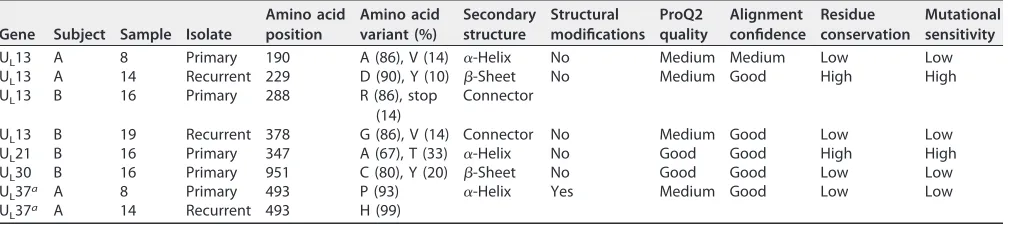
TABLE 6Phyre2 investigator tests
Gene Subject Sample Isolate
Amino acid position
Amino acid variant (%)
Secondary structure
Structural modifications
ProQ2 quality
Alignment confidence
Residue conservation
Mutational sensitivity
UL13 A 8 Primary 190 A (86), V (14) ␣-Helix No Medium Medium Low Low
UL13 A 14 Recurrent 229 D (90), Y (10) -Sheet No Medium Good High High
UL13 B 16 Primary 288 R (86), stop
(14)
Connector
UL13 B 19 Recurrent 378 G (86), V (14) Connector No Medium Good Low Low
UL21 B 16 Primary 347 A (67), T (33) ␣-Helix No Good Good High High
UL30 B 16 Primary 951 C (80), Y (20) -Sheet No Good Good Low Low
UL37a A 8 Primary 493 P (93) ␣-Helix Yes Medium Good Low Low
UL37a A 14 Recurrent 493 H (99)
aRepresented in Fig. 4.
on November 6, 2019 by guest
http://jvi.asm.org/
[image:11.585.42.547.618.731.2]going evolution. Such regions of higher sequence lability may allow a variant to emerge
that could, for example, evade host immune surveillance or adapt to a new host’s
genetic makeup. The regions with the highest levels of variation appeared in the
introns and intergenic sequences (Fig. 2; see Table S2 in the supplemental material),
consistent with recently published nearly complete HSV-2 genomes (30, 33). Numerous
ORFs contained SNVs relative to the reference sequence, but these nucleotides did not
differ between primary- and recurrent-disease isolates. Therefore, the primary-infection
sample may have fixed a polymorphism that confers a selective advantage or simply
may have tolerated diversity at a given location with little or no selective pressure.
Unique SNVs in ORFs of the primary or recurrent HSV-2 samples were primarily
concentrated in two genes, U
L13 and U
L14 (Table 5). U
L13, which encodes a protein
kinase, contained four unique nonsynonymous polymorphic residues. Two of the
polymorphic nucleotides specified changes in samples from the primary infection
(samples 8 and 16) that were subsequently purified to near homogeneity in the
recurrent-disease samples; the other two represent diversifying mutations in the
re-current samples (samples 16 and 19). U
L14, which encodes a tegument protein,
contained five unique SNVs. Two of these polymorphisms were nonsynonymous, one
of which generated more diversity in the recurrent sample (sample 19) while the other
resulted in near homogeneity in the recurrent sample (sample 14). Interestingly, 4 out
of 5 unique SNVs in U
L14 resulted in greater diversity in the recurrent sample (Table 5);
the U
L14 gene also showed a high degree of sequence variability in previous studies of
HSV-1 (38) and HSV-2 (27). The read depth heterogeneity at all 19 of the SNVs among
the four samples was
ⱖ
10%, lending credence to the interpretation that these were
true polymorphisms and not sequencing errors. Though our sample size was small, the
low number of unique SNVs suggests strong conservation of the genome sequences
between the primary and recurrent HSV-2 isolates from the same subject. While this
result implies substantial selection pressure on the viral genome during its history
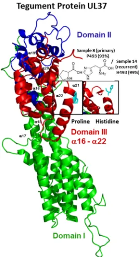
FIG 4Features of the amino acid variants resulting from nonsynonymous changes in the primary or
recurrent isolates of the tegument protein UL37. Assessment of the tertiary molecular structure of pUL37 is shown (with structural modifications) based on the crystal structure of the N-terminal half of pseudorabiesvirus pUL37 (PDB accession no.4K70; residues 24 to 570). Domains are numbered and represented in different colors. The polymorphic amino acids (P493H) (Table 6) are boxed and colored in cyan on the protein tertiary structures.
December 2017 Volume 91 Issue 23 e00942-17 jvi.asm.org 12
on November 6, 2019 by guest
http://jvi.asm.org/
[image:12.585.136.276.71.329.2]within individual hosts, it is also possible that virus isolated after several
recurrent-disease episodes represents reactivation of different neurons infected during the
primary infection event. Thus, even a fifth or sixth recurrent episode may produce virus
that is closer to the primary infection than the episode number would suggest.
Nonetheless, even with individual reactivation events from the latent pool, the virus
could mutate but apparently does not. It will be of interest to sample additional primary
and recurrent isolates from other subjects with longer histories of infection to solidify
the apparently low evolutionary rate in an infected individual.
The evolutionary implications of seven nonsynonymous SNVs observed in the
primary or recurrent samples from subjects A and B were analyzed by comparing the
secondary and tertiary structures or partial structures of HSV-2 pUL13, pUL21, pUL30,
and pUL37 with a previously crystallized protein homolog (similarity,
⬎
99%) (Table 6).
Because no homologous crystal structure models exist for proteins containing the
remaining six nonsynonymous changes (Table 5), their biological significance, if any, is
unclear. The three-dimensional structures of these multifunctional proteins provided a
FIG 5Bayesian majority rule consensus tree of 23 genomic HSV-2 sequences. The North American strains
were from Kolb et al. (31) (red) and Newman et al. (30) (blue). The four HSV-2 complete genomes sequenced in this research are in black. (A) Complete Bayesian tree using HSV-1 and ChHV-1 as outgroups. (B) Expansion of the HSV-2-specific node presenting the Bayesian posterior probabilities (⬎90%) and maximum-parsimony bootstrap (100 replicates; ⬎75%)/maximum-likelihood bootstrap (1,000 replicates;⬎75%) values above and below the branches, respectively.
on November 6, 2019 by guest
http://jvi.asm.org/
[image:13.585.98.310.58.476.2]detailed road map and an excellent opportunity to explore the structural/phenotypic
implications of the amino acid changes unveiled. Modeling of the polymorphisms in
pUL13, pUL21, and pUL30 did not reveal an appreciable change in structure (data not
shown), despite prediction of two polymorphisms as mutationally sensitive for proper
structure and function of the protein according to the Jensen-Shannon divergence (51)
and the SuSPect method (52). pUL13 is a Ser/Thr protein kinase conserved among
alpha-, beta-, and gammaherpesviruses (53) and has important roles in the herpesvirus
replication cycle (54, 55), tegument dissociation (56), viral-gene expression (57, 58) and
cell cycle regulation (59). Despite its many important roles, four genetic polymorphisms
unique to the primary or recurrent samples were found in subject A or B. However,
none of the four polymorphisms identified fell within a known kinase domain (60).
Unlike HSV-1 U
L13 mutants, HSV-2 U
L13-deficient mutants are highly compromised for
replication (57, 61, 62; M. Korom, J. E. Schrimpf, G. S. Delassus, and L. A. Morrison,
unpublished data). The stop codon predicted at pUL13 residue 288 in 14% of the reads
in primary-infection sample 16 would therefore decrease viral fitness, so it is not
surprising that this polymorphism disappeared in the recurrent-infection sample 19.
pUL37 is essential for HSV-1 replication, plays an important role in capsid trafficking
during virion entry, and interacts with the gK-UL20 protein complex to facilitate
cytoplasmic virion envelopment (63–67). The P493H substitution in helix
␣
21 lies in the
highly conserved helical bundle domain III of pUL37 and is predicted to disorder a
portion of the helix (Fig. 4). Residue 493 is close to a pUL37 self-association domain (aa
568 to 1123), which is one of its domains that appear to have distinct functions during
virus replication (67). Thus, the polymorphism observed in residue 493 may have
functional consequences, although the conservation and mutational sensitivity of the
residue are predicted to be low. Nonetheless, it is of interest that the proline that
predominated at this residue in the primary sample and is present in herpesviruses of
several animal species (68) had been replaced in 99% of the reads of the recurrent
sample. Its proximity to tyrosine 480, which interacts with gK to facilitate cytoplasmic
virion envelopment and infectious-virus production (67), suggests that pressure to
revert to histidine may exit to maximize capsid egress and envelopment.
This research presents for the first time a comparison of HSV-2 complete genome
sequences representing temporally distinct isolates from the same subjects based on
next-generation and Sanger sequencing techniques. Each subject’s primary isolate and
later recurrent isolate were highly homologous. The differences described among the
four low-passage-number HSV-2 strains newly sequenced in this research, the
high-passage-number HG52 strain, and the low-high-passage-number SD90e strain support the
use of one of the sequences reported here, derived from primary clinical isolates from
the United States, as a reference strain for future European and North American studies
because they represent a subset of sequences with greater similarity to HG52 than most
other sequences or partial sequences of North American isolates determined to date
(30, 31). Although we have been able to answer initial questions about the amount of
diversity among temporally distinct HSV-2 isolates from a single individual, and our
data provide a context for future evolutionary and virulence studies, further
investiga-tions of genome diversity among primary HSV isolates will be important to
develop-ment of a broadly effective prophylactic or therapeutic vaccine.
MATERIALS AND METHODS
Strain sampling, virus DNA isolation, and sequencing.HSV-2 sequences were generated from
DNA extracted from primary and recurrent isolates from two healthy, initially HSV-1/HSV-2-seronegative subjects (A and B) collected during the Herpevac Trial for Women (25). The times of initial infection and recurrent disease were well documented because the subjects became infected while participating in the Herpevac Trial. The viral isolates were designated sample 8 (primary isolate, subject A), sample 14 (recurrent isolate, fifth episode, subject A), sample 16 (primary isolate, subject B), and sample 19 (recurrent isolate, sixth episode, subject B) (Table 2 and Fig. 1). Subject A received HSV-2 gD vaccine and became infected approximately 8 weeks after the final dose; she experienced recurrences an average of every 20 days. Subject B received control vaccine, and her infection occurred approximately 6 weeks after the final dose; she experienced recurrences an average of every 44 days. The subjects provided written consent to future use of their samples, and sequencing of the isolates was approved by the Saint Louis
December 2017 Volume 91 Issue 23 e00942-17 jvi.asm.org 14
on November 6, 2019 by guest
http://jvi.asm.org/
University Institutional Review Board (IRB number 24706). Virus was isolated in Vero (African green monkey kidney) cell monolayers directly from the thawed clinical swabs in transport medium. The Vero cells were originally acquired from the laboratory of David Knipe. The isolates were then inoculated into larger flasks of Vero cells at a multiplicity of infection (MOI) of 0.1, and supernatant and cells were collected when the cytopathic effect reached 100%. Fresh DNA stocks were prepared by cesium chloride gradient sedimentation as previously described (69). Purified genomic DNA (500 ng) was submitted to the Genome Technology Access Center (St. Louis, MO). Sequence libraries were constructed by bar code addition to sheared DNA and run on a single lane on an Illumina Hi-Seq sequencing machine, producing 50-bp single-end reads. The quality and quantity of the DNA were assessed using an Agilent DNA high-sensitivity series chip assay (Agilent Technologies, Santa Clara, CA, USA) and a Qubit dsDNA kit (Life Technologies, Grand Island, NY, USA), respectively, and the libraries were standardized to 2 nM. The sequencing lane contained a PhiX plasmid spike-in; 0.6% of the reads were PhiX and demonstrated an error rate of 0.2% (1 per 500 bases).
HSV-2 genome sequence alignments using a reference-based approach. (i)De novo
pseudo-molecule assembly.The Velvet software suite (v1.2.08) (84) was utilized forde novocontig assembly of
all Illumina reads available for each sample (Table 1 and Fig. 1). Maximum K-mer length was set to 31, and the minimum contig length was set to 100. To obtain a genome scaffold (reference genome-oriented best-matching contigs), the Abacas (v1.3.1)/Nucmer software package (85, 86) was used applying the following consecutive steps. (Step 1) Repeat regions were handled via stepwise masking. The reference genome was masked in three separate locations in order to best align contigs to all repeat regions. The first unmasked region ranged from the beginning of the viral genome through the ULregion and up to the nucleotide base before the IRLregion. The second unmasked region ranged from the first base of the IRLregion through the IRSregion and up to the nucleotide base before the USregion. The final unmasked region covered the first nucleotide base of the USregion through the end of the genome. (Step 2) Contig mapping results for each partially masked reference genome comparison were combined and assembled into a contiguous pseudomolecule or DNA sequence assembly. Partially overlapping contigs were separated by 100 Ns. The base and read qualities of sequencing reads were assessed using the FastQC tool kit (v0.10.1). Finally, comparison of thede novo-assembled pseudomolecules with the HSV-2 HG52 genome using Nucmer indicated more than 97% coverage was achieved for each molecule (Table 1).
(ii) Programmatic alignments: Novoalign and Bowtie2.The Illumina reads were independently
aligned with the sequence of the high-passage-number clinical isolate HG52 laboratory strain (from Scotland) and analyzed using Novoalign (v3.02.06) and Bowtie2 (v2.1.0) mapping software (Table 2 and Fig. 1). For Novoalign, reads were selected for alignment if (i) 30 or more bases were of good quality, (ii) repetitive DNA sequences (homopolymers) had a score of⬎90, and (iii) dinucleotide sequences had a score of⬎120. The maximal alignment score was set to 254, a limit of 100 was set for the number of times a single read could be aligned, and repeats were reported in a random fashion. Bowtie2-based reference alignments utilized the -fast flag (implies -D 10 -R 2 -N 0 -L 22 -i S,0,2.50). Alignment quality control (QC) statistics for each binary alignment/map (BAM) file were obtained using RSeQC software (v2.3.7) (87). The resulting Novoalign BAM files were visualized against the HG52 reference genome using the Integrative Genomics Viewer (IGV) program (70) with a cap of 100 reads.
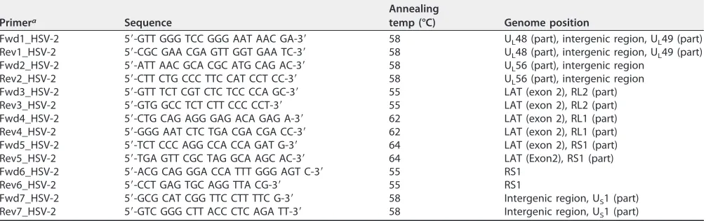
(iii) Sanger sequencing of regions with low read depth.Genome regions with low coverage in
[image:15.585.42.545.84.242.2]Novoalign reference mappings or incomplete/improper overlap inde novoassembly were filled using the Sanger sequencing method (Fig. 1). HSV-2 sequences were PCR amplified from 100 ng of the cesium chloride gradient-purified viral DNA using the primers listed in Table 7. Amplification reactions were performed using a reaction mixture containing 0.75l of forward and reverse primers (10 mM), 2.5l 10⫻AccuPrimePfxreaction mix (Invitrogen), 2.5l betaine solution (5 M) (Sigma), 2l MgCl2(50 mM), 1.5l dimethyl sulfoxide (DMSO), 0.5lTaqDNA polymerase, 2 to 4l of template DNA, and sterile ultrapure water (MilliQ) added to achieve a final volume of 25l. The amplification parameters consisted of a primary denaturing step of 2 min at 95°C, followed by 39 cycles of 20 s denaturing at 95°C, 30 s TABLE 7HSV-2 amplification and sequencing primers
Primera Sequence
Annealing
temp (°C) Genome position
Fwd1_HSV-2 5=-GTT GGG TCC GGG AAT AAC GA-3= 58 UL48 (part), intergenic region, UL49 (part)
Rev1_HSV-2 5=-CGC GAA CGA GTT GGT GAA TC-3= 58 UL48 (part), intergenic region, UL49 (part)
Fwd2_HSV-2 5=-ATT AAC GCA CGC ATG CAG AC-3= 58 UL56 (part), intergenic region
Rev2_HSV-2 5=-CTT CTG CCC TTC CAT CCT CC-3= 58 UL56 (part), intergenic region
Fwd3_HSV-2 5=-GTT TCT CGT CTC TCC CCA GC-3= 55 LAT (exon 2), RL2 (part)
Rev3_HSV-2 5=-GTG GCC TCT CTT CCC CCT-3= 55 LAT (exon 2), RL2 (part)
Fwd4_HSV-2 5=-CTG CAG AGG GAG ACA GAG A-3= 62 LAT (exon 2), RL1 (part)
Rev4_HSV-2 5=-GGG AAT CTC TGA CGA CGA CC-3= 62 LAT (exon 2), RL1 (part)
Fwd5_HSV-2 5=-TCT CCC AGG CCA CCA GAT G-3= 64 LAT (exon 2), RS1 (part)
Rev5_HSV-2 5=-TGA GTT CGC TAG GCA AGC AC-3= 64 LAT (Exon2), RS1 (part)
Fwd6_HSV-2 5=-ACG CAG GGA CCA TTT GGG AGT C-3= 55 RS1
Rev6_HSV-2 5=-CCT GAG TGC AGG TTA CG-3= 55 RS1
Fwd7_HSV-2 5=-GCG CAT CGG TTC CTT TTC G-3= 58 Intergenic region, US1 (part)
Rev7_HSV-2 5=-GTC GGG CTT ACC CTC AGA TT-3= 58 Intergenic region, US1 (part)
aUsed for amplification and sequencing.
on November 6, 2019 by guest
http://jvi.asm.org/
annealing, and 3 min extension at 68°C, followed by a final extension step of 5 min at 68°C. The PCR products were run on agarose gels to confirm the size and purity of the amplicon. The PCR products were then cleaned up using ExoSap-It (Affymetrix) following the protocol indicated by the manufacturer and sent to GeneWiz, Inc. (South Plainfield, NJ) for Sanger sequencing using their GC-rich sequencing and difficult-template protocols. Complementary strands were assembled and verified using Sequencher version 4.2.2 (Gene Codes Corp.).
(iv) Manual alignments.The consensus sequence of the Novoalign mapped reads was extracted
and saved as a FASTA file for each of the four samples (Fig. 1). The four FASTA files were imported into MEGA (v7.0.14) (71) software, along with sequences of the HG52 strain (from Scotland) and the low-passage-number clinical isolate SD90e (from South Africa), which were used as reference sequences. The Sanger sequences were then incorporated into the FASTA file of each sample. The genome sequence alignments obtained for each sample were improved manually using, consecutively, the nucleic acid and amino acid sequences. The full-length version of each sample genome was created by placing inverted copies of the IRLand IRSregions at the appropriate termini. The consensus sequence for each genome was generated by designating any variant of⬎90% prevalence as the consensus base. Variant alleles detected inⱖ10% of the read depth were annotated in the consensus sequence using the International Union of Pure and Applied Chemistry (IUPAC) nucleotide code. Variants with no clear majority allele (those with⬍10% prevalence) were omitted in the consensus sequence.
Evolutionary analyses. (i) Evaluation of SNVs and indels.To select the most plausible alignment
according to evolutionary theories (39, 40), SNVs and indels were tabulated based on the Novoalign and Bowtie2 programmatic alignments. The programmatic alignments, which included the high-quality Illumina reads and used the HG52 strain as a reference, were analyzed using SAMtools (v1.3) software (88). SAMtools mpileup was used on BAM-formatted mapped reads to generate information on match, mismatch, indel, strand, and mapping quality per reference genomic position. To reduce the impact of false positives on variant calling, only high-quality mapped reads and reads with base quality and mapping quality ofⱖ20 were utilized in the pile-up. SNVs and indels were also tabulated from the consensus sequence for each genome generated after manual comparison to HG52 and independently to SD90e as the reference strains, using the MEGA (v7.0.14) software.
(ii) Analysis of unique SNVs and modeling of protein structural changes.Some SNVs represented
inⱖ10% of the read depth appeared exclusively in the primary or recurrent sample from the same subject. Among these, amino acid variants resulting from nonsynonymous changes were mapped and analyzed based on the secondary and tertiary structures of a previously crystallized protein homolog with confidence of⬎99%. These comparisons were performed in the Phyre2 (Protein Homology/analogy Recognition Engine V2.0) Web portal (71). The Phyre investigator tool was used to analyze the features of the amino acid sequences of the HSV-2 proteins compared. The analyses included (i) prediction of the secondary structure of the HSV-2 protein of interest and structural modifications thereto by comparison to a crystallized protein homolog, (ii) assessment of the quality of the HSV-2 protein model predicted using ProQ2 (72), (iii) analysis of the consistency of the pairwise query template alignment (alignment confidence) based on the posterior probabilities calculated in the forward-backward algorithm (these values were calculated by Phyre investigator, scanning our sequence against a sequence database using the iterative sequencing program PSI-Blast), (iv) prediction of whether an altered amino acid residue was essential for proper structure and function of the protein (residue conservation) based on the Jensen-Shannon divergence (51), and (v) estimation of the effects of mutations at a particular position in our sequence (mutational sensitivity) using the SuSPect method (52). Lastly, the PyMOL Molecular Graphics System, version 1.8 (Schrödinger LLC) was used for rendering and editing the secondary and tertiary molecular structures found by the Phyre2 Web portal.
(iii) Phylogenetic analyses. A phylogenetic reconstruction was used to assess the evolutionary
relationships among our four samples, other North American strains published previously (30, 31), and the reference HSV-2 strains HG52 and SD90e. The HSV-1 strain 17 and ChHV-1 genome sequences were aligned and used as outgroups against the HSV-2 manually adjusted data set (Table 3). The final version of the whole genome of samples 8, 14, 16, and 19 was used, which included all SNV and indel events (ⱖ10% of the read depth) marked using the IUPAC code (Fig. 1). Bayesian inference (BI) analyses and maximum-likelihood (ML)- and maximum-parsimony (MP)-based searches (including coding and non-coding positions) were performed using, respectively, MrBayes 3.2.6 (73, 74), RAxML 7.2.8, and PAUP*4.0 beta 10 (75). Tests of goodness of fit for alternative nucleotide substitution models were performed through the Akaike information criterion (AIC) (76) and Bayesian information criterion (BIC) (77) tests and a decision-theoretic (DT) performance-based approach (78) in jModelTest 2 (79, 80). jModelTest 2 selected TVM⫹G as the optimal model. The closest GTR⫹G model was imposed in the respective partitions for the BI and ML inferences. BI analyses were run with 1 million generations using the Monte Carlo Markov chain (MCMC) algorithm. Trees were sampled every 1,000 generations, and 25% of the generations were discarded as burn in once stability in the likelihood values was attained. A half-compatible consensus Bayesian tree was computed from the 750 posterior probability saved trees. ML analysis was computed using 20 starting trees from 20 distinct randomized MP trees and 1,000 bootstrap replicates. MP analysis was based on heuristic searches of 10,000 random-order-entry trees, with tree bisection reconstruction (TBR) branch swapping and saving no more than 10 trees per replicate. The most parsimonious trees were used to compute the respective strict-consensus trees. Branch support was estimated through 1,000 bootstrap replicates (81) using the TBR-M (tree bisection reconstruction swapping, MULPARS off) strategy (82) as a method to reduce computational time. Strains with bootstrap support (BS) values of 75 to 100% or posterior probability support (PPS) values of 90 to 100% were considered moderately to strongly supported.
December 2017 Volume 91 Issue 23 e00942-17 jvi.asm.org 16
on November 6, 2019 by guest
http://jvi.asm.org/
(iv) Acyclovir sensitivity testing.Subjects A and B received valacyclovir oral therapy after HSV infection was diagnosed and after each subsequent episode of disease. Sequence changes were also observed in the polymerase (UL30 gene) of subject B. We therefore determined whether any alteration in the isolates’ sensitivity to the drug had occurred. All four samples were used to infect Vero cell monolayers in 24-well plates at an MOI of 0.01. After 1 h of adsorption, the wells were washed with PBS, and replicate wells were incubated in the presence of Dulbecco’s modified Eagle’s medium plus 2% newborn calf serum or supplemented to contain ACV at a concentration of 1.08, 3.6, 12, or 40M. The well contents were collected 44 h postinfection by scraping, and virus titers were determined by standard plaque assay.
Accession number(s).Sequence data (BAM files and FASTA files) were submitted to GenBank under
the accession numbers listed in Table 3.
SUPPLEMENTAL MATERIAL
Supplemental material for this article may be found at
https://doi.org/10.1128/JVI
.00942-17
.
SUPPLEMENTAL FILE 1,
PDF file, 0.7 MB.
ACKNOWLEDGMENTS
We thank Paul Cliften for helpful advice and discussion and Juan A. Villa for helpful
discussions about Phyre2 and the PyMOL Molecular Graphics System.
We gratefully acknowledge funding from the NIH Division of Microbiology and
Infectious Diseases contracts HHSN272200800003C to R.B.B. and HHSN272201300021I
to R.B.B. and L.A.M. and from the Pershing Trust and institutional funds to L.A.M.
REFERENCES
1. Davison AJ, Eberle R, Ehlers B, Hayward GS, McGeoch DJ, Minson AC, Pellett PE, Roizman B, Studdert MJ, Thiry E. 2009. The order Herpes-virales. Arch Virol 154:171–177.https://doi.org/10.1007/s00705-008 -0278-4.
2. Roizman B, Sears E. 1996. Herpes simplex viruses and their replication, p 1043–1107. In Fields BN, Knipe DM, Howley PM (ed), Fundamental virology, 3rd ed. Lippincott-Raven, Philadelphia, PA.
3. Taylor TJ, Brockman MA, McNamee EE, Knipe DM. 2002. Herpes simplex virus. Front Biosci 7:d752– d764.
4. Whitley RJ, Roizman B. 2001. Herpes simplex virus infections. Lancet 357:1513–1518.https://doi.org/10.1016/S0140-6736(00)04638-9. 5. Looker KJ, Magaret AS, Turner KM, Vickerman P, Gottlieb SL, Newman
LM. 2015. Global estimates of prevalent and incident herpes simplex virus type 2 infections in 2012. PLoS One 10:e114989.https://doi.org/10 .1371/journal.pone.0114989.
6. Xu F, Schillinger JA, Sternberg MR, Johnson RE, Lee FK, Nahmias AJ, Markowitz LE. 2002. Seroprevalence and coinfection with herpes simplex virus type 1 and type 2 in the United States, 1988-1994. J Infect Dis 185:1019 –1024.https://doi.org/10.1086/340041.
7. Bradley H, Markowitz LE, Gibson T, McQuillan GM. 2014. Seroprevalence of herpes simplex virus types 1 and 2—United States, 1999-2010. J Infect Dis 209:325–333.https://doi.org/10.1093/infdis/jit458.
8. Gupta R, Warren T, Wald A. 2007. Genital herpes. Lancet 370:2127–2137. https://doi.org/10.1016/S0140-6736(07)61908-4.
9. Whitley R, Kimberlin DW, Prober CG. 2007. Pathogenesis and disease. Chapter 32.In Arvin A, Campadelli-Fiume G, Mocarski E, Moore PS, Roizman B, Whitley R, Yamanishi K (ed), Human herpesviruses: biology, therapy, and immunoprophylaxis. Cambridge University Press, Cam-bridge, United Kingdom.
10. Wald A, Link K. 2002. Risk of human immunodeficiency virus infection in herpes simplex virus type 2-seropositive persons: a meta-analysis. J Infect Dis 185:45–52.https://doi.org/10.1086/338231.
11. Freeman EE, Weiss HA, Glynn JR, Cross PL, Whitworth JA, Hayes RJ. 2006. Herpes simplex virus 2 infection increases HIV acquisition in men and women: systematic review and meta-analysis of longitudinal studies. AIDS 20:73– 83.https://doi.org/10.1097/01.aids.0000198081.09337.a7. 12. Roizman B, Knipe DM, Whitley RJ. 2013. Herpes simplex viruses, p
1823–1897.InKnipe DM, Howley P (ed), Fields virology, 6th ed. Lippin-cott Williams and Wilkins, Philadelphia, PA.
13. Roizman B, Knipe DM. 2001. Herpes simplex viruses and their replication, p 2399 –2459.InKnipe DM, Howley PM, Griffin DE, Lamb RA, Martin MM, Roizman B, Straus SE (ed), Fields virology, 4th ed. Lippincott, Williams and Wilkins, Philadelphia, PA.
14. Braun DK, Batterson W, Roizman B. 1984. Identification and genetic mapping of a herpes simplex virus capsid protein that binds DNA. J Virol 50:645– 648.
15. De Bruyne T, Pieters L, Witvrouw M, De Clercq E, Vanden Berghe D, Vlietinck AJ. 1999. Biological evaluation of proanthocyanidin dimers and related polyphenols. J Nat Prod 62:954 –958.https://doi.org/10.1021/ np980481o.
16. Gupta R, Hill EL, McClernon D, Davis G, Selke S, Corey L, Wald A. 2005. Acyclovir sensitivity of sequential herpes simplex virus type 2 isolates from the genital mucosa of immunocompetent women. J Infect Dis 192:1102–1107.https://doi.org/10.1086/432766.
17. Tyring SK, Baker D, Snowden W. 2002. Valacyclovir for herpes simplex virus infection: long-term safety and sustained efficacy after 20 years’ experience with acyclovir. J Infect Dis 186(S