Int. J. Electrochem. Sci., 7 (2012) 6191 - 6201
International Journal of
ELECTROCHEMICAL
SCIENCE
www.electrochemsci.org
Nanoparticles-Assembly of LiMn
2O
4Hollow Microspheres with
Improved Rate Capability and Cycleability for Lithium Ion
Batteries
C. Y. Sun1, H. Y. Yang2, J. Xie1, G. S. Cao1, X. B. Zhao1, T. J. Zhu1,*
1
State Key Laboratory of Silicon Materials and Department of Materials Science and Engineering, Zhejiang University, Hangzhou 310027, China
2
Pillar of Engineering Product Development, Singapore University of Technology and Design 20 Dover Drive Singapore, 138682, Singapore
*
E-mail: [email protected]
Received: 23 May 2012 / Accepted: 4 June 2012 / Published: 1 July 2012
In this paper, we demonstrate the synthesis and application of spinel LiMn2O4 with unique
micro-/nano-hybrid structure as a promising cathode for high power Li-ion batteries. LiMn2O4 with high
crystallinity has been prepared successfully by solid-state lithiation of hexagonal MnO2 via a
template-engaged reaction. The product presents hollow microsphere with ultrathin outer shell assembled by nanoparticles. Electrochemical measurements indicate that LiMn2O4 hollow microspheres exhibit
superior rate capability and cycleability to the solid microspheres and commercial bulk counterparts. The initial discharge capacity of 91 mAh g-1 can be delivered at 20 C, and capacity retention of over 90% is obtained after 500 cycles at this high rate. The superior electrochemical performance is attributed to the unique micro-/nano-hybrid structure that facilitates the fast Li-intercalation kinetics and good structure stability.
Keywords:self-template reaction; micro/nano-hybrid structure; Li-ion batteries,cathode materials
1. INTRODUCTION
and better safety [1, 5]. However, bulk LiMn2O4 from conventional solid-state combustion often
suffers from low capacity and poor cycling under large current density due to severe polarization that is usually resulted from long Li-ion diffusion distance in the bulk [6-9]. To date, various approaches have been widely investigated to enhance high-rate performance. The design and synthesis of nanometer-sized materials have been verified to be an excellent solution because of their large surface area that can enhance the contact between active materials and electrolyte [10-13].
Recently, hollow spherical structure has attracted considerable attention due to its particular structure that facilitates the penetration of the electrolyte and buffers the volume expansion/contraction upon repeated cycling [14, 15]. Han et al. [16] has reported hollow microspheres of SnO2 with high
initial capacity and superior cycling stability. Hollow microspheres of CuO reported by Wang and co-workers [17] displayed specific capacity of over 600 mAh g-1 during up to 50 cycles. Si hollow nanospheres also exhibited improved cycle life and rate capacity [18]. Recently, LiMn2O4
micro-/nano-spheres have also been verified to be excellent spinel-based electrodes [19, 20].
In view of the particular advantages of hollow microspheres, synthesis and testing of LiMn2O4
hollow microspheres can be challenging but very attractive. In this work, we have succeeded in designing and synthesis of unique hollow microspheres of LiMn2O4 (HS-LMO) with
nanoparticles-assembled ultrathin shell by a self-support template. The electrochemical tests demonstrate that the as-obtained HS-LMO exhibits high initial capacity and durable high-rate capability.
2. EXPERIMENTAL
All the reagents were analytical grade and used without further purification. MnCO3
microspheres were prepared by the precipitation method reported [21] with modification. In a typical experiment, 100 ml of ethanol was added to the MnSO4 solution (500 mL, 0.01 M) with stirring to
form a homogeneous solution. Then, NH4HCO3 solution (500 mL, 0.1 M,) was slowly dropped into
the mixed solution under stirring. After the reaction, the white precipitate was collected, washed with distilled water and ethanol repeatedly and dried at 50 °C under vacuum for 12 h. The obtained MnCO3
microspheres serve as original template and manganese source for producing hollow microspheres of LiMn2O4 (HS-LMO). The detail is as follows: first, MnCO3 microspheres were pre-calcined at 300 °C
for 12 h to form hierarchical MnCO3@MnO2-shell/MnCO3-core; then, hollow microspheres of MnO2
(HS-MO) were obtained by dissolving MnCO3 with 0.1 M HCl; finally, HS-LMO was prepared by
calcining HS-MO with LiOH at 700 °C for 10 h in air. For comparison, electrochemical properties of other two kinds of LiMn2O4 were characterized. One was solid microsphere of LiMn2O4 (SS-LMO)
that was also prepared by same solid-state reactions using the prepared MnCO3 microsphere and LiOH
as precursors. The other was commercial LiMn2O4 from Aldrich (C-LMO).
The crystalline structure of the products was identified by X-ray diffraction (XRD) on a Rigaku D/Max-2550pc powder diffractometer equipped with Cu Kα radiation (λ = 1.5406 Å). The
was determined by inductively coupled plasma-atomic emission spectrometry (ICP-AES, IRIS Intrepid II XSP). Fourier transform infrared spectrum (FTIR) of the materials was carried out by a Bruker Tensor 27 spectrometer using KBr pellet technique.
The electrochemical properties of the materials were evaluated by using CR2025-type coin cells with lithium metal as the negative electrode. The cathode slurry was made by mixing active material, acetylene black and polyvinylidene fluoride (PVDF) in a weight ratio of 80: 10: 10 in N-methyl pyrrolidone (NMP) with magnetic stirring. The blended slurry was cast onto Al foil current collector and dried at 80 °C under vacuum for 12 h followed by pressing at 10 MPa. The cells were assembled in an argon-filled glove box. The electrolyte was 1 M LiPF6 in ethylene carbonate
(EC)-dimethyl carbonate (DMC) (1:1 in volume). Polypropylene micro-porous film (Celgard 2300) was used as the separator. Galvanostatic charging/discharging measurements were performed in the voltage range of 3.5-4.3 V vs. Li/Li+ at various current densities using a BTS battery tester (Neware, Shenzhen, China). Cycle voltammorgams (CVs) measurements were conducted on a CHI 660C electrochemistry workstation (Chenhua instrument Co, Shanghai, China) at a scan rate of 0.1 mV s-1.
3. RESULTS AND DISCUSSION
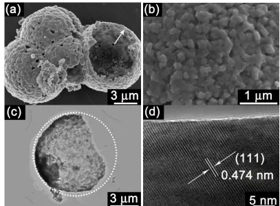
Figure 1. Morphology of HS-LMO: (a) low magnification SEM image, (b) SEM image for the surface of a microsphere, (c) TEM image of a broken microsphere, and (d) HRTEM image of a
[image:3.596.92.499.374.673.2]
Fig. 1 displays the typical morphologies of LiMn2O4 formed via template-engaged reaction,
using hollow microspheres of MnO2 served as the direct self-sacrifice template. Fig. 1a shows
LiMn2O4 hollow microspheres with a diameter of about 9
with the thickness of about 200 nm (2% of the diameter of microspheres). Outer shell is assembled by close-packed nanoparticles with a diameter of about 100 nm (Fig. 1b), which is different from smooth surface of LiMn2O4 hollow microsphere reported [19]. Typical TEM image also confirms hollow
structure and ultrathin shell of the product (Fig. 1c). HRTEM image focused on a nanoparticle (Fig. 1d) shows the crystalline character of the as-synthesized compound. The measured interplanar distance of 0.474 nm is consistent well with that of (111) plane of cubic spinel LiMn2O4, revealing high
crystallinity of as-obtained HS-LMO.
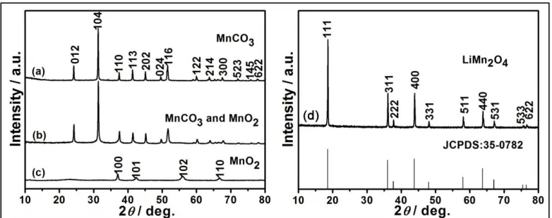
Figure 2. XRD patterns of (a) MnCO3 microspheres, (b) hierarchical structure (MnCO3@MnO2
-shell/MnCO3-core) obtained after pre-calcining MnCO3 at 300 °C for 12 h, (c) MnO2 obtained
after removal of MnCO3 using 0.1 M HCl and (d) LiMn2O4 obtained after template-engaged
lithiation process.
HS-LMO was formed by lithiation of hexagonal MnO2 with the prepared MnCO3 served as
manganese source and original template. The detail of transformation among phases is investigated by XRD patterns (Fig. 2). From Fig. 2a, the XRD pattern of the initial product by the precipitation method can be indexed to single phase MnCO3 (R-3c, JCPDS No. 44-1472). After pre-calcination at 300 °C for
12 h, the MnCO3 phase is still maintained and MnO2 phase is not detected (Fig. 2b). But after
dissolving MnCO3 by 0.1 M HCl, the diffraction peaks of hexagonal-symmetrical MnO2 are observed
clearly, such as (100), (101), (102), (110), which are consistent with JCPDS No. 30-0820 (Fig. 2c). From Fig. 2b and 2c, it is suggested that rhombohedral MnCO3 ispartially converted into hexagonal
MnO2 and the mixture of MnCO3@MnO2 is formed. Besides, compare with invisible diffraction peaks
of MnO2, diffraction peaks of MnCO3 are shown clearly, which can support that MnCO3 is the
dominating phase in mixture after pre-calcination. It may be due to low content of MnO2 in mixture
that results in its invisibility. It may also be attributed to particular hierarchical structure (MnCO3@MnO2) that MnO2 is not detected before removal of MnCO3 outer layer (Fig. 2b). In Fig. 2d,
[image:4.596.104.495.268.423.2]
spinel LiMn2O4 with a space group of Fd3m (JCPDS No. 35-0782). The composition of the
synthesized compound was further investigated by inductively coupled plasma-atomic emission spectrometry (ICP-AES) method. The result shows atomic ratio of Li/Mn is 0.52, indicating forming a lithium-rich spinel compound.
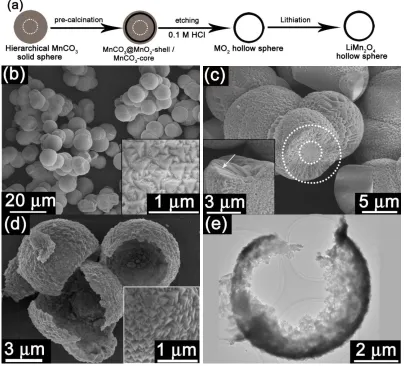
Herein, HS-LMO was prepared by a facile template-engaged reaction as illustrated in Fig. 3a, different from the synthesis of LiMn2O4 nanospheres by low-temperature chemical lithiation reaction
using LiI as lithium source in previous report [20]. Formation of HS-LMO can be mainly attributed to hierarchical structure of MnCO3 microspheres, suitable pre-calcination condition, removal of MnCO3
by HCl and subsequent high-temperature lithiation reaction. The uniform MnCO3 microspheres with a
diameter of about 9
of the microsphere consists of nanopyramids (the inset in Fig. 3b).
Figure 3. (a) Schematic illustration of the formation of HS-LMO, SEM images of (b) MnCO3
microspheres prepared by the precipitation method, the inset shows the outer surface of a microsphere, (c) a cross-sectional view of MnCO3 microsphere, the inset shows image of
further broken microsphere, and (d) HS-MO obtained after removing MnCO3 with 0.1 M HCl,
[image:5.596.97.501.291.657.2]
From the crossed-sectional view of a broken MnCO3 microsphere (Fig. 3c), different structures
between inner core and outer shell can be observed as marked by inner and outer dotted circles, contributing to hierarchical MnCO3@MnO2-shell/MnCO3-core after pre-calcination of MnCO3
microspheres [22]. Upon pre-calcination, the surface of MnCO3 shell was oxidized to MnO2 in
advance and processed from outer layer to inner layer due to more contact with air, contributing to formation of MnCO3@MnO2 shell [14, 22, 23]. In this case, only oxidation layer of MnO2 was formed
on the surface because MnCO3 could only be partially decomposed or under-calcined at this low
temperature, whereas inner core still kept as the MnCO3 salt [23]. Thus, hierarchical structure
(MnCO3-core/MnCO3@MnO2-shell) is formed, which is beneficial for further tailoring into a hollow
structure due to favour solubility of MnCO3 in diluted acid and difficult dissolution of MnO2. After 0.1
M HCl was employed to remove MnCO3 from the hierarchical strcture (MnCO3@MnO2-shell/MnCO3
-core), hollow spherical MnO2 was obtained and pyramid-like structure was well-preserved on the
surface of microspheres (Fig. 3d). Hollow structure can be further supported by TEM image (Fig. 3e). Ultrathin shell is formed because of low content of MnO2 shell in the mixture (Fig. 3d and 3e). After
calcinated with LiOH at 700 °C, HS-MO was converted to HS-LMO with well-preserved hollow spherical structure despite of formating some pores within the shell (Fig. 1). In addition, the morphology of outer shell converted from nanopyramids to nanoparticles as seen from Fig. 1b and 3d. The smooth dense shell was converted to nanoparticles-packed spherical shell with some nanopores, which could facilitate the penetration of electrolyte.
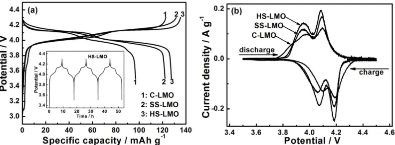
Figure 4. (a) The first charge/discharge voltage profiles of LiMn2O4/Li cells at 0.1 C between 3.5 and
4.3 V, the inset shows charge/discharge profiles of the HS-LMO/Li cell during first three cycles, and (b) CV plots between 3.5 and 4.5 V with a scan rate of 0.1 mV s-1.
[image:6.596.101.497.415.561.2]
coulombic efficiency of nearly 100%. Two pronounced charge/discharge plateaus can be observed for all three samples, demonstrating Li+ ions extraction/interaction reactions. Cycle voltammorgams were performed for three samples to further investigate potentials of Li-insertion/extraction. Two pairs of separated redox peaks can be clearly observed, which are associated with the potential plateaus in the charge/discharge curves, suggesting that Li+ ions are extracted or inserted from/into the spinel matrix with a two-step mechanism [24]. Among three samples, HS-LMO exhibits sharper and higher-symmetrical redox peaks than both C-LMO and SS-LMO, revealing higher crystallinity of HS-LMO and better interface with the electrolyte.
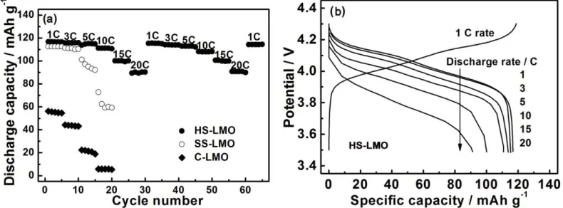
To further investigate potential application in high power devices, the cells are discharged at various rates ranging from 1 to 20 C after charging at 1 C rate (Fig. 5a). Discharge capacities of all the samples gradually decrease with improving discharge rate, indicating the diffusion-controlled kinetics process for electrode reaction [11]. Obvious difference can be observed easily among three cathodes, HS-LMO shows a lower fading rate than another two samples. In addition, the discharge capacity of HS-LMO can be recovered when current decreased from 20 to 1 C, indicating good reversibility of this cathode that may facilitate Li+ ions insertion/deinsertion into/from spinel matrix. Using 1 C charging rate, discharge profiles of HS-LMO/Li cell are shown in Fig. 5b. The average potential and discharge capacity decrease gradually with the improving discharge rate. At the same time, two discharge potential plateaus become sloped gradually, which may be due to electrode overpotentials and increased internal ohmic (IR) drop at high rates [10]. But HS-LMO electrode still displays discharge capacity of 91 mAh g-1 at 20 C rate. The superior rate capability of HS-LMO can be attributed to their structure features. Spinel LiMn2O4 has a three-dimensional lithium diffusion path due to favorable
crystal, resulting in easier lithium exchanging between active materials and electrode. It is also ascribed to nanoparticles which are beneficial for faster Li-insertion.
Figure 5. (a) Discharge capacity with cycle number under varied discharge rates after charging at 1 C, and (b) discharge voltage profiles at different current rates for HS-LMO.
[image:7.596.101.496.491.637.2]
110 mAh g-1, while another two materials fail to sustain high-rate requirement with much lower discharge capacity during 100 cycles.
Figure 6. (a) Comparison of discharge capacity among C-LMO, SS-LMO and HS-LMO at 10 C, and (b) discharge capacity retention and coulombic efficiency plots of HS-LMO/Li cell as a function of cycle number at 20 C.
To fully evaluate high-rate cycleability, HS-LMO based cells are discharged at higher rate (20 C) and conditions up to 500 cycles were performed using 1 C charge rate. As shown in Fig. 6b, the discharge capacity retention of HS-LMO/Li cell can still maintain over 90% after 500 cycles and coulombic efficiency can still maintain nearly 100% except the first cycle, displaying superior high-rate cycleability. This may be due to their special micro-/nano-hybrid structure: hollow structure with some nanopores facilitates the penetration of the electrolyte, which maximizes the contact of active material with electrolyte; outer shell can accommodate strain from the volume changes upon Li+ ions insertion/extraction, which is beneficial for sustaining structure stability during long cycling; nanoparticles on the shell can shorten Li+ ions diffusion distance and facilitate rapid Li+ ions diffusion, which can decrease polarization during charging/discharging under large current, leading to superior high-rate cycleability.
[image:8.596.105.501.134.270.2] [image:8.596.204.389.561.706.2]
Compare with hollow spherical LiMn2O4 prepared in the previous reports [19, 20], hollow
microspheres in this work exhibit higher capacity and better cycling stability. Discharge capacity of 91 mAh g-1 can be delivered under the current density of 2.96 A g-1, much higher than another two hollow microspheres [19, 20]. This is due to special preparation method in our work, without K+ cations involved during preparation and higher crystallinity from high-temperature calcination [25]. The presence of K+ cations may impede the diffusion of Li+ ions, resulting in a low capacity and inferior cycle life [26]. Besides, it is commonly considered that samples with high crystallinity usually display higher capacity and better cycling stability [27].
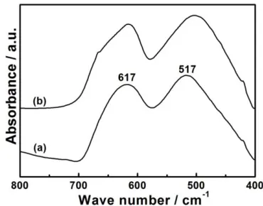
Structural stability of HS-LMO electrode was investigated after prolonged cycling under high-rate current. Spinel structure (AB2O4) can be investigated by FTIR spectra (Fig. 7). Before
charging/discharging, the FTIR spectra of HS-LMO displays two separated peaks (617 cm-1 and 517 cm-1), corresponding to the vibrations of tetrahedral LiO4 and octahedral MnO6 [28, 29]. After 200
cycles with discharging at 10 C, the FTIR characteristic is kept almost unchanged except a slight shift towards to lower frequencies. The slight shift and broadness of the peak located at 517 cm-1 may be due to the formation of lithium-poor spinel compound [28].
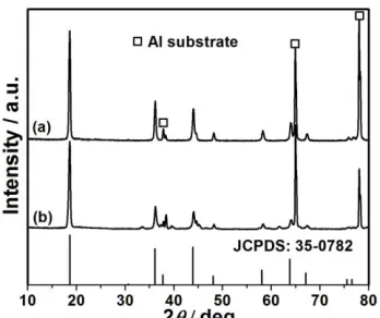
Figure 8. (a) XRD patterns of HS-LMO electrode after cycled for (a) 100 times and (b) 200 times with discharging at 10 C, the asterisk denotes aluminum substrate.
From Fig. 8, XRD patterns can also support that HS-LMO electrode still keeps cubic spinel structure and no possible impurity of Mn2O3 presents after cycling at high rate, displaying excellent
structure stability during fast Li-intercalation.
4. CONCLUSIONS
In summary, we succeeded in designing and synthesizing spinel LiMn2O4 with
micro-/nano-hybrid structure by a facile self-template method. The prepared LiMn2O4 presents hollow microspheres
with nanoparticles-assembled ultrathin shell. In this approach, the morphology of self-support MnO2
template could be well-preserved after high-temperature calcination. The synthesized LiMn2O4 hollow
[image:9.596.212.386.351.497.2]
long cycling under high-rate current. As cathodes for Li-ion batteries, they display superior rate capability and cycling stability, which is attributed to their special micro-/nano-hybrid structure and high crystallinity. Considering durable high-rate capability and facile fabrication method, nanoparticles-assembled hollow microspheres of LiMn2O4 can serve as a promising cathode for
high-power Li-ion batteries.
ACKNOWLEDGEMENT
The authors would like to acknowledge financial support from the National Natural Science Foundation of China (No. 51101139), the Qianjiang Talents Project of Science Technology Department of Zhejiang Province (2011R10021), the Ph.D. Programs Foundation of Ministry of Education of China (No. 20100101120024), and the Foundation of Education Department of Zhejiang Province (No. Y201016484). HYY thanks financial support from SUTD-ZJU/RES/042011 grant.
References
1. A. S. Arico, P. Bruce, B. Scrosati, J. M. Tarascon, W. van Schalkwijk, Nat. Mater. 4 (2005) 366-377.
2. S. Y. Chung, J. T. Bloking, Y. M. Chiang, Nat. Mater. 1 (2002) 123-128.
3. P. L. Taberna,; S. Mitra,; P. Poizot,; P. Simon,; J. M. Tarascon, Nat. Mater. 5 (2006) 567-573. 4. H. Li, Z. Wang, L. Chen, X. Huang, Adv. Mater. 21 (2009) 4593-4607.
5. P. Bruce, B. Scrosati, J. M. Tarascon, Angew. Chem., Int. Edit. 47 (2008) 2930-2946. 6. S. Lim, J. Cho, Electrochem. Commun. 10 (2008) 1478-1481.
7. M. M. Thackeray, J. Electrochem. Soc. 142 (1995) 2558-2563. 8. Y. Wang, G. Cao, Adv. Mater. 20 (2008) 2251-2269.
9. Y. G. Guo, Y. S. Hu, W. Sigle, J. Maier, Adv. Mater. 19 (2007) 2087-2091.
10. H. W. Lee, P. Muralidharan, R. Ruffo, C. M. Mari, Y. Cui, D. K. Kim, Nano Lett. 10 (2010) 3852-3856.
11. Y. L. Ding, J. Xie, G. S. Cao, T. J. Zhu, H. M. Yu, X. B. Zhao, Adv. Funct. Mater. 21 (2011) 348-355.
12. Y. G. Guo, J. S. Hu, L. J. Wan, Adv. Mater. 20 (2008) 2878-2887.
13. F. Cheng, H. Wang, Z. Zhu, Y. Wang, T. Zhang, Z. Tao, J. Chen, Energy Environ. Sci. 4 (2011) 3668-3675.
14. Y. L. Ding, X. B. Zhao, J. Xie, G. S. Cao, T. J. Zhu, H. M. Yu, C. Y. Sun, J. Mater. Chem. 21 (2011) 9475-9479.
15. A. M. Cao, J. S. Hu, H. P. Liang, L. J. Wan, Angew. Chem., Int. Edit. 44 (2005) 4391-4395. 16. S. Han, B. Jang, T. Kim, S. M. Oh, T. Hyeon, Adv. Funct. Mater. 15 (2005) 1845-1850. 17. S. Q. Wang, J. Y. Zhang, C. H. Chen, Scripta Mater. 57 (2007) 337-340.
18. H. Ma, F. Cheng, J. Y. Chen, J. Z. Zhao, C. S. Li, Z. L. Tao, J. Liang, Adv. Mater. 19 (2007) 4067-4070.
19. X. Xiao, J.; Lu, Y. Li, Nano Res. 3 (2010) 733-737.
20. J. Luo, L. Cheng, Y. Xia, Electrochem. Commun. 9 (2007) 1404-1409.
21. J. Fei, Y. Cui, X. Yan, W. Qi, Y. Yang, K. Wang, Q. He, J. Li, Adv. Mater. 20 (2008) 452-456. 22. J. Cao, Y. Zhu, L. Shi, L. Zhu, K. Bao, S. Liu, Y. Qian, Eur. J. Inorg. Chem. 2010, 1172-1176. 23. L. Wang, F. Tang, K. Ozawa, Z. G. Chen, A. Mukherj, Y. Zhu, J. Zou, H. M. Cheng, G. Q. Lu,
Angew. Chem., Inter. Edit. 48 (2009) 7048-7051.
24. Y. Xia, M. Yoshio, J. Electrochem. Soc. 143 (1996) 825-833.
26. J. Y. Luo, H. M. Xiong, Y. Y. Xia, J. Phys. Chem. C. 112 (2008) 12051-12057. 27. E. Hosono, T. Kudo, I. Honma, H. Matsuda, H. Zhou, Nano Lett. 9 (2009) 1045.
28. T. J. Richardson, S. J. Wen, K. A. Strichel, P. N. Ross Jr, E. J. Cairns, Mater. Res. Bull. 32 (1997) 609-618.
29. P. Kalyani, N. Kalaiselvi, N. Muniyandi, J. Power Sources 111 (2002) 232-238.