LIVER FUNCTION TESTS IN CONGESTIVE
CARDIAC FAILURE
DISSERTATION SUBMITTED TO
THE TAMIL NADU DR.M.G.R MEDICAL UNIVERSITY
IN PARTIAL FULFILLMENT OF THE REGULATIONS FOR THE AWARD OF THE DEGREE OF
M.D (GENERAL MEDICINE) BRANCH – I
TIRUNELVELI GOVT. MEDICAL COLLEGE & HOSPITAL
TIRUNELVELI
THE TAMILNADU DR.M.G.R MEDICAL UNIVERSITY
CHENNAI, TAMIL NADU
CERTIFICATE
This is to certify that this dissertation entitled “LIVER FUNCTION TESTS IN CONGESTIVE CARDIAC FAILURE” is the bonafide record work done by Dr. ANU RAJEE KRISHNAN, submitted as partial fulfillment for the requirements of M.D. Degree Examinations, General Medicine (Branch I) to be held in APRIL 2013.
Prof. Dr. S.S.NAZAR M.D Prof. Dr. R. GEETHARANI, M.D., Professor of Medicine, Professor and H.O.D,
Unit Chief M VII, Department of Medicine,
Department of Medicine, Tirunelveli Medical College hospital, Tirunelveli Medical College Hospital, Tirunelveli.
Tirunelveli.
THE DEAN,
DECLARATION
I solemnly declare that the dissertation titled “ LIVER FUNCTION TESTS IN CONGESTIVE CARDIAC FAILURE’’ was done by me at Tirunelveli Government Medical College and Hospital during 2011-2012 under the guidance and supervision of PROF. DR.S.S.NAZAR M.D.,Professor of Medicine.
This dissertation is submitted to the Tamil Nadu Dr.M.G.R Medical University towards the partial fulfillment of requirements for the award of M.D DEGREE (BRANCH-I) in General Medicine.
ACKNOWLEDGEMENT
I am extremely grateful to The DEAN, DR.MANOHARAN M.S, Tirunelveli Medical College for granting me permission to do this dissertation work in Tirunelveli Medical college Hospital, Tirunelveli.
I express my sincere gratitude to the professor and the Head of the Department of medicine Prof. Dr.R.GEETHARANI, M.D., for her valuable support and guidance in preparing this dissertation.
I am greatly indebted to my unit chief and beloved teacher Prof.Dr.S.S.NAZAR M D, who inspired, encouraged and guided me in every step of this study.
I am grateful to Professor Dr.RAVICHANDRAN EDWIN, M.D, D.M, Professor and Head of Department of Cardiology,Tirunelveli Medical College for his guidance in doing this study.
I am grateful to Dr.BALACHANDRAN, M.D, D.M, Assistant Professor of
Cardiology, Dr.KANDASAMY PRABHU, M.D,D.M, Assistant Professor of Gastroenterology, for their guidance and support throughout the study.
I am thankful to beloved Assistant Professors of my unit, Dr.RAJESH BABU, M.D. and Dr. SHAVANA, M.D., for their guidance
I thank the Department of Biochemistry for their help in investigation aspects.
CONTENTS
CONTENTS PAGE NO
1. INTRODUCTION 01
2. AIM OF THE STUDY 03
3. REVIEW OF LITERATURE 04
4. MATERIALS AND METHODS. 57
5. OBSERVATIONS AND ANALYSIS. 62
6. DISCUSSION 81
7. CONCLUSION 89
8. BIBLIOGRAPHY
9. ANNEXURE
1) PROFORMA 2) MASTER CHART
LIST OF ABBREVATIONS
1. LFT Liver function tests
2. CCF Congestive cardiac failure
3. CAHD Coronary artery heart disease
4. CP Cor pulmonale
5. RHD Rheumatic heart disease
6. CM Cardiomyopathy
7. HHD Hypertensive heart disease
8. AST/SGOT Aspartate amnotransferase/Serum glutamic
oxaloacetic transaminase
9. ALT/SGPT Alanine aminotransferase/Serum glutamic pyruvate
transaminase
10. ALP Alkaline phosphatase
11. GGT Gamma glutamyl transpeptidase.
12. ECG Electrocardiogram
13. CLN Centrilobular necrosis
INTRODUCTION
Liver is the biggest organ in human body with a mass of nearly 1.5 kg2. Liver has a massive functional reserve and regenerating capacity. It plays a major role in maintaining the normal physiology and metabolic homeostasis of human body. It is also referred to as the custodian of milieu interior7. Hence hepatic diseases can have a major impact on the homeostasis of body. Similarly disorders in other systems can adversely affect the liver as well.
Cardiac failure both chronic and acute can cause hepatic dysfunction 1. Twenty five percent of total cardiac output goes to the liver,hence any decrease in cardiac output leads to decreased liver perfusion.by means of vasoregulatory mechanisms and enhanced extraction of oxygen liver can tolerate variations in blood flow.
In the present study ,the consequence of congestive cardiac failure on liver function were studied in a group of 60 patients and was analysed and compared with various parameters.Differing causes of heart failure of various duration were included in the study. Follow up of the cases were done for a period of one week and measurements were repeated at the end of one week. Changes in the values were recorded and an attempt is done to find the prevalence of liver function abnormalities in these subjects,its correlation with various parameters and prognostic significance of liver function on cardiac failure.
A proper knowledge and understanding of structure and functions of liver,liver parameters,etiologies and different forms of heart failure and their presentation ,as well as the mechanism and pathology of liver in cardiac failure is inevitable before assessing the liver function abnormalities in congestive cardiac failure.
AIMS AND OBJECTIVES
1. To study the prevalence of liver function abnormalities in congestive cardiac failure patients attending Tirunelveli Medical College Hospital
2. To assess the correlation of liver function tests with etiology , duration and NYHA Class of cardiac failure
3. To study the pattern of elevation of liver enzymes in cardiac failure. 4. To study the prognostic importance of liver function abnormalities in
REVIEW OF LITERATURE
STRUCTURE AND FUNCTIONS OF LIVER
Blood circulation to the hepatic tissue has peculiar features.It has both systemic and portal circulation3.Arterial blood is supplied bythe hepatic artery, originating from the coeliac axis .The portal circulation consists of venous blood from spleen, intestines etc. The porta hepatis fissure, which lies on undersurface of the right lobe is the portal of entry for this vessels9. On entry into liver hepatic artery and portal vein divide into right and left lobe branches. Common bile duct is formed by confluence of right and left hepatic ducts in the porta. Sympathetic fibres from ganglia T7–T10 supplies the liver through the hepatic plexus1.
The CBD(common bile duct) is situated in front and right side of hepatic artery and in front of portalvein. Within the portal tracts intrahepatic bile ducts strictly track the path of intrahepatic arteries and portal veins. Right and left hepatic veins drain venous blood from the liver. They arise from
posterior aspect of liver and drain into IVC(inferior vena cava) close to its
entry point to right atrium. Collection of lymph nodes nearby portahepatis
and coeliac axis drain the lymph. The mediastinal glands receives superficial
hepatic lymph vessels traverse the diaphragm .One more group drains in
Except in three places the liver is entirely enclosed within peritoneum.
There is a bare area near the IVC fossa where liver is in straight contact with
the diaphragm. IVC fossa and gallbladder fossa lacks peritoneal covering1.
The peritoneal ligament and the intra-abdominal pressure keep the liver in
position.
FUNCTIONAL ANATOMY
The model of hepatic lobular architecture was put forward by
Kierman et al29 in 1833.Thepyramidal lobules comprises of a peripheral
portal tract made up of portal vein radicle, hepatic artery branch and the bile
columns of hepatocytes spread between above two structures. The hepatic
vein radicles and their adventitia occupy the central hepatic canals. They are enclosed by hepatocytes. The hepatic arteriole, the portal vein radicle and bile duct along with a few number of cells and connective tissue form the portal
triad. The major portion of liver about 60% is made up of hepatocytes3,9
.They are nearly 30mm in diameter and multilateral. The liver cells has three
surfaces: each fronting the canaliculus, the sinusoid and space of Disse,
neighbouring hepatocytes respectively. The lifetime of hepatocytes nearly
150 days. They lack basement membrane2.
Endothelial cells line the sinusoids. The sinusoids contain the Kupffer
cells(phagocytic cells of thereticulo-endothelial system), the fat storing
endothelial cells. Space of Disse is a tissue space between liver cells and
endothelium of sinusoids. Connective tissue around the portal region
contains liver lymphatics which has an endothelial lining. Lymphatics receive
lymph through the endothelium. The division of the hepatic artery delivers
blood to the contents in portal tracts forming a network surrounding biliary
ductules .These arterioles drains into sinusoids at multiple points .Portal veins
and hepatic arterioles do not anastamose directly2.
Bile canaliculi are the main portals of elimination of liver. They are
channels between the apposition surfaces of hepatocytes with no separate lining. The cytoskeleton of liver cells is made up of microfilaments supporting the cell membrane .Microvilli are found on the exterior of cells12.
Canals of Hering are thin-walled terminal bile ducts or ductules lined with
end in bile ducts in the portal canals Desmosomes, tight junctions and gap
junctions separate canaliculi from other cell surface.
Functionally liver is structured into acini, with hepatic arterial and portal venous blood entering the acinus from the portal areas which constitute zone1 and flows through the sinusoids to the terminal hepatic veins zone39 . The intervening liver cells constitute zone2. Hence zone3 of liver is susceptible to toxic and anoxic insult. Bile flows in the reverse direction from zone3 to zone1. The cells in the border of acini , zone3 differ in function from those of cells in zone 1. TCA cycle enzymes are more in periportal zone . Glutamic synthetase is present in zone 3. Perivenous zone is susceptible to anoxic injury. Congestive heart failure disturbs zone 3 of the hepatic acini leading to distinctive features 9.
LIVER FUNCTIONS
Liver cells execute many essential roles in maintaining homeostasis and well-being.
SYNTHETIC FUNCTIONS
The synthesis of proteins like carrier proteins, albumin, hormones, growth factors, coagulation factors, bile and transporters like bileacids, cholesterol, lecithin, phospholipids1.
DIGESTIVE FUNCTIONS
Nutrients like glucose, glycogen, lipids, cholesterol, amino acids stored and metabolized by liver1,2. Liver also carries out metabolism and conjugation of lipophilic compounds (bilirubin, anions, cations, drugs)
EXCRETORY FUNCTIONS
STORAGE
Storage of an array of various substances occur in the liver which includes glucose as glycogen, vitamin B12, vitamin A, vitamin K, vitamin D, copper and iron.
IMMUNE FUNCTIONS
The liver plays a major role in immunological reactions. In the liver RES (reticuloendothelial system) is constituted mainly by cells that are immunologically reactive. Liver acts as a 'sieve' for antigens reaching it through the portal circulation1.
LIVER FUNCTION TESTS
substituted by a mechanical pump ; dialysis membrane or fabrication of instilled proteins, growth factors and proteins1.
Tests Centered on Conjugation and Elimination Purposes
SERUM BILIRUBIN
Bilirubin, a yellow breakdown product of heme catabolism and is present in blood in two forms—conjugated and unconjugated. The unconjugated form or the indirect fraction, does not dissolve in water and is carried by albumin in the circulation. The conjugated or direct bilirubin fraction dissolves in water and is eliminated by renal system. Estimated by the van den Bergh method, the total serum bilirubin concentration is normally<17mol/L (1 mg/dL).Of these 30%, or 5.1mol/L (0.3 mg/dL) is constituted by direct-reacting bilirubin 48.
Rise of the indirect fraction of bilirubin is hardly due to hepatic dysfunction. Isolated increase of indirect bilirubin is found predominantly in hemolytic conditions and genetic disorders like Crigler-Najjar and Gilbert's syndromes. Isolated indirect hyperbilirubinemia (bilirubin increased but <15% conjugated) must warrant adiagnosis of hemolysis . An isolated indirect hyperbilirubinemia without hemolysis can be ascribed to Gilbert's syndrome and no additional assessment is needed.
form can be seen in almost any kind of hepatic disorder. Both direct and indirect forms of the bilirubin is likely to be elevated in most of liver disease.
SERUM ENZYMES
The liver comprises of numerous enzymes, of which some are found in the serum in small concentrations. These enzymes act like other serum proteins with no known function . These enzymes are dispersed in the interstitial fluid ,plasma and have distinctive half-lives, typically estimated in days. Enzymes are possibly cleared by cells in the reticuloendothelial system. The increase in particular enzyme action in the serum is assumed to chiefly mirror its increased rate of entry into serum from injured hepatocytes Serum enzyme tests can be assembled into three classes: (1) enzymes whose rise in serum mirrors injury to hepatic cells, (2) enzymes whose rise in serum mirrors cholestasis, and (3) enzyme tests with no specific pattern.
Enzymes that indicates destruction to Hepatocytes
circulation in larger amounts when there is injury to the hepatocyte membrane leading to easy permeability. Hepatocyte necrosis is not needed for the discharge of the aminotransferases, and the correlation between the amount of hepatocyte damage and the level of the aminotransferases is poor. Hence, the outright increase in aminotransferase has nil prognostic implication in acute hepatocellular conditions. Any kind of liver cell damage can produce a modest rise in the serum aminotransferases.Values of up to 300 U/L can be present in any kind of hepatic disorder and are nonspecific49 . Studies have revealed that fatty liver disease is the most likely explanation for mild ALT elevations in asymptomatic blood donors50.
Remarkable rises with aminotransferases > 1000 U/L is seen almost absolutely in conditions linked with widespread hepatocellular damage such as31
1. hepatic damage due to ischaemia
2. hepatic injury secondary to toxins and medications 3. (3)hepatitis due to viral infections.
pyridoxal phosphate.The transaminases are commonly not significantly raised in obstructive jaundice except in acute stage of biliary blockade due to the transport of a gallstone into the common bile duct where the transaminases can temporarily be raised upto 1000–2000 U/L limit48,49. Nevertheless, transaminases fall rapidly, and the hepatic function tests progress into cholestatic picture.
ENZYMES IN CHOLESTASIS
and smaller than threefold rise is demonstrated in nearly any kind of hepatic disorder. ALP rise more than four times seen mainly in patients with cholestatic hepatic diseases, carcinomas and amyloidosis, and Paget's disease of the bone. The liver isoenzyme is elevated in hepatic diseases.The identification of the cause of raised isoenzymes is useful, if increased ALP is the only anomalous outcome in an outwardly healthy subject .This can be advanced in numerous methods. The fractionation of the alkaline phosphatase by electrophoresis is one method. Another method is to differentiate isoenzymes by there varied susceptibility to inactivation by heat. A heat-stable fraction intensely points that either placenta or a tumor is the cause of the raised enzyme in serum. The heat labile fractions are liver, intestinal and bone ALPs. The most sensitive being bone. The measurement of serum 5'-nucleotidase or GGT is the best proven and most accessible method .In disorders other than hepatic disorders they are hardly raised. An isolated elevation of ALP of hepatic origin without icterus or raised transaminases usually indicates initial stages of cholestasis and rarely liver permeation by granulomas or malignancies1 . Other conditions that cause a sole rise in alkaline phosphatase are Diabetes, Hodgkin's disease, amyloidosis, hyperthyroidism, inflammatory bowel disease, congestive heart failure etc.
to cancer, common duct stone, sclerosing cholangitis, or bile duct stricture. Values are similarly increased in patients with intrahepatic cholestasis secondary to transplant rejection, hepatitis due to drugs, inflammatory bowel disease ,primary biliary cirrhosis and ethanol related steatonecrosis1,2,9. Values are also greatly elevated in hepatobiliary disorders seen in patients with AIDS (e.g., AIDS cholangiopathy due to cytomegalovirus or cryptosporidial infection and tuberculosis with hepatic involvement).
Tests centered on synthetic purposes of Liver
SERUM ALBUMIN
Hepatocytes produce and secrete 10 g of albumin per day. In patients with hepatic decompensation, albumin synthetic capacity decreases resulting in hypoalbuminemia. However, the serum albumin level also can be lowered by extrahepatic conditions such as malnutrition, enteropathy, renal disease, and hormonal disturbances. Therefore, hypoalbuminemia is not a precise pointer of liver dysfunction. As the half-life of serum albumin is nearly 20 days, the serum albumin cannot be taken as a consistent marker of liver synthetic function in subjects with acute hepatic damage. However, in subjects with chronic hepatic disease the serum albumin value is useful as a predictive marker of prognosis.
SERUM GLOBULINS
Serum globulins are a heterogenous collection of proteins consisting of immunoglobulins produced by B cells, plasma cells and globulins formed chiefly in liver cells. Globulins are increased in conditions of long standing hepatic disease like cirrhosis and chronic hepatitis. In cirrhosis, the increased serum gamma globulin concentration is the result of elevated production of Ig (immunoglobulins) directed against gut bacterial flora. This occurs because the cirrhotic liver fails to clear bacterial antigens that normally reach the liver through the hepatic circulation.
Increases in the concentration of specific isotypes of globulins are often helpful in the recognition of certain chronic liver diseases. Diffuse polyclonal increases in IgG levels are common in autoimmune hepatitis; increases >100% should alert the clinician to this possibility. Raised IgM levels were found in primary biliary cirrhosis, whereas rise in the IgA levels were seen in ethanol induced liver disease.
Clotting Factors
in the extrinsic coagulation pathway which includes factors II, V, VII, and X, which are all synthesized by the liver.
The differential diagnosis of an abnormally prolonged prothrombin time also includes vitamin K deficiency, therapeutic anticoagulation, and a consumptive coagulopathy. Vitamin K is needed for gamma-carboxylation and normal functioning of clotting factors 2, 7,9, and 10. Vitamin K deficiency can be associated with malnutrition, malabsorption, or antibiotic use and may lead to prolongation of the prothrombin time. The use of warfarin interferes with the vitamin K–induced gamma-carboxylation. The prothrombin time can be prolonged as a result of disseminated intravascular coagulation (DIC) and congenital deficiency of clotting factors. When prolongation of the prothrombin time is caused by liver disease, levels of factor VIII are normal or increased, whereas in DIC, factor VIII levels are decreased. Measurement of the serum factor V level and administration of vitamin K can be used to differentiate hepatic dysfunction and vitamin K deficiency as a cause of a prolonged prothrombin time. Factor V levels are decreased in liver disease but remain unaffected by vitamin K deficiency. Administration of vitamin K, 10 mg subcutaneously, results in correction of the prothrombin time by at least 30% within 24 hours in patients with vitamin K deficiency, but not in those with liver disease.
CONGESTIVE CARDIAC FAILURE
It is a clinical syndrome which may be due to a structural or functional defect of the heart causing impairment of the ventricle to receive or pump blood 4.Chronic heart failure is a systemic clinical syndrome which can have effects in other organs.The major manifestation includes
tiredness,breathlessness decreasing the exercise capacity and features of
The overall prevalence of Heart failure in the industrialized countries is two in hundred people. Incidence of heart failure rises as the age advances affecting nearly five to ten percent of population above sixty five years1. CHF is widely classified under two groups1,5;
1. systolic failure (Reduced ejection fraction less than 40%) 2. Diastolic failure (Ejection fraction preserved more than 40%)
Etiology
Conditions which cause alteration in LV structure or function become predisposing factors for a patient to develop HF5. The leading reason for cardiac failure in developed countries in the present times is Ischaemic Heart Disease
Causes for Systolic Failure(EF less than 40%) ¾ Ischaemic heart disease
¾ Myocardial infarction ¾ Myocardial ischemia
¾ Obstructive valvular disease ¾ Regurgitant valvular disease ¾ Chronic volume overload
¾ Chronic pressure overloadHypertension ¾ Hypertension
¾ Inherited disorders Invasive disorders ¾ Injury secondary to medications and toxins ¾ Disorders of Metabolism
¾ Arrhythmias
¾ Infections like viral and parasitic causes
Causes for Diastolic failure (EF More than 40%) Diseases causing hypertrophy pathologicaly
• Primary causes like HOCM(Hypertrophic cardiomyopathies)
• Secondary causes like Hypertension
Ageing
Conditions causing RCM(Restrictive cardiomyopathy) ¾ Invading diseases like Amyloidosis and Sarcoidosis ¾ Metabolic disorders like Hemochromatosis
• Disorders affecting the Endomyocardium including fibrotic conditions.
Diseases of pulmonary origin affecting heart ¾ Cor pulmonale
¾ Disorders of pulmonary vasculature
Hyperdynamic circulatory states ¾ Disorders of metabolism ¾ Hyperthyroidism
¾ V Shunting
¾ Anaemia of chronic durati
PRECIPITATING CAUSES OF CCF ¾ Cardiac arrhythmias
¾ Pulmonary embolism
¾ Infections, particularly lung infections ¾ Rheumatic or viral myocarditis
¾ Infective endocarditis ¾ Anaemia
¾ Conditions with increased metabolic demand like pregnancy, thyrotoxicosis
¾ Acute myocardial ischaema or infarction ¾ Accelerated hypertension
¾ Acute valvular regurgitation
¾ Severe physical work or emotional excess increase cardiac workload. ¾ Improper diet control
¾ Poor drug compliance ¾ Drugs worsening failure
• Calcium channel blockers
• Beta blockers
• NSAIDS
¾ Antiarrhythmic drugs. ¾ Alcoholism
In the present study the subjects were considered as suffering from congestive cardiac failure,if they satisfied a minimum of one major and minor criteria based on the FRAMINGHAM CRITERIA for diagnosis of heart failure.
FRAMINGHAM CRITERIA FOR DIAGNOSIS OF HEART FAILURE4,5
Major criteria
• Paroxysmal nocturnal dyspnea or orthopnea
• Cardiomegaly
• Jugular venous distention (or CVP > 16 mm Hg)
• Hepatojugular reflex
• Rales or acute pulmonary edema
• S3 gallop
• Response to diuretic (weight loss >4.5 kg in 5 days) Minor criteria
• Exertional dyspnea
• Nocturnal cough
• Ankle edema
• Pleural effusion
• Vital capacity < two thirds of normal
• Tachycardia (>120 bpm)
In this study symptomatic analysis of the subjects were done and they were classified according to the NEW YORK HEART ASSOCIATION
Classification of Cardiac Failure4,5 into 4 classes.
NEW YORK HEART ASSOCIATION (NYHA) FUNCTIONAL CLASSIFICATION
Class I
No restriction during ordinary actions Class II
Mild restriction during ordinary actions Class III
Marked restriction during normal actions and no symptoms at rest Class IV
Not able to carry out physical activity without symptoms and symptoms can be present at rest
PATHOGENESIS
or interrupting the force producing capability of the myocardium, eventually disrupting the normal contracting ability of heart6.
The inciting event can be of sudden onset (like MI) or insidious onset (like hemodynamic pressure or volume overload) or inherited ( like genetic cardiomyopathies)1,5 .Eventhough they are different in nature, ultimately they impair the pumping capacity of heart.
Cardiac failure patients seem to maintain and modify LV function for variable time ranging from months to years by means of several compensatory mechanisms which get triggered by the cardiac insult and dysfunction of left ventricle1,4.5. The following compensatory mechanisms are called into action
(1) The RAA axis (renin-angiotensin-aldosterone)and sympathetic nervous system activation which cause holding of salt and water and thus sustaining cardiac output
(3) Vasodilating molecules like the ANP and BNP ( atrial and brain natriuretic peptides) PGE2 and PGI2( prostaglandins) and NO (nitric oxide)
get activated counteracting the undue peripheral vasoconstriction. LEFT VENTRICULAR REMODELLING
Progressive activation of the above mechanisms produce a sequence of accommodative alterations in the myocardium which is jointly mentioned as LV remodeling eventually leading to the evolution to symptomatic failure5,6.
A succession of intricate events happen in molecular and cellular stages during remodeling of LV1,5. The factors stimulating these changes are
¾ Muscle fibre subjected to mechanical stretching
¾ Neurohormones in circulation (like noradrenaline, angiotensin II) ¾ Inflammatory cytokines [like TNF ( tumor necrosis factor) ] ¾ Growth factors and Peptide hormones (like endothelin) ¾ Oxygen free radical species (like superoxide and NO).
TYPES OF CARDIAC FAILURE
Heart failure may be acute or chronic, left-sided or right-sided, high-output or low-high-output, forward or backward, and systolic or diastolic5.
Acute and chronic failure
Left-sided and right-sided failure
Left sided heart failure is characterized by pulmonary congestion causing dyspnoea and orthopnoea. Usually occurs in conditions when there is a pressure overload on LV as in aortic stenosis or it is weakened due to a myocardial infarction1. Right-sided failure presents with features of systemic congestion which include elevated jugular venous pressure, congestive liver enlargement and lower extremity oedema. In long- standing cases of cardiac failure like valvular heart disease (aortic and mitral valve) and hypertension, features of biventricular failure are present4.
High-output failure and Low- output failure
High-output failure is encountered in severe anaemia, thyrotoxicosis, beriberi, arterio-venous fistulae, pregnancy and Paget’s disease. The heart has to deliver excessive amount of blood to provide sufficient supply of oxygen to the highly metabolically active tissue in these conditions6. Low output failure is seen conditions like coronary artery disease, dilated cardiomyopathy , hypertension , pericardial and valve diseases of heart.
Forward and backward failure
causing stimulation of the RAAS(renin-angiotensin-aldosterone system) with concomitant retention of salt and water producing oedema 4,5,6.
Systolic and diastolic failure
Systolic heart failure is featured by principal systolic ventricular contractile dysfunction chiefly due to myocardial dysfunction leading to an inadequate output and further symptoms. The main aetiologies include dilated cardiomyopathy , ischaemic heart disease, chronic excessive ventricular workload as in systemic hypertension, valvular heart disease (like mitral incompetence, aortic stenosis, aortic incompetence) and congenital heart disease (like ventricular septal defect, coarctation of aorta, congenital pulmonary and aortic stenosis).
Diastolic heart failure is characterized by lack of relaxation of ventricle causing rise of diastolic pressure in the ventricle and preserved diastolic volume. The commonest cause of diastolic dysfunction is ischaemic heart disease . Increased stiffness and thickening of ventricle as seen in restrictive cardiomyopathy due to amyloidosis or haemochromatosis also produces diastolic dysfunction .
In most patients with cardiac hypertrophy and dilatation, systolic and diastolic heart failure co-exist
Diastolic Heart Failure
¾ Elderly women more common
¾ Normal or increased ejection fraction ¾ S4 gallop
¾ Diastolic impairment by various echo measurements ¾ Treatment not well established
¾ Prognosis not as poor
¾ Myocardial ischemia common
Systolic Heart Failure ¾ Large, dilated heart
¾ Normal or low blood pressure
¾ Broad age group; more common in men ¾ Low ejection fraction
¾ S3 gallop
¾ Systolic and diastolic impairment by echo ¾ Treatment well established
¾ Poor prognosis
¾ Role of myocardial ischemia important
Clinical Manifestations
breathlessness is present , as failure advances lower levels of activity causes dyspnea and eventually progressing to dyspnea at rest. It has a multi-factorial origin which include pulmonary fluid collection , rising airway impedance ,declining lung compliance , anemia and breathing muscle fatigue. Dyspnea may become less frequent with the onset of right ventricular (RV) failure and tricuspid regurgitation22.
Orthopnea
Orthopnea or dyspnea in lying down posture is late feature of failure. The cause is increased venous return to the heart in supine position leading to an increase in pulmonary capillary pressure which stimulates the juxta-capillary J receptors resulting in dyspnea. Nocturnal cough is the most frequent and common symptom. It is relieved by maintaining an upright posture. Also seen in conditions other than cardiac disorders.
Paroxysmal Nocturnal Dyspnea (PND)
with PND continue to have symptoms even after assuming the erect position which differentiates it from orthopnea. Cardiac asthma is characterized by wheezing secondary to bronchospasm, and must be differentiated from primary asthma and pulmonary causes of wheezing.
Cheyne-Stokes Respiration
Cheyne-Stokes breathing is usually associated with low cardiac output. It is produced by a decreased stimulation of the central respiratory region to elevated arterial CO2 .It has the following phases: An apneic phase in which the there is a fall in the arterial PO2 and a corresponding rise in PCO2 which sensitizes the respiratory centre followed by hyperventilation and decrease in PCO2 levels which constitutes the hyperpneic phase. It is described as a brief cessation of breathing by the subject.
Other Symptoms
Physical Examination
General Appearance and Vital Signs
¾ In mild to moderate HF, the patient is comfortable at rest,with symptoms of orthopnoea
¾ In more severe HF, the patient dyspnoeic in siting posture . ¾ Decreased pulse pressure
¾ Systolic BP may be normal ,high or low according to the severity of cardiac failure
¾ Sinus tachycardia
¾ Cool peripheries , cyanosed lips and nails due to vasoconstriction ¾ Elevated Jugular Venous pressure
¾ Presence of hepatojugular reflex
Pulmonary Examination
• Pulmonary crackles due to the transudation of fluid from the intravascular space into the alveoli
• Expiratory wheezing (cardiac asthma)
• Pleural effusions result from the elevation of pleural capillary pressure and the resulting transudation of fluid into the pleural cavities.
a) pleural effusions occur commonly with biventricular failure.
Cardiac Examination
¾ Cardiomegaly with shifted apical impulse. A sustained apical impulse due to severe LV hypertrophy
¾ A third heart sound (S3) is audible and palpable at the apex due to
volume overload
¾ A sustained and prolonged left parasternal heave due to hypertrophied right ventricle.
¾ A fourth heart sound (S4) present diastolic dysfunction.
¾ The murmurs of mitral and tricuspid regurgitation heard in patients with advanced HF.
Abdomen and Extremities ¾ Tender hepatomegaly
¾ Pulsatile liver in systole in tricuspid regurgitation. ¾ Ascites
¾ Jaundice also occurs late with elevations of both direct and indirect bilirubin.
¾ Peripheral edema is usually symmetric and dependent in the ankles and pretibial region in ambulatory patients
¾ presacral edema and the scrotal edema in bed ridden cases.
Cardiac Cachexia
• Marked weight loss and cachexia.
Diagnosis of Heart Failure Laboratory Investigations
Subjects presenting with acute heart failure or exacerbation of chronic heart disease should undergo the following regular laboratory investigations. These include
• Complete blood count
• Renal function tests
• Serum electrolytes
• Liver function tests
• Urine analysis .
• Evaluation for DM(Diabetes Mellitus) which includes fasting blood glucose,post prandial glucose and in some cases OGTT(Oral glucose tolerance test)
• Fasting lipid profile to rule out dyslipidaemia
• Thyroid function tests including TSH(Thyroid stimulating hormone)
Electrocardiogram
¾ A regular twelve -lead Electrocardiogram (ECG ) is mandatory. ¾ To rule out left ventricule enlargement, arrhythmias, presence of old
myocardial infarction.
Chest X-Ray
¾ To rule out cardiomegaly
¾ To assess the pulmonary vasculature, ¾ To find out any noncardiac reasons
¾ Acute cases of cardiac failure show findings of pulmonary artery dilatation and features of pulmonary congestion.
¾ Long standing cases of cardiac failure often lack the above radiological findings. This may be accounted by the improved lymphatic drainage of fluid in lungs.
Left ventricular function Evaluation
9 The identification ,assessment and treatment of cardiac failure requires Non-invasive cardiac imaging .
9 The most valuable investigation is Two-D Echo or Doppler 9 It delivers a semi-quantitative evaluation of
a) Left ventricular dimensions and function b) To find out valvular lesions
c) To identify RWMA(regional wall motion abnormalities) which suggests an old MI.
d) To look for Left atrial enlargement and Left ventricular enlargement 9 Tissue and Pulse-wave Doppler beneficial for evaluation of heart
9 Presently MRI is the gold standard for evaluating LV dimensions and capacities giving a complete study of structure and function of heart. 9 The best valuable guide to assess LV performance is Ejection
Fraction(EF). Ejection Fraction=Stroke volume/ End-diastolic volume.
9 Ejection Fraction(EF) is convenient to calculate using non-invasive methods and simple to understand.
9 The major shortcoming of EF is it is easily affected by variations in end diastolic volume and vascular resistance.
Biomarkers
¾ Natriuretic peptides in circulation are valuable markers in identifying people with HF.
¾ B-type natriuretic peptide (BNP) and N-terminal pro-BNP are fairly sensitive indicators for detecting heart failure with depressed EF ¾ A lesser degree of elevation noted in heart failure cases with
maintained EF.
¾ Levels of Natriuretic peptide rise with age and kidney damage. ¾ Additional Biomarkers are
Exercise Testing
Treadmill or bicycle exercise testing valuable for evaluating the requirement of heart transplantation in severe advanced HF .
A greatest oxygen uptake (VO2) <14 mL/kg / min was found to have a
bad prognosis.
Cases with VO2<14 mL/kg /min were found to have prolonged survival
with transplantation .
Differential Diagnosis
Following conditions should be considered in differential diagnosis of cardiac failure
(1) Kidney Failure
(2) Noncardiogenic pulmonary congestion like ARDS( acute respiratory distress syndrome).
MECHANISM OF HEPATIC DYSFUNCTION IN HEART FAILURE
Heart failure causes:
a) reduced liver blood supply
b) elevated hepatic venous pressure causing swelling of sinusoids and atrophy of liver cells.
In cardiac failure there is elevated venous back pressure which is transferred to the hepatic veins and results in congested liver22.Another major factor causing injury is hypoxia23. Histological analysis shows atrophy of liver cells in zone 3 is caused by raised pressure24.
study that acute left heart failure led to necrosis of zone 3 liver cells in the absence of right heart failure showing that cardiac output is a major factor causing hepatic injury. Still congestion appears to be a cofactor in hepatic injury. It plays an important role in preparing the liver sensitive to ischaemic insults.
Liver ischaemia
Disparity between liver oxygen supply and need leads to liver ischaemia. As liver has a constant metabolism ,oxygen delivery is the major determinant of liver ischaemia26.Oxygen supply to liver depends on the hepatic blood flow and oxygen content of blood. It has been shown that cardiac output determines the blood supply to the liver .Hepatic blood supply is dual .Liver receives 20% of blood supply from hepatic artery and 80% from portal vein,hence major oxygen supply is from the portal vein.
With exercise usually cardiac output increases but in diseased heart cardiac output fails to rise and there is a fall in hepatic blood supply.In the normal state liver cells compensate by increasing the oxygen extracted.But in conditions of stress liver metabolic demands increase and oxygen extraction cannot be increased above a limit leading to liver cell hypoxia in the zone 3 of acini 27.
determining liver cell sensitivity to hypoxia . Ishaemia to liver cell causes damage to mitochondria ,decrease in ATP,increase in intracellular calcium and protease activation 29. Reperfusion injury also causes damage to liver cells by formation of free radicals and lipid peroxidation.
In the damaged mitochondria reactive oxygen species are formed which has direct toxic effect on the cells.Reperfusion causes transcription of numerous genes in liver cells that produces factors and various cytokines leading to liver cell damage .
.
HIOSTOPATHOLOGY OF LIVER IN CONGESTIVE HEART FAILURE
Macroscopic appearance
Microscopic appearance
Liver in cardiac failure shows microscopic features of congestion and necrosis in zone3 .In initial stages of congestion the zone 3 liver cells are flattened and atrophic and the nearby sinusoids are filled with blood. As the severity of congestion increases more significant atrophic changes are noted with further extension into the veins9 .Hepatocytes in centrilobular zone shows large concentrations of brown pigment.
even without shock or hypotension.An associated inflammatory reaction is noted with presence of lymphocytes ,neutrophils and plasma cells.
Peripheral zones show a rise in the liver cells leading to degenerative hyperplasia. This causes liver cell plate thickening. The cells are large with pale cytoplasm and pleomorphic nuclei. Certain cases show nodular hyperplasia.
The predominant histological features are central portal fibrosis with early nodularity in the parenchyma. Cardiac cirrhosis is reported to be a rare occurrence21.
Hepatic Congestion:
to necrosis. Fatty changes are also seen in certain liver cells. Zone 3 hepatocytes show a light brown pigment which is lipochrome23.
CLINICAL FINDINGS IN CONGESTIVE HEPATOPATHY:
Most leading cause of congestive heart failure producing liver abnormalities in the developed world is coronary artery disease. Incidence of hypertension is also increasing. Rheumatic heart disease leading onto cardiac failure which was previously more common has declined. Other uncommon causes are cor pulmonale, cardiomyopathies and congenital heart diseases. Hepatic abnormality in cardiac failure is usually asymptomatic and mild. Routine liver function tests may show abnormality in these patients. Clinically patients can have right hypochondrial pain due to enlargement of liver and stretching of the capsule. Other symptoms like anorexia, vomiting can also occur11,12. Usually acute heart failure or exacerbation of chronic heart failure presents with such symptoms.
Physical Examination:
Pedal edema and pleural effusion has also been reported in patients with right heart failure.
LIVER BIOCHEMICAL ABNORMALITIES IN HEART FAILURE Serum Bilirubin:
A wide range of liver function abnormalities has been noted in congestive cardiac failure. Nearly 75% of patients show a slight elevation in serum bilirubin values. Usually it does not exceed 3mg/dl and the indirect fraction is more than direct fraction. Factors producing this rise include liver cell dysfunction, RBC lysis, pulmonary infarction, infections and drugs. Sherlock et al observed a relation between right atrial pressure and serum bilirubin in his study. Hence the jaundice in right heart failure occurs predominantly due to congestion than ischemia. With the control of failure the bilirubin values become normal in a period of 1 week.
Serum Transaminase:
These enzymes show a rise in congestive cardiac failure. AST and ALT show an increase in nearly 30% of cases whereas LDH was found to be increased in 50% of cases.
were in the range of 40-90 IUs. The rise in ALT values were less significant. In cases of acute severe cardiac failure with hypotension and shock, AST levels as high as 1000-10000 IUs were reported. The rise in AST, ALT and LDH is in accordance with the rise in venous pressure, PCWP (pulmonary capillary wedge pressure) and cardiac output.
A recent occurrence of acute heart failure, already existing chronic heart failure and early onset of renal dysfunction suggest that hepatic dysfunction is due to circulatory failure and not due to viral or drug induced causes.
Serum Alkaline phosphatase:
In most cases of cardiac failure, ALP levels are within normal range. In certain studies an elevation of 10-20% has been reported. The rise in ALP values shows no relation with serum bilirubin or transaminases. In cases with massive hepatomegaly high values have been observed. Hence intrahepatic blockade secondary to congestion is considered to be the cause for abnormal levels. With the control of cardiac failure raised values become normal in nearly 1 week.
Serum proteins:
reduced absorption and poor nutrition. The values rarely go below 1.5 g/dl. The correction in serum albumin following control of cardiac failure takes a few months.
Increase in globulin was observed in 40-60% of cases with right heart failure. It is more common in acute rather than chronic failure. Usually a mild rise noted with values ranging from 3.5-4.1 g/dl in most of the cases. This increase was not found to return to normal even after control of heart failure.
Prothrombin time:
It is a sensitive marker of right heart failure as it was found to be prolonged in nearly 90% of subjects with congestive hepatomegaly due to cardiac failure. This prolongation may be due to reduced liver production or decreased Vitamin K dependent activation of clotting factors. Following treatment of right heart failure it takes two to three weeks for the resolution of prothrombin time.
Long-standing and recurrent congestive cardiac disease can produce cardiac cirrhosis. However the incidence is very low as most patients succumb to cardiac illness before the onset of cirrhosis.
REVIEW OF JOURNALS AND PAPERS
It was Kierman29 who first found an association between pathology of liver and congestive cardiac failure.He was the one who elaborated the term nutmeg liver.Later on after nearly seventy years Mallory gave the description of the characteristic microscopic appearance of congested liver with focal necrosis and centrilobular congestion13. Investigators like Lambert et al , Zimmerman et al observed the fatty alterations and exudation of fluid into pericapillary space causing pressure over the capillaries15,19.
In cardiac failure patients the biochemical and pathological findings of necrosis in zone 3 of hepatic acini was elaborated by Sherlock et al 12.. Subjects suffering from severe cardiac failure were found to have elevated bilirubin levels by Cantarow and Jolliffe et al who observed reduced clearance of bromsulfalein from circulation in cardiac failure cases12,14. Studies conducted in Montefiore medical centre also reported similar observations. Cardiac failure cases showed a three fold increase in the fibrotic changes in liver when compared to cases without cardiac decompensation. Gravin et al has reported cases of cardiac cirrhosis in autopsy findings of patients whose principal cause of death was heart failure21.
Studies conducted by Rich et al , Libman et al reported increased incidence of jaundice in cardiac failure secondary to pulmonary embolism and infarction
18,20. Other theories proposed in the pathophysiology of hepatic dysfunction
MATERIALS AND METHODS
PLACE OF STUDY
This study was conducted in the General Medical wards and Intensive care unit of Tirunelveli Medical college hospital,Tirunelveli.
PERIOD OF STUDY
November 2011 to November 2012.
DESIGN OF STUDY
This study is a Single Centre Cross-Sectional and Analytical Study. A total of 60 subjects were included in this study.
METHODOLOGY
A) SUBJECT SELECTION 1) Inclusion criteria
1.Congestive cardiac failure in all age groups of varying etiologies 2) Exclusion Criteria
1. History of alcoholism. 2. Past history of jaundice.
3. Recent intake of hepatotoxic and cholestatic drugs. 4. Presence of viral markers
5. Blood transfusion.
The following liver biochemical tests were carried out in this study: 1. Serum bilirubin
2. Serum transaminases
3. Serum alkaline phosphatase 4. Serum proteins
5. Prothrombin time
Serum Bilirubin:
Estimation of serum bilirubin was done by the Wandenburg reaction. In this test the bile pigments are diazotized by the sulphalinic acid and the products are estimated calorimetrically. This reaction can also differentiate indirect and direct fractions due to the differing solubilities of these fractions. The water soluble direct fraction produces the direct VB reaction when carried out in an aqueous medium. In ethanol the intramolecular hydrogen bonds of indirect bilirubin are fragmented and both direct and indirect fragments show reaction giving the total bilirubin value. The direct bilirubin I subtracted from the total bilirubin to get the indirect bilirubin value.
Serum enzyme assays: Serum AST and ALT:
to the gamma keto group of glutamate forming oxalo-acetic acid and pyruvic acid.
Serum ALP:
A variety of assays have been developed to measure alkaline phosphatase using different substrates. The substrates used were aminoantipyrine solution, alkaline phosphate buffer, potassium ferricyanide and substrate. Increased levels of ALP shows biliary tract dysfunction. Slight to moderate elevation in ALP activity can occur in congestive cardiac failure. The raised levels signify rise in the synthesis of ALP by liver cells and biliary epithelium.
Serum Proteins:
Widespread hepatic damage leads to reduction in serum levels of fibrinogen, prothrombin, albumin and other proteins which are produced exclusively by the liver cells. The estimation of serum proteins signify hepatic synthetic function. The most major protein synthesized by the liver is albumin. Normal level ranges from 3.5-5.5 mg/dl. It has a long half-life i.e 14-20 days and daily turn-over is less than 5%. Hence it is not an accurate indicator of acute hepatic damage.
Serum globulins include alpha and beta globulins as well as
Serum proteins are estimated by Biuret method. Substances which contain 2 or more peptide bonds and CO-NH2 groups give a purple or blue coloured precipitate with alkaline copper solution. Different proteins give differing amount of colour which helps in distinguishing them by this reaction.
Prothrombin time:
Clotting factors like fibrinogen, factor 2,5,7,9,10 are synthesized in the liver. Any dysfunction of the clotting factors can be determined by one stage prothrombin time. This method calculates the conversion rate of prothrombin to thrombin in the presence of calcium and thromboplastin. This reaction requires properly functioning Vitamin K dependent clotting factors. These clotting factors have a short half-life, hence prothrombin time can be taken as an earlier marker of hepatic injury and its prolongation in both acute and chronic hepatic damage signifies worse prognosis.
NORMAL VALUES OF LIVER FUNCTION TESTS
SL NO LIVER FUNCTION TESTS NORMAL VALUES
1. Serum BILIRUBIN 0.3 – 1.2mg/dl 2. Serum AST/SGOT 0 – 40 I.U 3. Serum ALT/SGPT 0 – 35 I.U
4. Serum ALP 20 – 140 I.U
5. Serum TOTAL PROTEINS 6 – 8.5 g/dl 6. Serum ALBUMIN 3.5 – 5.5 g/dl 7. Serum GLOBULIN 2.5 – 4.5 g/dl
8. PROTHROMBIN TIME
12 – 14 sec (control) abnormal if >1.5 times
OBSERVATION AND ANALYSIS
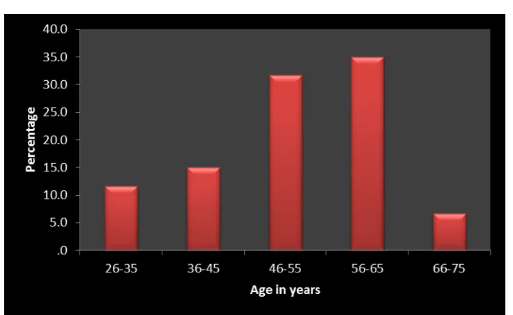
TABLE 1 - AGE DISTRIBUTION OF THE CASES Age group
of the cases in years
Frequency of cases in each age group
percentage
26-35 7 11.7
36-45 9 15.0
46-55 19 31.7
56-65 21 35.0
66-75 4 6.7
Total 60 100.0
AGE DISTRIBUTION
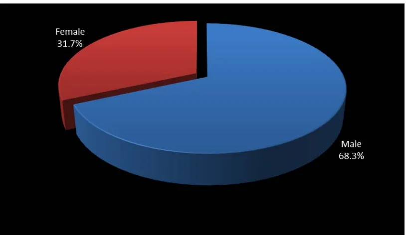
TABLE 2
SEX DISTRIBUTION OF THE CASES
Gender group of cases
Frequency of cases in each group
Percentage
Male 41 68.3 Female 19 31.7
Total 60 100.0
SEX DISTRIBUTION OF CASES
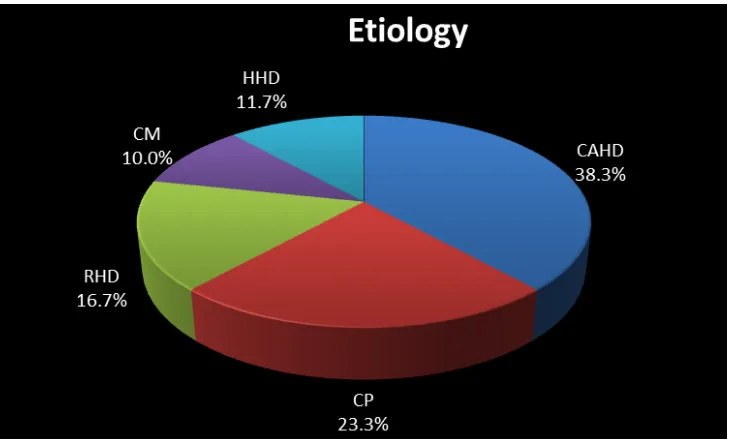
TABLE 3
ETIOLOGY OF CARDIAC FAILURE
Etiology of failure Frequency of cases Percentage Coronary artery disease 23 38.3
Cor Pulmonale 14 23.3
Rheumatic heart disease 10 16.7
Cardiomyopathy 6 10.0 Hypertensive heart disease 7 11.7
Total 60 100.0
ETIOLOGY OF CARDIAC FAILURE
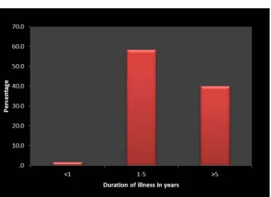
TABLE 5
DURATION OF CARDIAC FAILURE Duration of cardiac
failure
Frequency in each group
Percentage
<1 1 1.7
1-5 35 58.3
>5 24 40.0
Total 60 100.0
TABLE 4
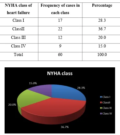
CASE DISTRIBUTION ACCORDING TO NEW YORK HEART ASSOCIATION CLASS OF HEART FAILURE
NYHA class of heart failure
Frequency of cases in each class
Percentage
Class I 17 28.3
ClassII 22 36.7
Class III 12 20.0
Class IV 9 15.0
Total 60 100.0
TABLE 6
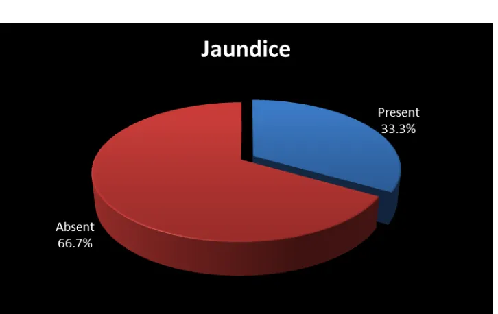
PRESENCE OF CLINICAL JAUNDICE IN THE CASES Presence of
jaundice Frequency Percentage
Present 20 33.3 Absent 40 66.7
Total 60 100.0
TABLE 7
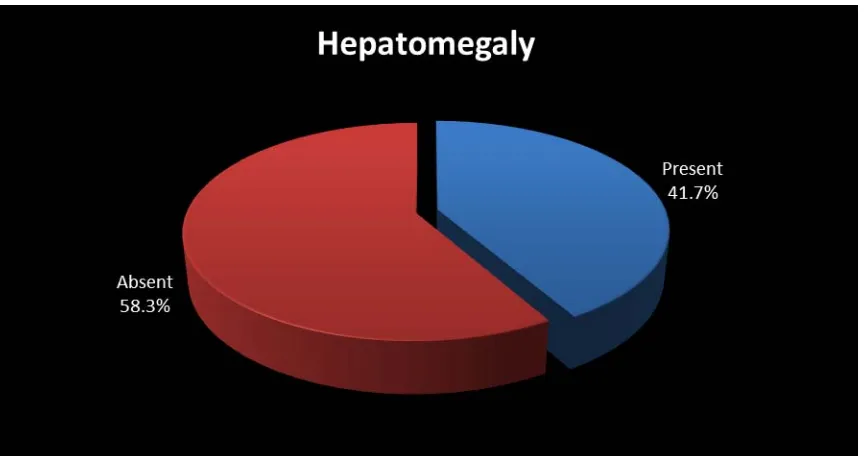
PRESENCE OF HEPATOMEGALY IN THE CASES Presence of
Hepatomegaly Frequency Percent
Present 25 41.7
Absent 35 58.3
Total 60 100.0
TABLE 8
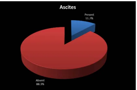
PRESENCE OF ASCITES IN THE CASES Presence of
Ascites Frequency Percent
Present 7 11.7 Absent 53 88.3
Total 60 100.0
TABLE 9
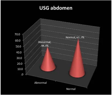
ULTRASOUND ABDOMEN SHOWING CONGESTIVE HEPATOMEGALY IN THE CASES
USG Abdomen with congestive liver
Frequency Percent
Present 23 38.3
Absent 37 61.7
Total 60 100.0
TABLE 10
LIVER BIOCHEMICAL ABNORMALITIES NOTED IN THE STUDY GROUP
Serum Bilirubin Frequency Percent
Abnormal 34 56.7 Normal 26 43.3
Total 60 100.0
LIVER PARAMETERS
Abnormal Normal FREQUENCY PERCENT FREQUENCY PERCENT
Serum
Bilirubin 34 56.7 26 43.3
Serum AST 32 53.3 28 46.7
Serum ALT 37 61.7 23 38.3
Serum ALP 8 13.3 52 86.7
Serum Total
Protein 12 20.0 48 80.0
Serum Albumin 23 38.3 37 61.7
Serum Globulin 4 6.7 56 93.3
Prothrombin
TABLE 11
PREVALENCE OF LIVER FUNCTION ABNORMALITY IN THE STUDY GROUP
LIVER FUNCTION
TESTS Frequency Percentage
Abnormal 34 56.7 Normal 26 43.3
TABLE 12
SHOWING COMPARISON OF MEAN BILIRUBIN VALUES WITH NYHA CLASS OF CARDIAC FAILURE
NYHA
CLASS FREQUENCY
Mean
BILIRUBIN S D F p value
Class I 17 1.1 .2
58.555 <0.001
ClassII 22 1.5 .4
Class III 12 2.3 .5
Class IV 9 2.9 .5
Total 60 1.7 .8
TABLE 13
SHOWING COMPARISON OF MEAN AST VALUES WITH NYHA CLASS OF HEART FAILURE
NYHA CLASS
FREQUENCY
Mean AST VALUES
S D F p value
Class I 17 36.9 3.5
5.171 0.003
ClassII 22 39.9 5.4
Class III 12 87.5 41.7 Class IV 9 188.8 271.8
Total 60 70.9 115.0
TABLE 14
SHOWING COMPARISON OF DIFFERENT LIVER PARAMETERS WITH NYHA CLASS OF HEART FAILURE Liver function tests Nyha
class
Frequency in each class
Mean
values S D F p value Serum ALT
Class I 17 32.4 4.0 5.933
.001
ClassII 22 34.9 6.0
Class III 12 81.0 34.5
Class IV 9 142.0 186.2
Total 60 59.5 80.7
Serum ALP
Class I 16 44.3 10.8 .336
.799
ClassII 22 39.5 15.2
Class III 12 42.0 18.6
Class IV 9 39.4 20.2
Total 59 41.3 15.5
Serum Total Protein
Class I 17 6.4 .2 20.476
.000
ClassII 22 6.3 .3
Class III 12 5.9 .2
Class IV 9 5.6 .4
Total 60 6.2 .4
Serum Albumin
Class I 17 3.5 .1 16.646
.000
ClassII 22 3.4 .2
Class III 12 3.1 .2
Class IV 9 2.9 .3
Total 60 3.3 .3
Prothrombin Time
Class I 17 13.8 .9 15.706 .000
ClassII 22 14.3 2.9
Class III 12 18.3 3.6
Class IV 9 20.2 3.5
Total 60 15.8 3.7
TABLE 15
COMPARISON OF SERUM BILIRUBIN WITH ETIOLOGY OF CARDIAC FAILURE
Etiology
SERUM BILIRUBIN Total
Abnormal Normal
Frequency Percent Frequency Percent Frequency Percent
CAHD 14 60.9 9 39.1 23 100.0 CP 8 57.1 6 42.9 14 100.0 RHD 7 70.0 3 30.0 10 100.0 CM 4 66.7 2 33.3 6 100.0 HHD 1 14.3 6 85.7 7 100.0 Total 34 56.7 26 43.3 60 100.0
χ2 =6.255 df =4 p=0.181 p value insignificant
TABLE 16
COMPARISON OF SERUM BILIRUBIN WITH DURATION OF CARDIAC FAILURE
Duration of illness
SERUM BILIRUBIN Total
Abnormal Normal
Frequency Percent Frequency Percent Frequency Percent
<1 0 .0 1 100.0 1 100.0
1-5 18 51.4 17 48.6 35 100.0
>5 16 66.7 8 33.3 24 100.0
Total 34 56.7 26 43.3 60 100.0
χ2 =2.676 df =2 p=0.262
p value is not significant
TABLE 17
SHOWING COMPARISON OF MEAN BILIRUBIN WITH OUTCOME OF CARDIAC FAILURE
Outcome Frequency Mean
Bilirubin S D T P value
Dead 4 2.95 .68 3.603 .001
Alive 56 1.65 .70
TABLE 18
SHOWING COMPARISON BETWEEN VALUES ON FIRST DAY OF STUDY AND SEVENTH DAY OF STUDY
LIVER FUNCTION
TESTS
DAY 1 DAY 7 McNemar
test P value Frequency Percent Frequency Percent
Serum Bilirubin 34 56.7 14 25.0 <0.001
Serum AST 32 53.3 16 28.6 0.002
Serum ALT 37 61.7 19 33.9 <0.001
Serum ALP 8 13.3 7 12.5 1.000
Serum TP 12 20.0 8 14.3 1.000
Serum Albumin 23 38.3 27 48.2 0.424
PROTHROMBIN
TIME 21 35.0 20 35.7 0.508
DISCUSSION
Large number of studies has been conducted in evaluating hepatic function in congestive heart failure .Umpteen number of studies are still going on in this arena of liver function.
In this study ,liver biochemical abnormalities and clinical features in cardiac failure because of differing etiologies in 60 patients were recorded ,analysed and compared within and correlated with numerous research papers.The principal intention of this study is to relate indian picture with international scenario.
AGE AND SEX DISTRIBUTION OF THE CASES
suggest that the occurrence of heart failure is nearly two percent of the adult population, with a sharp increase with age.
ETIOLOGY AND DURATION OF CARDIAC FAILURE
In this study out of the 60 patients studied 23 patients ( 38.3%) suffered from coronary artery disease, 14 patients from corpulmonale,10 from rheumatic heart disease,6 from cardiomyopathy,7 from hypertensive heart disease.In this study most common cause for heart failure was found to be coronary artery disease which is in correlation with the international scene. The incidence of rheumatic heart disease as the reason for heart failure has declined which is considered as a changing trend in the etiology of heart failure. Cowie et al and Fox et al has shown in there studies the solitary most common cause of heart failure in the developed world is coronary heart disease 1,5,44. Kannel et al , McMurray et al and levy et al in there studies suggest valvular heart disease and hypertension have come down as the principal reason for heart failure5.
Out of 60 subjects studied the duration of cardiac failure was greater than 5 years in 24 cases (40%),was in the range of 1- 5 years in 38 cases (58.3) and less than 1year in 1 case(1.7%).
NYHA CLASS
lower clases than higher classes which suggests improved quality of life with cardiac failure medications as reported by Luis et al.
JAUNDICE
Out of the 60 patients studied 20 subjects which is nearly 33% had jaundice. Other causes for jaundice like liver damaging drugs and alcohol related injury were excluded by taking proper history from the subjects. Serological tests were carried out to rule out viral causes for liver damage. Biegus et al has described jaundice clinically 33% of the cases. Similarly Kugel and Lichtman in 1933, Felder et al in 1950, chavez et al in 1943, Sherlock et al in 1951, Evans et al in 1952, Levine and Klatskin in 1964 has suggested in there papers that the serum bilirubin was frequently above the upper limit of normal range.
HEPATOMEGALY
ASCITES
Out of 60 subjects only seven persons showed clinically detectable ascites which is 11.7%. Norman et al demonstrated ascites and oedema in 15% of cases of cardiac failure studied12.
ULTRASOUND ABDOMEN
USG Abdomen was carried out in all the 60 subjects. 23 (38.3%) of them showed changes of congestive hepatomegaly in the abdominal scan.
HYPERBILIRUBINAEMIA
Felder et al has shown elevate bilirubin levels in 52% of the cases in his study 10. Similarly Wahi et al and Naresh bhu have reported hyperbilirubinaemia in 45% and 58% of subjects respectively. Kubo et al have reported that bilirubin values rarely increase more than 5mg% and are usually less than 3 mg%26. Sherlock et al have also reported similar findings with indirect fraction more than direct fraction in his study .Richman et al has reported that control of cardiac failure led to return of serum bilirubin values to basal levels in nearly one week which correlates with the findings in this study16.
In this study it was noted that as the NYHA Class of heart failure advanced the mean serum bilirubin level also progressively increased which suggests higher classes of heart failure were associated with higher degree of liver dysfunction This was supported by Luiz et al in their study43.
SERUM AMINOTRANSFERASES.
progressively as the NYHA class of cardiac failure progresses. Serum AST levels showed significant elevation in the class IV heart failure which correlates with the observations of the Luiz et al and walter et al. With control of cardiac failure more than 50% values came to the baseline values.
SERUM ALKALINE PHOSPHATASE
In this study out of the 60 subjects studied eight patients showed abnormal serum ALP (13.3%). Felder et al observed elevated serum ALP in ten – twenty percent of subjects studied10. Sherlock and Richman has recorded similar observations in their studies12. Rise in serum ALP does not relate with the elevation in serum transaminases and bilirubin. ALP values return to basal level with control of failure symptoms. Serum ALP values showed no correlation with the NYHA Classes of cardiac failure. It was observed that mean ALP levels showed no progressive change with worsening of heart failure.
In the present study serum AST and ALT showed marked elevation than serum ALP thus suggesting a predominant hepatocellular pattern of hepatic damage.
SERUM PROTEINS
serum albumin levels showed a progressive fall with class IV patients showing the lowest mean serum albumin value with a significant p value. Richman et al in his study has reported decreased albumin in thirty – fifty percent of cases.Mild decrease in albumin levels were observed with values ranging from 2.5g/dl to 2.9g/dl.it was observed by Dunn et al that cases with marked fluid retention showed albumin values less than 1.5g/dl24. Serum albumin values usually return to normal in a period of few months following control of cardiac failure.
Elevated globulin values were noted in 35-50% of subjects with right heart failure by Richman et al. A mild rise was noted in most of the cases ranging from 3.5 -4.1g/dl. A rise in globulins and fall in albumin causes reversal of Albumin globulin ratio.It was found that unlike other parameters globulin values did not return to normal value following control of cardiac failure.
PROTHROMBIN TIME
nearly 85% of the subjects.it was reprted that prothrombin time come to baseline values usually two – three weeks after control of cardiac failure.
OUTCOME
Out of 60 cases studied 4 cases succumbed to the cardiac illness during the course of illness .The mean bilirubin value of these cases were 2.95 which was more when compared to other subjects which was 1.65 with a p value of .001 which is significant suggesting that elevated bilirubin levels can be taken as a bad prognostic marker in congestive cardiac failure.
CONCLUSIONS
1. The most common cause of congestive cardiac failure in patients presenting to Tirunelveli Govt Medical College Hospital was found to be Coronary artery disease.
2. Elderly Male population were found to have an increased incidence of cardiac failure when compared to the opposite gender.
3. Liver function abnormalities were found in 56.7% of the total congestive cardiac failure patients included in this study.
4. Liver function abnormalities did not show any correlation with the aetiology and duration of cardiac failure though subjects with rheumatic heart disease showed a mild increase in incidence of abnormal liver function. Least incidence of liver function abnormalities were found in Hypertensive heart disease patients.
5. Serum AST was found to be abnormal in 53.3% of the subjects and serum ALT was abnormal in 61.7% of the cases whereas serum ALP was found to be abnormal only in 13.3% of cases.This suggest a predominant hepatocellular pattern of liver injury than cholestatic pattern.