LIVER FUNCTION TESTS
IN
CONGESTIVE CARDIAC FAILURE
Submitted to
THE TAMILNADU DR. M.G.R. MEDICAL UNIVERSITY
CHENNAI
In fulfillment of the regulations for the award of the degree of
M.D. (GENERAL MEDICINE)
BRANCH - I
KILPAUK MEDICAL COLLEGE
CHENNAI.
BONAFIDE CERTIFICATE
Certified that the dissertation titled “LIVER FUNCTION TESTS IN
CONGESTIVE CARDIAC FAILURE” is a bonafide work of the
candidate Dr.JAIGANESH.M, post graduate student, Department of
General Medicine, Kilpauk Medical College, Chennai – 10, done under my
guidance and supervision, in partial fulfillment of regulations of The
Tamilnadu Dr. MGR Medical University for the award of M.D. Degree
Branch I, (General Medicine) during the academic period from May 2007 to
March 2010.
Prof.G.Rajendran.M.D., Prof.N.Raghu M.D.,
Prof. & HOD Prof. of medicine and chief Department of General Medicine Department of General Medicine
Kilpauk Medical College, Chennai. Kilpauk Medical College, Chennai.
Prof. V.Kanagasabai, The Dean
Kilpauk Medical College
ACKNOWLEDGEMENT
I thank Prof. Dr. V. Kanagasabai, M.D., The Dean, Kilpauk Medical
College for kindly permitting me to use the resources and clinical material of
this hospital.
I am grateful to Prof. Dr. G. Rajendran, M.D., Professor and Head
of the Department of General Medicine, Govt. Royapettah Hospital for his
constant motivation and guidance for this study.
I profusely thank Prof. Dr. N. Raghu M. D., my beloved chief,
professor of medicine; Govt. Royapettah Hospital, for instilling ideas of this
study, guiding, supporting and rendering immense knowledge on preparing
this study.
I am grateful to Prof. Dr. N.Senguttuvan M.D.,D.M;(Cardiology)
Professor and H.O.D, Department of cardiology, Govt. Royapettah hospital,
Prof. Dr. Usha Srinivas M.D.; D.M., (Gastroenterology); Professor and
HOD, Dept Of Medical Gastroenterology; Prof. Dr.Gunasekaran M. D.,
professor of medicine for their guidance and support during the course of the
I am thankful to Dr. Shaik sulaiman; M.D., Dr. Govindarajulu;
M.D., Dr. Swarnalatha; M.D., my assistant professors for the support
extended to me during the course of the study.
I am indebted to the Assistant Professors in the Department of
Cardiology Govt. Royapettah Hospital, Chennai, Dr. Jayaprakash M.D ,
Dr. Manickam M.D, for the advise and help rendered to me.
I thank Mr. Siluvai, statistician for his valuable time spent in
analyzing the data and providing statistical support. I also thank my fellow
post graduate students and house surgeons for all the timely help they
rendered.
Last but not the least, with sincere gratitude; I thank all the patients
CONTENTS
SI.NO Title page
1. AIM OF STUDY 1
2. INTRODUCTION 2
3. REVIEW OF LITERATURE 4
4. MATERIALS AND METHODS 45
5. RESULTS 51
6. DISCUSSION 62
7. SUMMARY 69
8. CONCLUSION 72
9. PROFORMA i
10. MASTER CHART v
LIST OF ABBREVATIONS
1.
LFT Liver function tests
2.
AST/ SGOT Aspartate aminotransferase -serum
glutamic oxaloacetic transaminase
3.
ALT/SGPT Alanine aminotransferase-serum glutamic
pyruvate transaminase
4.
CPC chronic passive congestion
5.
CLN centrilobular necrosis
6.
CHC central hemorrhagic necrosis
7.
RHD Rheumatic heart disease
8.
CAHD Coronary atherosclerotic heart disease
AIMS OF THE STUDY
1. To study the liver function tests in congestive cardiac failure.
2. To observe any differences in liver function tests with the etiology of
congestive cardiac failure.
3. To study the relationship between liver function test and remission and
exacerbation of congestive cardiac failure.
4. To study whether liver function tests can be used as a prognostic
INTRODUCTION
The liver has been called the custodian of milieu interior [5]. So
any liver disorders will have far reaching consequences on body’s
homeostasis. Also, numerous pathologies of other systems can affect liver.
Both acute and chronic heart failure may result in abnormalities of liver.
Liver receives 25% of cardiac output, that a fall in cardiac output will result in
hepatic hypoperfusion. Liver has the capacity to withstand changes in blood
flow by vasoactive mechanisms and oxygen extraction from blood. However
when critical levels are reached, hepatic injury ensues[16].
Both Right and left sided heart failure can result in liver injury. In right
-sided heart failure, elevation of right heart pressure resulting in raised pressure
in hepatic sinusoids, hepatic congestion and liver cell hypoxia. In left sided
heart failure, decreased cardiac output results in hepatic hypo perfusion and
hypoxia. The common pathway is centrilobular hepatocellular necrosis. Zone 3
of the liver lobule is most vulnerable to hypoxic injury due organization of
hepatic blood flow.
In this study, the effects of congestive cardiac failure on liver and its
function is analyzed in 75 patients compared with 20 healthy individuals.
compared, based on their effects on liver functions. Remissions and
exacerbations have been tracked on the 7th day and the variations of liver
function have been recorded and an attempt has been made to find whether liver
function tests can be used as prognostic indicators of congestive cardiac failure.
Understanding anatomy and functions of liver, liver enzymes, causes and
various forms of congestive cardiac failure and their manifestations, pathology
of liver in heart failure are needed before evaluating abnormalities of liver
REVIEW OF LITERATURE
ANATOMY AND FUNTIONS OF LIVER
Liver is the largest organ of the body weighing around 1.5kg.
The size and shape of the liver vary and generally match the general body
shape—long and lean or squat and square. The liver is located in the right upper
quadrant of the abdomen under the right lower rib cage against the diaphragm
and projects for a variable extent into the left upper quadrant. The liver is held
in place by ligamentous attachments to the diaphragm, peritoneum, great
vessels, and upper gastrointestinal organs.
The liver has dual blood supply. The portal vein brings blood
from the gut and hepatic artery supplies arterial blood. Hepatic vein drains the
liver into inferior vena cava. Porta hepatis is fissure through which vessels enter
liver where, Portal vein and hepatic artery divides into branches to right and left
lobes. Similarly right and left hepatic ducts join here to form common hepatic
duct. Hepatic nerve plexus derives its supply from sympathetic chain T7-T11,
left and right vagus nerve and right phrenic nerve. Lymphatic drainage is into
small group of glands near porta hepatis. Liver receives 1500ml of blood per
FUNCTIONAL ANATOMY:
The majority of cells in the liver are hepatocytes, which
constitute two-thirds of the mass of the liver. The remaining cell types are
Kupffer cells (reticuloendothelial cells), stellate (Ito or fat-storing) cells,
endothelial cells and blood vessels, bile ductular cells, and supporting structures.
In light microscopy, the liver appears to be organized in lobules, with portal areas
at the periphery and central veins in the center of each lobule.
However, from a functional point of view, the liver is
organized into acini, with both hepatic arterial and portal venous blood entering
the acinus from the portal areas (zone 1) and then flowing through the sinusoids
to the terminal hepatic veins (zone 3); the intervening hepatocytes constitute zone
The advantage of viewing the acinus as the physiologic unit of the liver is that it
helps to explain the morphologic patterns and zonality of many vascular and
biliary diseases not explained by the lobular arrangement.
Portal areas of the liver consist of small veins, arteries, bile
ducts, and lymphatics organized in a loose stroma of supporting matrix and small
amounts of collagen. Blood flowing into the portal areas is distributed through
the sinusoids, passing from zone 1 to zone 3 of the acinus and draining into the
terminal hepatic veins ("central veins"). Hence zone 3 of the liver is vulnerable to
hypoxic injury. Secreted bile flows in the opposite direction, in a countercurrent
pattern from zone 3 to zone 1. The sinusoids are lined by unique endothelial cells
but not cellular elements. The plasma is thus in direct contact with hepatocytes in
the space of Disse.
Hepatocytes have distinct polarity. The basolateral side of the
hepatocyte lines the space of Disse and is richly lined with microvilli; it
demonstrates endocytotic and pinocytotic activity, with passive and active uptake
of nutrients, proteins, and other molecules. The apical pole of the hepatocyte
forms the cannicular membranes through which bile components are secreted.
The canniculi of hepatocytes form a fine network, which fuses into the bile
Kupffer cells usually lie within the sinusoidal vascular space
and represent the largest group of fixed macrophages in the body. The stellate
cells are located in the space of Disse but are not usually prominent unless
activated, when they produce collagen and matrix. White blood cells can migrate
through or around endothelial cells into the space of Disse and from there to
portal areas, where they can return to the circulation through lymphatics.
1. hepatic artery 2.bile duct 4. portal vein.
The relative functions of cells in the periphery of acini, zone 3
are different from those of cells in zone 1(periportal). Kreb’s cycle enzymes are
found highest in Zone 1. Glutamic synthetase is found in perivenous cells. Zone3
is vulnerable to hypoxic insult. Congestive cardiac failure affects zone 3 of the
liver resulting in characteristic features.
FUNCTIONS OF THE LIVER:
Hepatocytes perform numerous and vital roles in
maintaining homeostasis and health.
Synthetic functions:
These functions include the synthesis of most essential
serum proteins (albumin, carrier proteins, coagulation factors 2,7,9 and 10 , many
hormonal and growth factors), the production of bile and its carriers (bile acids,
cholesterol, lecithin, phospholipids).
Metabolism:
The regulation of nutrients (glucose, glycogen, lipids,
cholesterol, amino acids) such as uptake of glucose after a meal, aminoacids used
for synthetic functions, production of very low density lipoproteins, metabolism
of high and low density lipoproteins and conjugation of lipophilic compounds
Excretory functions:
Liver metabolises bilirubin, bile salts, many drugs and
alcohol
Storage functions:
Reticulo-endothelial function:
Liver acts as a microbe barrier for the portal venous blood
from the gut through kupffer and other cells. It also detoxifies toxins
Heat production:
Liver carries out various anabolic and catabolic reactions.
One of by- products of these reactions is heat, helping in thermostasis.
LIVER FUNCTION TESTS:
Measurement of these activities to assess liver function is
complicated by the multiplicity and variability of these functions. The most
commonly used liver "function" tests are measurements of serum bilirubin,
albumin, and prothrombin time.
The serum bilirubin level is a measure of hepatic
conjugation and excretion, and the serum albumin level and prothrombin time are
measures of protein synthesis. Abnormalities of bilirubin, albumin, and
prothrombin time are typical of hepatic dysfunction. Frank liver failure is
incompatible with life, and the functions of the liver are too complex and diverse
to be sub served by a mechanical pump; dialysis membrane; or concoction of
Tests Based on Detoxification and ExcretoryFunctions
1. Serum Bilirubin, a breakdown product of the porphyrin ring of
heme-containing proteins, is found in the blood in two fractions—conjugated and
unconjugated. The unconjugated fraction, also termed the indirect fraction, is
insoluble in water and is bound to albumin in the blood. The conjugated (direct)
bilirubin fraction is water soluble and can therefore be excreted by the kidney.
When measured by the original van den Bergh method, the normal total serum
bilirubin concentration is <17 micro mol/L (1 mg/dL). Up to 30%, or 5.1 micro
mol/L (0.3 mg/dL), of the total is direct-reacting (or conjugated) bilirubin.
Elevation of the unconjugated fraction of bilirubin is rarely due to liver
disease. An isolated elevation of unconjugated bilirubin is seen primarily in
hemolytic disorders and in a number of genetic conditions such as Crigler-Najjar
and Gilbert's syndromes .Isolated unconjugated hyperbilirubinemia (bilirubin
elevated but <15% direct) should prompt a workup for hemolysis In the absence
of hemolysis.
In contrast, conjugated hyperbilirubinemia almost always implies liver
or biliary tract disease. The rate-limiting step in bilirubin metabolism is not
conjugation of bilirubin, but rather the transport of conjugated bilirubin into the
type of liver disease. In most liver diseases, both conjugated and unconjugated
fractions of the bilirubin tend to be elevated.
2. Serum Enzymes
The liver contains thousands of enzymes, some of which are also present in the
serum in very low concentrations. These enzymes have no known function in the
serum and behave like other serum proteins. They are distributed in the plasma
and in interstitial fluid and have characteristic half-lives, usually measured in
days. Very little is known about the catabolism of serum enzymes, although they
are probably cleared by cells in the reticuloendothelial system. The elevation of a
given enzyme activity in the serum is thought to primarily reflect its increased
rate of entrance into serum from damaged liver cells.
Serum enzyme tests can be grouped into three categories: (1) enzymes whose
elevation in serum reflects damage to hepatocytes, (2) enzymes whose elevation
in serum reflects cholestasis, and (3) enzyme tests that do not fit precisely into
either pattern.
A. Enzymes that Reflect Damage to Hepatocytes
The aminotransferases (transaminases) are sensitive indicators of liver cell injury
and are most helpful in recognizing acute hepatocellular diseases such as
hepatitis. They include the aspartate aminotransferase (AST) and the alanine
muscle, kidneys, brain, pancreas, lungs, leukocytes, and erythrocytes in
decreasing order of concentration. ALT is found primarily in the liver. The
aminotransferases are normally present in the serum in low concentrations. These
enzymes are released into the blood in greater amounts when there is damage to
the liver cell membrane resulting in increased permeability. Liver cell necrosis is
not required for the release of the aminotransferases, and there is a poor
correlation between the degree of liver cell damage and the level of the
aminotransferases. Thus, the absolute elevation of the aminotransferases is of no
prognostic significance in acute hepatocellular disorders.
Any type of liver cell injury can cause modest elevations in the serum
aminotransferases. Levels of up to 300 U/L are nonspecific and may be found in
any type of liver disorder. Minimal ALT elevations in asymptomatic blood
donors rarely indicate severe liver disease; studies have shown that fatty liver
disease is the most likely explanation. Striking elevations—i.e.,
aminotransferases > 1000 U/L—occur almost exclusively in disorders associated
with extensive hepatocellular injury such as (1) viral hepatitis, (2) ischemic liver
injury (prolonged hypotension or acute heart failure), or (3) toxin- or
drug-induced liver injury.
The pattern of the aminotransferase elevation can be helpful diagnostically. In
most acute hepatocellular disorders, the ALT is higher than or equal to the AST.
alcoholic liver disease. The AST in alcoholic liver disease is rarely >300 U/L and
the ALT is often normal. A low level of ALT in the serum is due to an
alcohol-induced deficiency of pyridoxal phosphate.
The aminotransferases are usually not greatly elevated in obstructive jaundice.
One notable exception occurs during the acute phase of biliary obstruction caused
by the passage of a gallstone into the common bile duct. In this setting, the
aminotransferases can briefly be in the 1000–2000 U/L range. However,
aminotransferase levels decrease quickly, and the liver function tests rapidly
evolve into one typical of cholestasis.
B. Enzymes that Reflect Cholestasis
The activities of three enzymes—alkaline phosphatase, 5'nucleotidase, and
-glutamyl transpeptidase (GGT)—are usually elevated in cholestasis.
Elevation of liver-derived alkaline phosphatase is not totally specific for
cholestasis, and a less than threefold elevation can be seen in almost any type of
liver disease. Alkaline phosphatase elevations greater than four times normal
occur primarily in patients with cholestatic liver disorders, infiltrative liver
diseases such as cancer and amyloidosis, and bone conditions characterized by
rapid bone turnover (e.g., Paget's disease). In bone diseases, the elevation is due
to increased amounts of the bone isoenzymes. In liver diseases, the elevation is
If an elevated serum alkaline phosphatase is the only abnormal finding in an
apparently healthy person, or if the degree of elevation is higher than expected in
the clinical setting, identification of the source of elevated isoenzymes is helpful.
This problem can be approached in several ways. First, and most precise, is the
fractionation of the alkaline phosphatase by electrophoresis.
The second approach is based on the observation that alkaline
phosphatases from individual tissues differ in susceptibility to inactivation by
heat. The finding of an elevated serum alkaline phosphatase level in a patient
with a heat-stable fraction strongly suggests that the placenta or a tumor is the
source of the elevated enzyme in serum. Susceptibility to inactivation by heat
increases, respectively, for the intestinal, liver, and bone alkaline phosphatases,
bone being by far the most sensitive
Tests that Measure Biosynthetic Function of the Liver
A. Serum Albumin: Serum albumin is synthesized exclusively by
hepatocytes. Serum albumin has a long half-life: 18–20 days, with ~4% degraded
per day. Because of this slow turnover, the serum albumin is not a good indicator
of acute or mild hepatic dysfunction; only minimal changes in the serum albumin
are seen in acute liver conditions such as viral hepatitis, drug-related hepatoxicity,
and obstructive jaundice. In hepatitis, albumin levels < 3 g/dL should raise the
chronic liver disorders such as cirrhosis and usually reflects severe liver damage
and decreased albumin synthesis. One exception is the patient with ascites in
whom synthesis may be normal or even increased, but levels are low because of
the increased volume of distribution. However, hypoalbuminemia is not specific
for liver disease and may occur in protein malnutrition of any cause, as well as
protein-losing enteropathies, nephrotic syndrome, and chronic infections that are
associated with prolonged increases in levels of serum interleukin 1 and/or tumor
necrosis factor, cytokines that inhibit albumin synthesis. Serum albumin should
not be measured for screening in patients in whom there is no suspicion of liver
disease.
B. Serum Globulins
Serum globulins are a group of proteins made up of Gamma globulins
(immunoglobulins) produced by B lymphocytes and Alpha and Beta globulins
produced primarily in hepatocytes. Gamma Globulins are increased in chronic
liver disease, such as chronic hepatitis and cirrhosis. In cirrhosis, the increased
serum gamma globulin concentration is due to the increased synthesis of
antibodies, some of which are directed against intestinal bacteria. This occurs
because the cirrhotic liver fails to clear bacterial antigens that normally reach the
Increases in the concentration of specific isotypes of Gamma globulins are often
helpful in the recognition of certain chronic liver diseases. Diffuse polyclonal
increases in IgG levels are common in autoimmune hepatitis; increases >100%
should alert the clinician to this possibility. Increases in the IgM levels are
common in primary biliary cirrhosis, while increases in the IgA levels occur in
alcoholic liver disease.
C. Coagulation Factors
With the exception of factor VIII, the blood clotting factors are made exclusively
in hepatocytes. Their serum half-lives are much shorter than albumin, ranging
from 6 h for factor VII to 5 days for fibrinogen. Because of their rapid turnover,
measurement of the clotting factors is the single best acute measure of hepatic
synthetic function and helpful in both the diagnosis and assessing the prognosis
of acute parenchymal liver disease. Useful for this purpose is the serum
prothrombin time, which collectively measures factors II, V, VII, and X.
Biosynthesis of factors II, VII, IX, and X depends on vitamin K. The
prothrombin time may be elevated in hepatitis and cirrhosis as well as in
disorders that lead to vitamin K deficiency such as obstructive jaundice or fat
CONGESTIVE CARDIAC FAILURE
Congestive cardiac failure is defined as inability of heart to
maintain an output, at rest or during exercise, necessary for the metabolic needs
of the body and inability to receive blood into ventricular cavity at low pressure
during diastole.[18]
In our study, patients were included as having congestive cardiac
failure, if they had at least one Major and Minor criteria of Framingham
criteria. [19]
FRAMINGHAM CRITERIA
MAJOR
1. Paroxysmal nocturnal Dyspnea
2. Neck vein distension
3. Crackles
4. Cardiomegaly
5. Acute pulmonary edema
6. S3 gallop
7. Increased venous pressure (16cm H20)
MINOR
1. Extremity edema
2. Night cough
3. Dyspnea on exertion
4. Hepatomegaly
5. Pleural effusion
6. Vital capacity reduced by one third to normal
7. Tachycardia > 120/ min
8. Weight loss over 4.5kg over 5 days treatment.
Heart failure may be described as systolic, diastolic, high
output low output, acute or chronic and right or left sided. These separations blur
in the course of the disease.
CAUSES OF CONGESTIVE CARDIAC FAILURE:[20]
Depressed Ejection Fraction (<40%)
Coronary artery disease
Nonischemic dilated cardiomyopathy
Myocardial infarction
Toxic/drug-induced damage
Myocardial ischemia
Infiltrative disorders
Hypertension
Metabolic disorder
Viral
Obstructive valvular disease
Chronic volume overload
Chagas' disease
Disorders of rate and rhythm
Intracardiac (left-to-right) shunting
Chronic bradyarrhythmias
Preserved Ejection Fraction (>40–50%)
Pathological hypertrophy
Restrictive cardiomyopathy
Primary(hypertrophic cardiomyopathy)
Infiltrative disorders (amyloidosis, sarcoidosis)
Secondary (hypertension)
Fibrosis
Aging
Storage diseases (hemochromatosis)
Pulmonary Heart Disease
Pulmonary vascular disorders
High-Output States
Metabolic disorders
Excessive blood-flow requirements
Nutritional disorders (beriberi)
Chronic anemia
Systemic arteriovenous shunting
Thyrotoxicosis
Precipitating factors:
In evaluating heart failure, it is also important to identify
precipitating factors. [21]
1. Anemia
2. Pregnancy
3. Infections
4. Thyrotoxicosis
5. Arrythmia
6. Infective endocarditis
7. systemic hypertension
Forms of heart failure:
Systolic Versus Diastolic Failure
This classification relates to whether the principal
abnormality is the inability to contract normally and expel sufficient blood
(systolic failure) or to relax and fill normally (diastolic failure). The major
clinical manifestations of systolic failure relate to an inadequate cardiac output
with weakness, fatigue, reduced exercise tolerance and other symptoms of
hypoperfusion, while in diastolic failure they relate principally to an elevation of
filling pressures. In many patients, abnormalities of contraction and relaxation
coexist.
Diastolic heart failure may be caused by increased resistance
to ventricular inflow and reduced ventricular diastolic capacity (constrictive
pericarditis and restrictive, hypertensive, and hypertrophic cardiomyopathy),
impaired ventricular relaxation (acute myocardial ischemia, hypertrophic
cardiomyopathy), and myocardial fibrosis and infiltration.
High Output Versus Low Output Heart Failure
Low output heart failure occurs secondary to ischemic heart
disease, hypertension, dilated cardiomyopathy, and valvular and pericardial
disease. High output heart failure occurs in hyperthyroidism, anemia, pregnancy,
Acute Versus Chronic Heart Failure
The prototype of acute heart failure is the patient who is
entirely well but who suddenly develops a large myocardial infarction or rupture
of a cardiac valve. Chronic heart failure is typically observed in patients with
dilated cardiomyopathy or multivalvular heart disease that develops or progresses
slowly. Acute heart failure is usually largely systolic and the sudden reduction in
cardiac output often results in systemic hypotension without peripheral edema. In
chronic heart failure, arterial pressure tends to be well maintained until very late
in the course, but there is often accumulation of peripheral edema.
Right Sided Versus Left Sided Heart Failure
Patients in whom the left ventricle is mechanically
overloaded (e.g., aortic stenosis) or weakened (e.g., post myocardial infarction)
develop dyspnea and orthopnea as a result of pulmonary congestion, a condition
referred to as left sided heart failure. In contrast, when the underlying
abnormality affects the right ventricle primarily (e.g., pulmonic stenosis or
pulmonary hypertension), symptoms resulting from pulmonary congestion are
less common, and edema, congestive hepatomegaly, and systemic venous
distention, are more prominent. However, when heart failure has existed for
months or years, biventricular failure usually results. For example, patients with
edema, congestive hepatomegaly, and systemic venous distention late in the
course of their disease.
Backward Versus Forward Heart Failure
The concept of backward heart failure contends that in heart
failure, one or the other ventricle fails to discharge its contents or fails to fill
normally. As a consequence, the pressures in the atrium and venous system
behind the failing ventricle rise, and retention of sodium and water occurs as a
consequence of the elevation of systemic venous and capillary pressures and the
resultant transudation of fluids into the interstitial space. In forward failure, salt
and water retention is a consequence of diminished renal perfusion and excessive
proximal tubular sodium re-absorption and of excessive distal tubular re-
absorption through activation of the renin-angiotensin-aldosterone system.
Irrespective of the type of failure, Congestive heart failure affects liver
structurally and various functions of liver.
In this study, changes in liver function tests in various causes of congestive
cardiac failure such as rheumatic, congenital, ischemic, cardiomyopathic, cor
pulmonale and hypertensive heart diseases are noted in 75 patients presenting
with heart failure and compared with themselves and a control of 20 apparently
are performed again and compared with admission values, to note whether
remission or exacerbation has any impact on LFTs.
Review of other Studies and journals:
One of the most common manifestations of congestive heart failure is
enlargement of the liver. This fact has led several investigators such as Jollife;
Robertson [1] [2] [3] [4] to study liver function tests in an attempt to evaluate
hepatic dysfunction in congestive heart failure.
Historically, the first association of liver pathology and congestive
heart failure was noted by Kiernan [6] who described the "nutmeg liver." Seventy
eight years later Mallory [7] described the typical microscopic appearance of
central congestion with focal necrosis. Other authors pointed out the fatty
changes [8] and the compression of capillaries by edema fluid accumulated
between the liver cell cords and capillaries [9] [10].
The three main theories of the pathogenesis of the altered liver
anatomy are: infection,[7]. mechanical compression [11] [12] and hypoxia
with secondary nutritional deficiency [13] [14] [15] The deficiency in oxygen
supply to liver cells in heart failure seems to be due not only to the slowing of
blood flow through the liver but even more so to arterial unsaturation resulting
from pulmonary lesions. This was stressed by Rich [14] and others [13] [22] [15]
who pointed out that the jaundice of heart failure is especially apt to develop
removal rates by means of hepatic vein catheterization, and the studies of blood
flow to other organs in congestive heart failure'5' 16, 67, 68, 71, 97 make it
reasonable to assume that there is a decrease in blood flow to the liver in the
presence of cardiac decompensation.
From the Medical Division of the Montefiore Hospital, there is other
evidence for impaired liver function in congestive heart failure. Hepatic fibrosis
has been found three times as great in patients with heart failure as in patients
without heart failure.[24] [35] [26]. More significantly, central fibrosis so
common in decompensated cardiac patients, did not occur at all in patients who
were not in heart failure.
Hyperbilirubinemia has been found in patients who were in severe
chronic heart failure. Jolliffe[37] and Cantarow[6] noted that bromsulfalein was
not cleared from the blood at a normal rate in the presence of heart failure. Blood
cholesterol levels do not seem to be altered by circulatory embarrassment.[40]
Gravin [25] in 1943 observed 35 cases of cardiac cirrhosis in 790 autopsy cases
in whom heart failure was chief cause of death.The consensus is that there is a
severe aberration in liver function in association with a failing heart.
S.Sherlock[43]described the clinical and biochemical features of zone
3 necrosis in patients with heart failure.
Killip T. Payne in 1960 [ 38] described massive elevation of serum
Mechanism of liver injury in congestive cardiac failure:
Congestive heart failure causes:
1. Decreased hepatic blood flow
2. Increased hepatic venous pressure leading to edema of the sinusoids and
atrophy of hepatocytes.
3. Decreased arterial saturation which could result in hepatocellular
hypoxia.[39]
Single most important factor is Hypoxia of liver cells resulting in centrilobular
necrosis (zone3).[44] Also ,in heart failure passive congestion of liver results due
to increased venous pressure transmitted to small hepatic veins that drain
Acini[42].
Microscopic studies reveled increased pressure can cause atrophy zone 3
hepatocytes.[45]. In addition, elevated venous pressure results in sinusoidal
congestion enlargement of sinusoidal fenestrae , through which protein rich fluid
then enters into the space of disse. [50]. The resulting perisinusoidal edema may
impair diffusion of oxygen and nutrients into the hepatocytes.[45]
Excess fluid in the space of disse normally drains into hepatic lymphatics, but
when lymph formation exceeds the capacity of the lymphatics, high protein fluid
exudes from the surface of the liver into the peritoneal cavity and the typical high
In the chronic hepatic congestion, fibrosis may develop in perivenular area ,zone
3 and space of disse and further impair the diffusion of oxygen and nutrients from
blood to hepatocytes. The fibrosis varies from one region of the liver to the other.
This variability may be partly explained by the fibrogenic effects of focal thrombi
within the sinusoidal hepatic venules and portal veins that are a consequence of
chronic vascular stasis [47].
Passive congestion alone does not seem to be sufficient to cause
significant hepatic necrosis, decreased hepatic blood flow is also necessary [42].
This conclusion is supported by the observation of S.Sherlock [43], of the lack of
correlation between right atrial pressure and the degree of zone 3 necrosis in
patients with congestive heart failure. Moreover, significant zone 3 necrosis
caused by acute left sided heart failure has been documented in the complete
absence of right sided heart failure, a finding that suggests cardiac output is the
principal determinant of hepatic ischemia [47]. However passive congestion
seems to be an important co factor in most cases of clinical apparent hepatic
ischemia. In a recent study all patients with the clinical diagnosis of ischemic
hepatitis were found to have clinically significant cardiac disease and 94% have
right sided heart failure [ 48].
In another study, ischemic hepatitis in patients with chronic
with ischemic hepatitis had a central venous pressure above 10 cmH20.[49].
These findings suggests that chronic passive congestion major role in sensitizing
the liver to hemodynamic and hypoxic insults
Hepatic ischemia:
Hepatic ischemia like ischemia involving other organs results from the
imbalance between hepatic oxygen supply and demand. Because of the metabolic
rate of the liver is relatively constant, oxygen supply, not demand is principal
determinant of hepatic ischemia [52]. Hepatic oxygen delivery is the function of
both the oxygen content of the blood and total hepatic blood flow.
Systemic hypoxia alone is usually insufficient to cause significant
Hepatic injury [53][54],but severe hypoxemia was reported to cause hepatic
injury in a patient with obstructive sleep apnea[55]
And in a series of patients with exacerbation of chronic respiratory failure usually,
in the setting of passive hepatic congestion resulting from right sided heart
failure.[49].
Most critical factor in hepatic ischemia is total hepatic blood flow.
Numerous studies have shown that hepatic blood flow is determined primarily by
cardiac output and that the splanchnic bed receives relatively constant 25% of the
of the blood flow from the hepatic artery and 66 – 83 % via the low pressure
portal vein, in which the oxygen saturation is relatively high [57] [58], thus
much of its oxygen supply from the portal vein.
If patients cannot increasse their cardiac output there is a fall in
splanchnic blood flow with exercise and a reduced hepatic blood flow, the
oxygen requirement of the liver does not change and the oxygen extraction
increases[56] [59]. In the resting state, increased extraction of oxygen by
hepatocytes compensates for the reduction of hepatic blood flow. However, under
situations of systemic stress, such as sepsis or increased physical activity in
which the cardiac output cannot increased to supply extra hepatic metabolic
demands and the hepatic and splanchnic perfusion falls. Hepatic oxygen
extraction is often unable to increase above an already elevated baseline, and
hepatocellular hypoxia results, especially in zone3 of the hepatic acini.[51].
Data from in vitro and animal models suggest that because of their high
metabolic rate, hepatocytes are vulnerable to hypoxia at physiological
temperatures than other cell types in liver such as kupffer cells, sinusoidal
endothelial cells and biliary epithelial cells.[60]
Furthermore, nutritional status has an important influence on hepatic
hepatic ischemia, possibly because their diminished glycogen stores provides less
substrate for anaerobic glycolysis, so that the intra cellular production of lactic
acid is decreased.[61]. The cellular mechanism of ischemic injury, which occurs
when hepatocytes injured appears to be related to a disruption of mitochondrial
respiration, depletion of adenosine triphosphate rise in levels of intracellular
calcium and activation of cellular proteases.[62] [66]
Further contributing hepatic damage under these conditions is reperfusion
injury which occurs when hepatocytes injured by ischemia are exposed to oxygen.
Reperfusion injury is mediated by generation of free radicals, causing injury
through lipid peroxidation.[60]
Within injured hepatocytes, mitochondria generate toxic oxygen species
from reduced electron carriers. In addition, ischemia promotes the conversion of
cytosolic xanthine dehydrogenase to xanthine oxidase which results in turn in
production of superoxide and hydrogen peroxide from accumulated xanthine.[63].
In addition to the direct toxic effects of reactive oxygen species, ischemia and
reperfusion induce the transcription of multiple genes in the hepatocytes via
transcription factors, heat shock factor and nuclear factors, the product of these
genes, including various cytokines, may contribute to hepato cellular injury [64].
PATHOLOGY OF LIVER IN CONGESTIVE CARDIAC FAILURE
Macroscopic features:
The liver in congestive heart failure is enlarged and purple in
colour with rounded edges. At autopsy, a cut section usually shows the nutmeg
appearance associated with venous distension [43]; regular deep brown
centrilobular zones alternating yellow or pale tan periportal zones. When cardiac
sclerosis is present, the capsular surface is pitted and granular, on the cut surface;
poorly circumscribed areas of parenchymal granularity or nodularity emerge
from periportal zones, the nodules being less than 1-2mm in diameter. This
picture differs from true cirrhosis because of the very small size of nodules, their
non uniform distribution, poor circumscription and their origin from periportal
regions.
Microscopic features:
Heart failure may cause the histological changes of passive congestion
and centrilobular necrosis but hepatic morphology may be normal. In a study
presented in American heart journal, among 75 patients with liver failure, 47 had
normal liver biopsy [67]. With mild congestion due to an elevated venous pressure,
centrilobular hepatocytes become compressed and atrophic and the adjacent
sinusoids are engorged with blood.
If there has not been severe or chronic congestion, little else is seen [68]. With
increasing congestion more marked hepatocellular compression with atrophy is
seen wit an extension further from the central veins. Increased brown pigment in
the centrilobular liver cells is a consistent finding.
With increasing congestion centrilobular necrosis increases and fibrosis
develops bridging central veins [70]. However, centrilobular necrosis can occur in
severe heart failure without hypotension or shock [71] suggesting that it is a low
cardiac output, causing decreased blood flow and parenchymal hypoxia which is
main prerequisite for centrilobular necrosis.[ 72]. Shock is an extreme form of low
NORMAL LIVER
Centrilobular necrosis in Heart failure may be associated with an inflammatory
reaction consisting of polymorphonuclear leukocytes or lymphocytes and plasma
cells, and this picture may depend on the duration of ischemia. Cardiac sclerosis
consists of fibrosis of central veins and at the centrilobular region, with or without
bridging to other central veins or portal tracts. In one study cardiac sclerosis was
the most common form of hepatic fibrosis, occurring in 48% of the cases[79].
Regenerative hyperplasia may be seen in peripheral zones, the
parenchyma showing a variable increase in the number of hepatocytes within the
liver cell plates. In most cases plate thickening alone, “Twinning” of liver cells
with a pale cytoplasm and enlarged and pleomorphic nuclei. Nodular regenerative
hyperplasia has been found in a small number of cases.
Cardiac cirrhosis appears to be relatively uncommon and can be
considered as a progression from cardiac cirrhosis. Central- central and central-
portal fibrosis and fibrous scars adjacent to parenchyma showing early nodularity,
are the main histological findings.
Passive congestion (Passive hyperemia) (liver).
Passive congestion of the liver. (H&E, ob. x10)
Passive congestion of the liver. Central veins, spaces of Disse and
central vascular sinusoids are dilated, compressing the hepatocytes which
are atrophied and, with progression, will necrotize - central hemorrhagic
hepatocytes may present fatty change (hypoxic mechanism); periportal
hepatocytes are normal
If the passive congestion is pronounced, then there can be centrilobular
necrosis, because the oxygenation in zone 3 of the hepatic lobule is not great.
The light brown pigment seen here in the necrotic hepatocytes around the
central vein is lipochrome.
Congestive hepatopathy is characterized by:
• Perisinusoidal fibrosis,
• Hepatic venule dilation, and
Clinical Features of congestive liver disease:
In the mid of 20th century the leading cause of congestive
cardiac failure leading to liver dysfunction was Rheumatic heart disease
[67][68], especially, mitral stenosis and tricuspid regurgitation. In the industrial
world it is the atherosclerotic heart disease the most common cause.
Hypertensive heart disease is also on the rise. Other causes are as dilated/
Restrictive cardiomyopathy congenital heart disease and corpulmonale.
Liver dysfunction in congestive heart failure is usually mild and
asymptomatic and often detected incidentally on routine liver biochemical
investigations. The clinical presentation arises mainly from the cardiac disease.
Some patients may experience right upper quadrant pain secondary to stretching
of hepatic capsule from an enlarged liver. This symptom may be present in
acute right- sided heart failure or with exacerbation of chronic heart failure.
Anorexia, nausea and vomiting may also occur, but it is unclear whether they
are caused by hepatic congestion, intestinal congestion or other factors such as
medication, hypoxia or shock.[51].
Heart failure is rare but reported cause of hepatic
encephalopathy and coma[67] and hepatic congestion can also cause severe
hypoglycemia, which may induce stupor[74][75]. Fulminant hepatic failure
may result from previously unrecognized cardiomyopathy and treatment of
On physical examination jaundice is present in approximately
20% of patients with congestive liver disease and is dependent on severity of
heart failure [68]. Of interest, edematous tissue do not appear jaundiced because
bilirubin is protein bound, and therefore does not enter the edematous areas
which have a low protein content [77].
Hepatomegaly occurs in 95-99% of patients with moderate to
severe right heart failure [51][67]. The enlarged liver may be palpated 5 cms
below costal margin in as many as 50% of patients [73].
Hepatojugular reflex can be elicited by applying manual
pressure over the liver, which increases venous return to the heart. Because the
already impaired right heart is unable to accommodate the increased blood flow,
the resultant back pressure is transmitted to the jugular vein, which is visible on
physical examination. In the presence of tricuspid regurgitation, a pulsatile liver
can be palpated. This is the result of transmission of back pressure from the
regurgitant flow from the hepatic veins [51]. Other etiologies of hepatic
pulsation in the right sided heart failure include cor-pulmonale and constrictive
pericarditis.
Peripheral edema is seen in 70-77% of patients with right sided
failure, pleural effusion in 12-25% of patients but can be as high as 79% in
patients with cardiac cirrhosis. [51,73] . Ascites is reported in 7 – 49%of
patients [67][73]. In cardiac ascites protein concentration is usually >2.5g/dl
CAUSES OF CARDIAC FAILURE
Causes of cardiac failure in studies conducted by 2 physicians; in the study of
liver function abnormalities.
White et al Richman et al
ETIOLOGY
No. of
patients
Percentage
(%)
No. of
patients
Percentage
(%)
Atheroscelerotic HD 12 16 58 33.1
Hypertensive HD 23 30.7 38 21.7
Rheumatic HD 33 44.7 56 32
Cor pulmonale 2 2.7 10 5.7
Constrictive
pericarditis 2 2.7 7 4.0
Others
Congenital 3 1.7
Syphilitic 1 1.3 1 0.6
Scleroderma 1 0.6
Thyroid 2 2.7
Ball valve thrombosis 1 0.6
LIVER FUNCTION TESTS IN CARDIAC FAILURE:
SERUM BILIRUBIN:
Congestive heart failure results in abroad range of liver biochemical
abnormalities. The most common is a mild increase in serum bilirubin levels,
which occurs inn about 70% of patients [51]. The total serum bilirubin is usually
less than 3mg/dl with a high unconjugated fraction [51]. However
strikinglyhyperbilirubinemia may develop in patients with severe usually acute
right sided heart failure.The exact mechanism of hyperbilirubinemia is unclear
and multiple factors are thought to contribute,including hepatocellular
dysfunction, hemolysis,pulmonary infarction secondary to distented hepatic
veins,medications,superimposed sepsis.
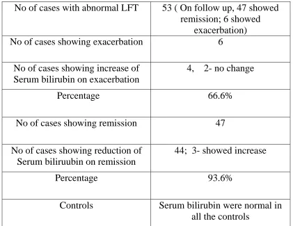
S. Sherlock [43] found a correlation of the serum bilirubin level with
right atrial pressure,not the cardiac output.Therefore,the jaundice of the right
sided heart failure seems to be clinically and pathophysiologically distinct from
that associated ischaemic hepatitis and results from congestion rather than
diminished hepatic perfusion.Upon improvement of the right sided heart failure
elevated serum bilirubin levels return back to normal quite rapidly over a period
SERUM AMINOTRANSFERASES:
Serum aminotransferases, glutamate dehydrogenase, lactic
dehydrogenase are all elevated in right sided heart failure [80,73]. Asparate
transaminase, alanine transaminase rise in up to a third of patients and show,
similar increases, an increased lactate dehydrogenase is found in 20 to 60% of
patients.
In a series of 175 patients with acute and chronic heart failure
[ 73] asparate transaminase was elevated in 49% of those with acute heart failure,
but only in 5% of those with chronic heart failure;80% of the high levels were
between 40 and 80 I.U. Alanine transaminase results paralleled, but were less
marked than, those of asparate transaminase. Very high values of asparate
transaminase (1000-10,000I.U) can occur with an acute onset of severe heart
failure especially if it is associated with hypotension or shock [43, 72] and they
correlate well with the degree of centrilobular hepatic necrosis.
Increased venous congestion and decreased hepatic perfusion
both contribute to the elevation of aminotransferases, although centrilobular
hypoxia and or necrosis are probably the major factors.
The increase in asparate transaminase and alanine
transaminase and lactate dehydrogenase correlate with increase in systemic
correlation coefficients are low suggesting that other factors must also be
involved [80 ]
Very high enzyme levels in congestive heart failure
are often misinterpreted as evidence of viral or drug induced hepatitis [ 81].The
following clinical features suggest that circulatory failure is the cause of liver cell
necrosis [82].
I. The presence of chronic heart failure
II. A recent episode of acute circulatory failure
III. The early appearance of renal insufficiency(this tends to be a late
development in severe viral hepatitis)
SERUM ALKALINE PHOSPHATASE
Serum alkaline phosphatase rises by about 10-20% in
patients with congestive cardiac failure [43, 51,73 ]. However in most patients
the levels are within normal limits; rarely do they exceed twice normal. Elevation
in serum alkaline phosphatase levels do not correlate with increases in serum
bilirubin or aminotrnsferases. The highest elevations are usually seen in patients
with marked liver enlargement. The etiology of abnormal serum level, therefore
is thought to be a result of intra hepatic obstruction secondary to hepatic
congestion.[73] . With the improvements in cardiac status, serum alkaline
PROTHROMBIN TIME:
The prothrombin time is prolonged in 80 – 90% of patients
with congestive cardiac failure with congestive hepatomegaly and is therefore a
sensitive index of right-sided heart failure [43,67,73]. Parenteral administration
of vitamin k results in little or no correction, suggesting that decreased hepatic
synthesis or decreased activation of vitamin k dependent clotting factors may
play a role.[67, 73]. Resolution of the prolonged prothrombin time usually takes
2-3 weeks after successful treatment of the right sided heart failure. Caution
should therefore be exercised when treating patients in heart patients with
warfarin or coumarin derivatives [83].
SERUM PROTEINS:
Serum albumin is low in about 30-50% of patients with
congestive hepatomegaly [51, 73]. And the incidence and degree of change
appears to be similar in acute and chronic failure. The changes were not marked,
in 75% of those with reduced albumin the values were between 2.5- 2.9gm/dl.
The lowest values were in patients with right- sided pressures due to rheumatic
heart disease or cor pulmonale.
Serum albumin concentrations below 1.5 gm/dl are rarely
observed and are often associated with marked ascites and edema. The etiologies
decreased absorption from the congested intestine and poor nutrition. With the
resolution of underlying cardiac disease, improvement in serum levels usually
occurs over a period of few months. The serum albumin levels do not correlate
with the degree of biological damage to the liver.
Hyperglobulinemia occurs in 37- 60% of patients with the
right- sided heart failure, and is more common in patient with acute than with
chronic heart failure[73]. The elevations tend to be mild with levels between 3.5
and 4.1 g/dl in the majority of patients. In contrast to other liver tests, the
hyperglobuminemia usually does not return to normal after treatment of
congestive heart failure.
Prolonged or recurrent episodes of congestive heart disease
can lead to cardiac cirrhosis. The incidence is rare, however, most patients die of
MATERIALS AND METHODS
All cases of congestive cardiac failure (75), of varied etiologies
observed in patients from july 2008 - december 2009. 20 healthy individuals
were taken as controls. Liver function tests are performed to both controls and
cases, serum bilirubin, AST, ALT, SAP, Serum proteins and Prothrombin time
admitted at Govt. Royapettah Hospital during the period from july 2008 to both
on day 1 and day 7 of admission.
This study is an observational study, comparing the liver functions
between cases (various causes of heart failure) and between cases and controls.
Cases:
Heart failure patients due various etiologies including Coronary heart
disease, rheumatic heart disease, hypertensive heart disease, cor pulmonale and
cardiomyopathy meeting the following inclusion criteria and not possessing
following exclusion criteria were selected
INCLUSION CRITERIA
1. Cases of congestive cardiac failure, as per Framingham criteria; of various
age groups and etiologies such as
• Rheumatic valvular Heart Disease
• Ischemic Heart Disease
• Hypertensive Heart Disease
• Cardiomyopathies
• Corpulmonale
2. Congestive cardiac failure of varied presentation either acute or chronic.
EXCLUSION CRITERIA
1. Known alcoholic
2. Past History of jaundice
3. Recent intake of Hepatotoxic drugs or drugs causing raised liver parameters,
such as Rifampicin, INH, Steroids, chlorpromazine, amiadarone, statins,
hydralazine , phenytoin and valproate.
4. positive viral markers
Controls:
Healthy individuals between 20-60 yrs of age without any known history of
medical illness such as heart and liver diseases were selected.
Consent:
The study groups thus identified were instructed about the nature of the
study. Willing participants were taken up after getting a written informed
In this study the following liver functions tests were done:
1. Serum bilirubin
2. serum transaminases
3. serum alkaline phosphatase
4. serum proteins
5. prothrombin time.
Results were entered in Microsoft Excel Spreadsheet and analyzed.
Significance values were analyzed using Minitab software, Epi.info software.
Chi- square test, Student ‘t’ values was applied for significance. Significance was
considered, if the ‘p’ value was below 0.05.
SERUM BILIRUBIN
Serum bilirubin was estimated by the vanden bergh reaction. In
this reaction the bilirubin pigments are diazotized by sulphalinic acid and the
chromatographic products are measured calorimetrically. Vanden bergh reaction
can be used to distinguish between conjugated and unconjugated bilirubin
because of the different solubility properties of the pigments. When the reaction
is carried out in an aqueous medium, the water soluble conjugated bilirubin
reacts to give the direct van den Bergh reaction. When the reaction is carried out
in ethanol, the intramolecular hydrogen bonds of unconjugated bilirubin are
broken. Thus both conjugated and unconjugated pigments react giving the total
bilirubin level. The total minus direct reacting bilirubin gives the indirect value
SERUM ENZYME ASSAYS
A number of serum enzymes have been used to distinguish and
assess the cellular injury and biliary tract dysfunction or obstruction. All have
limitations in sensitivity and specificity and not truly distinguished
the processes definitely. Elevations in enzyme activities may be
hepatic as well as non hepatic disorders like circulatory failure. With
and careful interpretation, a number of serum enzymes provide important
clinical tools.
In this study, the serum enzymes namely SGOT and SGPT was
done by enzymatic substrate method. The substrate used are SGOT and
SGPT substrate respectively along with D,N,P,H Di nitro-phenyl hydrannel
Principle
:
These enzyme catalyse the transfer of the gamma amino groups
and alanine respectively to the gamma keto group of keto glutamase to the
formation of oxalo acetic acid and pyruvic acid.
SERUM ALKALINE PHOSPHATASE
Human serum contains several forms of alkaline phosphataseA
plasma membrane derived enzyme of uncertain physiologic function
Which hydrolyses acetic phosphate enters at pH 9. A number of different
substrate used were Amino-antipyrine solution, potassium ferri cyanide, alkaline
phosphate buffer and substrate. Elevated levels of
alkaline phosphatase activity usually reflect impaired biliary tract function. The
Increased levels reflect increased synthesis of the enzyme by hepatocytes and
biliary tract epithelium rather than restoration of the enzyme due to obstruction.
Slight to moderate increased alkaline phosphatase activity can occur secondary to
congestive cardiac failure.
SERUM PROTEINS
Extensive liver injury may lead to decreased blood levels of
albumin, fibrinnogen, prothrombin, and other proteins synthesized exclusively by
hepayocytes. In contrast to serum enzymes measurements serum protein level
estimation reflect liver synthetic function rather than hepatocellular injury. Some
proteins are neither early nor sensitive indicators of liver disease, because of the
extent of hepatic reserve and their half life. Serum albumin is quantitatively the
most important serum protein synthesized by the liver. The normal value ranges
from 3.5 – 5.5 mg/dl. Albumin has a fairly long half life [14 -20 days] with less
than 5% turnover daily. It is therefore not a good indicator of acute or mild liver
injury.
Serum globulins are a heterogeneous group of proteins, normally 2.0 –
3.5 mg /dl includes alpha and beta globulins as well as serum immunoglobulins.
periphery reticulo-endothelial compartment due to shunting of antigens passed
the liver and impaired clearance by hepatic kupffer cells.
Serum proteins were estimated by the Biuret method. In this
substances which contain two CO –NH2 groups joined together directly or
through a single carbon nitrogen atom and those which contain two or more
peptide links give a blue or purple colored compound with alkaline copper
solution. The amount of colour given by the biuret reaction also varies
appreciably for different proteins.
PROTHROMBIN TIME
Liver synthesizes six coagulation factors namely Fibrinogen
[Factor– Prothrombin(Factor–II) and Factors V,VII,IX,X. Abnormalities of these
coagulation factors can be most efficiently determined by the one stage
prothrombin time which measures the rate of prothrombin conversion to
thrombin in the presence of thromboplastin and calcium and requires the
integrity of vitamin K dependent coagulation factors. The prothrombin time is
dependent on normal hepatic synthesis of clotting factors and sufficient intestinal
uptake of vitamin K. Acute or chronic parenchymal liver injury may lead to
prolongation of the prothrombin time due to impaired synthesis of clotting
proteins, because these proteins have a shorter half life, the prothrombin time
may be regarded as an earlier indicator of severe liver injury and its elevation in
RESULTS
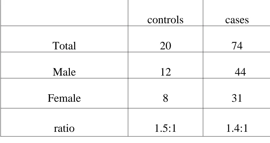
Table 1 – Sex distribution of cases and controls
controls
cases
Total
20
74
Male
12
44
Female
8
31
ratio
1.5:1
1.4:1
CASES
female 41%
male
male 59%
female
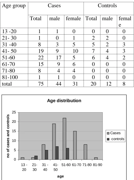
Table -2 Age distribution of cases and controls
Cases
Controls
Age group
otal male female Total male femal
T
e
13 -20
1
1
0
0
0
0
21- 30
1
0
1
2
2
0
31 -40
8
3
5
5
2
3
41- 50
19
9
10
7
4
3
51-60
22 17 5
6
4
2
61-70
15 9 6
0
0
0
71-80
8
4 4
0
0
0
81-100
1
1 0
0
0
0
total
75
44 31
20 12
8
Age distribution
0 5 10 15 20 25 13 -20 21-30 31 -40 41-5051-60 61-70 71-80 81-90
age
n
o
o
f cases an
d co n tr o ls Cases controls
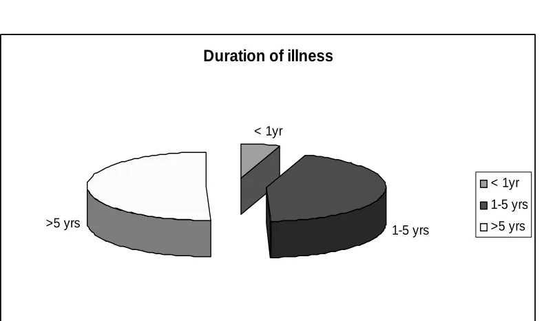
Table 3-
Duration of illness
Years of Heart No of ca es percentage
s failure
< 1yr 4 5.3%
1-5 yrs 33 44%
>5 yrs 38 50.6%
Duration of illness
< 1yr
1-5 yrs >5 yrs
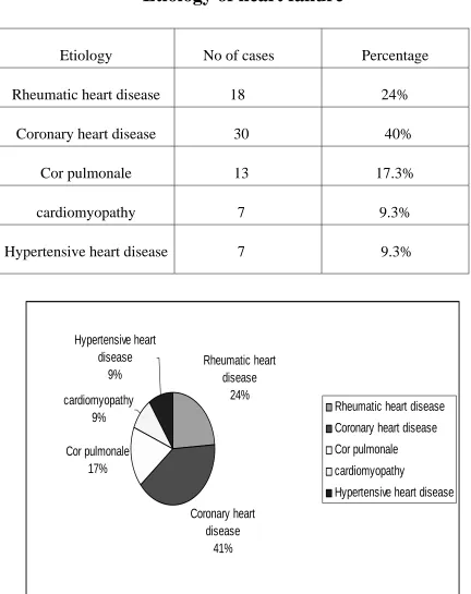
Table – 4
Etiology of heart failure
Etiology No of cases Percentage
Rheumatic heart disease 18 24%
Coronary heart disease 30 40%
Cor pulmonale 13 17.3%
cardiomyopathy 7 9.3%
Hypertensive heart disease 7 9.3%
Rheumatic heart disease
24%
Coronary heart disease
41% Cor pulmonale
17% cardiomyopathy
9%
Hypertensive heart disease
9%
Rheumatic heart disease
Coronary heart disease
Cor pulmonale
cardiomyopathy
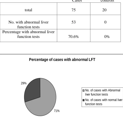
Table 5
Percentage of abnormal liver function tests
Cases controls
total 75 20
No. with abnormal liver function tests
53 0
Percentage with abnormal liver
70.6%
function tests 0%
Percentage of cases with abnormal LFT
71% 29%
No. of cases with Abnormal liver function tests
No. of cases with normal liver function tests
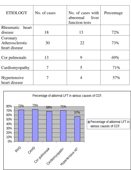
Table 6
Abnormal liver function tests as per etiology
ETIO
abnormal liver nction tests
e LOGY No. of cases No. of cases with Percentag
fu
R rt
disease 18 72%
heumatic hea
13 Coronary
ease
30 22 73% Atherosclerotic
heart dis
Cor pulmonale 13 9 69%
Cardiomyopathy 7 5 71%
Hypertensive 7 4 57% heart disease
Percentage of ablormal LFT in various causes of CCF.
72% 73% 69% 71%
57% 0% 10% 20% 30% 40% 50% 60% 70% 80%
RHD CAHD
Cor pulm onal e Card iom yopa thy Hype rten sive HD
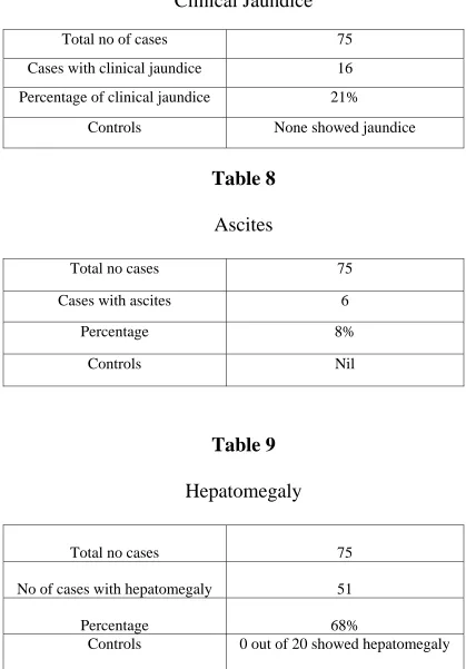
Table 7
Clinical Jaundice
Total no of cases 75
Cases with clinical jaundice 16
Percentage of clinical jaundice 21%
Controls None showed jaundice
Ascites
[image:63.612.87.506.106.707.2]Total no cases 75
Table 8
Cases with ascites 6
Percentage 8%
Controls N il
Table 9
Hepatomegaly
Total no cases 75
No of cases with hepatomegaly 51
Percentage 68%
Tab
No of cases with hepatomegaly showing increased serum
alkaline phosphatase
le 10
No of Cases with Hepatomegaly 51 ( out of 75) Cases with raised SAP 10 ( out of 75) No of cases with raised SAP in
Hepatomegaly and CCF
10 ( out 0f 51)
No of cases with raised ALP without hepatomegaly
0 ( out of 24)
Percentage of raised SAP in CCF 12.5% Percentage of raised SAP in patients
with CCF having hepatomegaly
19.6%
Hepatomegaly without raised
SAP
hepatomegaly and raised SAP No hepatomegaly
or raised SAP Hepatomegaly without raised SAP
hepatomegaly and raised SAP
No hepatomegaly or raised SAP
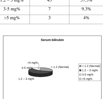
Table 11
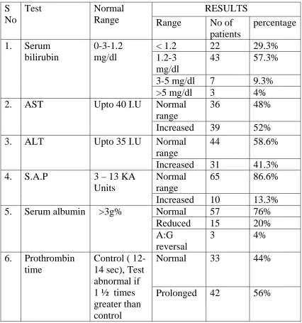
Serum bilirubin
Serum bilirubin
No of cases
Percentage
< 1.2 (Normal)
22
29.3%
1.2 – 3 mg%
43
57.3%
3-5 mg%
7
9.3%
>5 mg%
3
4%
Serum bilirubin
< 1.2 (Normal)
1.2 – 3 mg% 3-5 mg%
>5 mg%