PREVENTION & MANAGEMENT
OF CERVICAL SPONDYLOSIS IN
COMPUTER PROFESSIONALS.
SUBMITTED TO
THE
TAMILNADU
Dr.
M.G.R
‐
MEDICAL
UNIVERSITY
CHENNAI
‐
32.
FOR
THE
FULFILLMENT
OF
M.D., (PMR)
PREVENTION & MANAGEMENT
OF CERVICAL SPONDYLOSIS IN
COMPUTER PROFESSIONALS.
This Thesis work is submitted to “THE TAMILNADU
DR. M.G.R MEDICAL UNIVERSITY” CHENNAI – 32 is part of
fulfillment of the M.D., (PMR) (one time measure) Examination
held during MAY 2011.
Work done by:
NAME:
CONTENTS
1.
INTRODUCTION
-
1
2.
DEFINITION
-
2
3.
AETIOLOGY
-
3
4.
PATHOLOGY
- 5
5.
PATHOGENESIS
- 6
6.
ANATOMY
-
7
7.
BIOMECHANICS
- 14
8.
CLINICAL
FEATURES
- 18
9.
INVESTIGATION
S
- 22
10.
COMPLICATIONS - 24
11.
CONSERVATIVE
MANAGEMENT
- 25
12.
ASSESSMENT
- 28
13.
PHISIOTHERAPHY MANAGEMENT
-
34
14.
PREVENTION
- 47
CONSERVATIVE
INTRODUCTION
The human body is one of the greatest wonder of nature. From birth to death it works as a restless machine in perfect co-ordination and harmony and enables us to lead our lives.
According to various estimates, about 10% of the world population is affected by one or more disabilities.
Man identifies himself separate from other animals by his erect posture. Erect posture is considered by highness, improve his personality and braveness.
Cervical spine region is one of the most common areas of dysfunction, be it acute trauma or degenerative disorders or chronic postural strains.
DEFINITION
¾ Cervical spondylosis is a degenerative condition of cervical spine that affect the vertebral bodies and intervertebral discs of neck, as well as contents of spinal cord.
¾ SPONDYLO is a Greek word – meaning vertebra. Spondylo generally means changes in the vertebral joint characterized by increased
AETIOLOGY
¾ TRAUMARepetitive sub clinical trauma probably influences the onset and progression of spondylosis
¾ BAD POSTURE
Poor posture can lend to mechanical problems, dysfunction and pain. As the computer users work for a prolonged period of time with forward head posture.
¾ AGING
As the age advances, there is progressive degeneration of disc leading to pain and restricted range of motion.
¾ OTHER PRE DISPOSING FACTORS
• Occupation requiring repetitive motion.
• Previous injuries with fracture or disc prolapse.
• Hemi vertebra or fused vertebra.
• People who carry heavy loads. E.g.: coolie, porters in railway station.
PERSONALITIES AT RISK FOR CERVICAL
SPONDYLOSIS
Old people.
Computer professionals
People doing work that demands minute concentration Persons who constantly work bending their neck Bike users.
Travelers who travel a long distance and sleep in sitting, Watching T.V in abnormal positions or by lying down. Habits of holding neck in one position.
EPIDEMOLOGY
INCIDENCE
¾ AGE
Aging results in progressive wear and tear leading to bone loss which begins at 30-40 years of age continues henceforth. But in this modern sophisticated world, even youth of 20 seem to suffer from such complaints.
¾ SEX
Both sexes are affected equally; it starts usually earlier in men than in women
¾ LIFE STYLE
Sedentary life and not using the spine properly, generally, predisposes to such problems.
¾ RACE
No correlation between race and cervical spondylosis.
¾ OCCUPATION
PATHOLOGY
The primary event is a progressive decrease in the degree of hydration, resulting in loss of disc height, which leads to disc fibrosis and annular weakening.
It manifests itself by ubiquitous and inevitable degenerative changes including desiccation of cervical discs, disc protrusion due to irregular wear of the facet joints, narrowing of the facet joints, leading to reactive hypertrophy of facets and thickening of inter laminar ligaments, as well as attendant inflammation of these regions.
Rarely large posterior osteophytes may cause pressure on the anterior portion of the spinal cord and produce mixed symptoms of the upper limb nerve root pain and lower extremity weakness i.e. cervical myelopathy.
Cervical spondylosis
PATHOGENESIS
The normal disc due to aging or trauma undergoes disc degeneration and collapse of the disc. The causes protrusion of the annulus fibrosis portion of the disc.
This leads the development of abnormal sharp outgrowths of the bones called the osteophytic formation. Osteophytes narrow the spinal canal by occupying it. There may even be a congenital cause leading to narrowing of spinal cord.
On the other hand, the protrusion of annulus fibrosis causes apophyseal joint damage, narrowing of facet joints leading to – instability of the joints, joint hypertrophy as a reaction of inflammation, thickened ligamentum flavum. All these factors in turn lead to narrowing of spinal cord.
Sometimes the osteophytic protrusion may compress the spinal nerve roots and causes radiating pain over the respective dermatomal distribution – radiculopathy.
ANATOMY OF VERTEBRAL COLUMN
The vertebral column provides a base of support for the head and internal organ; a stable base for attachment for ligaments, bones, and muscles of extremities, rib cage and the pelvis; a link between upper and lower extremities and mobility for trunk, in addition it supports the spinal cord.
¾ STRUCTURE
The vertebral column is divided in to 33 short bones called vertebrae and 23 intervertebral discs.
The vertebrae increase in size from cervical to lumbar region and decrease in size from sacral to coccygeal region.
¾ PRIMARY AND SECONDARY CURVES
When the column of an adult is viewed from the side, four distinct antero – posterior curves are evident. 2 curves (thoracic and sacral)are primary curves, whereas the two curves (cervical and lumbar)
Curves that have a posterior convexity are kyphotic and those that have anterior are lordotic curves.
¾ THE INTERVERTEBRAL DISC
They make about 20% to 33% of the total length of the vertebral column, increasing in size from cervical to lumbar region, approximately 3mm in cervical to 9mm in lumbar.
The discs in the cervical and lumbar region contribute to greater mobility than those of that in thoracic region.
The disc is composed of two parts, a central portion called the nucleus pulposis and a peripheral portion called the annulus fibrosis.
Nucleus pulposis: The fluid content and composition of the disc varies with aging. The fluid content of nucleus pulpous accounts for 88% app.
¾ THE CERVICAL VERTEBRA
The cervical vertebras are identified by the presence of foramina transversaria. There are 7 cervical vertebrae out of which 3-6 are typical and the remaining 3 (1st – atlas, 2nd – axis, 7th) are typical. The 7th cervical vertebra is a transitional vertebra with characteristics of both cervical and thoracic.
¾ FIRST CERVICAL VERTEBRA
It is called atlas as it supports the globe if the head. It is ring shaped and has no body. Its Centrum is fused with the centrum of second cervical vertebra. It has no spine. It has a short anterior arch and long posterior arch, too large lateral masses and two transverse processes.
¾ THE SECOND CERVICAL VERTEBRA
It is called as the axis because the head (with atlas) rotates around it. Axis is identified by the presence of DENS (odontoid Process), a strong, tooth like process projecting upwards from the body. Dens represent the centrum of the atlas. Superior surface of the body is fused with dens.
¾ THE SEVENTH CERVICAL VERTEBRA
It is also known as VERTEBRA PROMINENS because of its long spinous process, the tip of which can be felt through the skin. Spine is thick, long and nearly horizontal and it is not bifid, but ends in a tubercle. Foramina transversarium is relatively small, sometimes double or may even be absent.
¾ THE ATLANTO – AXIAL JOINT
The atlanto-axial joint is composed of three separate joints; the median atlanto axial joint (atlanto-odontoid) between the dens and the atlas and the 2 lateral joints between the superior zygapophyseal facets of the axis and the inferior zygapophyseal facets of the atlas. The median joint is the synovial pivot joint in which the dens of the axis rotate in an osteoligamentous ring formed by the anterior arch of the atlas and the transverse – atlantao ligament. The two lateral joint are plane synovial joints.
¾ THE ATLATO-OCCIPITAL JOINT
This is a plane synovial joint and is composed of two concave superior zygapophyseal facets of the atlas that articulate with two convex occipital condyles of the skull.
¾ LIGAMENTS OF CERVICAL SPINE
• Anterior and posterior longitudinal ligaments
• Ligamentum flavum
• Interspinous ligament
• Supraspinous ligament
• Inter transverse ligament
• Zygapophyseal joint capsules
• Ligamentum nuchae
• Transverse atlantal ligament
• Alar ligament
¾ MUSCLES OF CERVICAL REGION
22 complex muscles support the head.
• Scaleneous anterior
• Scaleneous medius
• Scaleneous posterior
• Sterno cleido mastoid
¾ SUB- OCCIPTAL MUSCLES
¾ SUPRA – HYOID MUSCLES
• Diagastric
• Stylohyoid
• Mylohyoid
• Hypolossus
¾ INFRA- HYOID MUSCLES
• Sterno hyoid
• Thyro hyoid
• Omohyoid
• Prevertebral neck muscles
• Rectus capitis lareralis
• Rectus capitis anterior
• Longus capitis
• Longus coli inferior oblique
• Longus coli superior oblique
• Longus coli medial
¾ VASCULAR SUPPLY
Vertebral artery
¾ NERVE SUPPLY
• Posterior primary rami
BIO-MECHANICS OF CERVICAL SPINE
¾
STABILITY
• The cervical region differs from the thoracic and lumbar regions in that the cervical region bears less weight and is generally more mobile. Although the cervical region demonstrates the most flexibility if any of the regions of the vertebral column, stability of the cervical region especially of the atlanto-occipital and the atlanto-axial joints, is essential of the support of the head and protection of the spinal cord.
• The design of the atlas is such that it provides more space for the spinal cord. The extra space is made to ensure that the spinal cord is not impinged during the large amount of motion that occurs here.
• The bony configuration of the atlanto- occipital articulation confers some stability, but the application of small load produces large rotations across the occipital- atlanto-axial complex and also across the lower cervical spine.
• The existence of a large neutral zone implies that the ligaments and joint capsules are lax and the muscles play an important role in stability for the occipital –atlanto- axial complex. The muscles providing stability are the multifidus, interspinalis, semispinalis capitis, and spinalis cervicis.
• No discs are present at either the atlanto-occipital or atlanto-axial
• The loads imposed in the cervical region vary with the position of the head and the body and are minimal in a well supported reclining body posture. The compressive loads are relatively low in erect stance and sitting postures and high during the end ranges of flexion and extension.
• Cervical motion segments in bending and axial torsion exhibit lower stiffness than the lumbar motion segments but exhibit similar stiffness in compression. The joint capsules in the cervical region and lax and therefore provide less restriction to motion than in the thoracic and lumbar region.
¾
MOBILTY
The cervical spine is designed for a relatively large amount of mobility. Normally the neck moves 600 times every hour whether we are awake or sleep. The motion of flexion, extension and rotation are permitted in the cervical spine. These motions are accompanied by translations that increase the magnitude from c2 to c7. However the predominant translation occurs in the sagittal plane during flexion and extension.
The atlanto-occipital joint permits primarily a nodding motion of the head in a sagittal plane around a coronal axis. However a small amount of axial rotation and lateral flexion is also possible. Flexion at the atlanto-occipital joints is limited by osseous contact of anterior ring of foramen magnum of the skull on the dens. Extension is checked by the membrane.
Lateral flexion is limited by the uncinate process whereas the hyperextension is limited by the bony contact of spinous processes. The height is large in comparison to the antero-posterior and transverse diameters of cervical discs. Therefore a large amount of flexion extension and lateral flexion may occur at each segment.
¾
MUSCLES AND THEIR ACTIONS
Stability is provided by the following muscles:
• Multifidus
• Interspinalis
• Semi spinalis capitis
• Semi spinalis cervicis Flexion of head at atlanto-occipital joint:
• Longus capitis
• Rectus capitis anterior
• Sternocleiodomastoid Extension at atlanto-occipital joint:
• Recti capiti posterior major and mirror
• Oblique capitis superior
• Semi spinal capitis
• Sterno mastoid
• Trapezius Rotation:
• Oblique capitis superioris
• Rectus capitis posterior minimus
• Splenius capitis
• Sterno mastoid
Flexion of lower cervical region is produced by:
• Longus coli
• Scalene muscles
Extensors of head and neck that produce rotation to opposite side:
• Multifidus
• Rotators
• Semi spinalis
Extensor muscles causing rotation to same side:
• Oblique capitis
CLINICAL FEATURES
• Persons who constantly work bending their neck
• Pain in the neck which can radiate to back, shoulders and down to upper arm, forearm and hand.
• Early morning stiffness of the neck and shoulders.
• Weakness of the hands with numbness and tingling sensation.
• Cracking sounds are heard when moving the neck.
• Swelling and tenderness of the neck may noted over the spinous process along with spots of inflammation.
• Restriction of the movements of the neck.
• Pain gets intensified with movement, bending, coughing and sneezing.
• Pain increases on hyperextension, and by giving pressure against the top of the head.
RADICULOPATHY
¾
PAINA sharp stabbing pain, worse on coughing may be supimposed on a more constant deep ache radiating over the shoulders and down the arm.
¾
PARAESTHESIANumbness or tingling sensation follows a nerve root distribution.
MOTOR CHANGES
Muscles weakness and wasting in appropriate muscle groups. E.g. c5, c6 – biceps, deltoid jerk. C7 – triceps jerk.
TROPHIC CHANGES
In long standing root compression, skin becomes dry scaly, inelastic sometimes blue and cold.
MYELOPATHY
Compression causes segmental damage at the involved level and long tract signs below the level.
ARMS
• Lower motor signs and symptoms at the level of lesion, upper motor signs below the level of lesion.
• In c5 lesion- deltoid and biceps weakness, wasting, diminished biceps jerk, increased finger jerks. LEGS
Sphincter disturbance is seldom a prominent early feature. As the disease advances, losses of bladder control manifest.
ROOT MOTOR REFLEX SENSATION
C5(c4-c5) lesions C6(c5-c6) lesions C7(c6-c7) lesions C8(c7-c8)lesions Deltoid muscle weakness.
Weakness of wrist extensor muscles Weakness of wrist flexors. Weakness of finger flexors. Diminished biceps reflex. Diminished brachio- radialis reflex. Diminished triceps reflex. None.
Numbness in the deltoid region. Numbness in the dorso- lateral aspect of thumb and index finger. Numbness over index, middle finger and the dorsum of hand. Numbness in the ring finger, little finger and medial border of forearm.
IN ADVANCED STAGES OF DISEASE Loss of balance and fear of falling.
CLINICAL ASPECTS OF THE DISEASE
As in the lumbar and thoracic spine, herniation of the contents of an intervertebral disc may occur when a tear occurs when a tear occurs in the annulus fibrosus.
The symptoms and signs are the result of root compression spinal cord compression, or both. Patients often hold their hands elevated and behind the head, presumably because this maneuver reduces the tension in the nerve root and thus lessens the pain.
In most cases the onset of is pain is awakening, without identifiable trauma or other predisposing factors.
The c6-c7 disc most frequently herniated, of about 2/3rd of cervical herniations.
LHERMITTE’S SIGN
Refers to sudden electrical sensation down the neck and back triggered by neck flexion. This sign is positive is cervical spondylosis.
BRACHIAL NEURALGIA
Brachium means arm i.e., from shoulder to elbow. Thus neuralgic pain is medically termed as brachial neuralgia. This is otherwise called cervico-brachial neuralgia or thoracic outlet syndrome where brachial plexus is irritated or inflamed or compressed or pinched to cause symptoms in shoulder, arm, forearm and hand.
This syndrome is caused due to losing of normal curvature of spine with friction loss, degeneration, distortion and herniation of the disc.
INVESTIGATIONS
¾
PLAIN X-RAYS
• Narrowing of disc space.
• Anterior and posterior marginal lipping of vertebral bodies-osteophyte formation.
• Sub- chondral bone sclerosis.
• Narrowing of intervertebral foramen.
• Loss of cervical lordosis is an early finding.
• Spinal cord narrowing correlates with myelopathy.
• Spondylosis (arthritis) of the facet joints.
¾
MAGNETIC RESONANCE IMAGING
• MRI is a powerful tool in the assessment of patients with cervical spondylosis. Images from MRI can help to identify disc lesions, osteophytes and joint arthrosis.
¾ MYELOGRAM/ COMPUTED TOMOGRAPHY
• CT/ MYELOGRAPHT is a “golden standard” for imaging of cervical spine during evaluation for spondylosis and disc disease. It provides an excellent definition of herniated discs and spondylotic ridges and demonstrates their relationships to nerve roots and spinal cord.
• It is very useful in delineating bone spurs from safe disc herniations. But it is an invasive process.
• Thin section ( 1.5 to 2mm) high resolution computed tomography is suited for patients with cervical radiculopathy, since it demonstrates laterally suited osseous spurs and foraminal stenonis. CT after intra thecal contrast medium injection produces the most reliable technique of investigation.
¾
DISCOGRAPHY
DIFFERENTIAL DISGNOSIS
Herniated disc
Traumatic myelopathy Amyotrophic latera sclerosis Syringomyelia
Cervical rib
Neurological disorders Neo plastic disorders Infammatory disorders Infection
Primary
Shouderand upper exeremity problems Psychogenic disorder
Visceral and miscellaneous disorders
COMPLICATIONS
• Degenerative changes can cause permanent deformity of the spine-KYPHOSIS
• Paralysis of hands or legs due to compression of nerve root.
• Lack of bladder or bowel control depending upon the intensity of spondylosis.
MANAGEMENT
¾
CONSERVATIVE TREARTMENT
After conducting necessary test to identify the problem in cervical spine, a treatment plan will be developed.
Conservative treatment is the more accepted from of treatment in cervical disc syndromes.
REST
This implies not total rest but selective rest avoidance of unnecessary activities and strains to the neck.
SUPPORT AND IMMOBILIZATION
This enables the diseased part to heal, provides rest prevents deformities, relives pain and supports the patient psychologically.
But using cervical collar in the long run cause fixation of bones and restriction of movement.
MEDICATIONS
For pain relief-analgesics-NSAIDS
• Diclofenac 50-100mg
• Aceclofenac 100-200mg
• Indomethacin 25-75mg
• Nabuflam 500mg
• Pyroxicam 20mg
• Mefenamic acid 500mg
MUSCES RELAXANTS
To treat muscles spasm-e.g. BACLOFAN.
CORTICOSTEROIDS
Oral prednisolone or single intra muscular injection. Non-narcotics- to relieve pain and swelling.
Anti depressant drugs.
Nowadays people commonly use external applications or sprays like soothing agents which alleviates pains in minutes. This will temporarily mask the complaints or pain.
Symptoms of radiculopathy, whether acute or chronic, usually respond to these conservative measures, progression of a disabling neurological deficit however demands surgical intervention.
MEDICAL PITFALLS
• Use of bed that is too hard.
• Over use of muscle relaxation or pain medication.
• Prolonged rest, inactivity or vigorous exercise.
• Failure to recognize depression.
• Failure to recognize chronic syndrome.
PROGNOSIS
Cervical spondylosis is a slowly progressive chronic joint disability, especially when it is associated with neuronal compression.
The gradual progression of cervical spondylosis cannot be stopped, however it doesn’t always cause symptoms. For individuals who do experience problem, conservative treatment is very effective in managing the symptoms. Approximately 75% of patients suffering from neck pain find relief through physiotherapy alone.
PHYSIOTHRAPY ASSESSMENT
ASSESSMENT CHART
• NAME
• AGE
• SEX
• OCCUPATION
• CHIEF COMPLAINTS
• HISTORY OF PATIENT
PRESENT ILLNESS: ACUTE:
SUB ACUTE: CHRONIC:
HISTORY OF PAIN:
• ONSET : SUDDEN OR GRADURAL OR LATE ONSET
• NATURE OF PAIN : STABBING OR
DULL ACHING OR BURNING
• EXTENT : LOCAISED PAIN OR RADIATING PAIN
AGGREVATING AND RELIEVING FACTORS:
• PROLONGED SITTING :
• WRITING :
• SUPINE LYING :
• PRONE LYING :
• FLEXION :
• EXTENSION :
• ROTATION :
• COUGHING AND
SNEEZING :
• REST :
VISUA ANALOG SCALE:
PAST MEDICAL HISTORY
Any surgery, medication, physiotherapy treatment, any improvement from it.
OCCUPATIONAL HISTORY:
• PROLONGED WRITNG:
• LIFTING OBJECTS OVER HEAD:
• WORKING IN FLEXED POSITION OF NECK:
• HEAVY MANUAL LABOURING:
• WORKING IN FRONT OF COMPUTER:
PHYSICAL EXAMINATION
:
(a)
INSPECTION:
1. POSTURE: EAR LOBES
SHOULDER LEVEL SPINE OF SCAPULA
INFERIOR ANGLES OF SCAPULA ILLIAC CREST
2. GAIT:
(B) PALPATION: TENDERNESS
SPASM NODULES
TONE OF MUSCLES REDNESS
SWELING
(C) EXAMINATION: NEUROLOGICA EXAMINTION:
1. RANG OF MOTION: USING INCH TAPE FLEXINN -30 Degrees EXTENSION -30 Degrees LATERAL FLEXION - 40 degrees ROTATION - 30 Degrees 2. REFEXES: BICEPS JERK
BRACHIO RADIAIS JERK TRICEPS JERK
3. MUSCLE POWER:
Muscle power is tested using M R C grading and the muscles are graded from G0 to G5 for flexors, extensors, lateral flexors, rotators.
0 - NO CONTRACTION
1 - FLICKER OF CONTRACTION
3 - FULL ROM AGAINST GRAVITY
4 - FULL ROM AGAINST GRAVITY WITH MINIMAL RESISTANCE 5 - FULL ROM AGAINST GRAVITY WITH MAXIMAL RESISTANCE
SENSORY EXAMINATION:
• BY USING COTTON OR WOOL
• BY USING PINS OR NEEDES
• BY USING HOT AND COLD WATER TEST TUBES This examination is done along the affected dermatomes.
SPECIA DIAGNOSTIC TESTS
1. FEXTION COMPRESSING
2. FORAMINAL COMPRESSION TEST 3. DISTRACTION TEST
4. VERTEBRA ARTERY 5. ADSON’S TEST
¾ SPURLING’S TEST : FORAMEN COPRESSION TEST : POSITION OF THE PATIENT: sitting
¾ DISTRACTION TEST:
POSITION OF THE PATIENT: sitting
PROCEDURE: examiner carefully pulls the head upwards. INFERENCE: disappearance of symptoms of nerve compression.
¾ VERTEBRAL ARTERY TEST:
POSITION OF THE PATIENT: supine lying.
PROCEDURE: Examiner passively takes the patient’s head and neck into extension and side flexion. After this movement is achieved, the examiner rotates the neck to the same side and holds it for approximately 30 seconds.
INFERENCE: dizziness or nystagmus indicates compressions of the vertebral artery.
¾ ADSON’S MANEUVER (done to differentiate from cervical ribsyndrome)
POSITION OF THE PATIENT: sitting.
PROCEDURE: the examiner locates the radial pulse. The patient head is rotated the face the shoulder. The patient then extends the head while examiner laterally rotates and extends the patient’s shoulder. The patient is asked to take a deep breath and hold it.
INFERENCE: Disappearance or diminishing of the pulse indicates a cervical rib.
INVESTICATION:
DIAGNOSIS:
GOAL-SHORT TERM/LONG TERM
TREATMENT PLAN:
PHYSIOTHERAPY MANAGEMENT
SHORT TERMGOALS:
1. To relive pain 2. To relive spasm
3. To improve the mobility of cervical spine 4. To subside the inflammatory
5. To stretch any tight structures that hinders mobility
LONG TERM GOAL:
1. To strengthen weak muscles
2. To educate the patient about importance of risk care and to introduce the patient to a fitness programme
3. To improve functional abilities 4. To create postural awareness
5. To make the patient to actively involved in his job as quickly as possible 6. To prevent further recurrence
MEANS:
• PHYSICAL AGENT AND MASSAGE
• CERVICAL TRACTION
• MANIPULATION
• EXERCISES
• CERVICAL COLLAR
• POSTURAL AND ERGONOMIC ADVICE
Control stiffness of soft tissue and joints Assist mobility
Increase blood supply and to relieve ischemia
¾
SUPERFICAL THERMO THERAPY MODALITIES
Wax bath, hydro collator packs, hot water bags, etc.
¾ INFRA RED RADIATION
These are electromagnetic waves with wavelengths of 750nm . 400,000n m. it produces heating effect and pain relief.
¾
DEEP HEATING MODALITIES
• SHORT WAVE DIATHERMY:
These waves have the frequency of 27.12MHz and produces wavelength of 11 m.
Position of the patient: arm lean sitting. Acute : 10 minutes twice daily Sub acute : 10 to20 minutes daily Chronic : 20 to30 minutes daily
• MICROWAVE
• ULTRASONIC TREATMENT
It is based on reverse piezo-electric. The machine produce 1 MHz frequency.
Position of the patient : arm lean sitting.
Frequency : 0.5MHz to 3.5MHz.
• CRYO THERAPY
IT is the form of ice massage and ice packs. Application of ice Beta endorphins and enkephanlins (opiate like substance) which reduce pain.
• TRANSCUTANEUS ELECTRICAL NERVE STIMULATION
TENS is application of pulsed rectangular wave current via surface electrodes on patient’s skin.
Pulse shape - rectangular
Pulse width - 100 to 200 micro seconds. Frequency - 150 Hz commonly used Intensity - 0 to 60 mA.
Three types of TENS: HIGH TENS, LOW TENS, BURST TENS.
• INTERFERENTIAL THERAPY
Uses of magnetic field in an extremely low frequency and ultra low intensities have been found to be effective for the relief of pain of musculo- skeletal system.
Frequency : 1 Hz to 10 Hz
• MASSAGE
Deep and sedative massage and manipulations effectively reduce spasm and pain thereby inducing relaxation.
Techniques : Effleurage, circular kneading, frictions to the localized area.
• CERVICAL TRACTION
It is modality of choice for many dysfunctions. It is indicated in wide range of problems from sprains to fractures and dislocations of cervical vertebra.
TYPES OF TRACTION
(a) CONTINOUS TRACTION
The traction is usually continued for 24 hours. The force of traction is minimal i.e. 5 to 15 lbs. sustained small pull for long hours induced relaxation of the muscle spasm and gradual vertebral separation. It relieves compression on the nerve roots or spinal cord. It offers immobilization. It may help the prolapsed disc to move back into its place.
(b) STATIC TRACTION
A traction with a constant pull varying from 10 to 30 be is applied for 20 to 25 mins. It is indicated in the presence of definite neurological signs, including radiating pain, which is not relieved by other
conservative modes. The signs and symptoms gradually improve with the traction on.
(c) INTERMITENT TRACTION
A traction with alternate phases of pull and relaxation is the popular mode of traction. It produces the effect of massage on the muscular, ligamentous and capsular structures. It promotes circulation, reduces swelling there by reducing inflammation, pain and spasm. It also prevents adhesions between dura sleeves of the nerve roots.
ANGLE OF PULL OF TRACTION
TRACTION IN FLEXION
Maximum pull and vertebra separation occurs posteriorly at the lower segment of cervical spine when the traction is given in 24 degrees. TRACTION IN NETURAL
It offers maximum pull and intervertebral separation at the upper segments of cervical spine. Traction in hyper extension also produces the same effect.
DURATION OF TRACTION
It varies as per the underlying pathology .static traction – 20to25 minutes once a day or alternate day’s intermittent traction-15 to 20minutes on alternate days.
FORCE OF TRACTION
Tractive force could vary between 1 and 10th to 1/7th of patient’s body weight. Higher the force of traction, lesser should be its duration and vice versa.
PATIENT’S POSITION
Overhead traction – sitting position.
CONTRA INDICATIONS TO TRACTION
• Marked ligamentous instability immediately inferior to the specific traction level.
• Malignancy in cervical or thoracic spine.
• Signs and symptoms of spinal cord involment.
• Rheumatoid arthritis with necrosis of ligaments adjacent to the traction level.
• MANIPULATION
Reduction of the intra articular displacement in cervical spondylosis by manipulation is very effective, provided the spondylosis is not too advanced.
Manipulation is always carried out during strong traction. It needs to be maintained correctly to prevent recurrence.
CONTRA INDICATION
• Evidence of impaired pyramidal function
• Root palsy excluding minor par aesthesia.
• Primary postero – lateral on set, when symptoms appear in reverse order. E.g. paraesthesia occurring first in the digits then goes up to the shoulder.
• Cervical movements provoking brachial pain instead of scapular region pain.
• CERVICAL COLLAR
The use of cervical collar is on the decline. It is indicated in acute disc prolapsed, acute pain due to sprain, strain, fractures and dislocations, and for temporary immobilization.
ROLE OF COLLAR
• Reduces pain and spasm of the neck muscles.
• Provides immobilization to the neck.
• It helps to do the isometeric exercise.
• It helps to correct the deformity.
• It provides additional support, comfort and psychologica well being.
Proper fitting of the collar is necessary e.g. its fitting in neck flexion is advocated where separation of facets and opening of foramina is the aim.
DRAWBACKS OF THE COLLAR
• POSTURAL CORECTION
• Maintenance of proper neck posture straight neck with chin tucked in during sitting, standing, walking, and working is of paramount
importance.
• Proper sized pillows with correct height during sleep should be used to maintain the position of netural in relation to shoulders.
• EXECRISES – MEASURES TO STRENGHTEN AND MOBILIZE THE
NECK :
Exercise play vital role in strengthening the neck muscles and mobilizing the neck once the pain has subsided.
• EXECRISES FOR NECK MOBILIZATION
Relaxed passive exercises for all the movements that is in flexion, extension, side flexion, rotation.
• EXECRISES TO STRENGHTEN THE NECK MUSCLES
Strong isometrics help to achieve this when the movement is contra indicated. POSITION: Sitting or standing.
FEXION: Place both hands on the force head and press the fore head into the palms but do not allow the motion to occur.
These exercises can be done by the therapist or the patient himself.
EXERCISES TO STRENGTHEN THE WEAK MUSCLES
Strengthening the week muscles can be done by active assisted exercises to the neck, active self resisted isometric exercises.
FREE EXERCISE
EXTENSION OF HEAD AND NECK
IN SITTING: Head dropping forward and raising to vertical. IN PRONE: Head dropping forward raising to vertical. FLEXION OF THE HEAD AND NECK
SUPINE LYING: head lifted and bent forwards till the chin touches the streno – clavicular joint.
SIDE FELEXION OF NECK
RESISTED EXERCISES FOR HEAD AND NECK
EXTENSION: manually – in crook lying. Mechanically by spring or weight, but this must not be too heavy.
FLEXION: manually by giving pressure on the forehead. SIDE FLEXION: manual resistance from low grasp sitting.
SELF STRETCHING OF THE NECK
• Let your neck bend side to side, and with your hand apply a little pressure to your head to help bringing it to the same direction. Do until you feel a tight stretch and hold it for six seconds. Repeat the same three times and then to the opposite side.
• Let your head hang forward, as if you are touching your chin to your chest. Lace your fingers behind your head to apply a little pressure to bring your chin further down. Stop when you feel a tight stretch and maintain it for six seconds. Do three times.
OTHER EXERCISE RELATED TO NECK
• Shoulders shrugging exercises
• Shoulder bracing exercises
• Shoulder lateral rotating exercises.
PROPRIOCEPTIVE NEURO MUSCUAR FACILITATION EXECISES
AIM
To simulate the maximum number of motor ends and thereby to initiate muscle fiber space to contract
To prevent muscle wasting.
EXTENTION WITH ROTATION TO RIGHT
Starting position, Patient in supine lying with shoulders leveled with the end of plinth and head is flexed and rotated to left. Therapist stands in lunge position with right leg forwards, at the head end. Then grips the lateral surface of the lateral surface of the right half of mandible by the thumb of his right hand and rest of the hand is not in contact with patient. He places his left hand on the occiput with thumb adducted and fingers down.
COMMANDS:”NOW”
“PUSH AND LOOK TO THE RIGHT”
MOVEMENT: head and neck will go into extension and rotation to right side.
FLEXION AND ROTATION TO THE RIGHT
and with left foot forward, with left hand on the mandible and right hand on the occiput.
COMMANDS:”NOW”
“PULL YOUR CHIN UP TOWARDS YOUR BREAST POCKET” MOVEMENT: The head and neck will go in flexion and rotation to right side.
ACQUATIC THERAPHY EXCERCISES(HYDROTHERAPY)
• Total relaxation in float support lying, togetherwith the warmth of the water gains relief of the muscle spasm.Head, neck and trunk side flexion performed slowly through full range gains mobility and ensures that the muscles lengthen and shorten fully.
• To stretch tight Para vertebral muscles, the patient practices tucking the chin in and pushing the c4, c5 level into the neck float.
• Float support lying, pushing one hand the other towards the feet helps to relax the upper fibers of trapezius.
ERGONOMIC ADVICE
Using several criteria for assessing epidemiologic evidence, the national institution for occupational safety and health (NISOM) revived the literature to
determine the work relatedness of musculo-skeletal disorders of the neck. The literature concluded that there is evidence of a casual relation between highly repetitive work and occurrence of neck disorders.
Because more people are interacting with computer at work and at home, the effects of prolonged VISUAL AND PRECISION and tasks have become a public concern. The ergonomic interfaces need to be considered are:
• The viewing conditions.
• The manipulative requirements
• The positioning of the body
All the above three, affects the posture and the forces exerted by neck and shoulder muscles.
VIEWING CONDITIONS
They depend on the quality of the image displayed for a task and the visual acquity of the exposed person
MANIPULATION
This involves the hand and other implements as we as assembly or precision tasks at work or in recreational activities.
PERFORMING TASKS
For thought and resource fullness help control the cost of intervention. The adjustment of the interface is VISUAL AND PRECISION tasks may not entail expensive
equipments.
• Tilting a monitoring may remove glare
• Repositioning a monitor or chair may improve viewing distance placing a copy holder close to the monitor may reduce repetitive neck rotation
• Active short breaks for stretching may bring relief from static exertion of the neck, or at least help maintain productivity levels.
The concept of “NECK HYGEINE” ergonomic at least on an empirical clinical basis. It can be translated to the following set of empowering action for the patient.
1) Beware of your posture and change position regularly. 2) Avoid extreme posture of the neck.
3) Avoid maintaining the neck in a fixed posture (prolonged static work) 4) Adjust the work bench or station to your size, eye sight and task 5) Learn and use relaxation and stretching exercise for the neck.
PATIENT RELATED MODIFICATIONS
COMPUTERS are a fact of modern life. But spending hours each day in front of computers causes pain in neck and shoulders.
• Machines when used favorably as we need, they prove to be a boon to us. MONITOR
• If possible use reading monitor to reduce the strain on neck muscle. The screen should be flicker, glare and reflection free with clear well defined characters.
• Top of the screen should be in level with fore such that the eyes move through an angle of 0-15 degrees downwards from the horizontal.
• The distance of eye to screen should be 50-90 cm (about an arm’s length).
CHAIR
• Sitting posture is also important in maintaining static position that are painless.
• Knees should be slightly higher than the hips, arm rests and chair back should support arms and spine, and reclined posture are acceptable with adequate support of the head.
• Work should be at the same height as the elbow.
• Height of the chair should be adjusted in relation to the key board such that upper arms hang vertically with forearms horizontal. The wrist in level, and hands straight on the key board
• In an ergonomically, efficient typing, the elbows should be height above nor below the key board but on same plane
• The depth of the seat should be less than the length of the sitter’s chair.
• The chair should be large enough to allow easy change of position yet small enough to give support where necessary.
• The neck rest should provide
• The back rest should be at an angle of 105 degrees to 110 degrees to the seat
• The seat should not slope backwards excessively.
KEY BOARD
• The space on the work surface in front of keyboard must be sufficient to provide supportive rest for the hands, i.e, between 6 and 8cm of wrist rest equal in height.
MOUSE POSITION
• Elbow bent at 90 degress.
• Wrist straight are slightly bent up.
• Shoulder relaxed and arms at the side.
• Elbow supported on the arm rest.
DO’S
• Sleep in a straight position by spreading the arms sideways.
• Use a small pillow that is not more than 3 to 4 inches in thickness.
• Maintain good posture while reading or working with computers.
• Always work comfortably while doing any jobs.
• Wear a collar while traveling to avoid pain and deterioration.
• Wear warm scarf on cold days.
• Warm up slowly to relax neck stiffness, if any
• Do simple and gentle exercises regularly.
DONT’S
• Don’t put your hands under the head while sleeping.
• Don’t lift any heavy weight or do any strenuous exercise.
• Don’t massage violently with analgesics, since it can aggravate the complaints.
• Avoid cold exposures and cold water baths.
• Don’t keep your head too close or too away from the monitor of the computer.
• Don’t protrude your chin forward while looking at the monitor.
NECK CARE
Maintaining a good posture and performing regular exercise also help in preventing in neck problems.
NECK CARE AND PREVENTION OF NECK PROBLEMS
1) GOOD POSTURE: Aim at maintaining normal curves of the spine when performing activities of daily living.
a)
POSTURE IN SITTING:• Use a chair with appropriate height, stable base and proper back rest to support the back
• Use a table with appropriate height to reduce stress on the neck.
• Avoid napping on chairs.
b) POSTURE IN STANDING
Maintain the neck in a relaxed position when standing so that the head is upright and the chin tucked in (retracted)
c)POSTURE IN LYING
• Avoid using pillow that is too high or too low.
• Avoid lying prone.
• Avoid reading while in bed
2) REGULARLY EXERCISE
Proper and regular neck exercise to maintain or improve flexibility and strengthen the muscles. Practicing neck exercise everyday can slow down the degeneration of
cervical spine.
3) TIPS FOR MAINTAINING A HEALTHY NECK
• Arrange your work environment favorably
• Take rest breaks to prevent neck strains.
• Stop the task and take rest immediately for a short while if you experience neck pain.
NECK TIPS
Do not lean over your work, hobbies, projects etc. Angle your work so that you can look straight ahead, or sit closer to your work area.try to take frequent “MINI BREAKS “to relive tension and avoid overhead reaching.
EXERCISE CAUTION
CONCLUSION
Conclusion operates are more commonly affected due to the adaptation of poor posture and prolonged sitting with unsupported back and rotation of the neck which causes various neck problems.
Physiotherapeutic measures like cervical traction, exercises, modalities, posture correction help to a great extent to improve the condition.
• Cervical traction relives Compression over the nerve root.
• Exercises like free exercises, stretching help to relive pain.
• Resisted exercises strengthen the weakened muscles of neck.
• Long term management is taken care by advising the patient to maintain proper neck posture.
CASH SHEETS :
CASE 1:
NAME: MR. X AGE: 30 YRS SEX: MALE
OCCUPATION: IT PROFESSIONAL
CLINICAL FEATURES: Pain in the back of the neck.
Pain radiating to the whole of right upper limb DURATION: 2 months (sub acute)
PAIN ASSESSMENT: ONSET: Gradual.
FREQUENCY: constant. NATURE: shooting. EXTENT: Radiating pain.
AGGRAGATING AND RELIEVING FACTORS:
AGGRAGATING FACTORS: RELIEVING FACTORS: Prolonged sitting. Supine lying.
Writing. Rest.
VISUAL ANOLOGUE SCALE:
7 PAST MEDICAL HISTORY:
Any surgery - No
Medications – Analgesics P.T Treatment – No Other Treatment – No Result – No relief
OCCUPATIONAL HISTORY: Prolonged writing
Prolonged sitting Prolonged neck flexion
PHYSICAL EXAMINATION: A.INSPECTION
TONE – Increased over Para vertebral muscles. TEMPERATURE: No warmth.
NUUROLOGICAL EXAMINATION:
A. RANGE OF MOTION: CERVICAL SPINE FLEXION – Limited ROM on the both sides. EXTENSION - Limited ROM on the both sides.
LATERAL FLEXION - Limited ROM on the right side, left side is normal. ROTATION – Normal on right side, limited ROM on left side.
Movements are restricted due to pain in the right upper limb.
B. REFLEXES - RIGHT SIDE LEFT SIDE Biceps Weak Normal Brachio radialis Weak Normal Triceps Normal Normal
C. MUSCLES POWER: CERVICAL REION: Flexion –G4
Extension – G4
Lateral flexion- G4
SENSORY EXAMINATION:
i)
Numbness over thumb and index finger right sideii)
Sensory deficit over deltoid and lateral aspect of upper arm and forearm on the right side.INVESTIGATION: X-RAY:
i) Osteo porotic changes seen (c5c6)
ii) Narrowing of disc space (c5c6)
DIAGNOSIS:
Cervical spondylosis with radiating pain over right sight. GOALS:
i) To relieve pain
ii) To relieve para vertebral spasm.
iii) To increase the ROM of the cervical spine. iv) To improve the functional activity.
EXERCISE PLAN
Isometric flexions, extension, lateral extension, shoulder shrugging and bracing exercises, correction of posture.
HOME ADVICE:
i) Avoid prolonged neck flexion. ii) Wear a cervical collar.
iii) Use thin or butterfly pillow.
CASE 2:
NAME: MR. Y AGE: 31 YRS SEX: MALE
OCCUPATION: SOFTWARE TESTING PROFESSIONAL CLINICAL FEATURES: Pain in the back of the neck.
Pain radiating to the whole of right upper limb Tightness at posterior neck region.
DURATION: 2 months (sub acute) PAIN ASSESSMENT:
ONSET: Sudden.
FREQUENCY: At intervals.
NATURE: Dull aching pain at neck and throbbing pain at right upper arm. SITE: Posterior aspect of neck.
EXTENT: Radiation to right upper arm.
AGGRAGATING AND RELIEVING FACTORS:
AGGRAGATING FACTORS: RELIEVING FACTORS: Pain provoked during over strain. Pain relieved when at rest. VISUAL ANOLOGUE SCALE:
PAST MEDICAL HISTORY: Any surgery - No
Medications – Analgesics P.T Treatment – No Other Treatment – No Result – No relief
OCCUPATIONAL HISTORY: Prolonged sitting
Viewing of monitor for a long time.
PHYSICAL EXAMINATION: A.INSPECTION
POSTURE: Chin protruded in the front. GAIT: Normal
BODY BUILT: Normal
TONE – Increased over Para vertebral muscles: TEMPERATURE: No warmth.
NUUROLOGICAL EXAMINATION:
A. RANGE OF MOTION: CERVICAL SPINE FLEXION – Limited ROM on the both sides EXTENSION - Limited ROM on the both sides
LATERAL FLEXION - Limited ROM on the right side, left side is normal. ROTATION – Normal on right side, limited ROM on left side.
Limited abduction movement of shoulder.
B. REFLEXES - RIGHT SIDE LEFT SIDE Biceps Normal Normal Brachio radialis Normal Normal Triceps Normal Normal C. MUSCLES POWER: CERVICAL REION:
Flexion –G5
Extension – G5
Lateral flexion- G5
SENSORY EXAMINATION:
i) Numbness over thumb and index finger
ii) Sensory deficit over deltoid and lateral aspect of upper arm and forearm on the right side.
INVESTIGATION: X-RAY:
i) Osteo porotic changes seen (c5c6)
ii) Narrowing of disc space (c5c6)
DIAGNOSIS:
Cervical spondylosis with radiating pain over right sight. GOALS:
i) To relieve pain
ii) To relieve Para vertebral spasm.
iii) To increase the ROM of the cervical spine. iv) To improve the functional activity.
TREATMENT PLAN: Interferential therapy. Shortwave diathermy. Cervical traction.
Isometric flexions, extension, lateral extension, shoulder shrugging and bracing exercises, correction of posture.
HOME ADVICE:
i) Avoid prolonged neck flexion. ii) Wear a cervical collar.
iii) Use thin or butterfly pillow.
iv) Working table should be of correct height to avoid straining the neck. v) Don’t lie on the affected side.
CASE 3:
NAME: MR. Z AGE: 40 YRS SEX: MALE
OCCUPATION: ACCOUNTANT
CLINICAL FEATURES: Pain in the back of the neck.
Pain during all the movement of spine. Unable to write for a long time due to pain. DURATION: 5 months (sub acute)
PAIN ASSESSMENT: ONSET: Sudden.
FREQUENCY: Constant.
NATURE: sharp stabbing pain at neck. SITE: Posterior aspect of neck.
EXTENT: localised.
AGGRAGATING AND RELIEVING FACTORS:
VISUAL ANOLOGUE SCALE:
8 PAST MEDICAL HISTORY:
Any surgery - No
Medications – Analgesics P.T Treatment – No Other Treatment – No Result – No relief
OCCUPATIONAL HISTORY: Prolonged writing
Prolonged sitting Prolonged neck flexion
PHYSICAL EXAMINATION: A.INSPECTION
POSTURE: Normal. GAIT: Normal
MUSCLES WASTING: deltoid. B.PALPATION:
TENDERNESS – Over c5 c6, c6 c7 region.
TONE – Increased over Para vertebral muscles: TEMPERATURE: No warmth.
NUUROLOGICAL EXAMINATION:
A. RANGE OF MOTION: CERVICAL SPINE FLEXION – Limited
EXTENSION - Limited
LATERAL FLEXION - Limited ROM on the right side, left side is normal. ROTATION – Normal on right side, limited ROM on left side.
Movements are restricted due to pain in the right upper limb.
B. REFLEXES - RIGHT SIDE LEFT SIDE Biceps weak Normal Brachio radialis weak Normal C. MUSCLES POWER: CERVICAL REION:
Flexion –G4
Extension – G4
Lateral flexion- G4
SENSORY EXAMINATION:
i) Numbness over thumb and index finger
ii) Sensory deficit over deltoid and lateral aspect of upper arm and forearm on the right side.
INVESTIGATION: X-RAY:
i) Osteo porotic changes seen (c5c6)
ii) Narrowing of disc space (c5c6)
DIAGNOSIS:
Cervical spondylosis.
GOALS:
i) To relieve pain
ii) To relieve Para vertebral spasm.
iii) To increase the ROM of the cervical spine. iv) To improve the functional activity.
LONG TERM TREATMENT: Cervical collar.
Maintain good neck posture. EXERCISE PLAN
Isometric flexions, extension, lateral extension, shoulder shrugging and bracing exercises, correction of posture.
HOME ADVICE:
i) Avoid prolonged neck flexion. ii) Wear a cervical collar.
iii) Use thin or butterfly pillow.
iv) Working table should be of correct height to avoid straining the neck.
CASE 4:
NAME: MR. A AGE: 27 YRS SEX: FEMALE
OCCUPATION: IT PROFESSIONAL
CLINICAL FEATURES: Pain in the back of the neck.
Pain radiating to the whole of right upper limb Difficulty in abducting arm.
Unable to do work for long time. DURATION: 3 months (sub acute)
PAIN ASSESSMENT: ONSET: Gradual.
FREQUENCY: Constant. NATURE: Dull pain. EXTENT: Radiating Pain.
AGGRAGATING AND RELIEVING FACTORS:
VISUAL ANOLOGUE SCALE:
7 PAST MEDICAL HISTORY:
Any surgery - No
Medications – Analgesics P.T Treatment – No Other Treatment – No Result – No relief
OCCUPATIONAL HISTORY: Prolonged writing
Prolonged sitting Prolonged neck flexion
PHYSICAL EXAMINATION: A.INSPECTION
POSTURE: Normal. GAIT: Normal B.PALPATION:
TENDERNESS – Over c5 c6
SPASM – Present over Para vertebral muscles . NODULES – Absent
TEMPERATURE: No warmth.
NUUROLOGICAL EXAMINATION:
A. RANGE OF MOTION: CERVICAL SPINE FLEXION – Limited ROM on the both sides EXTENSION - Limited ROM on the both sides
LATERAL FLEXION - Limited ROM on the right side, left side is normal. ROTATION – Normal on right side, limited ROM on left side.
Arm abduction ROM is reduced.
B. REFLEXES - RIGHT SIDE LEFT SIDE Biceps weak Normal Brachio radialis weak Normal Triceps Normal Normal C. MUSCLES POWER: CERVICAL REION:
Flexion –G4
Extension – G4
Lateral flexion- G4
INVESTIGATION: X-RAY:
i) Osteo porotic changes seen (c5c6)
ii) Narrowing of disc space (c5c6)
DIAGNOSIS:
Cervical spondylosis with right side brachial neuralgia. GOALS:
i) To relieve pain
ii) To relieve Para vertebral spasm.
iii) To increase the ROM of the cervical spine. iv) To improve the functional activity.
TREATMENT PLAN: Interferential therapy. Shortwave diathermy. Cervical traction.
LONG TERM TREATMENT: Cervical collar.
Isometric flexions, extension, lateral extension, shoulder shrugging and bracing exercises, correction of posture.
HOME ADVICE:
i) Avoid prolonged neck flexion. ii) Wear a cervical collar.
iii) Use thin or butterfly pillow.
CASE 5:
NAME: MR. X AGE: 30 YRS SEX: MALE
OCCUPATION: IT PROFESSIONAL
CLINICAL FEATURES: Pain in the back of the neck.
Pain radiating to the whole of right upper limb. DURATION: 2 months (sub acute)
PAIN ASSESSMENT: ONSET: Gradual.
FREQUENCY: Constant. NATURE: Shooting. EXTENT: Radiating Pain.
AGGRAGATING AND RELIEVING FACTORS:
AGGRAGATING FACTORS: RELIEVING FACTORS: Prolonged sitting. Supine lying. Writing. Rest.
Prone lying. Flexion.
VISUAL ANOLOGUE SCALE:
PAST MEDICAL HISTORY: Any surgery - No
Medications – Analgesics P.T Treatment – No Other Treatment – No Result – No relief
OCCUPATIONAL HISTORY: Prolonged writing
Prolonged sitting Prolonged neck flexion
PHYSICAL EXAMINATION: A.INSPECTION
POSTURE: Normal. GAIT: Normal B.PALPATION:
TENDERNESS – Over c5 c6
A. RANGE OF MOTION: CERVICAL SPINE FLEXION – Limited ROM on the both sides EXTENSION - Limited ROM on the both sides
LATERAL FLEXION - Limited ROM on the right side, left side is normal. ROTATION – Normal on right side, limited ROM on left side.
Movement are restricted due to pain in the right upper limb.
B. REFLEXES - RIGHT SIDE LEFT SIDE
Biceps weak Normal
Brachio radialis weak Normal C. MUSCLES POWER: CERVICAL REION:
Flexion –G4
Extension – G4
Lateral flexion- G4
Rotation – G4
SENSORY EXAMINATION:
i) Numbness over thumb and index finger
ii) Sensory deficit over deltoid and lateral aspect of upper arm and forearm on the right side.
INVESTIGATION: X-RAY:
i) Osteo porotic changes seen (c5c6)
DIAGNOSIS:
Cervical spondylosis with right side brachial neuralgia. GOALS:
i) To relieve pain
ii) To relieve Para vertebral spasm.
iii) To increase the ROM of the cervical spine. iv) To improve the functional activity.
TREATMENT PLAN: Interferential therapy. Shortwave diathermy. Cervical traction.
LONG TERM TREATMENT: Cervical collar.
Maintain good neck posture. EXERCISE PLAN
Isometric flexions, extension, lateral extension, shoulder shrugging and bracing exercises, correction of posture.
HOME ADVICE:
BIBLIOGRAPHY
1) ANOTOMY – B D CHAURASIA.
2) BIO MECHANICS – JOINT STRUCTURE AND FUNCTION BY CYNTHIA C. NORKIN.
3) CLINICAL ORTHOPEDIC EXAMINATION – RONALD, MC RAC. 4) MUSCLE TESTING TECHNIQUES OF MANUAL EXAMINATION –
LUCILLE DANIELS.
5) THERAPEPEUTIC EXERCISE FOUNDATION AND TECHNICS – CAROLYN KINSER.
6) ESSENTIALS OF ORTHOPEADICS AND APPILIED PHYSIOSTHERAPY – GIANTH JOSHI.
7) PHYSIOTHERAPY IN MEDICAL AND SURGICAL CONDITIONS – GOEL’S PHYSIOTHERAPHY.
8) TIDY’S PHYSIOTHERAPY – ANN THOMPSON, ALISON SKINNER, JOAN PIERCY.
9) CASH,S TEXT BOOK OF ORTHOPEDICS AND RHEUMATOLOGY FOR PHYSIOTHERAPISTS.
10)RANDAL L.BRADDOM – PHYSICAL MEDICINE & RE HABILIATION. SECOND EDITION – W.B. SAUNDERS COMPANY.
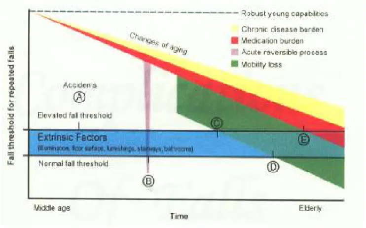
FIGURE 1.
FACTORS THAT CONTRIBUTE TO THE RISK FALLS IN
THE ELDERLY POPULATIONS
KEY:
A = Patient with an accidental fall and no intrinsic or extrinsic risk factors
B = Patient with acute illness.
C = Patient with moderate illness, loss of mobility and some prescription
medications who falls because of an extrinsic factor.
The balance system works with the visual skeletal systems (the muscle and joints and
their sensors) to maintain orientation or balance. For example, visual signals are sent to
the brain about the body’s positioning relation to its surroundings. These signals are
processed by the brain, and compared to information from the vestibular and the
skeletal system. An example of interaction between the visual and vestibular system is
Called the vestibular-ocular reflex. The nystagmus (an involuntary rhythmic Eye
movement) that occurs when a person is spun around and then Suddenly stops is in
example of avestibular-occur reflex.
Balance control
ITEM DESCRIPTION:
1.SITTING TO STANDING:
INSTRUCTIONS: Please stand up. Try not to use your hand for
Support.
( ) 4 able to stand without using hands and stabilize independently.
( ) 3 able to stand independently using hands.
( ) 2 able to stand using hands after several tries.
( ) 1 needs minimal aid to stand or stabililize.
( ) 0 needs moderate or maximal assist to stand.
REACHING FORWARD WITH OUTSTRETCHED ARM WHILE
STANDING
9.PICK UP OBJECT FROM THE FLOOR FROM A STANDING
POSITION:
INSTRUCTION:Pick up the shoe/slipper, which is place in front of
Your feet.
( ) 4 able to pick up slipper safely and easily.
( ) 3 able to pick up slipper but needs supervision.
( ) 2 unable to pick up but reaches 2-5 cm (1-2 inches) from slipper
and keeps balnce independently.
( ) 1 unable to pick up and needs supervision while trying.
( ) 0 unable to try/needs assist to keep from balance or falling.
PICKING UP OBJECT FROM THE FLOOR FROM A STANDING
POSITION
10. TURNING TO LOOK BEHIND OVER LEFT AND RIGHT
SHOULDERS WHILE STANDING:
INSTRUCTION:Turn to look directly behind you over toward the
left shoulder. Repeat to the right.Examiner may pick an object to
look at directly behind to encourage a better twist turn.
( ) 4 looks behind from both sides and weight shifts well.