JOURNAL OFVIROLOGY, May 2007, p. 5192–5201 Vol. 81, No. 10 0022-538X/07/$08.00⫹0 doi:10.1128/JVI.02447-06
Copyright © 2007, American Society for Microbiology. All Rights Reserved.
CTCF-Dependent Chromatin Boundary Element between the
Latency-Associated Transcript and ICP0 Promoters in the
Herpes Simplex Virus Type 1 Genome
䌤
Qi Chen,† Lan Lin,† Sheryl Smith, Jing Huang, Shelley L. Berger, and Jumin Zhou*
The Wistar Institute, 3601 Spruce Street, Philadelphia, Pennsylvania 19104
Received 6 November 2006/Accepted 24 January 2007
Cells latently infected with herpes simplex virus (HSV) contain nucleosomal DNA similar to that of host cell chromatin. Recent studies have demonstrated that histones in the latency-associated transcript (LAT) pro-moter and intron regions contain histone modifications permissive for transcription. However, those histones
associated with the lytic-specificICP0gene, which lies only 5 kb away, contain modifications typical of silenced
chromatin. How this active chromatin is kept separate from the repressed chromatin in the nearbyICP0region
remains crucial to the understanding of the HSV lytic cycle. In this study, we show that the LAT intron region contains an insulator. Specifically, we show that an 800-bp region from the LAT intron can block enhancers
in both tissue culture cells and Drosophila melanogaster embryos. Importantly, the 800-bp HSV insulator
protects a LAT transgene from positional effects in Drosophilaeye tissue. The 800-bp region contains nine
copies of 16-bp repeats. In vitro electrophoretic mobility shift assay revealed that CTCF interacts with the CTCCC sequence within the repeats. In vivo chromatin immunoprecipitation assay demonstrated that CTCF interacts with these repeats in latently infected trigeminal ganglion neurons. The deletion of these repeats
impaired insulator activity in human K562 cells andDrosophilaembryos. Finally, double-spaced RNA
knock-down of CTCF disrupts enhancer-blocking activity of the LAT insulator in transfectedDrosophilaS3 cells.
These results strongly support the hypothesis that the 800-bp DNA in the LAT intron region works as a chromatin boundary during latency to separate active chromatin associated with the LAT promoter region from repressed chromatin in the ICP0 gene.
Human herpes simplex virus (HSV) is the main cause of cold sores and genital sores. The HSV type 1 (HSV-1) genome contains 152 kb of double-stranded DNA encoding close to 100 transcripts, more than 70 of which have been shown to code for proteins. During latent infections, the viral genome is main-tained in sensory neurons. Only one large transcript, the latency-associated transcript (mLAT), is actively transcribed during latency (5, 11, 26, 37, 46). The highly regulated cascade of transcription during the productive phase of infection does not occur, since these genes are silenced. However, the mechanism of maintaining latency is not well understood.
The latent HSV genome is circular, episomal, and chroma-tinized (12, 25, 32). Recently, it has been shown that the LAT region bears histone covalent modifications known to be asso-ciated with activated genes in mammalian cells (24, 38). Spe-cifically, the LAT region is enriched with histone H3K9 and H3K14 acetylation, whereas the surrounding regions, such as the lytic-specific gene ICP0, contain histones that are hy-poacetylated and methylated at H3K9 and H3K14. These dis-tinctive histone modification patterns are observed in several well-characterized loci, including the vertebrate-globingene locus and theSaccharomyces cerevisiaemating type loci, where the open, transcriptionally competent region is rich in
acety-lated histones compared with the surrounding repressed re-gions.
Importantly, insulators are present at the boundary between these regions (6). At the yeast mating type loci, deleting the boundary element leads to the spread of heterochromatin into transcriptionally active regions (28, 29). In the chicken-globin
locus, the HS4 insulator also demarcates the boundary be-tween the heterochromaticfolatereceptor gene and the active locus control region (LCR) (4, 10, 40). In addition, a recent report indicated that an enhancer-blocking activity might be associated with the LAT intron (2). These studies suggest that insulators may surround the LAT region of HSV-1 and protect it from epigenetic silencing present in most of the genome during latency.
Insulators or chromatin boundary elements usually contain two measurable activities. First, insulators block the activity of an enhancer when inserted between the enhancer and its pro-moter; second, insulators prevent regulatory activities on one side of the element from affecting regions on the other side. Thus, insulators can protect transgenes from positional effects when inserted into the genome. Insulators were first identified from theDrosophila melanogasterheat shock locus, where the
scs,scs⬘elements (14, 20, 42) protect the active chromatin in this locus from invading neighboring genes during heat shock. To date, at least two dozen insulators have been characterized in organisms ranging from yeast to humans (39). At the func-tional level, there is a remarkable conservation of insulator function across species despite the apparent diversity of inter-acting proteins and lack of conserved DNA sequences among different insulators. For example, theDrosophila scsandscs⬘
* Corresponding author. Mailing address: The Wistar Institute, 3601 Spruce Street, Philadelphia, PA 19104. Phone: (215) 898-3988. Fax: (215) 898-0663. E-mail: [email protected].
† These authors contributed equally to this work. 䌤Published ahead of print on 31 January 2007.
5192
on November 8, 2019 by guest
http://jvi.asm.org/
elements have been shown to block enhancer-promoter inter-actions in injectedXenopuseggs (13). In contrast, the-globin
insulator has been shown to protect the mini-white gene in
Drosophila against positional effects (10, 33). Recently, the
-globininsulator was reported to protect genes inserted into
an adenovirus vector (35, 41).
The evolutionary conservation of insulator function suggests the conservation of proteins. In collaboration with others, we identified theDrosophilaorthologue of vertebrate CTCF and demonstrated that it mediates the function of theFab-8 insu-lator in flies (27). Antibody staining of dCTCF on polytene chromosomes from the third instar larvae reveals roughly 200 binding sites for the protein, with approximately 30% of these bands overlapping with another insulator protein, Mod(mdg4) (S. Smith et al., unpublished results) (15). These studies sug-gest that theDrosophilagenome contains many CTCF-depen-dent insulators, which may explain why vertebrate insulators function appropriately in flies. In addition, these studies sug-gest that Drosophilacould be an excellent model system for testing insulator elements from various species.
Very little is known about the chromatin structure of viral genomes, including viral insulators. However, given the com-pact genome size and highly regulated cascade of gene expres-sion, it is clearly necessary to shield one type of chromatin from adjacent regions or one transcription unit from the next. In several studies, inserting vertebrate insulators into the viral genome protects transgenes from silencing or positional effects (35). More importantly, a recent study has identified a
CTCF-dependent insulator-like element from the Epstein-Barr virus genome (9). These studies suggest that there may be no fun-damental difference between viral chromatin and the host chromatin in terms of insulator function and that insulators may function both in viral genomes and in host chromatin. In this study, we report the identification and characterization of an insulator from the HSV-1 LAT intron region that functions via CTCF.
MATERIALS AND METHODS
Plasmid constructs.For theDrosophila enhancer-blocking assay, the 2-kb HSV-1 LAT intron and its truncated 1.4-kb 5⬘fragment, the 0.8-kb 3⬘fragment of 1.4 kb, and the CTCF binding site deletion fragment of 0.8 kb were cloned into 2⫻phycoerythrin (PE)-I5 construct at the BamHI restriction site with PCR-generated BamHI tails (GC-rich PCR kit; Invitrogen), respectively. 2⫻PE-I5 vector with a l-kbspacer orFab-8insulator cloned at the BamHI site work as negative and positive controls, respectively. ForDrosophila positional effect assay, two copies of 250-bp chicken-globinHS4 minimal insulators or 0.8-kb HSV-1 LAT insulator (Fig. 1) were cloned into the NsiI site (downstream of the whitegene) and BglII site (upstream ofwhite gene between H1 and NEE enhancers) of the HGN vector (45) with PCR-generated fragments with corre-sponding restriction enzyme site tails, respectively. For the tissue culture enhancer-blocking assay, the 2-kb LAT, 800-bp LAT, 200-bp CTCF binding sites, 800-bp LAT with CTCF deletion (800⌬), S5, andFab-8were generated by PCR and cloned into the pNI (11) vector at the SacI site.
Pelement transformation and genetic crosses.Pelement transformation vec-tors containing themini whitegene were introduced into theyw67
[image:2.585.108.470.66.334.2]fly embryos by microinjection as described previously (34). For each of the transgenic con-structs, independent transformants were selected and maintained as transgenic stocks.
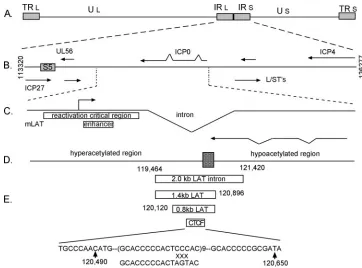
FIG. 1. Structure of the LAT region of the HSV. (A) Structure of the HSV genome. TR, terminal repeat; IR, internal repeats. (B) Detailed map showing different genes within the internal repeats. S5 is a 1-kb DNA fragment from 114,919 to 115,904. (C) LAT region showing LAT promoter, LAT enhancer, and LAT intron. (D) Histone acetylation across the LAT region based on reference 23. (E) Different tested DNAs from the LAT region. The rectangular bars indicate the location of the 2.0-kb intron, 1.4-kb intron, 0.8-kb LAT insulator, and 200-bp region containing 9 repeats of CTCF binding sites. The 200-bp sequence is shown at the bottom of the figure. Mutation to the putative CTCF binding sites in the region is also shown.
on November 8, 2019 by guest
http://jvi.asm.org/
RNA in situ hybridization.Whole-mount RNA in situ hybridizations were performed as described previously (36, 45). Briefly, 2- to 4-h embryos were collected, dechorionated with 50% bleach for 5 min, and fixed with 4% formal-dehyde in phosphate-buffered saline and heptane for 20 min. Methanol was added to the fixed embryos to remove the vitelline membranes. These embryos were incubated with prehybridization buffer (50% formamide, 4⫻SSC [1⫻SSC is 0.15 M NaCl plus 0.015 M sodium citrate], 1⫻Denhardt’s, 250g/ml tRNA, 250g/ml single-stranded DNA, 50g/ml heparin, 0.1% Tween 20, and 5% dextran sulfate) at 55°C for 1 h and 12 to 16 h when antisense RNA probes for the specific reporter genes were added to final concentration of 0.25g/ml. Embryos were washed with wash buffer (50% formamide, 2⫻SSC, 0.1% Tween 20) at 55°C for 4 to 6 h, and phosphate-buffered saline with 0.1% Tween 80 for 30 min. Anti-digoxigenin-alkaline phosphatase antibody (Roche) was added to the embryo at a 1:2,000 dilution. The color reaction was carried out at room temperature with alkaline phosphatase staining buffer containing nitroblue tet-razolium and 5-bromo-4chloro-3-indolylphosphate (BCIP; Roche). Stained em-bryos were rinsed, fixed with ethanol, and mounted on glass slides.
K562 cell transfection and colony formation assay.K562 (human erythromy-eloblastoid leukemia cell line) was a gift kindly supplied by V. Lobanenkov, cultured in the Iscove’s modified Eagle’s medium (Invitrogen) supplemented with 10% fetal bovine serum (Invitrogen) and 1⫻penicillin-streptomycin in a 37°C incubator supplied with 5% CO2. Cells (1⫻10
7
) were harvested for each transfection. Twenty microliters of Lipofectamine 2000 (Invitrogen) and 2g of SalI linearized plasmid DNA were separately diluted into 2 ml OPTI-MEMI (Invitrogen) reduced serum minimum, shortly incubated, then gently mixed, and incubated at room temperature for 20 min. Harvested cells were resuspended in transfection medium (Iscove’s modified Eagle’s medium plus 10% fetal bovine serum) to a final concentration 1⫻107cells/ml, and each 1-ml aliquot was
diluted in 20 ml of transfection medium. DNA/Lipofectamine mixtures were loaded onto cells, gently mixed, and then incubated at 37°C for 24 h. The transfected cells were collected and resuspended in 10 ml of complete medium with 750g/ml Geneticin (Invitrogen), and a 5-ml aliquot was separately diluted into 30 ml of warmed medium with 0.3% tissue culture agar (A9915; Sigma). The cells were quickly inverted and poured onto 15-cm petri dishes. Colonies were counted after 2 to 3 weeks of incubation at 37°C.
Mice infection and TG dissection.Six- to eight-week-old female BALB/c mice (Jackson Laboratories) were anesthetized with pentobarbital (50 to 90 mg/kg), ketamine (80 mg/kg), and xylazine (10 mg/kg), followed by corneal scarification and infection with HSV-1 at 3.7⫻106PFU/mouse. At 30 days postinfection,
mice were sacrificed and trigeminal ganglia (TG) were removed. The average time to isolate TG from one mouse is 1 to 2 min. After dissection, TG were snap-frozen in dry ice and stored at⫺80°C.
EMSA.Electrophoretic mobility shift assays (EMSA) reaction mixtures (20l) contained either human CTCF protein (1g) produced in baculovirus (provided by P. Lieberman) orDrosophilaCTCF (4l) produced by using a TNT-T7 Quick-Coupled Transcription/Translation system (Promega). Reactions were carried out using PCR-amplified DNA from the putative repetitive binding region for CTCF within the 2-kb LAT intron of HSV-1 strain 17 (coordinates 120467 to 120671) engineered with EcoRI restriction sites on both ends. DNA generated by PCR was digested with EcoRI and labeled with 20 Ci of [␣-32
P]dATP and unlabeled dTTP using 1 U Klenow fragment (New England Biolabs). Unincorporated nucleotides were removed using a MicroSpin G-25 spin column (Amersham Biosciences), and 1 to 2 ng labeled probe was used in each reaction. EMSA reactions additionally contained 0.5g/l poly(dI-dC) (Sigma), 0.5g/l bovine serum albumin, 5% glycerol, 20 mM HEPES (pH 7.6), 10 mM MgCl2, 1 mM dithiothreitol, 30 mM KCl, and 0.1 mM ZnSO4in a final
reaction volume of 10l.Competition assays were carried out using unlabeled
wild-type (full length or 26mer, 5⬘-CCCACGCACCCCCACTCCCACGCAC C-3⬘[the CTCF binding motif is underlined]) or mutant (26mer, 5⬘-CCCACG CACCCCCACTAGTACGCACC-3⬘) fragments. Reactions were carried out at 25°C for 30 min and resolved using a 6% DNA retardation gel (Invitrogen). Bands were visualized by exposure of dried gels to X-OMAT Blue XB-1 film (Kodak) for 8 h at⫺80°C. Films were developed using a Kodak X-OMAT 1000A processor.
ChIP assay.TG were digested with serum-free medium containing 0.125% Trypsin-EDTA, 0.195 mg/ml collagenase (Sigma), and 5 mM CaCl2at 37°C for
1 h. Cells were passed through a cell strainer (100m; BD Bioscience) and fixed with 1% formaldehyde at room temperature for 10 min. Cells were then col-lected by spinning at 1,200 rpm for 5 min, washed once with ice-cold phosphate-buffered saline, and subjected to chromatin immunoprecipitation (ChIP) assay. ChIP assays were carried out essentially by following the procedures described previously (17), except that sodium deoxycholate was removed from the lysis buffer. Eight TG (four mice), 2g H3 antibody (polyclonal; Abcam), and 2.5l
CTCF antibody (cocktail of monoclonal, a kind of gift from Victor Lobanenkov) were used for each immunoprecipitation. Immunoprecipitated DNA was reverse-cross-linked and purified with the QIAquick 8 PCR purification kit (QIAGEN). The amount of DNA was measured by real-time PCR as previously described (17). Primers for thymidine kinase (TK), ICP0 promoters were mentioned pre-viously (22). Primers for mouse glyceraldehyde-3-phosphate dehydrogenase (GAPDH), CTCF binding site, and LAT promoter are mouse GAPDH (for-ward, 5⬘-CAATGTGTCCGTCGTGGATCT-3⬘; reverse, 5⬘-TTGAAGTCGCA GGAGACAACC-3⬘), CTCF binding site (forward, 5⬘-AGACAGGGAAAAGA TACAAAAGTAAACC-3⬘; reverse, 5⬘-TTTGCTGTCTGTTGGGAAATAAA G-3⬘), LAT promoter (forward, 5⬘-GTGCCCGCGAGATATCAATC-3⬘; reverse, 5⬘-GGAAAGTCTTTCGCACCACC-3⬘). The input signal with mouse GAPDH and TK primers was used to calculate relative genomic signal of HSV-1 of infected to uninfected TG.
Insulator assay and dsRNA interference knockdown of CTCF in transfected
DrosophilaS3 cells.2XR, a red fluorescent protein (RFP)/green fluorescent protein (GFP) dual-fluorescence vector, was a gift, kindly supplied by Haini Cai from the University of Georgia at Athens. This vector was modified as follows. The metallothionein enhancer between the DsRed and enhanced GFP genes was replaced by an EcoRI/NotI double digest with the 2⫻PE enhancer from the 2⫻ PE-IAB5 construct (previously described). Two tandem copies of either 800-bp LAT or its counterpart with CTCF binding sites deleted were inserted at the NotI site between the 2⫻PE enhancer and enhanced GFP gene to make RPLG or RPLDG constructs, respectively. S3 cells were cultured and transfected in HyQ-SFX insect medium (HyClone) supplemented with 0.5⫻ penicillin-strep-tomycin. One milliliter of 5⫻105
cells/ml was seeded per well using a 12-well plate for each transfection. One microgram of DNA construct and 2.5l of Cellfectin (Invitrogen) were separately diluted into 100l of media, shortly incubated for 10 min, and then mixed and incubated for 45 min at room tem-perature. The cells were then rinsed, subsequently loaded with DNA/Cellfectin mix and 800l media, and then incubated at 25°C for 5 h. The transfection cocktail was then replaced with 2 ml of media, and the fluorescence signal was assessed after 24 h of incubation at 25°C, using an inverted microscope. A 500-bp CTCF sequence was amplified with T7 promoter-tailed primers (forward, T7-A GACTACGCCCAAGAAGCAA; reverse, T7-CTTGTCGGCATTCTCATCCT) and then transcribed using an in vitro RNA transcription kit (MEGAscript T7 kit; Ambion). Double-stranded (dsRNA)-CTCF was then purified using an RNAeasy kit (QIAGEN), and the optical density at 260 nm (OD260) was
mea-sured to determine the concentration. For transfections involving dsRNA inter-ference, cells were seeded and 5g of dsRNA-CTCF was loaded directly into the medium 3 days prior to transfection.
RESULTS
Insulator elements usually exhibit two measurable activities: blocking the action of an enhancer (when inserted between the enhancer and its promoter) and preventing the spread of active or repressed chromatin structure along the chromosome. Most of the insulators tested have both functions. Because LAT is the only actively transcribed gene from the virus in latently infected cells, we hypothesize that insulators may be present near the LAT promoter to prevent it from being silenced by nearby inactive chromatin. The LAT region is located between two early lytic genes,ICP0 and ICP27 (Fig. 1). The mLAT transcript extends approximately 8.5 kb. ICP0 is transcribed from the opposite strand and overlaps with the mLAT tran-scribed region. The 8.5-kb mLAT also contains a stable 2-kb intron, which is thought to be important for maintaining the survival of the infected neuron (1, 18, 31). Recent studies (23, 24) using ChIP to detect the histone modification status of the LAT region from latently infected animals demonstrated that hyperacetylated histones exist from the LAT promoter to the 2-kb intron. TheICP0region, however, is hypoacetylated. An-other active histone modification, H3K4 methylation, is also detected in the LAT promoter and intron regions. Based on the ChIP studies, the boundary between transcription-permis-sive and -nonpermistranscription-permis-sive regions is located near the 3⬘end of
5194 CHEN ET AL. J. VIROL.
on November 8, 2019 by guest
http://jvi.asm.org/
theICP0, which overlaps with the LAT 2-kb intron by about 700 bp (2).
The LAT intron contains an enhancer-blocking activity.To
test for the presence of a potential insulator element, we fo-cused on the 2-kb LAT intron region (Fig. 1) due to its location between the inactiveICP0 and the actively transcribed LAT promoter during latency. We took the entire 2-kb LAT, the first 5⬘1.4 kb, and the last 700-bpICP0-overlapping sequence and tested them in aDrosophilaembryonic enhancer-blocking assay. The Drosophila transgene vector contains two diver-gently transcribed genes,white(w) andlacZ. Between the two gene promoters, we inserted two embryonic enhancers—PE from thetwistgene (19) and IAB5 from theAbd-Blocus (7). Multiple cloning sites were included between the two enhanc-ers for test DNAs. These P element transformation vectors were introduced into theDrosophilagerm line by injectingyw67
embryos as described previously (36). Multiple independent transformants were obtained for the recombinantPelement shown in Fig. 2. We collected and fixed 2- to 4-h-old embryos. In situ hybridizations using DigU-labeled antisense RNA probes against white or lacZ were performed essentially as described in previous reports. Transgene expressions were vi-sualized using anti-DigU antibody-labeled alkaline phosphate, followed by adding substrates nitroblue tetrazolium and BCIP.
Drosophilaembryonic enhancers are regulated by
segmen-tation genes, which are expressed along the anterior-posterior or dorsal-ventral gradient. Enhancers integrate segmentation gene gradients and result in bands or stripes of reporter gene activity (44). The 2⫻PE enhancer directs transgene expression in the ventral-most region of the embryo and produces an anterior-posterior stripe (Fig. 2A). The IAB5 enhancer acti-vates the transgene in the posterior third of the embryos, seen as a vertical band (Fig. 2B).
When these two enhancers were both included in the trans-gene, separated by a control 1-kb spacer, they produced additive activities on both the leftwardwgene and the right-ward lacZgene (Fig. 2A and B). However, when they were separated by the Drosophila Fab-8 insulator, the expression pattern changed. The additive activities were no longer ob-served. The proximal 2⫻PE enhancer was still functional with respect to thewpromoter, but the distal IAB5 enhancer was no longer active (Fig. 2C). On the contrary, thelacZpromoter was found to be solely activated by IAB5, whereas 2⫻PE did not activatelacZ(Fig. 2D). This result indicated that the Fab-8
element blocked the IAB5-w and 2⫻ PE-lacZ interactions. This selective block only affected the insulator-distal enhancer for a given promoter. The promoter-proximal enhancer was not affected (2⫻PE forwand IAB5 forlacZ). Therefore, the
Fab-8 insulator blocks enhancer-promoter interaction only when inserted between the enhancer and its promoter.
When the 2-kb LAT intron was tested in the same manner, it exhibited a similar effect on the enhancer-promoter interac-tion: it blocked 2⫻PE-wand IAB5-lacZinteractions, although to a lesser extent thanFab-8(Fig. 2E, F). We then tested the 5⬘1.4-kb LAT in the enhancer-blocking assay and found that it blocked enhancer-promoter interactions to an extent similar to that of the 2-kb LAT intron (Fig. 2G, H). However, when the 3⬘0.7-kb LAT intron was tested, it showed no enhancer block-ing; the staining pattern ofwandlacZwas similar to that of the control (data not shown). These results suggest that the LAT intron contains enhancer-blocking activity that maps to the 5⬘ 1.4-kb region.
The LAT intron DNA blocks the LCR enhancer in tissue
culture.In the aboveDrosophilaexperiments, the LAT intron
DNA functioned as an insulator element, but it was not clear whether this element also blocks enhancer-promoter interac-FIG. 2. Enhancer blocking by LAT intron DNAs. Two- to four-hour-old embryos carrying each of the transgenes shown were collected and subjected to in situ hybridization using antisense RNA probes to either thew(A, C, E, G, I, K) or thelacZ(B, D, F, H, J, L) gene. All embryos were oriented anterior to the left and dorsal side up. (A, B) Spacer control; (C, D)Fab-8insulator fromDrosophila Abd-Blocus; (E, F) 2.0-kb LAT intron; (G, H) 1.4-kb LAT intron; (I, J) 0.8-kb LAT intron insulator; (K, L) 0.8⌬, 0.8-kb LAT intron insulator with CTCF binding sites deleted.
on November 8, 2019 by guest
http://jvi.asm.org/
tions in mammalian cells. To accomplish this, we tested the LAT DNA in a well-established colony assay (10, 27). The transfection vector pNI contains the neomycin-resistant gene
neodriven by mouse␥-globinpromoter and an LCR enhancer element that activates its expression in K562 cells (Fig. 3A, vector kindly provided by Victor Lobanenkov’s group from NIH). Since the transfected vector would integrate into the genome as an array of multiple units, a 1.2-kbHS4insulator was inserted at the 5⬘of the␣-neogene to protect the enhancer in one vector unit from activating the promoter in the neigh-boring unit. Test DNAs were cloned between␣-neo and the LCR enhancer at SacI site. When the vectors were transfected into K652 cells, molten soft agar supplemented with Geneticin was poured with the transfected cells. The cells were immobi-lized by agar. In approximately 2 to 3 weeks, transfected cells expressing ␣-neo formed individual colonies, whereas non-transfected cells or low-neo-expressing cells either died out or formed tiny colonies. When an insulator element is inserted between the LCR enhancer and ␣-neo, it should block the LCR and lead to no, or very low levels of␣-neoexpression. In such transfections, a low colony number is expected. In con-trast, when a spacer DNA is inserted, a larger number of colonies would be expected because the LCR enhancer could activate␣-neoin all transfected cells.
We tested five DNA fragments, a 1-kbspacer DNA, the 1.2-kbHS4-globininsulator, the 2-kb LAT intron, theFab-8
insulator (43), and a 1-kb HSV DNA (S5) near the 3⬘end of the ICP27 gene (Fig. 1). We found that the -globin HS4
insulator, the 2-kb LAT, and Fab-8 blocked neo expression,
whereas the control 1-kbDNA, HSV sequence S5, did not (Fig. 3B). As can be seen in Fig. 3B, the positive control, the 1.2-kbHS4insulator, reduced the LCR enhancer by twofold. Similarly, the 2-kb LAT intron DNA reduced the LCR en-hancer 2.5-fold. TheDrosophila Fab-8insulator, on the other hand, gave a threefold reduction to the LCR enhancer. The S5 sequence did not appreciably affect␣-neotranscription. These results suggest that the 2-kb LAT intron DNA functions as an insulator. This experiment also confirms our previous observa-tion that theDrosophila Fab-8insulator blocks enhancer-pro-moter interactions in mammalian cells (27).
The 800-bp LAT insulator blocks positional effects in
Dro-sophila. Since the LAT intron insulator is located between active and repressive chromatin, it may potentially provide the chromatin boundary to restrict the silenced chromatin from inactivating LAT transcription during latent infection or pre-vent the active LAT chromatin from ectopically activating si-lenced genes such as ICP0. Therefore, it is important to test whether the LAT insulator also functions as a chromatin boundary or chromatin barrier. For this reason, we analyzed the 800-bp LAT insulator for the protection of positional ef-fects in theDrosophilaeye. Position effects are caused by trans-gene integration into active or repressed genomic chromatin. As a result, the transgenes are expressed at various levels due to local chromatin structure near the integration site (10, 21). Two copies of the 800-bp LAT DNA, including the CTCF binding sites (Fig. 4E), were inserted both upstream and down-stream of thewgene. In addition, two embryonic enhancers, H1 and NEE were also inserted so that the NEE (but not H1) FIG. 3. Colony assay for insulator activity in K562 cells. Test DNA fragments were inserted into a SacI site between the LCR enhancer and the neomycin-resistant gene␣-neodriven by the mouse␥-globinpromoter. The 5⬘HS4 insulator was needed to prevent cross regulation between neighboring vectors. Transfections were overlaid with soft agar in the presence of Geneticin. Colonies appeared in 2 to 3 weeks and were counted. An insulator would reduce the number of colonies as the LCR enhancer was blocked and could not activate␣-neo. Results represent an average of 6 to 9 experiments. (A) Reporter gene construct showing theneoresistance gene, the LCR enhancer, and the test DNA. If the test DNA blocks enhancer-promoter interaction, the transfection would produce a smaller number of colonies. (B) Different test DNA fragments were tested and the results are shown: pNI vector with 1-kbinsert; pJC5-4 vector carrying 1.2-kbHS4insulator (3); 2-kb LAT, 800-bp LAT, 200-bp LAT, and 800-bp LAT represented the different LAT fragments tested; 800-bp⌬CTCF is the 800-bp LAT with CTCF binding repeats deleted;Fab-8(43) is the 680-bpFab-8insulator from theDrosophila Abd-Blocus; S5 is a 1-kb DNA from the 3⬘region of ICP27, which is also located upstream of the LAT promoter. The experiments were done in duplicates and repeated at least six times. Results are relative␣-neo activity following normalized colony numbers against control DNA (set to 100).
5196 CHEN ET AL. J. VIROL.
on November 8, 2019 by guest
http://jvi.asm.org/
was separated fromwby the LAT insulator (Fig. 4E). A similar control construct was made by replacing LAT with two copies of 250-bp minimalHS4 insulator from the chicken -globin
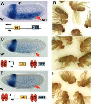
LCR (Fig. 4C). As can be seen from Fig. 4A, the transgenic enhancers (H1 and NEE) activated additivewgene expression in early embryos. In adults, different strains of flies exhibited a great variation of eye colors. Five representative strains are shown in Fig. 4B, where eye color ranged from pale yellow to dark red. When the control HS4 insulator was inserted in the transgene, the distal NEE enhancer was blocked from activat-ingw(Fig. 4C). Because HS4 flanked thewgene, protecting it from positional effects, adult flies showed little variation of eye color, a result consistent with a previous published study (10) (Fig. 4D). When the 800-bp LAT element was inserted, it also selectively blocked the NEE enhancer without affecting the proximal H1 (Fig. 4E). Importantly, this element showed an even stronger protection against positional effects compared to HS4 (Fig. 4F). These results strongly suggest that the 800-bp LAT insulator may function as a chromatin boundary to pre-vent the spread of active or silenced chromatin in the Drosoph-ilaeye tissue. The stronger insulation against positional effects
[image:6.585.136.449.68.425.2]echoes the stronger enhancer-blocking effect seen in Fig. 2 and Fig. 3 compared to the HS4 insulator, which is probably due to the higher number of CTCF binding sites presented in the repeats (see below).
CTCF interacts with repeated regions within the LAT
in-tron.Because almost all well-studied vertebrate insulators use
CTCF to function, we examined the LAT intron DNA for potential CTCF binding sites. CTCF is known to interact with diverse target sequences, and a consensus could not be reliably established. However, most CTCF binding sites are rich in G or C residues (3, 8, 16, 30). This is in part due to the fact that CTCF appears to use different Zinc fingers for different target sites (30). Indeed, a stretch of 9 repeats of a 16-bp DNA within the 1.4-kb LAT insulator near the 3⬘end ofICP0is rich in GC residues, which resembles the pattern of many known CTCF binding sites (Fig. 1) (30). To test if these repeats interact with CTCF proteins, we conducted EMSA with Drosophila and human CTCF. As shown in Fig. 5, theDrosophilaCTCF spe-cifically bound the 216-bp DNA sequence containing the re-peats (lanes 2, 3). This binding can be blocked by cold unla-beled competitor DNA corresponding to one of the repeats FIG. 4. Enhancer blocking and protection of positional effect by the 800-bp LAT insulator. (A) In the control transgene, the embryonic enhancers H1 and NEE activated thewpromoter in early embryos. (B) Adult flies carrying this transgene showed different eye colors varying from strain to strain. Five representative strains are shown. (C) The HS4 insulator from chick-globinLCR blocked the NEE enhancer (arrow). (D) Five representative transgenic strains from panel C are shown here with very little eye color variation. (E) The 800-bp LAT insulator also blocked NEE (arrow) but did not affect H1 activity. (F) The transgene carrying LAT showed pale yellow eye color with almost no variations.
on November 8, 2019 by guest
http://jvi.asm.org/
but not by a mutated competitor (lanes 4 to 9, CTCCC changed to CTAGT), indicating that dCTCF indeed binds specifically to the repeat. When the same competition assay was done with human CTCF, a similar result was obtained (data not shown). Similarly, human CTCF binding to the 216-bp probe (lanes 11, 12) can be specifically competed by a cold (unlabeled) 216-bp fragment (lanes 13 to 15) but not by an equimolar amount of nonspecific competitor (dI-dC) using 850 bp as a median base pair value (lanes 16 to 18). These data clearly demonstrate that the LAT insulator interacts with both
Drosophilaand human CTCF, suggesting that LAT insulator
activity may be mediated by the conserved, CTCF-dependent enhancer-blocking mechanism.
A CTCF-dependent enhancer-blocking activity resides within
the 800-bp LAT intron.To test the role of the putative CTCF
binding site, we assayed three DNA fragments, an 800-bp DNA, which includes the CTCF binding sites, flanked by 300 bp on either side, a 200-bp DNA fragment that includes the nine copies of the 16-bp repeats, and a 600-bp fragment made from deleting the 200-bp CTCF sites from the 800-bp element. We first tested these DNAs in the colony assay (Fig. 3) and found that the 800-bp DNA blocked colony formation effi-ciently, while the 200-bp DNA only slightly affected colony number. In contrast, deleting the CTCF binding sites from the 800-bp DNA dramatically reduced the enhancer-blocking tivity. This result indicates that the LAT intron insulator ac-tivity mostly resides within the 800-bp region and the CTCF binding sites are essential for insulator activity. However, these sites are not sufficient for insulator activity, suggesting that additional DNA flanking the CTCF sites may be necessary. Supporting this possibility, removing 100 bp of DNA from the 3⬘ end of the 1.4-kb LAT fragment reduced its enhancer-blocking activity in fly embryos (data not shown). This notion is also consistent with our recent study of theFab-8insulator, where the minimal 100-bp DNA containing the CTCF binding sites does not block enhancer-promoter interactions even though CTCF is necessary for the function of this insulator (Q. Chen and J. Zhou, unpublished).
[image:7.585.46.282.65.197.2]Because the colony assay could not clearly distinguish a transcriptional silencer from an insulator, we therefore tested the 800-bp LAT DNA and the 600-bp LAT⌬(800-bp LAT with CTCF binding repeats deleted) inDrosophilaembryos. Insert-ing the 800-bp DNA in the Drosophila vector lead to the blocking of both PE enhancer-lacZpromoter interaction (Fig. 2I) and IAB5 enhancer-wpromoter interaction (Fig. 2J), con-firming that the insulator activity maps within this DNA. When the 200-bp CTCF binding sites were removed from the 800-bp DNA, the enhancer-blocking activity was greatly reduced. This can be seen by comparing Fig. 2K with Fig. 2I or comparing Fig. 2L with Fig. 2J. This result confirms that CTCF binding
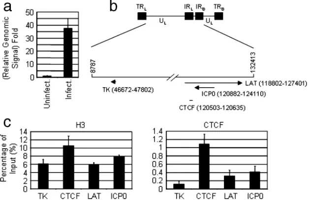
FIG. 6. CTCF interacts with the LAT intron in latently infected TG by HSV-1. (a) DNA from uninfected and infected TG were prepared and purified as described in Materials and Methods. Real-time PCR signals with TK primer from uninfected and infected TG were normalized to the corresponding signal with the mouse GAPDH primer. The value of uninfected TG was set to 1. (b) Schematic of HSV-1 genome. (c) Recruitment of histone H3 and CTCF to various regions of HSV-1 during latent infection.
FIG. 5. CTCF interacts with the LAT intron insulator in vitro. EMSA analyses of dCTCF binding to LAT (lanes 1 to 9). Human CTCF (lanes 10 to 18) binding to the LAT intron insulator. Both
Drosophilaand human CTCF bound LAT (lanes 2, 11, 12). This bind-ing was almost completely obliterated by wild-type (WT) bindbind-ing sites (lane 5, 14, 15) but not by mutant binding sites (lane 8) or nonspecific competitor (comp.) (lanes 3, 7, 8, 16, 17, 18).
5198 CHEN ET AL. J. VIROL.
on November 8, 2019 by guest
http://jvi.asm.org/
[image:7.585.136.450.483.687.2]sites are essential for enhancer-blocking activity of the 800-bp LAT insulator.
CTCF interacts with the LAT region in latently infected
mouse TG. Since CTCF interacts with LAT in vitro and is
required for its enhancer-blocking activity, it is clearly possible that CTCF interacts with the LAT region of the HSV genome in latently infected cells. To this possibility, we performed a ChIP assay on the TG from mice latently infected with the HSV-1 F strain as previously shown (17, 23, 38) (see Materials and Methods). We first compared the relative genomic signal from uninfected and infected TG to determine whether the latent infection was successful. Using quantitative real-time PCR and primers for thymidine kinase, we consistently ob-served that the relative genomic signal from infected TG was about 37-fold higher than that from uninfected TG (Fig. 6a), indicating that HSV-1 successfully established latency in TG under our experimental conditions. ChIP assays were then performed in infected TG to detect the recruitment of histone H3 and CTCF to different regions of HSV-1 (Fig. 6b and c). The recruitment of histone H3, serving as a positive control, to different regions of HSV-1 was comparable, although the sig-nal for CTCF binding site was slightly higher than those for the other sites (Fig. 6b, left panel). CTCF, however, was found to associate with its binding site significantly stronger (3- to
10-fold) than to the other sites (Fig. 6c, right panel). Therefore, our results show that the putative CTCF binding site in HSV-1 is the physiological binding site of CTCF in the latently in-fected TG.
CTCF is essential for the enhancer-blocking activity of the
LAT intron insulator.To finally demonstrate that the CTCF
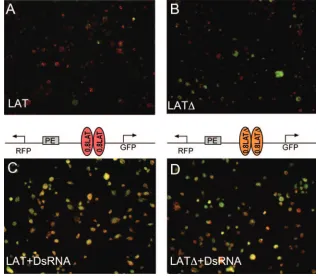
[image:8.585.132.450.70.346.2]protein is essential for the enhancer-blocking activity of the LAT insulator, we took advantage of the recently developed dual fluorescent protein enhancer-blocking vector (M. Li and H. Cai, personal communication) and modified it so that it includes a constitutive PE enhancer (Fig. 7). Between the PE enhancer and the GFP promoter, we inserted two copies of either the 800-bp LAT insulator or the 600-bp LAT⌬(CTCF binding sites deleted). When the Drosophila S3 cells were transfected with these plasmids, the PE enhancer could acti-vate the leftward transcribed RFP gene but only minimally activate the rightward transcribed GFP gene; as a result, the merged image showed mostly red cells (Fig. 7A). In contrast, when the CTCF interacting repeats were deleted from the 800-bp element, many more cells also began to express GFP in addition to RFP, and the merged image shows that most cells exhibit overlapping expression pattern (yellow). This result indicated that the LAT insulator blocks the PE enhancer in S3 cells and that the block is mostly dependent on the CTCF FIG. 7. CTCF is necessary for enhancer-blocking activity of the LAT insulator in transfected S3 cells. (A) Merged picture showing both GFP and RFP expression in S3 cells transfected with plasmids carrying two copies of the 800-bp LAT insulator. Most cells appear red or orange, indicating the fact that these cells predominantly express RFP. GFP expression is observed at high levels in a few cells. (B) Similar to panel A, except these cells were transfected with plasmid carrying a mutant LAT insulator, which has the CTCF binding repeat deleted. The majority of the cells appear yellow to green, indicating the elevated expression of GFP. A few cells express high levels of RFP, suggesting that the enhancer-blocking activity is compromised by the deletion of CTCF binding sites. (C) Transfection was done as for panel A, except the cells were treated with dsRNA directed to CTCF. Most of the cells appeared yellow, suggesting that GFP is highly activated due to the inactivation of the CTCF insulator. (D) Transfections were performed as for panel B, except the cells were treated with dsRNA against CTCF. Most cells express both GFP and RFP at high levels, with a few cells predominantly expressing either GFP or RFP. The number of cells that express GFP is slightly higher than that shown in panel B, suggesting that the LAT⌬still has residual CTCF-dependent enhancer-blocking activity.
on November 8, 2019 by guest
http://jvi.asm.org/
binding repeats. This result is also consistent with results shown in Fig. 2 and Fig. 3.
We then performed an RNA interference knockdown of CTCF by adding 5g dsRNA directed against CTCF mRNA to the medium 3 days prior to the transfection. The dsRNA treatment greatly increased GFP expression in cells transfected with plasmids carrying the LAT insulator, as most of the trans-fected cells appeared yellow due to increased expression of GFP (Fig. 7C). Adding dsRNA to CTCF also increased GFP expression in the cells transfected with plasmid carrying mu-tant insulator, but the increase is moderate compared to the wide-type insulator (Fig. 7D). It is possible that the mutant insulator may have cryptic CTCF binding sites that provide the element with residual enhancer-blocking activity shown in this figure and Fig. 2 and 3. It is also possible that CTCF may be important for the function of other insulator proteins that may interact with the mutant insulator. Taken together, these re-sults strongly suggest that knocking down CTCF disrupts the enhancer-blocking activity of the LAT insulator.
DISCUSSION
We have presented several lines of evidence demonstrating that the 2-kb LAT intron region of the HSV-1 genome con-tains an 800-bp insulator element that blocks enhancer activi-ties in Drosophila embryos, Drosophila S3 cells and human K562 cells. The 800-bp element also prevents thewhite trans-gene from chromosomal positional effects in Drosophilaeye tissue. The enhancer-blocking activity requires the repeated 16-bp DNA sequences, which interact with CTCF both in EMSAs and in latently infected TG. The knocking down of CTCF in S3 cells disrupts the enhancer-blocking activity by the LAT insu-lator. These results strongly suggest that the DNA sequence present in the LAT intron provides a CTCF-dependent chro-matin boundary in the endogenous HSV genome during latent infection.
The possible function of the insulator during latent infection may be to confine the silenced chromatin to the ICP0 region, separated from the activated, acetylated chromatin in the LAT promoter region. It is also possible that the LAT insulator may prevent enhancer elements such as the LAT enhancer from acting on lytic-specific gene promoters such as the ICP0 pro-moter downstream of LAT intron and IE, ICP27 propro-moters located upstream of the LAT promoter. Our findings support the hypothesis that insulators exist in compact viral genomes and that insulator mechanisms are shared between the virus and the host, implying that there is great similarity between the virus and host genome in terms of regulation of higher-order chromatin structure. Our findings also provide evidence that
Drosophilaand vertebrates share a conserved mechanism of
CTCF-dependent insulation and show that Drosophila is an excellent model system in which insulator elements from di-verse organisms can be studied.
Our study is in part consistent with a recent independent study by Amelio et al. (2). This study identified enhancer-blocking activity from a 1.4-kb DNA, which include the 800-bp region we have examined from the LAT intron region and detected CTCF binding in latently infected dorsal root gan-glion. However, in contrast to our findings, their study did not find a role for CTCF binding sites in the enhancer-blocking
activity of the 1.4-kb LAT element. We found that both CTCF binding sites and CTCF itself are essential for the enhancer-blocking activity of the 800-bp LAT insulator. Our conclusions are supported by three different enhancer-blocking assays, in-cluding the standard colony assay using human K562 cells, the widely used enhancer-blocking assay in Drosophila embryos and a newly developed assay in S3 cells. In addition, we con-firmed the genetic function of CTCF using an RNA interfer-ence experiment.
The discrepancies between the two studies could derive from different assay systems and different DNA fragments tested. For example, Amelio et al. used a 1.4-kb element, while we used a smaller 800-bp element. Amelio et al. also reported repressor activity associated with the larger 1.4-kb element. We did not observe obvious transcriptional silencing activity (which could inactivate enhancers or promoters located nearby) from the LAT region. For example, the 800-bp LAT insulator did not silence the RFP gene in transfected S3 cells. This again could be due to the use of different DNA fragments, vectors, or culture systems. Either silencing activity observed by Amelio et al. was not captured in our assays or silencing activity resides mostly outside the 800-bp fragment we tested. In summary, our study is a systematic characterization and molecular definition of a viral insulator that appears to func-tion as chromatin boundary element to separate the regulatory activity of the latency specific LAT gene away from the neigh-boring, silenced lytic specific genes. Together with studies by Amelio et al. (2) and Chau et al. (9), we present the first demonstrations that CTCF may play an important role in reg-ulating the functions of viral chromatin in ways similar to the host chromatin. In the neurotrophic HSV-1 genome, insulators could potentially serve as barriers to keep active and inactive chromatin domains separate during different phases of the infection cycle and to localize the effects of enhancer or si-lencer elements to the intended promoters. In the lymphotro-phic Epstein-Barr virus, the CTCF binding element may either silence or block enhancer activity, emanating from the origin of replication, and prevent the activation of the EBNA2-6 genes, which are important in the establishment of latency. Thus, it is likely that CTCF may be an important part of the mechanisms that regulate viral infection cycles.
ACKNOWLEDGMENTS
We thank Victor Lobanenkov for providing the K562 cells as well as monoclonal antibodies to human CTCF. We thank the laboratory of Nigel Fraser of the University of Pennsylvania Medical School for providing HSV-1, plasmids, and expertise on the mouse-HSV latency system. We thank Haini Cai from the University of Georgia at Athens for sharing unpublished results and vectors. We also thank Nigel Fraser, Victor Lobanenkov, Dmitri Loukinov of NIH, and Charles Chau of The Wistar Institute for helpful discussions.
This work was supported by NIH program project NS033768 to Nigel Fraser and in part by NIH GM65391 to J.Z. This work is partially supported by funds from the Commonwealth Universal Research En-hancement Program, Pennsylvania Department of Health.
REFERENCES
1.Ahmed, M., M. Lock, C. G. Miller, and N. W. Fraser.2002. Regions of the herpes simplex virus type 1 latency-associated transcript that protect cells from apoptosis in vitro and protect neuronal cells in vivo. J. Virol.76:717– 729.
2.Amelio, A. L., P. K. McAnany, and D. C. Bloom.2006. A chromatin insulator-like element in the herpes simplex virus type 1 latency-associated transcript
5200 CHEN ET AL. J. VIROL.
on November 8, 2019 by guest
http://jvi.asm.org/
region binds CCCTC-binding factor and displays enhancer-blocking and silencing activities. J. Virol.80:2358–2368.
3.Bell, A. C., and G. Felsenfeld.2000. Methylation of a CTCF-dependent boundary controls imprinted expression of the Igf2 gene. Nature405:482– 485.
4.Bell, A. C., A. G. West, and G. Felsenfeld.1999. The protein CTCF is required for the enhancer blocking activity of vertebrate insulators. Cell
98:387–396.
5.Bloom, D. C.2004. HSV LAT and neuronal survival. Int. Rev. Immunol.
23:187–198.
6.Burgess-Beusse, B., C. Farrell, M. Gaszner, M. Litt, V. Mutskov, F. Recillas-Targa, M. Simpson, A. West, and G. Felsenfeld.2002. The insulation of genes from external enhancers and silencing chromatin. Proc. Natl. Acad. Sci. USA99(Suppl. 4):16433–16437.
7.Busturia, A., and M. Bienz.1993. Silencers in abdominal-B, a homeotic Drosophila gene. EMBO J.12:1415–1425.
8.Chao, W., K. D. Huynh, R. J. Spencer, L. S. Davidow, and J. T. Lee.2002. CTCF, a candidate trans-acting factor for X-inactivation choice. Science
295:345–347.
9.Chau, C. M., X. Y. Zhang, S. B. McMahon, and P. M. Lieberman.2006. Regulation of Epstein-Barr virus latency type by the chromatin boundary factor CTCF. J. Virol.80:5723–5732.
10.Chung, J. H., M. Whiteley, and G. Felsenfeld.1993. A 5⬘element of the chicken beta-globin domain serves as an insulator in human erythroid cells and protects against position effect in Drosophila. Cell74:505–514. 11.Deatly, A. M., J. G. Spivack, E. Lavi, and N. W. Fraser.1987. RNA from an
immediate early region of the type 1 herpes simplex virus genome is present in the trigeminal ganglia of latently infected mice. Proc. Natl. Acad. Sci. USA
84:3204–3208.
12.Deshmane, S. L., and N. W. Fraser.1989. During latency, herpes simplex virus type 1 DNA is associated with nucleosomes in a chromatin structure. J. Virol.63:943–947.
13.Dunaway, M., J. Y. Hwang, M. Xiong, and H. L. Yuen.1997. The activity of the scs and scs’ insulator elements is not dependent on chromosomal context. Mol. Cell. Biol.17:182–189.
14.Gaszner, M., J. Vazquez, and P. Schedl.1999. The Zw5 protein, a compo-nent of the scs chromatin domain boundary, is able to block enhancer-promoter interaction. Genes Dev.13:2098–2107.
15.Gerasimova, T. I., D. A. Gdula, D. V. Gerasimov, O. Simonova, and V. G. Corces.1995. A Drosophila protein that imparts directionality on a chroma-tin insulator is an enhancer of position-effect variegation. Cell82:587–597. 16.Hark, A. T., C. J. Schoenherr, D. J. Katz, R. S. Ingram, J. M. Levorse, and
S. M. Tilghman. 2000. CTCF mediates methylation-sensitive enhancer-blocking activity at the H19/Igf2 locus. Nature405:486–489.
17.Huang, J., J. R. Kent, B. Placek, K. A. Whelan, C. M. Hollow, P. Y. Zeng, N. W. Fraser, and S. L. Berger.2006. Trimethylation of histone H3 lysine 4 by Set1 in the lytic infection of human herpes simplex virus 1. J. Virol.
80:5740–5746.
18.Inman, M., G. C. Perng, G. Henderson, H. Ghiasi, A. B. Nesburn, S. L. Wechsler, and C. Jones.2001. Region of herpes simplex virus type 1 latency-associated transcript sufficient for wild-type spontaneous reactivation pro-motes cell survival in tissue culture. J. Virol.75:3636–3646.
19.Jiang, J., C. A. Rushlow, Q. Zhou, S. Small, and M. Levine.1992. Individual dorsal morphogen binding sites mediate activation and repression in the Drosophila embryo. EMBO J.11:3147–3154.
20.Kellum, R., and P. Schedl.1992. A group of scs elements function as domain boundaries in an enhancer-blocking assay. Mol. Cell. Biol.12:2424–2431. 21.Kellum, R., and P. Schedl.1991. A position-effect assay for boundaries of
higher order chromosomal domains. Cell64:941–950.
22.Kent, J. R., P. Y. Zeng, D. Atanasiu, J. Gardner, N. W. Fraser, and S. L. Berger.2004. During lytic infection herpes simplex virus type 1 is associated with histones bearing modifications that correlate with active transcription. J. Virol.78:10178–10186.
23.Kubat, N. J., A. L. Amelio, N. V. Giordani, and D. C. Bloom.2004. The herpes simplex virus type 1 latency-associated transcript (LAT) enhancer/rcr is hyperacetylated during latency independently of LAT transcription. J. Vi-rol.78:12508–12518.
24.Kubat, N. J., R. K. Tran, P. McAnany, and D. C. Bloom.2004. Specific histone tail modification and not DNA methylation is a determinant of herpes simplex virus type 1 latent gene expression. J. Virol.78:1139–1149.
25.Mellerick, D. M., and N. W. Fraser.1987. Physical state of the latent herpes simplex virus genome in a mouse model system: evidence suggesting an episomal state. Virology158:265–275.
26.Mitchell, W. J., S. L. Deshmane, A. Dolan, D. J. McGeoch, and N. W. Fraser.
1990. Characterization of herpes simplex virus type 2 transcription during latent infection of mouse trigeminal ganglia. J. Virol.64:5342–5348. 27.Moon, H., G. Filippova, D. Loukinov, E. Pugacheva, Q. Chen, S. T. Smith, A.
Munhall, B. Grewe, M. Bartkuhn, R. Arnold, L. J. Burke, R. Renkawitz-Pohl, R. Ohlsson, J. Zhou, R. Renkawitz, and V. Lobanenkov.2005. CTCF is conserved from Drosophila to humans and confers enhancer blocking of the Fab-8 insulator. EMBO Rep.6:165–170.
28.Nakayama, J., J. C. Rice, B. D. Strahl, C. D. Allis, and S. I. Grewal.2001. Role of histone H3 lysine 9 methylation in epigenetic control of heterochro-matin assembly. Science292:110–113.
29.Noma, K., C. D. Allis, and S. I. Grewal.2001. Transitions in distinct histone H3 methylation patterns at the heterochromatin domain boundaries. Science
293:1150–1155.
30.Ohlsson, R., R. Renkawitz, and V. Lobanenkov.2001. CTCF is a uniquely versatile transcription regulator linked to epigenetics and disease. Trends Genet.17:520–527.
31.Perng, G. C., C. Jones, J. Ciacci-Zanella, M. Stone, G. Henderson, A. Yukht, S. M. Slanina, F. M. Hofman, H. Ghiasi, A. B. Nesburn, and S. L. Wechsler.
2000. Virus-induced neuronal apoptosis blocked by the herpes simplex virus latency-associated transcript. Science287:1500–1503.
32.Rock, D. L., and N. W. Fraser.1983. Detection of HSV-1 genome in central nervous system of latently infected mice. Nature302:523–525.
33.Roseman, R. R., V. Pirrotta, and P. K. Geyer.1993. The su(Hw) protein insulates expression of the Drosophila melanogaster white gene from chro-mosomal position-effects. EMBO J.12:435–442.
34.Rubin, G. M., and A. C. Spradling.1982. Genetic transformation of Dro-sophila with transposable element vectors. Science218:348–353.
35.Steinwaerder, D. S., and A. Lieber.2000. Insulation from viral transcriptional regulatory elements improves inducible transgene expression from adenovi-rus vectors in vitro and in vivo. Gene Ther.7:556–567.
36.Tautz, D., and C. Pfeifle. 1989. A non-radioactive in situ hybridization method for the localization of specific RNAs in Drosophila embryos reveals translational control of the segmentation gene hunchback. Chromosoma
98:81–85.
37.Wagner, E. K., and D. C. Bloom.1997. Experimental investigation of herpes simplex virus latency. Clin. Microbiol. Rev.10:419–443.
38.Wang, Q. Y., C. Zhou, K. E. Johnson, R. C. Colgrove, D. M. Coen, and D. M. Knipe.2005. Herpesviral latency-associated transcript gene promotes assem-bly of heterochromatin on viral lytic-gene promoters in latent infection. Proc. Natl. Acad. Sci. USA102:16055–16059.
39.West, A. G., M. Gaszner, and G. Felsenfeld.2002. Insulators: many functions, many mechanisms. Genes Dev.16:271–288.
40.West, A. G., S. Huang, M. Gaszner, M. D. Litt, and G. Felsenfeld.2004. Recruitment of histone modifications by USF proteins at a vertebrate barrier element. Mol. Cell16:453–463.
41.Ye, X., M. Liang, X. Meng, X. Ren, H. Chen, Z. Y. Li, S. Ni, A. Lieber, and F. Hu.2003. Insulation from viral transcriptional regulatory elements en-ables improvement to hepatoma-specific gene expression from adenovirus vectors. Biochem. Biophys. Res. Commun.307:759–764.
42.Zhao, K., C. M. Hart, and U. K. Laemmli.1995. Visualization of chromo-somal domains with boundary element-associated factor BEAF-32. Cell81:
879–889.
43.Zhou, J., H. Ashe, C. Burks, and M. Levine.1999. Characterization of the transvection mediating region of the abdominal-B locus in Drosophila. De-velopment126:3057–3065.
44.Zhou, J., H. N. Cai, S. Ohtsuki, and M. Levine.1997. The regulation of enhancer-promoter interactions in the Drosophila embryo. Cold Spring Harb. Symp. Quant. Biol.62:307–312.
45.Zhou, J., and M. Levine.1999. A novel cis-regulatory element, the PTS, mediates an anti-insulator activity in the Drosophila embryo. Cell99:567– 575.
46.Zwaagstra, J. C., H. Ghiasi, S. M. Slanina, A. B. Nesburn, S. C. Wheatley, K. Lillycrop, J. Wood, D. S. Latchman, K. Patel, and S. L. Wechsler.1990. Activity of herpes simplex virus type 1 latency-associated transcript (LAT) promoter in neuron-derived cells: evidence for neuron specificity and for a large LAT transcript. J. Virol.64:5019–5028.