White Rose Research Online URL for this paper: http://eprints.whiterose.ac.uk/123399/
Version: Accepted Version
Article:
Bowen, T, Aakerøy, L, Eisenkolb, S et al. (10 more authors) (2017) Exercise Training Reverses Extrapulmonary Impairments in Smoke-exposed Mice. Medicine & Science in Sports & Exercise, 49 (5). pp. 879-887. ISSN 0195-9131
https://doi.org/10.1249/MSS.0000000000001195
© 2017 by the American College of Sports Medicine. This is an author produced version of a paper published in Medicine & Science in Sports & Exercise. Uploaded in accordance with the publisher's self-archiving policy. Unauthorized reproduction of this article is prohibited.
[email protected] https://eprints.whiterose.ac.uk/ Reuse
Items deposited in White Rose Research Online are protected by copyright, with all rights reserved unless indicated otherwise. They may be downloaded and/or printed for private study, or other acts as permitted by national copyright laws. The publisher or other rights holders may allow further reproduction and re-use of the full text version. This is indicated by the licence information on the White Rose Research Online record for the item.
Takedown
If you consider content in White Rose Research Online to be in breach of UK law, please notify us by
Exercise training reverses extrapulmonary impairments in smoke-exposed mice 1
2
T Scott Bowen PhD1*, Lars Aakerøy MD2,3*, Sophia Eisenkolb1, Patricia Kunth1, Fredrik Bakkerud 3
MSc2, Martin Wohlwend MSc2, Anne Marie Ormbostad MSc2, Tina Fischer1,Ulrik Wisloff PhD2,
4
Gerhard Schuler MD1, Sigurd Steinshamn MD PhD 2,3, Volker Adams PhD1, Eivind Bronstad MD 5
PhD 2,3 6
7
Department of Internal Medicine and Cardiology, Leipzig University-Heart Center, Leipzig, 8
Germany1; K.G. Jebsen Center of Exercise in Medicine, Department of Circulation and Medical
9
Imaging, Faculty of Medicine, Norwegian University of Science and Technology, Trondheim, 10
Norway2; Department of Thoracic Medicine, Clinic of Thoracic and Occupational Medicine, St 11
Olavs University Hospital, Trondheim, Norway3 12
13
* Equal contribution 14
15
Running title: Smoke-induced extrapulmonary impairments
16
17
Corresponding Author: T Scott Bowen, PhD
18
Address: University of Leipzig - Heart Center, Strümpellstrasse 39, 04289 Leipzig, Germany 19
Telephone: +49 341 865 1671
20
Fax: +49 341 865 1461
21
Email: [email protected]
Abstract 23
Purpose: Cigarette smoking is the main risk factor for chronic obstructive pulmonary disease and
24
emphysema. However, evidence on the extrapulmonary effects of smoke exposure that precede
25
lung impairments remains unclear at present, as are data on non-pharmacological treatments such
26
as exercise training.
27
Methods: Three groups of mice including control (n=10), smoking (n=10), and smoking with 6
28
weeks of high-intensity interval treadmill running (n=11) were exposed to 20 weeks of fresh air or
29
whole-body cigarette smoke. Exercise capacity (peak oxygen uptake) and lung destruction
30
(histology) were subsequently measured, while the heart, peripheral endothelium (aorta), and
31
respiratory (diaphragm) and limb (extensor digitorum longus and soleus) skeletal muscles were
32
assessed for in vivo and in vitro function, in situ mitochondrial respiration, and molecular
33
alterations.
34
Results: Smoking reduced body weight by 26% (P<0.05) without overt airway destruction
35
(P>0.05). Smoking impaired exercise capacity by 15% while inducing right ventricular dysfunction
36
by ~20%, endothelial dysfunction by ~20%, and diaphragm muscle weakness by ~15% (all
37
P<0.05), but these were either attenuated or reversed by exercise training (P<0.05). Compared to
38
controls, smoking mice had normal limb muscle and mitochondrial function (cardiac and skeletal
39
muscle fibers), however diaphragm measures of oxidative stress and protein degradation were
40
increased by 111% and 65% (P<0.05), but these were attenuated by exercise training (P<0.05).
41
Conclusions: Prolonged cigarette smoking reduced exercise capacity concomitant with functional
42
impairments to the heart, peripheral endothelium, and respiratory muscle that preceded the
43
development of overt emphysema. However, high-intensity exercise training was able to reverse
44
these smoke-induced extrapulmonary impairments.
Key words: Diaphragm; Skeletal muscle; Endothelium; High-intensity interval training;
46
Mitochondrial function; COPD
Introduction 48
Cigarette smoking is the main risk factor for chronic obstructive pulmonary disease (COPD). This
49
is characterized not only by airway destruction and airflow limitation but also extrapulmonary
50
impairments to the heart (12, 25), peripheral vasculature (6, 27), and skeletal muscles (2, 3, 18, 24),
51
which collectively exacerbate the key symptom of exercise intolerance. Interestingly, initial
52
evidence suggests that smokers at risk of COPD may also suffer extrapulmonary impairments
53
similar to those demonstrated in smokers with CODP (11, 31). However, data detailing the
54
extrapulmonary effects of prolonged smoke exposure in the period preceding COPD remain
55
unclear at present, as are data on potential favorable therapeutic interventions. For example, it still
56
remains unknown whether smoke-induced extrapulmonary impairments in the heart, endothelium,
57
and respiratory and limb skeletal muscle occur in parallel as a direct consequence to cigarette
58
smoke (i.e., primary effect) or as an indirect consequence following the subsequent development
59
of emphysema (i.e., secondary effect) (2, 3, 7, 13). In addition, data on whether prolonged smoke
60
exposure induces skeletal muscle dysfunction in the lower limbs remains controversial, with some
61
(24, 28) but not all (7, 34) studies reporting impairments. Further, whether smoking directly induces
62
respiratory (i.e., diaphragm) fiber contractile dysfunction remains unknown, despite the diaphragm
63
playing a direct role in the pathogenesis of dyspnea in smokers (11) and also not being affected by
64
disuse (as is common for the lower limb muscles (7)). As such, more evidence is needed to clarify
65
the role of cigarette smoke on respiratory and limb skeletal muscle contractile function.
66
67
Interestingly, physical inactivity has recently been demonstrated to exacerbate the extrapulmonary
68
impairments associated with prolonged cigarette smoking (7), while exercise training has been
69
shown to reverse smoke-induced cardiac dysfunction in mice (15). Yet whether exercise training
70
can also reverse numerous smoke-induced systemic impairments related not only to cardiac
function but also to the peripheral endothelium as well as respiratory and locomotor skeletal
72
muscles remains unknown. In this study, therefore, we exposed mice to cigarette smoke followed
73
by the intervention of high-intensity exercise training and used an integrative approach by
74
combining measures of the lung and exercise capacity in parallel with multiple-extrapulmonary
75
organs. We hypothesized that cigarette exposure would impair cardiac, endothelial, respiratory and
76
limb muscle function prior to the development of overt emphysema, and that exercise training
77
would be able to attenuate such impairments.
78
79
80
Materials and Methods 82
Animals and smoke exposure 83
Ten week old A/JOlaHsd female mice (Harlan Laboratories, UK) were used in this study, as
84
approved by the Norwegian Animal Research Authority and in accordance with ACSM’s animal
85
care standards. This study lasted a total of 26 weeks: 0-20 weeks mice were exposed to either
fresh-86
air or cigarette smoke; 20-26 weeks mice performed exercise training or remained sedentary; week
87
26 mice were sacrificed. Briefly, mice were randomly separated into three groups to receive either:
88
(1) fresh air (control mice; n=10); (2) cigarette smoke (smoking mice; n=10); and (3) cigarette
89
smoke and high-intensity interval exercise training (smoking+HIIT mice; n=11). The exposure
90
period consisted of 6 hours per day for 5 days a week and this was achieved by placing mice in a
91
chamber connected to a smoking device (TE-10, Teague Enterprises, California, US), which
92
provided fresh air or cigarette smoke (Research Cigarettes 3R4F, University of Kentucky,
93
Lexington, US). The total particulate matter (TPM) was 100 mg/m3 for the first 2 weeks and 200
94
mg/m3 for the remaining exposure time (a level of 250 TPM has been suggested to be equivalent 95
to humans smoking around 15 packs per day (21)), which was monitored weekly and measured
96
with gravimetric method as previously described (20). The exposure period was terminated after
97
20 weeks and the exercise training intervention was then performed.
98
99
Exercise training and exercise capacity 100
High-intensity interval training (HIIT) was performed on a treadmill (25° incline) 5 times per week
101
over 6 weeks. Each session consisted of 10 intervals (4 min at ~90 % peak work rate) separated by
102
2 min recovery (~60% peak work rate). Exercise capacity was assessed in all mice by measuring
103
peak oxygen uptake (VO2peak) in ml/min via a closed metabolic chamber, as previously described 104
(17).
106
In vivo cardiac function and hemodynamics 107
Echocardiography was performed as previously described (15). Briefly, in the week before
108
sacrifice mice were anesthetized with 2 % isoflurane adjusted to maintain similar heart- and
109
respiration rates between animals. The echocardiography system with a 24 MHz probe was used
110
to determine both right and left ventricular function (Vevo 2100 Visual Sonics, Toronto, Canada).
111
Right ventricular systolic function was assessed by measuring the tricuspid annular plane systolic
112
excursion (TAPSE), which was defined as the differences in systolic and diastolic displacement of
113
the right ventricular base, as obtained from the apical four chamber view in B-mode. This included
114
the M-mode cursor being oriented to the junction of the tricuspid valve plane in the right ventricular
115
free wall and through the apex. Right ventricular systolic velocity (RVS’) was obtained from the
116
apical four chamber view using tissue Doppler from the lateral wall close to the tricuspid inflow.
117
Pulmonary acceleration time (PAP), defined as the onset of pulmonary flow to the peak flow, was
118
measured using pulsed-wave Doppler in the short-axis view at the level of the aortic valve. In
119
addition left ventricular function was assessed using B-mode images in the parasternal short axis
120
view to calculate left ventricular ejection fraction (LVEF), while left ventricular fractional
121
shortening (LVFS) was calculated in M-mode. In vivo hemodynamics of the right ventricular
122
systolic pressure was also determined as previously described (33). Briefly, immediately prior to
123
sacrifice the mice were anesthetized with 2 % isoflurane under spontaneous breathing and the
124
external jugular vein was dissected free and a 1.2 F pressure catheter inserted to determine right
125
ventricular systolic pressure (Scisense pressure catheter FTH-1211B-0018, Scisense Inc, Canada).
126
Data were acquired with Advantage PV System ADV500 (Transonic Inc, Canada). Body
127
temperature was measured with a rectal probe, which was kept constant at 37.5°C during the
128
experiment. The animal was observed until stable readings before recording. The animals were
killed immediately following the recordings by removal of the heart. Data were analyzed with
130
LabChart version 7 (ADInstruments, UK). Right ventricular systolic pressure was used an index
131
of pulmonary hypertension, which is usually developed during emphysema.
132
133
Lung emphysema 134
The right lung was inflated by instilling a 4 % formaldehyde phosphate buffered solution via the
135
trachea at a pressure of 25 cm H2O. It was maintained to fixate like this for a minimum of 15 min 136
before ligating the right main bronchus and putting the whole specimen in formaldehyde solution.
137
Later, it was paraffin-embedded, sectioned, and stained with hematoxylin, eosin and saffron. Light
138
microscopy was then used to identify suitable areas free from artefacts and 4-8 images per animal
139
were selected on a random basis. A reference grid was placed on images in order to count the points
140
of intercepts between the grid lines and alveolar walls, followed by dividing this number by the
141
total length of all grid lines, in order to determine the mean linear intercept length (Lm), as
142
previously detailed (10).
143
144
In vitro skeletal muscle function 145
A diaphragm strip, the soleus, and the extensor digitorum longus (EDL) were isolated and
146
stimulated across a range of frequencies for measurement of in vitro isometric contractile function,
147
as previously described (5). Muscle force (N) was normalized to muscle cross-sectional area (cm2)
148
by dividing muscle mass (g) by the product of Lo (cm) and estimated muscle density (1.06) (8), 149
which allowed specific force in N/cm2 to be calculated.
150
151
To provide an index of vascular function, aortic rings (n=4) were dissected from mice and mounted
153
in a buffer-filled (in mmol/L: 118 NaCl, 25 NaHCO3, 4.7 KCl, 1.2 KH2PO4, 1.2 MgSO4, 2.5 CaCl2, 154
5.5 glucose) organ bath between a hook and a force transducer, as previously described (1). After
155
30 min of equilibrium, the maximal constriction was achieved by adding KCl (final concentration
156
100 mmol/L) to the buffer. After several rinses, aortic rings were pre-constricted by adding
157
increasing concentrations of phenylephrine (Sigma, Taufkirchen Germany; 10-9 to 10-3 mol/L) to
158
around 70 % of maximal KCl constriction. Relaxation to increasing concentrations of acetylcholine
159
(Sigma, Taufkirchen Germany; 10-9 to 10-2 mol/L) and sodium nitroprusside (Sigma; 10-9 to 10-2
160
mol/L) were recorded until no further changes were observed in order to determine
endothelial-161
dependent and endothelial-independent vasodilation, respectively.
162
163
In situ mitochondrial respiration 164
Mitochondrial respiration was measured in situ from permeabilized cardiac and diaphragm fibers
165
using high resolution respirometry (Oxygraph-2k: Oroboros Instruments, Innsbruck, Austria), as
166
previously described (5). Dissected fibers were immediately placed in ice-cold BIOPS (2.77 mM
167
CaK2EGTA buffer, 7.23 mM K2EGTA buffer, 20 mM imidazole, 20 mM taurine, 50 mM 2-(N-168
morpholino)ethanesulfonic acid hydrate, 0.5 mM dithiothreitol, 6.56 mM MgCl2·6H2O, 5.77 mM 169
Na2ATP and 15 mM Na2PCr (pH 7.1)) and under a microscope carefully dissected in 2 ml BIOPS 170
at 4oC and permeabilized in saponin (50 µg/ml) for 30 min. Fiber bundles were then transferred to
171
2 ml ice cold mitochondrial respiration medium (MiR05; 0.5 mM EGTA, 3mM MgCl2·6H2O,
172
60mM Lactobionic acid, 20mM taurine, 10mM KH2PO4, 20mM HEPES, 110mM D-Sucrose, 1 173
g/L BSA essentially fatty acid free, pH 7.1) and gently agitated for 10 minutes at 4oC before placed 174
in a 2 mL oxygraph chamber incubated in MIR05 at 37°C oxygenated at ~300-400 µM
(Oxygraph-175
2k, Oroboros, Innsbruck, Austria). All experiments were performed in duplicates. A protocol
evaluating both leak (L) and oxidative phosphorylation (P) respiration states in complex I (CI) and 177
complex I+II (CI+II) was assessed by addition of malate (2mM) and glutamate (10mM), followed 178
by saturating ADP (5mM), and then succinate (10mM), respectively. All measurements were
179
normalized to muscle wet weight and are presented as pmol O2.sec-1.mg-1. In addition, the 180
respiratory control ratio (RCR) was calculated as the ratio of maximal CI (P CI) to leak state (L CI) 181
respiration, and also a substrate control ratio (SRC) was calculated as P CI to P CI + II. 182
183
Tissue analyses 184
Fiber typing and cross sectional-area analyses were performed on diaphragm sections, as
185
previously described (5). Protein expression of MuRF1, MAFbx, NADPH oxidase (subunit
186
gp91phox), myosin heavy chain, and eNOS (phosphorylated and acetylated) was assessed by western
187
blot, while enzyme activity of NADPH oxidase, catalase, and superoxide dismutase was quantified
188
in a photometric manner, as previously described (5). All data are presented as fold change relative
189
to sham.
190
191
Statistical analyses 192
Data are presented as mean±SEM. Between-group differences in terms of physical characteristics,
193
cardiac function, VO2peak, mitochondrial function, protein expression, and enzyme activity were 194
assessed by one-way ANOVA (Bonferroni post hoc) or non-parametric Kruskal-Wallis (Dunn’s
195
post hoc). Endothelial function (group X concentration) as well as force-frequency (group X
196
frequency) and fatigue protocols (group X time) from each skeletal muscle were assessed by
two-197
way repeated measures ANOVA with Bonferroni post hoc analysis. Statistical significance was
198
accepted as P<0.05.
199
Results 201
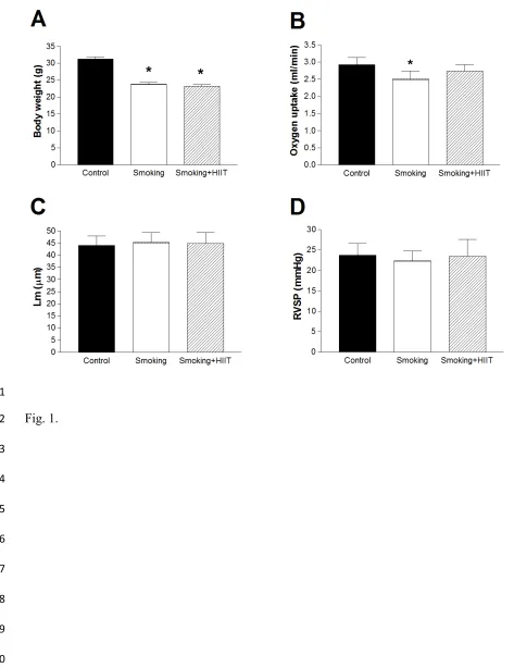
Smoke exposure in mice reduced body weight by 26 % (P<0.05; Fig. 1A) and impaired exercise
202
capacity by 15 % (P<0.05; Fig. 1B), however the latter was normalized following exercise training
203
(P>0.05; Fig. 1B). In addition, in line with our hypothesis cigarette smoke exposure did not cause
204
the development of overt emphysema, as demonstrated by no discernable alveolar destruction (i.e.,
205
mean linear intercept remained unchanged; P>0.05; Fig. 1C) and by the absence of pulmonary
206
hypertension (i.e., right-ventricular systolic pressures were unchanged; P>0.05; Fig. 1D).
207
Similarly, no differences were found between groups in terms of tibia length or skeletal muscle wet
208
weights (P>0.05).
209
210
Smoke-induced extrapulmonary impairments 211
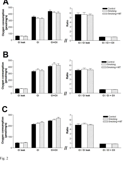
Heart: Heart weight remained unaltered following cigarette smoking (P>0.05). In addition,
212
while echocardiography revealed that left ventricular function was not impaired by smoking
213
(P>0.05; Table 1), right ventricular systolic function was reduced compared to controls by ~20 %
214
as assessed by TAPSE (P<0.05; Table 1). To determine whether mitochondrial dysfunction may
215
underlie this impairment, we further assessed mitochondrial respiration in fibers from the right and
216
left ventricle, but found no differences (P>0.05; Fig. 2A-B).
217
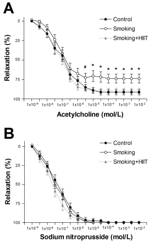
Peripheral endothelium: The effect of smoking on peripheral endothelial function was
218
assessed from isolated aortic rings for both endothelial-dependent and -independent vasodilation
219
(i.e., relaxation). Stimulation of aortic rings with increasing concentrations of acetylcholine
220
revealed that smoking was associated with impaired endothelial-dependent vasodilation (P<0.05;
221
Fig. 3A), with smoke-exposed mice achieving only 70±7 % of their maximal dilation compared to
222
control mice values of 91±3 % (P<0.01). In contrast, smoke exposure did not impair
endothelium-223
independent vasodilation (P>0.05; Fig. 3B), with maximal vasodilation values of 100% achieved
in both groups (P>0.05). To determine potential mechanisms underlying these functional
225
impairments, we further assessed protein expression of phosphorylated eNOS and acetylated eNOS
226
in the aorta but found no differences between control and smoke-exposed mice (P>0.05).
227
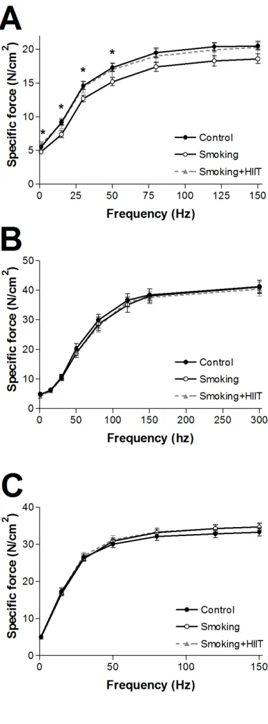
Respiratory and limb skeletal muscles: Compared to controls, diaphragm fiber bundles
228
from smoking mice were weaker by an average of 15±2 % (range: 11 - 19%; P<0.05; Fig. 4A),
229
which occurred at the lower and more clinically-relevant frequencies of 1, 15, 30, and 50 Hz.
230
However, no differences were discerned during the fatigue protocol between groups. In contrast to
231
the diaphragm, smoking had no adverse effect on limb muscle contractile function for either the
232
glycolytic EDL muscle (Fig. 4B) or oxidative soleus muscle (Fig. 4C), or on relative force during
233
the fatigue protocol (P>0.05; data not shown). As these data suggested the diaphragm was more
234
susceptible to cigarette smoke-induced impairments, we focused thereafter on this specific skeletal
235
muscle. Histological analyses of the diaphragm between control and smoke-exposed mice revealed
236
no structural changes in terms of fiber cross-sectional area (fast and slow twitch: 1077±102 vs.
237
1222±82 m2 and 895±123 vs. 877±76 m2, respectively; P>0.05) or fiber proportions (fast and
238
slow twitch: 92±1 vs. 92±1 % and 8±1 vs. 8±1 %, respectively; P>0.05). In contrast, while the
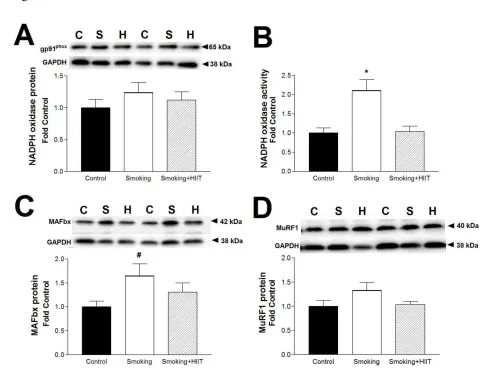
239
protein expression of NADPH oxidase, a marker of oxidative stress, was unaltered between groups
240
(P>0.05; 5A), the more functional measure of enzyme activity was increased by 111 % (P<0.05;
241
Fig. 5B). Similarly, the protein expression of MAFbx (a key marker of protein degradation) was
242
also increased by 65 % in smokers compared to controls (P<0.05; 5C). In contrast, however, the
243
protein expression of MuRF1 (another marker of protein degradation) was not different between
244
groups (P>0.05; 5D), as was the case for the contractile protein myosin heavy chain (1.00±0.17 vs.
245
0.90±0.15; P>0.05). We further assessed whether mitochondrial dysfunction could be contributing
246
to the diaphragm muscle weakness following smoke exposure, however in situ mitochondrial
247
respiration of permeabilized fibers was not different between groups (P>0.05; Fig. 2C).
249
Effects of high-intensity exercise training in smoke-exposed mice 250
Six weeks of exercise training normalized the smoke-induced reduction in exercise capacity (Fig.
251
1B), while also improving left ventricular systolic function (i.e., ejection fraction and fractional
252
shortening) when compared to sedentary smoke-exposed mice (P<0.05; Table 1). In addition,
253
exercise essentially reversed the smoke-induced impairment of TAPSE, suggesting right
254
ventricular function was normalized (P<0.05; Table 1), while also reversing peripheral endothelial
255
dysfunction such that maximal endothelium-dependent dilation was normalized to control values
256
(P>0.05; Fig. 3A). In line with this, we found the protein expression of phosphorylated eNOS to
257
be increased in the aorta of smoke-trained mice compared to both control and smoke-exposed
258
sedentary mice (P<0.05). Importantly, smoke-induced diaphragm contractile dysfunction was also
259
reversed by exercise training to similar values of that observed in control mice (P>0.05; Fig. 4A),
260
which was also the case for both the enzyme activity of NADPH oxidase (P>0.05; Fig. 5B) and the
261
protein expression of MAFbx (P>0.05; Fig. 5C). Nevertheless, no differences were subsequently
262
found between control, smoking, and smoke-trained mice in terms of the anti-oxidative enzyme
263
activity of superoxide dismutase (1.00±0.09 vs. 1.22±0.22 vs. 1.30±0.23, respectively; P>0.05) or
264
catalase (1.00±0.13 vs. 1.01±0.13 vs. 0.83±0.12, respectively; P>0.05). Furthermore, exercise
265
training had no influence on the expression of other proteins (e.g., myosin heavy chain, MuRF1,
266
NADPH oxidase), on fiber properties (i.e. cross-sectional area and type), or on mitochondrial
267
function (Fig. 2C; all P>0.05).
268
Discussion 270
The main findings of this study include: 1) cigarette smoking induced functional impairments to
271
the heart, peripheral endothelium, and respiratory muscle, which occurred in the absence of overt
272
emphysema and these likely converged to reduce exercise capacity; and 2) high-intensity exercise
273
training reversed these smoke-induced extrapulmonary impairments in line with normalizing
274
exercise capacity. Collectively, therefore, our findings may have important clinical implications as
275
they suggest that cigarette smokers in the absence of emphysema likely develop simultaneous
276
extrapulmonary impairments to the heart, peripheral endothelium, and diaphragm that exacerbate
277
exercise intolerance (11, 31), but such maladaptations could be reversed by the
non-278
pharmacological intervention of exercise training.
279
280
Smoke exposure and skeletal muscle function 281
Skeletal muscle dysfunction is a key component of COPD (19) and is associated with numerous
282
adaptations (e.g., fiber atrophy, mitochondrial dysfunction, and increased ROS) (2, 3, 18, 24), yet
283
numerous studies have yielded contradictory results with some (24, 28), but not all (7, 34), showing
284
contractile impediments in the lower limbs. In these previous studies, however, overt emphysema
285
had either developed (7, 24) or was not reported (18, 28), leaving it difficult to discriminate whether
286
the cigarette smoke or emphysema was the causal factor underlying skeletal muscle dysfunction.
287
Our study indicates that cigarette exposure in the absence of emphysema does not induce limb
288
muscle contractile dysfunction, as supported by findings in both highly oxidative (i.e. soleus) and
289
glycolytic (i.e., EDL) muscles. In addition, muscle disuse may have also confounded previous
290
studies where limb muscle dysfunction was reported, as recent data demonstrated physical
291
inactivity combined with smoke-induced emphysema significantly exacerbates limb muscle
292
impairments (7). In the current experiments, therefore, we also assessed the main muscle of
respiration, the diaphragm, which is not only constantly active (i.e., thus attenuating any potential
294
confounding factors associated with disuse) but also is thought to play a direct role in the
295
pathogenesis of dyspnea (a key symptom in smokers even without overt COPD (11)). In the present
296
study, we provide novel evidence that prolonged smoking induces diaphragm contractile
297
dysfunction that is independent of lung structural damage, with fiber bundles ~15-20 % weaker
298
compared to control mice. These data suggest, therefore, a greater sensitivity of respiratory rather
299
than locomotor striated muscle to cigarette smoke-induced dysfunction. Furthermore, this loss of
300
function was not due to muscle atrophy as force was normalized to muscle density, which suggests
301
fiber weakness induced by smoking was the result of intracellular impairments (i.e., contractile
302
dysfunction).
303
304
While diaphragm muscle weakness in emphysema has previously been observed in diaphragm
305
fibers from humans (23) and animals (32), we are the first to provide direct evidence that cigarette
306
smoke exposure can lead to diaphragm fiber weakness in the period preceding lung structural
307
damage. The mechanisms underlying the smoke-induced diaphragm muscle weakness may be
308
related to an increase in oxidative stress and protein degradation, as NADPH oxidase activity (but
309
not protein content) and MAFbx protein expression was increased in the diaphragm of smoking
310
mice, but these were attenuated in the smoke-exercise trained mice in parallel with normalized
311
contractile function. These findings compliment previous studies, which also reported increased
312
levels of oxidative stress and oxidative protein modifications in the diaphragm following prolonged
313
cigarette smoking in mice (2, 3). However, contrary to data from the soleus muscle (29),
314
mitochondrial dysfunction was not present in diaphragm fibers following prolonged smoke
315
exposure, which suggests a low oxidative phosphorylation capacity was unlikely mediating the
316
smoke-induced respiratory muscle weakness. Taken together, therefore, our data support a role for
cigarette smoking as a causal mechanism in diaphragm muscle weakness, potentially mediated by
318
elevated oxidative stress levels and activation of proteolytic pathways, which likely predisposes
319
many smokers to respiratory dysfunction in the period preceding emphysema (11).
320
321
Smoke exposure and cardiovascular function 322
Following prolonged smoke exposure cardiac dysfunction is known to develop, with right
323
ventricular function particularly compromised (25), while peripheral vascular endothelial
324
dysfunction is also manifest (27). Our study confirmed that right ventricular function is reduced
325
and peripheral endothelial function is impaired following cigarette exposure, which occurred in the
326
absence of overt emphysema. In addition, we also confirmed previous findings that right
327
ventricular dysfunction can be reversed by exercise training (15). In order to better understand the
328
mechanisms of these smoke-induced cardiac impairments, we assessed mitochondrial function in
329
cardiac fibers. However, and in contrast to a previous study reporting impaired respiration in fibers
330
from the left ventricle (30), we found no differences between groups in either the left or right
331
ventricle, which suggests mitochondrial dysfunction is unlikely a key mechanism of
smoke-332
induced right ventricular dysfunction, at least in relation to the present animal model. In terms of
333
the peripheral endothelium, we provide novel data that high-intensity exercise training can reverse
334
smoke-induced peripheral vascular impairments, as endothelium-dependent vasodilation in aortic
335
rings was normalized to control values following exercise training. We also found a significant
336
increase in the protein expression of phosphorylated eNOS in smoke-trained mice, which may help
337
explain how exercise mediated its benefits, as smoking is well known to reduce NO bioavailability
338
and eNOS activity (4). However, why endothelial dysfunction was only observed at the higher
339
rather than the lower acetylcholine concentrations in smoke-exposed mice is unclear, but it may be
340
related to differences in membrane receptor sensitivity or density. Nevertheless, that data collected
from both animals (27) and humans (16, 26) show a similar trend after smoke exposure supports
342
the physiological relevance of the present findings.
343
344
If we take an integrative approach, it could be speculated that diaphragm muscle weakness induced
345
by smoke-exposure is mediated, in part, by upstream impairments related to cardiac and peripheral
346
endothelial dysfunction that limit blood flow to induce local hypoxia, with the latter suggested as
347
a key mechanism underpinning skeletal muscle dysfunction in COPD (19). Hypoxia has been
348
shown in diaphragm fiber bundles to potentiate ROS production which, in turn, directly impair
349
subsequent force generation (22). As such, our overall findings of impaired central and peripheral
350
vascular function coupled with diaphragm muscle weakness in concert with elevated markers of
351
ROS suggest hypoxia could be a putative mechanism for the smoke-induced respiratory muscle
352
weakness.
353
354
Animal model of cigarette smoke exposure 355
The uncertainty related to the role of cigarette smoke exposure on extrapulmonary impairments
356
had previously been clouded by the fact that emphysema was usually developed secondary to
357
cigarette exposure (2, 3, 7, 13) or that lung indices of emphysema were not measured (18, 28). In
358
the current study we selected a period of 20 weeks for smoke exposure so animals would not
359
develop overt emphysema, which is generally documented after 24 weeks in mice (9, 13).
360
Furthermore, the A/J mouse strain we studied is moderately susceptible to lung destruction
361
following smoke exposure (21), but not as sensitive (e.g., C57BL/6J) or resistant as other strains
362
(e.g., NZWLac/J) (14, 35). Collectively, therefore, the present data support our objective and
363
reinforce the clinical translation of our data, as prolonged smoke exposure resulted in no signs of
364
emphysema, while numerous multi-organ impairments converged concomitant with an impaired
exercise capacity of ~20%, with the latter value similar to that reported in patients at high risk of
366
developing emphysema (11). Nevertheless, we acknowledge the possibility our mouse model may
367
have still induced lung injury and airflow limitation despite the absence of emphysema
368
histologically. As such, the addition of lung functional measurements including measuring airway
369
resistance would have provided further clarity on this issue. Furthermore, as the present study
370
assessed female mice only, it also remains unclear whether the observed multi-organ impairments
371
would have been reduced or even exacerbated in males exposed to the same amount of cigarette
372
smoke.
373
374
Conclusion 375
High-intensity exercise training reversed cigarette smoke-induced extrapulmonary impairments to
376
the heart, the peripheral vascular endothelium, and the diaphragm as well normalizing exercise
377
capacity, which occurred before the onset of overt emphysema. These findings suggest, therefore,
378
that cigarette smokers in the absence of emphysema likely develop extrapulmonary impairments
379
that exacerbate exercise intolerance, but exercise training can reverse these detrimental
380
consequences.
Acknowledgments 382
None
383
384
385
Conflict of interest 386
The authors have no conflicts of interest to disclose in relation to professional relationships with
387
companies or manufacturers who will benefit from the results of the present study. The authors
388
state that the results of the present study do not constitute endorsement by ACSM. The authors
389
declare that the results of the study are presented clearly, honestly, and without fabrication,
390
falsification, or inappropriate data manipulation
391
392
393
References 395
1. Adams V, Alves M, Fischer T et al. High intensity interval training attenuates endothelial
396
dysfunction in a Dahl salt-sensitive rat model of heart failure with preserved ejection
397
fraction (HFpEF). J Appl Physiol (1985). 2015; 119(6): 745-52.
398
2. Barreiro E, del Puerto-Nevado L, Puig-Vilanova E et al. Cigarette smoke-induced oxidative
399
stress in skeletal muscles of mice. Respir Physiol Neurobiol. 2012;182(1):9-17.
400
3. Barreiro E, Peinado VI, Galdiz JB et al. Cigarette smoke-induced oxidative stress: A role
401
in chronic obstructive pulmonary disease skeletal muscle dysfunction. Am J Respir Crit 402
Care Med. 2010;182(4):477-88.
403
4. Barua RS, Ambrose JA, Eales-Reynolds LJ, DeVoe MC, Zervas JG, Saha DC.
404
Dysfunctional endothelial nitric oxide biosynthesis in healthy smokers with impaired
405
endothelium-dependent vasodilatation. Circulation. 2001;104(16):1905-10.
406
5. Bowen TS, Rolim NP, Fischer T et al. Heart failure with preserved ejection fraction induces
407
molecular, mitochondrial, histological, and functional alterations in rat respiratory and limb
408
skeletal muscle. Eur J Heart Fail. 2015;17(3):263-72.
409
6. Celermajer DS, Sorensen KE, Georgakopoulos D et al. Cigarette smoking is associated with
410
dose-related and potentially reversible impairment of endothelium-dependent dilation in
411
healthy young adults. Circulation. 1993;88(5 Pt 1):2149-55.
412
7. Cielen N, Maes K, Heulens N et al. Interaction between Physical Activity and Smoking on
413
Lung, Muscle, and Bone in Mice. Am J Respir Cell Mol Biol. 2016;54(5):674-82.
414
8. Close RI. Dynamic properties of mammalian skeletal muscles. Physiol Rev.
415
1972;52(1):129-97.
416
9. D'Hulst A I, Vermaelen KY, Brusselle GG, Joos GF, Pauwels RA. Time course of cigarette
417
smoke-induced pulmonary inflammation in mice. Eur Respir J. 2005;26(2):204-13.
10. Dunnill MS. Quantitative methods in the study of pulmonary pathology. Thorax.
419
1962;17(4):320-28.
420
11. Elbehairy AF, Guenette JA, Faisal A et al. Mechanisms of exertional dyspnoea in
421
symptomatic smokers without COPD. Eur Respir J. 2016.
422
12. Freixa X, Portillo K, Pare C et al. Echocardiographic abnormalities in patients with COPD
423
at their first hospital admission. Eur Respir J. 2013;41(4):784-91.
424
13. Gosker HR, Langen RC, Bracke KR et al. Extrapulmonary manifestations of chronic
425
obstructive pulmonary disease in a mouse model of chronic cigarette smoke exposure. Am 426
J Respir Cell Mol Biol. 2009;40(6):710-6.
427
14. Guerassimov A, Hoshino Y, Takubo Y et al. The development of emphysema in cigarette
428
smoke-exposed mice is strain dependent. Am J Respir Crit Care Med. 2004;170(9):974-80.
429
15. Hassel E, Berre AM, Skjulsvik AJ, Steinshamn S. Effects of exercise training on pulmonary
430
vessel muscularization and right ventricular function in an animal model of COPD. Respir 431
Res. 2014;15:117.
432
16. Heitzer T, Yla-Herttuala S, Luoma J et al. Cigarette smoking potentiates endothelial
433
dysfunction of forearm resistance vessels in patients with hypercholesterolemia. Role of
434
oxidized LDL. Circulation. 1996;93(7):1346-53.
435
17. Kemi OJ, Loennechen JP, Wisloff U, Ellingsen O. Intensity-controlled treadmill running in
436
mice: cardiac and skeletal muscle hypertrophy. J Appl Physiol (1985). 2002;93(4):1301-9.
437
18. Kruger K, Dischereit G, Seimetz M, Wilhelm J, Weissmann N, Mooren FC. Time course
438
of cigarette smoke-induced changes of systemic inflammation and muscle structure. Am J 439
Physiol Lung Cell Mol Physiol. 2015;309(2):L119-28.
19. Maltais F, Decramer M, Casaburi R et al. An official American Thoracic Society/European
441
Respiratory Society statement: update on limb muscle dysfunction in chronic obstructive
442
pulmonary disease. Am J Respir Crit Care Med. 2014;189(9):e15-62.
443
20. March TH, Barr EB, Finch GL et al. Cigarette smoke exposure produces more evidence of
444
emphysema in B6C3F1 mice than in F344 rats. Toxicol Sci. 1999;51(2):289-99.
445
21. March TH, Bowen LE, Finch GL, Nikula KJ, Wayne BJ, Hobbs CH. Effects of strain and
446
treatment with inhaled aII-trans-retinoic acid on cigarette smoke-induced pulmonary
447
emphysema in mice. COPD. 2005;2(3):289-302.
448
22. Mohanraj P, Merola AJ, Wright VP, Clanton TL. Antioxidants protect rat diaphragmatic
449
muscle function under hypoxic conditions. J Appl Physiol (1985). 1998;84(6):1960-6.
450
23. Ottenheijm CA, Heunks LM, Sieck GC et al. Diaphragm dysfunction in chronic obstructive
451
pulmonary disease. Am J Respir Crit Care Med. 2005;172(2):200-5.
452
24. Rinaldi M, Maes K, De Vleeschauwer S et al. Long-term nose-only cigarette smoke
453
exposure induces emphysema and mild skeletal muscle dysfunction in mice. Dis Model 454
Mech. 2012;5(3):333-41.
455
25. Seimetz M, Parajuli N, Pichl A et al. Inducible NOS inhibition reverses
tobacco-smoke-456
induced emphysema and pulmonary hypertension in mice. Cell. 2011;147(2):293-305.
457
26. Sharif MA, Bayraktutan U, Arya N et al. Smoking impairs endothelial function in human
458
saphenous vein in an ex vivo model. Ann Vasc Surg. 2009;23(1):116-21.
459
27. Talukder MA, Johnson WM, Varadharaj S et al. Chronic cigarette smoking causes
460
hypertension, increased oxidative stress, impaired NO bioavailability, endothelial
461
dysfunction, and cardiac remodeling in mice. Am J Physiol Heart Circ Physiol.
462
2011;300(1):H388-96.
28. Tang K, Wagner PD, Breen EC. TNF-alpha-mediated reduction in PGC-1alpha may impair
464
skeletal muscle function after cigarette smoke exposure. J Cell Physiol.
2010;222(2):320-465
7.
466
29. Thatcher MO, Tippetts TS, Nelson MB et al. Ceramides mediate cigarette smoke-induced
467
metabolic disruption in mice. Am J Physiol Endocrinol Metab. 2014;307(10):E919-27.
468
30. Tippetts TS, Winden DR, Swensen AC et al. Cigarette smoke increases cardiomyocyte
469
ceramide accumulation and inhibits mitochondrial respiration. BMC Cardiovasc Disord.
470
2014;14:165.
471
31. van den Borst B, Koster A, Yu B et al. Is age-related decline in lean mass and physical
472
function accelerated by obstructive lung disease or smoking? Thorax. 2011;66(11):961-9.
473
32. van Hees H, Ottenheijm C, Ennen L, Linkels M, Dekhuijzen R, Heunks L. Proteasome
474
inhibition improves diaphragm function in an animal model for COPD. Am J Physiol Lung 475
Cell Mol Physiol. 2011;301(1):L110-6.
476
33. Woldbaek PR, Stromme TA, Sande JB, Christensen G, Tonnessen T, Ilebekk A. Evaluation
477
of a new fiber-optic pressure recording system for cardiovascular measurements in mice.
478
Am J Physiol Heart Circ Physiol. 2003;285(5):H2233-9.
479
34. Wust RC, Morse CI, de Haan A, Rittweger J, Jones DA, Degens H. Skeletal muscle
480
properties and fatigue resistance in relation to smoking history. Eur J Appl Physiol.
481
2008;104(1):103-10.
482
35. Yao H, Edirisinghe I, Rajendrasozhan S et al. Cigarette smoke-mediated inflammatory and
483
oxidative responses are strain-dependent in mice. Am J Physiol Lung Cell Mol Physiol.
484
2008;294(6):L1174-86.
485
486
Figure Legends 488
Fig. 1 Body weight (A), exercise capacity (B), mean linear intercept (Lm; C) and right
489
ventricular systolic pressure (RVSP; D) of control, smoking, and smoking mice that performed
490
high-intensity interval training (HIIT). That lung destruction (C) or pulmonary hypertension (D)
491
did not differ between groups supports the contention that overt emphysema was not developed in
492
smoke-exposed mice. *P<0.05 vs. Control.
493
494
Fig. 2 In situ mitochondrial respiration states and the respective control ratios in
495
permeabilized fibers of the left ventricle (A), right ventricle (B), and diaphragm (C), as assessed
496
from control, smoking, and smoking mice that performed high-intensity interval training (HIIT).
497
Respiration states were assessed, in turn, including: CI leak, complex I leak respiration following 498
addition of malate and glutamate; CI, complex I phosphorylated state following addition of ADP; 499
CII, complex II phosphorylated state following addition of succinate. No significant differences 500
were found between groups for all measures.
501
502
Fig. 3 Peripheral in vitro endothelial-dependent (A) and -independent (B) function in
503
response to increasing acetylcholine and sodium nitroprusside (SNP) concentrations, as assessed
504
in isolated aortic rings from control, smoking, and smoking mice that performed high-intensity
505
interval training (HIIT). *P<0.05 vs. Control and Smoking+HIIT; #P<0.05 vs. Smoking+HIIT
506
507
Fig. 4 In vitro respiratory and lower limb skeletal muscle contractile function assessed
508
across a range of stimulation frequencies in the diaphragm (A), EDL (B), and soleus (C) for control,
509
smoking, and smoking mice that performed high-intensity interval training (HIIT). *P<0.05 vs.
510
Smoking.
512
Fig. 5 Protein expression (A) and enzyme activity (B) of NADPH oxidase as well as
513
protein expression of MAFbx (C) and MuRF1 (D) in the diaphragm from control, smoking, and
514
smoking mice that performed high-intensity interval training (HIIT). Representative blots for each
515
protein are also provided in the upper panel, which were normalized to the loading control GAPDH
516
for control (c), smoking (s), and smoking mice that performed HIIT (H). *P<0.05 vs. Control and
517
Smoking+HIIT; #P<0.05 vs. Control. 518
520
[image:27.612.48.521.97.711.2]521
Fig. 1.
522
523
524
525
526
527
528
529
531
Fig. 2
532
534
[image:29.612.70.382.75.584.2]535
Fig. 3
536
538
Fig. 4
Fig. 4
540
541
542
Fig. 5.
[image:31.612.59.543.83.465.2]