Activation of Electron-Deficient
Quinones Through
Hydrogen-Bond-Donor-Coupled Electron Transfer
The Harvard community has made this
article openly available. Please share how
this access benefits you. Your story matters
Citation Turek, Amanda Katherine. 2015. Activation of Electron-Deficient Quinones Through Hydrogen-Bond-Donor-Coupled Electron
Transfer. Doctoral dissertation, Harvard University, Graduate School of Arts & Sciences.
Citable link http://nrs.harvard.edu/urn-3:HUL.InstRepos:23845499
Terms of Use This article was downloaded from Harvard University’s DASH repository, and is made available under the terms and conditions applicable to Other Posted Material, as set forth at http://
Dissertation Advisor: Professor Eric N. Jacobsen Amanda Katherine Turek
iii
Activation of Electron-Deficient Quinones
Through Hydrogen-Bond-Donor-Coupled Electron Transfer
Abstract
Quinones are organic oxidants that play important roles in biological contexts and find
wide application in organic synthesis. They are known to be activated toward electron transfer
through hydrogen bonding, which has largely been observed for Lewis basic, weakly oxidizing
quinones. Comparable activation through H-bonding is more difficult to achieve when more
reactive, electron-deficient quinones are used, as these intrinsically weaker Lewis bases are less
prone to engage in H-bonding interactions.
Herein, we describe the successful application of HBD-coupled electron transfer as a
strategy to activate electron-deficient quinones. A systematic investigation of several
small-molecule HBDs allowed examination of the effects of H-bonding on electron transfer to
o-chloranil, an electron-deficient quinone that lacks the intrinsic reactivity necessary to oxidize
many organic substrates of synthetic interest. This study has led to the discovery that dicationic
HBDs have an exceptionally large effect on the rate and thermodynamics of these electron
transfer reactions.
Favorable modulation of the thermodynamics occurs as a result of the stabilization
provided to the reduced quinone (Q•–) by the HBD. Electrochemical experiments have allowed
quantification of the binding affinity for Q•–to each of the HBDs, as well as elucidation of the
Dissertation Advisor: Professor Eric N. Jacobsen Amanda Katherine Turek
iv
with 2:1 stoichiometry, whereas dicationic HBDs bind in a 1:1 complex. Dicationic
bis-amidinium salts exhibit significantly improved binding to Q•–, offering more thermodynamic
stabilization to this reduced state.
The effects of HBDs on the kinetics of electron transfer have also been evaluated under
homogenous conditions. Reactions between o-chloranil and ferrocene derivatives exhibit
pronounced HBD-dependent rate enhancements, with dicationic HBDs displaying the greatest
effect. Relative to neutral dual HBDs, the bis-amidinium salts accelerate the rate of electron
transfer by > 1012. Binding stoichiometries within the rate-limiting transition states corroborate
the results determined electrochemically, and binding affinity correlates with rate enhancement
was observed across the series of HBDs evaluated.
Application of HBD-coupled electron transfer in an oxidative lactonization illustrates that
this strategy is applicable to catalysis of organic reactions. A dicationic HBD catalyst affords the
lactone product in nearly quantitative yield within 24 h, whereas o-chloranil alone was
ineffective (< 5% yield). The rates of lactonization with several HBD catalysts correlate well
with the thermodynamic and kinetic trends described above. This trend indicates that the rate of
the oxidative lactonization is related to the ability of the HBD to promote an electron transfer
step. Potential strategies for application in enantioselective transformations and possibilities for
v
Table of Contents
Abstract iii
Table of Contents v
Acknowledgments ix
List of Abbreviations and Symbols xv
Chapter 1. An overview of hydrogen-bonding and protonation effects in electron
transfer reactions 1
1.1 Introduction 1
1.2 Proton-coupled electron 2
1.2.1 Fundamentals of PCET 2
1.2.2 PCET in organic synthesis 3
1.3 PCET chemistry of quinones 4
1.3.1 Mechanisms for PCET to quinones
4
1.3.2 Biological significance of PCET involving quinones 6
1.3.3 Intermediacy of H-bonded complexes in PCET mechanisms 8
1.4 Binding studies of quinones to weak HBDs 11
1.4.1 H-bonding between quinones and alcohols 11
1.4.2 H-bonding between quinones and amides 13
1.4.3 Influence of cationic additives on electron transfer to quinones 14
1.4.4 Binding of quinones to neutral dual HBDs 15
vi
Chapter 2. Activation of electron-deficient quinones through HBD-coupled electron
transfer 19
A. Electrochemical quantification of o-chloranil binding to HBDs 19
2a.1 Introduction 19
2a.2 Theoretical basis 21
2a.2.1 Thermodynamic analysis 21
2a.2.2 Mechanistic tools for the study of HBD-coupled electron transfer 23
2a.3 Mechanisms and notation 24
2a.4 Experimental design 25
2a.4.1 Determination of conditions for CV experiments 25
2a.4.2 General approach to electrochemical experiments and data analysis 28
2a.5 Results and discussion 28
2a.5.1 Mechanistic analysis of HBD-coupled electron transfer using N,N’
-dicyclohexylguanidinium salts 28
2a.5.2 Mechanistic analysis of HBD-coupled electron transfer using N,N’
-diphenylguanidinium salts 34
2a.5.3 Mechanistic analysis of HBD-coupled electron transfer using neutral
ureas 38
2a.5.4 Comparison of binding abilities for HBDs 1–3 39
2a.5.5 Mechanistic analysis of HBD-coupled electron transfer using
dicationic bis-amidinium salts 41
2a.5.6 Comparison of bis-amidinium salt 4 with HBDs 1–3 45 2a.5.7 Control experiments and comparison with bis-amidinium salt 4 47 2a.5.8 Effect of conformational rigidity on binding ability 53
2a.5.9 Mechanistic analysis of HBD-coupled electron transfer using
vii
2a.6 Conclusions from electrochemical experiments 61
2a.7 Experimental details 63
2a.7.1 General information 63
2a.7.2 Procedures for the syntheses of HBDs 1–9 64
2a.7.3 Cyclic voltammetry experiments 75
2a.7.4 Measurement of binding constants for neutral Q 94
2a.7.5 Geometry optimizations for (HBD)n·Q•– complexes 100
B. Kinetic analysis of electron transfer to o-chloranil promoted by HBDs 108
2b.1 Introduction 108
2b.2 Design of kinetic experiments 109
2b.3 Determination of kinetic order in HBD for electron transfer reactions 110
2b.4 Method of continuous variations for determination of 4·Q stoichiometry 114 2b.5 Relative rate constants for HBD-coupled electron transfer 116
2b.6 Large rate acceleration provided by diphenylformamidinium salts 119
2b.7 Thermodynamic analysis of homogeneous electron transfer processes 120
2b.8 Experimental details 122
2b.8.1 General information 122
2b.8.2 Beer’s Law plots 124
2b.8.3 Initial rates experiments for determination of order in HBD 128
2b.8.4 Job plot for determination of 4·Q stoichiometry 133
2b.8.5 Pseudo-first-order experiments for determination of relative rate
constants 134
C. Synthetic application of HBD-coupled electron transfer 137
viii
2c.2 Application of HBD-coupled electron transfer in a synthetic transformation 138
2c.2.1 Development and catalysis of an oxidative lactonization 138
2c.2.2 Structure-activity relationship studies on 4 140
2c.2.3 Effect of dimeric HBDs 142
2c.3 Kinetic isotope effect experiment and proposed mechanism 143
2c.4 Experimental details 145
2c.4.1 Procedures for the syntheses of HBDs 10–12 145
2c.4.2 Oxidative lactonization procedures and substrate synthesis 150
2c.4.3 KIE experiment 152
D. Outlook for future work 153
2d.1 Expanding the scope of HBD-coupled electron transfer 153
2d.1.1 Outlook for investigation with respect to HBD design 153
2d.1.2 Preliminary results for HBD-coupled electron transfer to
electron-deficient p-quinones 154
2d.2 Outlook for synthetic application in enantioselective catalysis 160
2d.2.1 Conceptual design 160
2d.2.2 Oxidative lactonization catalyzed by chiral, dimeric bis-guanidinium
salts 162
2d.3 Proposed application of HBD-coupled electron transfer in a redox-neutral
reaction 165
2d.3.1 Reactivity principle 165
2d.3.2 Proposal for enantioselective approach 166
2d.4 Conclusions 167
ix
Acknowledgments
I have had the great fortune of having several mentors in my professional life who are not
only brilliant scientists but are also wonderful human beings. First and foremost among these is
my advisor, Eric Jacobsen. I’ve learned more from him about being a chemist, teacher, and
mentor than the G1-version of me—who joined his group six years ago—ever thought I would. I
am honored to have had the opportunity to study with him, and am proud of the chemist and
person this opportunity has allowed me to become. Above all, I thank Eric for his unwavering
confidence in me, for which I am truly grateful.
Professor Ted Betley has been a supportive member of my GAC, and in my committee
meetings has challenged me to think more deeply about my science. I think one of my biggest
regrets from the past six years is that I didn’t get to know him better. I also thank Professor Dan
Nocera for joining my thesis committee and agreeing to collaborate with us on this project. I
have learned a lot from him and his group, and this project has matured greatly thanks to this
collaboration. I also appreciate his enthusiasm for the project, which came at a time when it was
sorely needed.
As an undergraduate, I was lucky enough to have two fantastic advisors. Professor
Tehshik Yoon took me into his lab and under his wing as a sophomore. I look back on my time
in the Yoon group with some embarrassment, remembering when I ran into Tehshik’s office at 9
PM to show him an NMR spectrum—I had run a Swern reaction and was so excited that my
crude product was clean. Despite how busy he surely was, Tehshik was nothing but enthusiastic,
x
and encouragement throughout my time in graduate school, always willing to lend an “ear” via
e-mail or over the phone.
Professor Bassam Shakhashiri has been a true source of inspiration to me as well. He has
challenged me to think broadly about my responsibility to society as a chemist, and has taught
me to understand the critical importance of clear, respectful communication with the non-science
community. Bassam has traveled to Boston many times over the past six years, and I’m grateful
to him for always carving out time in his schedule for me. I’ve enjoyed our long conversations
over lunch, brunch, and dinner, and have always left feeling inspired to be a better scientist, and
lucky to know Bassam as a mentor and a friend.
I thank Nicole Minotti for her encouragement and friendship over the past few years. The
Jacobsen group is truly lucky to have her around, especially for help with embedding fonts. I’m
especially thankful for her willingness to deal with the people from a certain company that will
remain unnamed, so I didn’t have to do it myself. I will miss our long-winded conversations over
a wide range of topics encompassing both the mundane and (more often) the absurd.
I have been joined on my project by two collaborators. David Hardee was a welcome
addition to Team Super Grover in my third year, offering support, ideas, discussion, and
commiseration (when needed). Although he has since moved on to “real life,” he has continued
to provide advice and encouragement as I wrap up my graduate work. Andrew Ullman was my
collaborator in the Nocera group, and taught me everything I know about electrochemistry. I’m
appreciative of his willingness to spend countless hours teaching me about CV experiments and
electrochemical simulations and discussing results, analysis, and electrochemical theory.
Rob Knowles was a terrific mentor during my first full year in the lab, always patient
xi
confidence in my ideas and hypotheses, allowing me to become an independent experimentalist.
He remains one of the most inspirational people I know, and I consider myself fortunate to have
worked with him so closely in the Jacobsen group.
While every member of the Jacobsen group has had an impact on me in his or her own
way, a few of them warrant individual acknowledgment, starting with my classmate and friend,
Mike Witten. Our honest relationship has made all the difference in graduate school. Mike has
been a great friend to me, always up for getting lunch at Grendel’s, watching the Packers even
though he’s a Patriots fan, driving me and some friends to Cape Cod for my birthday, and even
flying out to Milwaukee to attend my wedding.
Pam Tadross has also been a hugely supportive influence during graduate school, and has
also become a very dear friend. She has patiently helped me through tough times, and always
encourages me to be confident and proud of my work. Charles Yeung constantly encourages me
to be optimistic, and it’s hard—even for me—not to give in (at least sometimes). His advice,
wisdom, and friendship have been invaluable. I’m also glad to know Baye Galligan, and I
commend her for choosing a path in life that she believes will make her truly happy.
We don’t get to choose our officemates and bay-mates, so I’m especially lucky to have
shared a workspace with some great people. Bekka Klausen welcomed me to M202D, passing on
her wisdom as a senior grad student and introducing me to Catan. I’m glad that Rose Kennedy
later moved into M202D and joined me in the battle against various olfactory offenses. She is an
inspiring person and I am excited to see what the future holds for her.
Gary Zhang has been my bay- and desk-neighbor for five years, which has led to many
interesting discussions, both chemical and philosophical in nature. He has been consistently
xii
bay-mate Song Lin offered to give me foods that I’m allergic to, I still miss his face. Working
across from YP has been delightful. It turns out that those shelves separating two benches don’t
provide much of a barrier to snarky insults. Regardless, I’ll still miss his angry bump trap e-mails
and lopsided fantasy football trades.
Extra thanks to the members of the Jacobsen group, both current and former, who spent
some of their personal time reading over parts of this thesis and offering invaluable feedback:
Rose Kennedy, David Hardee, Eric Woerly, Gary Zhang, Andrew Bendelsmith, YP (Park), and
Libby Hennessy, and extra-extra thanks to Charles Yeung, who read this thesis in its entirety.
Special thanks to some old friends who made the past six years more pleasant: Tess
Anderson and Souriya Vang, who were nearby in Providence and traveled to Boston for many
calorie-laden visits to the dumpling house. I’m also glad that Lucy Liu went to medical school at
Yale, which was very convenient for trips to New York City to see musicals, even though we
didn’t get Nathan Lane’s autograph. Happily, I’ve been able to visit my former Yoon Group
coworkers, Tamas Benkovics—who “adopted” me when I was an “orphan” undergraduate—and
Juana Du, in New Jersey. And I can always count on them for cheerleading via text message. I’m
also thankful for friends from Wisconsin who came to visit—Julie Ripple for visiting twice and
going on an adventure to Tanglewood with me, and Juli Held for forcing me to finally check out
Fenway Park. I also thank Linda Cutting for helping me to sustain and develop my love of piano
over the past six years.
One of the biggest challenges presented by graduate school has been the separation from
my family. I can’t possibly adequately thank my parents here, so I’ll just say I’m certain that,
with Pam and Gary Turek, I definitely won the parent lottery. I’m also grateful to my sister, Sam,
xiii
hope for, and have brightened many a gloomy day. I know the distance has been especially hard
on them. Grandpa and Uncle Glen are two of the most inspirational people in my life, and I strive
to follow their example on how to lead a fulfilling, meaningful life. Finally, I suspect the
separation may have been hardest on Grandma Judy, although our separation now is even more
difficult to bear. On particularly frustrating days, a simple phone call with her would help me to
get through—she always knew the right thing to say. My heart aches for her to be here and share
in this accomplishment with me.
My best friend throughout graduate school—and in the eleven years I’ve known him—
has been Wei An, who encourages me in all I do and in every instance puts my well-being above
his own. Over the past six years, Wei has taken my successes and, more often, my frustrations
perhaps more personally than I have. I wish our individual pursuit of professional success didn’t
mean that we would be separated by a thousand miles, but I’m grateful to have a partner who
supports me despite this unhappy reality. Having Wei by my side—sometimes figuratively,
xiv
xv
List of Abbreviations and Symbols
APCI atmospheric pressure chemical ionization
aq aqueous
α symmetry factor
Å angstrom
B3LYP Becke-3-Lee-Yang-Parr
BArF24 tetrakis[3,5-bis(trifluoromethyl)phenyl]borate
bRC bacterial reaction center
BrFc bromoferrocene
Br2Fc 1,1’-dibromoferrocene
°C degree Celsius
calcd. calculated
CBz benzyloxychloroformate
CE chemical step–electron transfer
CEC chemical step–electron transfer–chemical step
cm centimeter
CV cyclic voltammetry
D diffusion coefficient
d days
d deuterium
DCM dichloromethane
xvi DFT density functional theory
DIPEA N,N-diisopropylethylamine DME dimethoxyethane
DMF dimethylformamide
DMSO dimethylsulfoxide
E reduction potential
EC electron transfer–chemical step
ECC electron transfer–chemical step–chemical step
EDC N-(3-dimethylaminopropyl)-N’-ethylcarbodiimide hydrochloride equiv. equivalents
ESI electrospray ionization
ET electron transfer
Et2O diethyl ether
EtOAc ethyl acetate
ε molar absorptivity/extinction coefficient
Fc ferrocene
FTIR fourier transform infrared spectroscopy
G Gibbs free energy
g gram
h hour
HAT H-atom abstraction
HBD hydrogen-bond donor
xvii
IR infrared
K Kelvin
kcal kilocalorie
Kn equilibrium constant
kobs observed rate constant
krel relative rate constant
ks heterogeneous rate constant
KIE kinetic isotope effect
L liter
m milli
M molar
M06-2X Minnesota 2006 hybrid meta density functional theory
Me methyl
Me2Fc 1,1’-dimethylferrocene
MeOH methanol
MePTZ N-methylphenothiazine
min minute
mol mole
MS mass spectrometry
μ micro
NBS N-bromosuccinimide nBu4N tetrabutylammonium
xviii NMR nuclear magnetic resonance
o ortho
p para
PCET proton-coupled electron transfer
PCM polarizable continuum model
ppm parts per million
p-Q p-chloranil PSII photosystem II
Q o-chloranil
rds rate-determining step
rt room temperature
s second
sat. saturated
tBu tert-butyl
TLC thin-layer chromatography
THF tetrahydrofuran
TBME tert-butyl methyl ether
TMPD N,N,N’,N’-tetramethylphenylenediamine TOF time-of-flight
TolSQ 2-tolylsulfinyl-1,4-benzoquinone
UV-vis ultraviolet-visible
V volts
xix
“And this I believe: that the free, exploring mind of the individual human is the most valuable thing in the world.”
1
Chapter 1
An overview of hydrogen-bonding and protonation
effects in electron transfer reactions
1.1 Introduction
Hydrogen-bonding interactions have a substantial effect on the energetics of organic
reactions by offering stabilizing interactions to transition structures and reactive intermediates.1
This stabilizing effect also plays an important role in electron transfer chemistry, in which
H-bonding—and, at the extreme, protonation—can substantially alter the thermodynamics and
kinetics of this reaction.2 Hydrogen-bond-donor-coupled electron transfer is a process analogous
to proton-coupled electron transfer (PCET), in which H-bonding interactions—in contrast to full
proton transfer—are responsible for this modulation of the thermodynamics and kinetics. In this
chapter, we present a brief description of PCET and its effect on electron transfer to quinones in
biological systems. An overview of the literature precedent regarding the intermediacy of
1 (a) Taylor, M. S.; Jacobsen, E. N. Angew. Chem. Int. Ed. 2006, 45, 1520–1543. (b) Knowles, R. R.;
Jacobsen, E. N. Proc. Natl. Acad. Sci. USA 2010, 107, 20678–20685.
2 (a) Cukier, R. I.; Nocera, D. G. Annu. Rev. Phys. Chem. 1998, 49, 337–369. (b) Huynh, M. H. V.; Meyer,
2
bonding within PCET processes is also provided, and we conclude with a review of the studies
that examine the effect of synthetic hydrogen bond donors (HBDs) on electron transfer to
quinones.
1.2 Proton-coupled electron transfer 1.2.1 Fundamentals of PCET
PCET has a significant impact on the thermodynamics and kinetics of many redox
reactions2 and plays a critical role in biological pathways involving radical transport and
catalysis.3 The three primary mechanisms by which PCET can occur for a 1 H+/1 e– process are
described by a square scheme (Figure 1.1). Stepwise pathways for PCET consist of two
elementary steps—proton transfer and electron transfer—which can occur in either order.
Irrespective of the specific route followed, the first step is generally endergonic—reduction will
generate a species of increased basicity; protonation will generate a stronger oxidant. The energy
differences between these two intermediates dictate the pathway by which PCET occurs,
favoring the route that proceeds through the lower energy intermediate.4 In the event that both intermediates are significantly high in energy, a concerted route—in which a proton and electron
are transferred concurrently—may be more favorable. However, due to the strict kinetic
requirements governing such a process, concerted pathways for PCET are rarer than their
stepwise counterparts.2b,c
3 (a) Reece, S. Y.; Nocera, D. G. Annu. Rev. Biochem. 2009, 78, 673–699. (b) Kaila, V. R. I.; Verkhovsky,
M. I.; Wikström, M. Chem. Rev. 2010, 110, 7062–7081. (c) Dempsey, J. L.; Winkler, J. R.; Gray, H. B.
Chem. Rev. 2010, 110, 7024–7039. (d) Migliore, A.; Polizzi, N.; Therien, M. J.; Beratan, D. N. Chem. Rev, 2014, 114, 3381–3465.
4 (a) Mayer, J. M.; Rhile, I. J. Biochim. Biophys. Acta 2004, 1655, 51–58. (b) Warren, J. J.; Tronic, T. A.;
3
Figure 1.1. Square scheme describing pathways for PCET.
1.2.2 PCET in organic synthesis
PCET has recently been employed in synthetic transformations using phosphoric acid
catalysts (Figure 1.2).5 In these reactions, ketones are activated toward reduction through proton transfer, with a proposed intermediate step involving H-bonding. In the absence of acid,
formation of the intermediate ketyl radical is endergonic by 18 kcal/mol. The incorporation of an
acid catalyst, however, provides a pathway for concerted PCET, enabling reactivity that is
inaccessible by direct electron transfer. Stepwise pathways are discounted on the basis of the rate
law, indicating that proton transfer is involved in the rate-limiting step, and the large pKa
differences between a protonated ketone and the acid catalyst. A chiral phosphoric acid catalyst
is employed to achieve an enantioselective version of this reaction.
5 Tarantino, K. T.; Liu, P.; Knowles, R. R. J. Am. Chem. Soc. 2013, 135, 10022–10025. (b) Rono, L. J.;
4
Figure 1.2. Ketyl-olefin cyclization mediated by PCET
1.3 PCET chemistry of quinones
1.3.1 Mechanisms for PCET to quinones
In aqueous solutions, quinones undergo a 2 H+/ 2 e– reduction that is described by a
nine-membered square scheme (Figure 1.3).6 The specific pathway by which PCET proceeds is
dependent on the pH of the aqueous solution, and determination of the specific mechanism can
be complicated.7 Furthermore, under unbuffered aqueous conditions, the mechanistic scenarios
are not limited to simple proton transfer, and H-bonding also plays a role.8
6 Chambers, J. Q. Electrochemistry of Quinones. In The Chemistry of the Quinonoid Compounds; Patai,
S.; Rappoport, Z.; Eds.; Wiley: New York, 1988; Vol. II, Chapter 12, pp 719–757.
7 Costentin, C. Chem. Rev. 2008, 108, 2145–2179.
5
Figure 1.3. Nine-membered square scheme describing quinone reduction under aqueous or protic conditions.
Under nonaqueous, aprotic conditions, quinone reduction is considerably simplified, and
proceeds through two sequential single-electron reductions, as shown on the rightmost side of the
square scheme (Figure 1.4). However, in nonaqueous media, addition of protic or acidic
additives can influence and complicate the mechanism of quinone reduction,7,9–10 which can
again be described by the nine-membered scheme for a 2-electron reduction.
Figure 1.4. Quinone reduction in aprotic media.
9 Gupta, N.; Linschitz, H. J. Am. Chem. Soc. 1997, 119, 638–6391.
10 Gómez, M.; Gómez-Castro, C. Z.; Padilla-Martínez, I. I.; Martínez-Martínez, F. J.; González, F. J. J.
6
Generally, concerted pathways are disfavored for reduction of quinones due to the
moderate basicity of the intervening semiquinone radical anions (Q•–) .4 For this reason, quinones
are often observed to proceed through a stepwise pathway involving electron transfer followed
by proton transfer. In one landmark study regarding the influence of H-bonding and protonation
on quinone electrochemistry,9 it was found that use of strongly H-bonding/acidic additives and/or
basic quinones afforded cyclic voltammograms (CVs) that indicated H-bonding with the quinone
prior to reduction. These results emphasize that the specific properties of the quinones and
additives used dictate the pathways by which reduction of the quinone occurs, and will be
discussed in detail in section 1.4.1.
1.3.2 Biological significance of PCET involving quinones
Quinones are especially important cofactors that are known to undergo PCET in many
biological systems, including photosystem II (PSII)11 and the related bacterial reaction center
(bRC).12 Ubiquinone (Figure 1.5) serves as the terminal electron acceptor in a chain of electron transfer events in photosynthesis, and accepts an electron from another ubiquinone molecule,
(QA). This penultimate electron acceptor, QA, behaves as an electron shuttle, consecutively
transferring two electrons to QB in a net PCET process.13
11 Ferreira, K. N.; Iverson, T. M.; Maghlaoui, K.; Barber, J.; Iwata, S. Science 2004, 303, 1831–1838. 12 Graige, M. S.; Paddock, M. L.; Bruce, J. M.; Feher, G.; Okamura, M. Y. J. Am. Chem. Soc. 1996, 118,
9005–9016.
13 (a) Okamura, M. Y.; Paddock, M. L.; Graige, M. S.; Feher, G. Biochim. Biophys. Acta. 2000, 1458,
7
Figure 1.5. General structure of ubiquinone.
Because QA and QB are structurally identical, there is no intrinsic difference in their
reduction potentials. Despite this lack of inherent bias for electron transfer in a single direction,
PCET occurs exclusively toward QB in a stepwise manner with an initial electron transfer step.
This directionality is apparently coupled to the formation of H-bonds with QB, and more
specifically, its reduced semiquinone state (QB•–).14 X-ray crystal structures obtained by Stowell
for the bRC of Rb. sphaeroides revealed that QB undergoes a conformational shift associated
with electron transfer. The structure obtained under dark conditions—under which no electron
transfer occurs—showed QB involved in a single H-bonding interaction with an amide within its
binding site (Figure 1.6A). When the structure was obtained under light conditions, after
electron transfer has occurred, it was found that QB•– reorients to accommodate additional
H-bonding interactions within the binding site—to a cationic histidine residue and the hydroxyl
side chain of a serine residue (Figure 1.6B). More recent work has ruled out the serine residue as
a H-bond donor, finding instead that an additional bond forms to the amide backbone (Figure
1.6C).15 This conformational gating mechanism provides additional stabilization to Q
B•– through
14 Stowell, M. H. B.; McPhillips, T. M.; Rees, D. C.; Soltis, S. M.; Abresch, E.; Feher, G. Science 1997,
276, 812–816.
15 (a) Martin, E.; Baldansuren, A.; Lin, T.-J.; Samoilova, R. I.; Wraight, C. A.; Dikanov, S. A.; O’Malley,
8
hydrogen-bonding, and is proposed to constitute the rate-limiting step for electron transfer to
QB.16
Figure 1.6. Rendition of conformational gating in the QB binding site of Rb. Sphaeroids.14
1.3.3 Intermediacy of H-bonded complexes in PCET mechanisms
Discrete H-bonded states have been found to be significant in PCET pathways, as H-
bonding is on the continuum toward proton transfer. Electron transfer across H-bonded interfaces
has played a critical role in the development of PCET theory (Figure 1.7).2a,17
Figure 1.7. Photoinduced electron transfer across salt bridges.17a
16 Graige, M. S.; Feher, G.; Okamura, M. Y. Proc. Natl. Acad. Sci. USA 1998, 95, 11679–11684.
17 (a) Roberts, J. A.; Kirby, J. P.; Nocera, D. G. J. Am. Chem Soc. 1995, 117, 8051–8052. (b) Kirby, J. P.;
9
Excitation of these complexes causes electron transfer to occur from the donor moiety to
the acceptor moiety. Both the nature of the salt bridge and its orientation with respect to the
donor and acceptor fragments influenced the rate of this electron transfer. When the electron
transfer opposes the dipole of the salt bridge, PCET occurs at a faster rate and is more
thermodynamically favorable than electron transfer in the direction of the dipole. Electon
transfer across a symmetric salt bridge, which lacks a dipole, occurs at an intermediate rate.
These results illustrate that Coulombic attraction and repulsion has a significant effect on
electron transfer by affecting solvent orientation and the specific location of the protons involved.
Subsequent studies demonstrated that the degree of ionization within the salt bridge, in addition
to orientation of the dipole, also has a strong influence on the rate of electron transfer. The rate of
electron transfer increases as the salt bridge becomes more fully ionized, though the
thermodynamics appear to be unaffected.17c The effect on rate is attributed to differences in
electronic coupling between donor and acceptor, as well as to factors governing solvent
reorganization.
Electrochemical studies have also been used to investigate PCET pathways proceeding
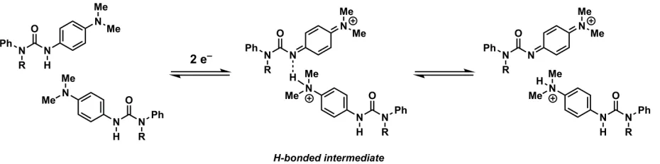
through stable hydrogen-bonded intermediates.18 The urea shown in Figure 1.8 participates in a
complicated PCET mechanism, with oxidation of the phenylenediamine moiety coupled to
proton transfer from the urea moiety. A series of electrochemical and spectroelectrochemical
experiments conclusively showed the intermediacy of a H-bonded complex in the course of this
stepwise PCET mechanism, which forms after the electron transfer step and prior to proton
transfer. H-bonding, however, appeared to play an important role only in less polar solvents,
such as dichloromethane, and did not appear to affect the mechanism in acetonitrile. This study
18 Clare, L. A.; Pham, A. T.; Magdaleno, F.; Acosta, J.; Woods, J. E.; Cooksy, A. L.; Smith, D. K. J. Am.
10
led to the formalization of a “wedge scheme” for PCET, describing mechanisms in which these
stable, long-lived H-bonded intermediates play an integral role in the overall mechanism. Mayer
and coworkers have also presented a quantitative analysis of the influence of H-bonded
intermediates on the overall thermodynamics and kinetics of a bimolecular PCET reaction.19
Figure 1.8. Hydrogen-bonded intermediates within a PCET mechanism.18
The involvement of H-bonded intermediates in PCET reactions of quinones has been
rigorously studied in one specific example involving reduction of 3,5-di-tert-butyl-o
-benzoquinone.20 In the two-electron reduction of this quinone, the characteristic CV traces
involving two reversible waves are absent, and instead are replaced with more complicated CV
traces involving irreversible waves. In an attempt to elucidate the mechanism for quinone
reduction in this case, formation of a stable H-bonded intermediate between adventitious water
and the semiquinone was determined to precede reduction to and protonation of the quinone
dianion.
19 Mader, E. A.; Mayer, J. M. Inorg. Chem. 2010, 49, 3685–3687.
[image:29.612.77.542.210.326.2]11
A study of quinone reduction in unbuffered aqueous solution illustrates the importance of
H-bonding in determining the mechanism by which electron transfer proceeds.8 Even in aqueous,
non-acidic media, quinones undergo a one-step, two-electron reduction, largely due to the role of
H-bonding. In aqueous media, the CV waves corresponding to the first- and second-electron
reductions of quinones essentially merge into a single wave. This is attributed to a large degree
of thermodynamic stabilization provided to the dianon through H-bonding with water.
1.4 Binding studies of quinones to weak HBDs 1.4.1 H-bonding between quinones and alcohols
One of the first systematic investigations of the continuum between H-bonding and
proton transfer, and the corresponding effect on quinone reduction, was carried out by Linschitz
and coworkers in 1997.9 This study surveyed many quinones with a broad scope of reduction
potentials, and examined the effect of H-bonding additives on their electrochemistry in polar,
aprotic solvents (Figure 1.9).
Weak HBDs, such as ethanol, had essentially no impact on the first electron transfer, but
had a substantial impact on the second electron transfer. Determination of units describing Keq
for this association is not straightforward, due to the fact that multiple alcohols can associate
with the quinone and this number varies depending on the specific quinone. However, the largest
effects are observed for the least oxidizing quinones, which are also the most Lewis basic. In all
12
Figure 1.9. Quinones examined by Linschitz and coworkers.9
When stronger HBDs, such as trifluoroethanol, are used with the same quinones, a range
of effects are observed by CV, demonstrating that pathways for H-bonding and proton transfer
can manipulate the mechanisms of electron transfer and PCET. More electron-deficient quinones
start to show a shift in E1/2 corresponding to the first electron transfer, although this effect is still
quite small. The more basic quinones, however, display irreversibility in their CV traces,
consistent with protonation of the quinone dianion post-electron-transfer. This effect is
exacerbated when yet stronger H-bond donors like hexafluoroisopropanol are used, and the CV
traces in this case are consistent with a mechanism involving either a protonated or
hydrogen-bonded complex prior to reduction to the dianion. Finally, strongly acidic donors like
trifluoroacetic acid show unambiguous protonation effects consistent with a PCET pathway
involving electron transfer, followed by protonation, followed by a second electron transfer. In a
closely related study, this work was extended to even more basic quinones, and similar effects
were observed.21
21 Macías-Ruvalcaba, N. A.; González, I.; Aguilar-Martínez, M. J. Electrochem. Soc. 2004, 151, E110–
[image:31.612.89.526.71.131.2]13
1.4.2 H-bonding between quinones and amides
The effect of amides on electron transfer to p-chloranil, an electron-deficient quinone, has been studied as well, but still no effect on the first electron transfer was observed.10 An o
-phenolic benzamide, capable of acting as a dual HBD, displayed the largest effect on the second
electron transfer (Figure 1.10A). A separate study examined the effect of a different amide, o -bis(phenylcarbamoylmethyl)benzene, on electron transfer to the same quinone.22 In this case, the
first electron transfer is sufficiently affected such that the quinone can mediate an electron
transfer from cobalt tetraphenylporphyrin in the presence of the amide (Figure 1.10B), and the
amide is determined to bind the reduced quinone in a 1:1 complex. Additionally, an effect on the
rate of electron transfer was also observed, but was not quantified. However, as the reduction
potentials of the two redox partners are only separated by 40 mV, the neutral quinone and
electron donor are in equilibrium with the electron transfer complex. Consistent with the trends
regarding Lewis basicity of quinones (vide supra), a larger effect is observed when the less electron-deficient 2,6-dichloro-p-benzoquinone is used. Under these circumstances, electron
transfer is considerably less favorable (ΔEred = –0.21 V).
14
Figure 1.10. Effect of amides on electron transfer to p-chloranil.
1.4.3 Influence of cationic additives on electron transfer to quinones
Simple ammonium salts have been shown to affect the kinetics of electron transfer to
benzoquinone and naphthoquinone.23 Photoinduced electron transfer from tetraphenylporphyrin to the quinone exhibits a second-order dependence on the concentration of NH4PF6, indicating
that the reduced quinones are stabilized in a 2:1 complex with cationic ammonium ions. An
effect on the reduction potentials of the quinones was also observed, although the specific
binding constants in each case were not determined.
A mechanistic study designed to distinguish stepwise pathways from a concerted
pathway employed protonated histidine as a means of activating a sulfinyl-substituted quinone
toward reduction (Figure 1.11). 24
15
Figure 1.11. Electron transfer between TolSQ and 1,1’-dimethylferrocene, promoted by protonated histidine.
The binding constant between histidine and the quinone was calculated from a single
titration point and was found to be 4.2 x 10¹⁰ M–1 for a 1:1 complex. This
2-tolylsulfinyl-1,4-benzoquinone (TolSQ) is still a fairly weak oxidant (E1/2 = –0.64 V vs. Fc+/0) and is quite basic,
as evidenced by the irreversibility observed by cyclic voltammetry indicating that the H-bonded
complex is unstable and undergoes decomposition. Protonated histidine promotes electron
transfer between TolSQ and ferrocene, which in the absence of electron transfer is
thermodynamically disfavored (ΔGET = 15.5 kcal/mol). Other, less-reducing ferrocenes were examined in kinetic isotope effect (KIE) experiments using deuterated histidine, which revealed
that the KIE decreases as driving force increases. This observation lends support for a proposed
mechanistic changeover from a concerted pathway—in which H-bond formation would be part
of the rate-determining step—to a stepwise pathway involving rate-determining electron transfer,
in which no KIE would be observed.
1.4.4 Binding of quinones to neutral dual HBDs
Several studies involving binding between Lewis basic quinones and neutral dual HBDs
have been performed. When 9,10-phenanthrenequinone is titrated with 1,3-diphenylurea, CV
16
shift in apparent potential (Figure 1.12A).25 In an extension of this study, it was found that this
shift corresponds to a binding constant of 905 M–1 describing the association between the
quinone and the urea.26 It was also proposed that the structure of the quinone determines strength
of binding to dual HBDs—that o-quinones are better suited for H-bonding as both oxygen atoms
are accessible to the urea N–H bonds. However, the potential for p-quinones to form 2:1
complexes between the HBD and the quinone appears not to have been considered. Subsequent
studies with 1,4-dinitrobenzene do provide evidence for formation of these complexes.27 An
interesting structure-activity relationship that demonstrates the importance of the dual H-bonding
motif was carried out using pyridylureas (Figure 1.12B). 4-Pyridiylurea can provide two
H-bonds to the quinone, and is as effective as 1,3-diphenylurea at stabilizing the semiquinone.
However, 2-pyridylurea is expected to have an intramolecular H-bond that renders one N-H bond
inaccessible to the quinone. As a result, this quinone has a minimal effect on the first electron
reduction of the quinone.
Figure 1.12. (a) Binding of 9,10-phenanthrenequinone semiquinone to 1,3-diphenylurea (b) Structure-activity relationship using 2- and 4-pyridyl-3-phenylurea.
17
Thioureas have been observed to have a measurable, albeit small, effect on the first
electron transfer to ubiquinone. 28 Titration of ubiquinone with one equivalent of N,N’
-dialkylthiourea afforded a 15 mV shift in redox potential (the binding constant was not
quantified) as a result of H-bonding between the thiourea and the semiquinone (Figure 1.13A). In
contrast, diphenylthiourea—which is considerably more acidic—enabled direct two-electron
reduction of ubiquinone to the dianion (Figure 1.13B). This example further demonstrates the
importance of H-bonding in regulating the various mechanisms by which quinone reduction can
proceed.
Figure 1.13. Binding of ubiquinone to thioureas.
1.5 Conclusions
H-bonding interactions exhibit a profound influence on electron transfer pathways,
specifically when quinone oxidants are used. The studies described here have demonstrated that
small-molecule HBDs are capable of stabilizing the reduced quinone through binding, thereby
facilitating electron transfer. Effects on rate have also been reported. While the literature
precedent describing these interactions is vast, no attempts have been made to extend this effect
to a synthetically interesting context, in which quinone oxidants are used to mediate an organic
18
transformation. Doing so would require both the use of synthetically useful quinones as well as
design of HBDs capable of stabilizing such a quinone in its reduced state. Mechanistic studies
aimed at developing such a HBD-coupled electron transfer, as well as its application in an
19
Chapter 2
Activation of electron-deficient quinones through
HBD-coupled electron transfer
A. Electrochemical quantification of
o
-chloranil binding to HBDs
2a.1 Introduction
Because the ability of quinones to participate in electron transfer is strongly influenced by hydrogen bonding, we hypothesized that small-molecule hydrogen-bond donors (HBDs) could activate quinone oxidants for application in a synthetically interesting context. While HBDs are known to couple with electron transfer to enhance the reactivity of quinone oxidants, this effect was only observed with weakly oxidizing quinones (E1/2 < –0.6 V vs. Fc+/0) that are good Lewis bases (Figure 2.1).1
1 For discussion, see Chapter 1. (a) Gupta, N.; Linschitz, H. J. Am. Chem. Soc. 1997, 119, 638–6391. (b)
Gómez, M.; Gómez-Castro, C. Z.; Padilla-Martínez, I. I.; Martínez-Martínez, F. J.; González, F. J. J. Electroanal. Chem. 2004, 567, 269–276. (c) Macías-Ruvalcaba, N. A.; González, I.; Aguilar-Martínez, M.
J. Electrochem. Soc. 2004, 151, E110–E118. (d) Fukuzumi, S.; Kitaguchi, H.; Suenobu, T.; Ogo, S. Chem. Commun. 2002, 1984–1985. (e) Okamoto, K.; Ohkubo, K.; Kadish, K.M.; Fukuzumi, S. J. Phys. Chem. A.
20
Figure 2.1. Differences between electron-rich and electron-deficient quinones.
In contrast, the HBDs studied had little discernible effect on the electron transfer to electron-deficient quinones. Electron-deficient quinones bear electron-withdrawing substituents, which increase their oxidizing ability but diminish their Lewis basicity and binding ability. In turn, this presents a challenge to the activation of electron-deficient quinones through H-bonding. This effect is even more pronounced in combination with weaker HBDs (e.g. alcohols), which are unlikely to have any discernible effect on weak hydrogen bond acceptors. However, the success of our HBD-coupled electron transfer strategy rests on the ability to activate more strongly oxidizing quinones in order to achieve synthetically useful levels of reactivity.2,3
We report herein a systematic evaluation of a series of small-molecule hydrogen bond donors, with the intention of identifying molecules that are capable of modulating the thermodynamics of electron transfer to an electron-deficient quinone (Figure 2.2). o-Chloranil (Q) (E1/2 = –0.35 V vs. Fc+/0) was selected as the oxidant, as it is an electron-deficient quinone that nonetheless lacks the intrinsic reactivity necessary to oxidize many organic substrates of
2 Becker, H.-D.; Turner, A. B. Quinones as oxidants and dehydrogenating agents. In The Chemistry of the Quinonoid Compounds; Patai, S.; Rappoport, Z.; Eds.; Wiley: New York, 1988; Vol. II, Chapter 23, pp 1352–1384.
21
synthetic interest. Our examination of the influence of H-bonding on the single-electron transfer chemistry of o-chloranil has led to the discovery that dicationic bis-amidinium salts can exert remarkable influence on the thermodynamics of electron transfer.
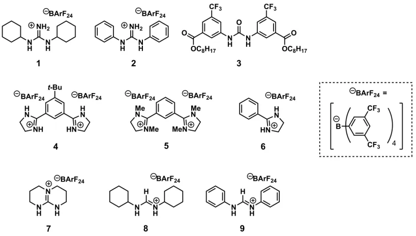
Figure 2.2. Series of hydrogen bond donors and additives examined in this chapter.
2a.2 Theoretical basis
2a.2.1 Thermodynamic analysis
HBD-coupled electron transfer using quinone oxidants can be split into two elementary steps: electron transfer between the quinone (Q) and an electron donor, and binding of the reduced quinone to a HBD (Figure 2.3). The specific mechanism for this process may vary, but this general analysis aids in defining both the challenge that is presented to achieving favorable
electron transfer reactions (ΔGnet < 0) using HBDs and the benefits of using electron-deficient
[image:40.612.102.511.181.413.2]22
Figure 2.3. Thermodynamic analysis of hydrogen-bond-donor-coupled electron transfer.
The activating effect of a HBD on the overall reaction can be understood in terms of binding of the HBD to the reduced quinone (Q•–). This interaction (ΔGbind) must offset the
thermodynamic price of the electron transfer (given a ΔGET > 0), which depends on the specific substrate and the intrinsic oxidizing ability of the quinone. Oxidation of organic substrates of interest—including olefins and aromatic rings—by electron-rich quinones is so unfavorable that an unattainably high binding energy would be necessary in order for HBDs to facilitate the overall reaction. Electron transfer in synthetically interesting contexts with electron-deficient
quinones is less unfavorable.4 However, as noted above, these quinones and their reduced
counterparts are inherently weak H-bond acceptors. As such, the success of the proposed HBD-coupled electron transfer strategy relies on finding the appropriate balance of HBD strength and quinone reactivity.
23
2a.2.2 Mechanistic tools for the study of HBD-coupled electron transfer
In aprotic media, quinones undergo two sequential single electron transfers, proceeding
through the semiquinone radical anion intermediate, Q•–.5 Protic and H-bonding additives
influence the mechanism by which electron transfer proceeds—this study is concerned principally with the effect of HBDs on the first electron transfer step. To quantify the ability of a HBD to modulate the thermodynamics of electron transfer, we need to understand how well an HBD can bind to Q•–—or, how strongly the HBD favors this reduced state over the neutral,
oxidized state.
The mechanistic tools used to quantify PCET are readily applied to the study of HBD-coupled electron transfer. Equation (1),6 which is related to the Nernst equation, shows that the apparent potential of a quinone involved in PCET will undergo a pH-dependent shift (ΔE1/2), where KQ and KQ•– correspond to the acid dissociation constants of the neutral quinone and
singly-reduced semiquinone, respectively.
∆E1/2 = 0.059 𝑉𝑉𝑙𝑙𝑙𝑙𝑙𝑙 1+[𝐻𝐻+]
𝐾𝐾𝑄𝑄•–
1+[𝐻𝐻+]𝐾𝐾𝑄𝑄 (1)
We can adapt equation (1) to describe HBD-coupled electron transfer, in which the ΔE1/2 is dependent on the HBD concentration and the association constants for the binding of the quinone and semiquinone (K’Q and K’Q•–, respectively) to the HBD (equation (2)).
5 Chambers, J. Q. Electrochemistry of Quinones. In The Chemistry of the Quinonoid Compounds; Patai,
S.; Rappoport, Z.; Eds.; Wiley: New York, 1988; Vol. II, Chapter 12, pp 719–757.
24
∆E1/2 = 0.059 𝑉𝑉𝑙𝑙𝑙𝑙𝑙𝑙
1+𝐾𝐾′Q•– [𝐻𝐻𝐻𝐻𝐻𝐻]
1+𝐾𝐾′𝑄𝑄[𝐻𝐻𝐻𝐻𝐻𝐻] (2)
This relationship between ΔE1/2 and the association constants shows that, as long as K’Q•–
>K’Q, increasing concentration of the HBD results in a more positive ΔE1/2, effectively creating a more potent oxidant by favoring the reduced state through binding. The equilibrium constants that govern this shift in the potential can be elucidated electrochemically through cyclic
voltammetry and provide a quantitative measure of the stabilization provided by the HBD to Q•–,
which in turn is related toΔGbind.
2a.3 Mechanisms and notation
HBDs affect the oxidizing ability of Q through several different pathways that can involve complex binding stoichiometries. If two HBDs are involved in binding, the square scheme is extended in order to describe all mechanistic possibilities (Figure 2.4).
Figure 2.4. Square scheme describing HBD-coupled electron transfer to quinones when
25
As Figure 2.4 illustrates, a pathway for HBD-coupled electron transfer involves a series of chemical binding (C) and electron transfer (E) steps, each of which has an associated
equilibrium constant. To determine the binding constants that describe the stabilization of Q•– by
a given HBD, identification of the specific pathway by which HBD-coupled electron transfer proceeds and elucidation of the corresponding equilibrium constants is required. Simulations of electrochemical data can be used to distinguish between the possibilities for HBD-coupled electron transfer, and provide support for a specific mechanistic pathway. Through these simulations, values for the equilibrium constants are also obtained.
2a.4 Experimental design
2a.4.1 Determination of conditions for CV experiments
The electrochemical studies outlined in this chapter are performed with o-chloranil, but preliminary investigations with p-chloranil (p-Q) provided the basis for determination of our experimental conditions. Selection of an appropriate electrolyte was critical as the identity of the electrolyte proved to have a significant impact on the CV data. For example, when nBu4NPF6 was employed as the electrolyte, a minimal effect on the apparent potential of the p-Q/p-Q•–
26
Figure 2.5. Experimental CVs of p-chloranil (initial concentration = 0.5 mM) recorded in 0.1 M
nBu4PF6/CH2Cl2(glovebox) in the presence of increasing [1].
Speculating that this small effect was due to inhibition of the HBD by –PF6, we repeated this experiment using an electrolyte with a non-coordinating anion.7 A switch to nBu4NBArF248 affords more substantial shifts in the recorded CVs of p-Q upon titration with 1 (Figure 2.6).
7 (a) LeSuer, R. J.; Buttolph, C.; Geiger, W. E. Anal. Chem. 2004, 76, 6395–6401. (b) Barrière, F.; Geiger,
W. E. J. Am. Chem. Soc. 2006, 128, 3980–3989.
8 Nishida, H.; Takada, N.; Yoshimura, M.; Sonoda, T.; Kobayashi, H. Bull. Chem. Soc. Jpn. 1984, 57,
[image:45.612.142.476.71.437.2]27
Figure 2.6. Experimental CVs of p-chloranil (initial concentration = 0.5 mM) recorded in 0.1 M
nBu4NBArF24/CH2Cl2(glovebox) in the presence of increasing [1].
[image:46.612.142.476.71.438.2]28
2a.4.2 General approach to electrochemical experiments and data analysis
A systematic investigation of HBDs was undertaken, with the aim of understanding how H-bonding interactions can stabilize Q•– and thereby promote electron transfer to
electron-deficient quinones are used. Titrations of Q were carried out with each HBD, using cyclic
voltammetry to record the ΔE1/2. In each case, a significant, measurable effect was observed on
the apparent potential that corresponds to the first electron transfer. Additionally, the reversibility of the CVs recorded in all titration experiments indicate that the ΔE1/2 is the result of H-bonding to Q•– and not protonation, which would manifest as irreversibility in the CV traces.
The experimental data in each case were analyzed using simulations to determine the pathway and equilibrium constants associated with HBD-coupled electron transfer. The equations and parameters used for each simulation are outlined in the subsequent sections. Discrepancies in current magnitude between experiment and simulation are noted throughout the various titration experiments, and are attributed to variability in diffusion coefficient across the range of species involved in the simulation. This value, which was not determined by the simulations, has no bearing on ΔE1/2 and therefore does not impact the simulated equilibrium constants.
2a.5 Results and discussion
2a.5.1 Mechanistic analysis of HBD-coupled electron transfer using N,N’
-dicyclohexylguanidinium salts
29
Figure 2.7. Experimental CVs of o-chloranil (initial concentration = 0.5 mM) recordedin 0.1 M
nBu4NBArF24/CH2Cl2(glovebox) in the presence of increasing [1].
To elucidate the equilibrium constants that describe binding of Q•– to 1, the full set of
30
[Q] would afford a corresponding increase in current due to reduction of this complex, with a concomitant decrease in current due to reduction of Q.
Figure 2.8. Experimental CVs of o-chloranil (initial concentration = 0.5 mM) recorded in 0.1 M
nBu4NBArF24/CH2Cl2 (glovebox) in the presence of increasing [1]. Only data collected at low [1] are shown.
This pre-association hypothesis was borne out in the simulations, in which a mechanism involving binding of neutral Q to 1 provides the best fit to the experimental data. Specifically, these data are accurately represented by a CEC mechanism, in which this binding event (K1)
precedes electron transfer (E2), and another binding event between 1 and Q•– follows (K4)
[image:49.612.148.468.152.405.2]31
values using a Benesi-Hildebrand analysis are consistent with those obtained from the simulations, providing additional support for the CEC mechanism. These data, along with simulated mechanisms that represent poor fits to the experimental data, are presented in section 2.7.3.
Figure 2.9. CEC mechanism.
Figure 2.10. Experimental CVs of o-chloranil (initial concentration = 0.5 mM) recorded in 0.1
M nBu4NBArF24/CH2Cl2 (glovebox) in the presence of increasing [1] and comparison with
[image:50.612.150.461.373.628.2]32
Figure 2.11. Experimental CVs of o-chloranil (initial concentration = 0.5 mM) recorded in 0.1
M nBu4NBArF24/CH2Cl2 (glovebox) in the presence of increasing [1] and comparison with
simulation of a CEC mechanism. Only data collected at low [1] are shown.
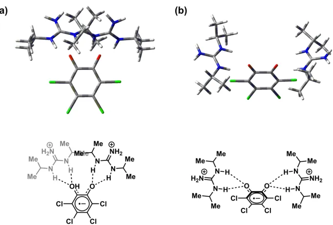
Simple DFT calculations indicate that the geometry of the (1)2·Q•– complex involves
33
Figure 2.12. Geometry calculated for the (1)2·Q•– complex, optimized using (a) B3LYP/6-31G(d,p) level of theory; the gray HBD molecule is oriented behind the other HBD molecule; (b) M06-2X/6-31G(d,p) level of theory.
The bottom of the extended square scheme in Figure 2.4 describes the stabilization of Q•–
through binding to two HBD molecules. Therefore, K3K4—the product of the equilibrium constants corresponding to the two binding events—quantitatively describes the equilibrium favoring the reduced state and provides a measure of the oxidizing strength of Q in the presence of a given HBD. Explicit determination of K1, E2, and K4 is afforded by simulation of a CEC mechanism, and E1 is experimentally determined. Therefore, the thermodynamically redundant parameter K3 can be calculated from these values. The equations and parameters used to simulate the experimental data, including the elucidated equilibrium constants, are shown in Table 2.1.9
From these values, K3K4 describing formation of the (1)2·Q•– complex was calculated to be
6.1x10⁸ M–2.
9 In this simulation and subsequent simulations, k
f for each binding step was fixed at the
diffusion-controlled limit of 1x10¹⁰ s–1. This was due to the fact that the k
f was consistently simulated to be slightly
[image:52.612.127.468.79.309.2]34
Table 2.1. Equations and best-fit parameters used to simulate CV data for 0.5 mM o-chloranil (Q) in 0.1 M nBu4NBArF24/CH2Cl2 (glovebox) in the presence of increasing [1]. Italicized values were fixed in the simulation.
Electron transfer
steps E1/2 (V vs. Fc
+/0) α k
s (cm/s)
Q + e–→Q•– –0.33 0.5 0.022
1·Q + e–→1·Q•– –0.17 0.5 0.21
Chemical steps Keq (M–1) kf (s–1)
Q →1·Q 66 1 x 10¹⁰
1 + 1·Q•–→ (1)
2·Q•– 1.8 x 10⁴ 1 x 10¹⁰
2a.5.2. Mechanistic analysis of HBD-coupled electron transfer using N,N’
-diphenylguani-dinium salts
Titration of Q with N,N’-diphenylguanidinium salt 2 also affords a shift in the apparent potential of the Q/Q•– redox couple (Figure 2.13). At low [2], the recorded CVs display features
similar to those observed in the titration with 1 (Figure 2.14), indicating that HBD-coupled electron transfer involving 2 may also proceed via a CEC mechanism. In addition to these curve shapes, other current peaks are observed at low potential as [2] increases (e.g. scans (c)–(d) in
Figure 2.13). These peaks correspond to the second electron reduction of Q (e.g. the Q•–/Q2–
redox couple). The apparent potential corresponding to this process undergoes a substantial shift
upon addition of 2, due to the strong Lewis basicity of the dianion. Because we are only
35
Figure 2.13. Experimental CVs of o-chloranil (initial concentration = 0.5 mM) recorded in 0.1
M nBu4NBArF24/CH2Cl2 (glovebox) in the presence of increasing [2] and comparison with
simulation of a CEC mechanism. In scans (c) and (d), the additional current peaks at lower
36
Figure 2.14. Experimental CVs of o-chloranil (initial concentration = 0.5 mM) recorded in 0.1
M nBu4NBArF24/CH2Cl2 (glovebox) in the presence of increasing [2] and comparison with
simulation of a CEC mechanism. Only data collected at low [2] are shown.
As is the case with 1, the data recorded in the presence of 2 are best simulated by a CEC mechanism, which replicates both the overall ΔE1/2 and the distinct curve shapes at low [2]. Computations on this 2:1 complex also show bifurcated H-bonding interactions with distances of 1.81 Å and 1.86 Å calculated with B3LYP, and 1.75 Å and 1.89 Å calculated with M06-2X. In the calculation using M06-2X, an apparent slipped pi-stacking interaction between the arenes of the two HBD molecules is observed, with a distance between the arenes of 3.4Å (Figure 2.15).
The value of K3K4 describing formation of this (2)2·Q•– complex was calculated to be 1.8x10¹⁰
37
[image:56.612.81.533.74.289.2]Figure 2.15. Geometry calculated for the (2)2·Q•– complex, optimized using (a) B3LYP/6-31G(d,p) level of theory; the gray HBD molecule is oriented behind the other HBD; (b) M06-2X/6-31G(d,p) level of theory; (c) Pi-stacking interaction between the arenes on the HBD.
Table 2.2. Equations and best-fit parameters used to simulate CV data for 0.5 mM o-chloranil (Q) in 0.1 M nBu4NBArF24/CH2Cl2 (glovebox) in the presence of increasing [2]. Italicized values were fixed in the simulation.
Electron transfer
steps E1/2 (V vs. Fc
0/+) α k
s (cm/s)
Q + e–→Q•– –0.34 0.5 0.022
2·Q + e–→2·Q•– –0.056 0.5 1.5
Chemical steps Keq (M–1) kf (s–1)
2 + Q →2·Q 5.5 1 x 10¹⁰
2 + 2·Q•–→ (2)
2·Q•– 5.2 x 10⁴ 1 x 10¹⁰
38
2a.5.3 Mechanistic analysis of HBD-coupled electron transfer using neutral ureas
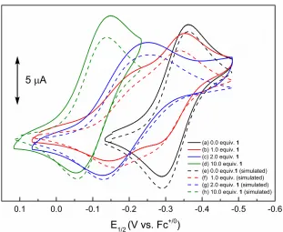
Titration of o-chloranil with urea 3 affords the CV traces shown in Figure 2.16. The same features observed at low [1] and [2] are also present at low [3], although the effect is significantly less pronounced. A CEC mechanism again provides the best fit to the experimental data, demonstrating that in the case of neutral, dual HBDs, a 2:1 complex is formed with the reduced quinone. The parameters and equations used to simulate the titrations with 3 are summarized in Table 2.3. Analysis of the best-fit parameters yields a K3K4 value of 1.0x10⁷ M–2
[image:57.612.75.540.295.664.2]for formation of (3)2·Q•–.
Figure 2.16. Experimental CVs of o-chloranil (initial concentration = 0.5 mM) recorded in 0.1
M nBu4NBArF24/CH2Cl2 (glovebox) in the presence of increasing [3] and comparison with

![Figure 2.5. Experimental CVs of p-chloranil (initial concentration = 0.5 mM) recorded in 0.1 M nBu4PF6/CH2Cl2 (glovebox) in the presence of increasing [1]](https://thumb-us.123doks.com/thumbv2/123dok_us/7925232.192583/45.612.142.476.71.437/figure-experimental-chloranil-concentration-recorded-glovebox-presence-increasing.webp)
![Figure 2.6. Experimental CVs of p-chloranil (initial concentration = 0.5 mM) recorded in 0.1 M nBu4NBArF24/CH2Cl2 (glovebox) in the presence of increasing [1]](https://thumb-us.123doks.com/thumbv2/123dok_us/7925232.192583/46.612.142.476.71.438/figure-experimental-chloranil-concentration-recorded-glovebox-presence-increasing.webp)
![Figure 2.7. Experimental CVs of no-chloranil (initial concentration = 0.5 mM) recorded in 0.1 M Bu4NBArF24/CH2Cl2 (glovebox) in the presence of increasing [1]](https://thumb-us.123doks.com/thumbv2/123dok_us/7925232.192583/48.612.109.501.76.431/figure-experimental-chloranil-concentration-recorded-glovebox-presence-increasing.webp)
![Figure 2.8. Experimental CVs of o-chloranil (initial concentration = 0.5 mM) recorded in 0.1 M nBu4NBArF24/CH2Cl2 (glovebox) in the presence of increasing [1]](https://thumb-us.123doks.com/thumbv2/123dok_us/7925232.192583/49.612.148.468.152.405/figure-experimental-chloranil-concentration-recorded-glovebox-presence-increasing.webp)
![Figure 2.11. Experimental CVs of o-chloranil (initial concentration = 0.5 mM) recorded in 0.1 M nBu4NBArF24/CH2Cl2 (glovebox) in the presence of increasing [1] and comparison with simulation of a CEC mechanism](https://thumb-us.123doks.com/thumbv2/123dok_us/7925232.192583/51.612.151.468.71.322/experimental-chloranil-concentration-presence-increasing-comparison-simulation-mechanism.webp)