An affinity-based scoring scheme for
predicting DNA-binding activities of
modularly assembled zinc-finger proteins
The Harvard community has made this
article openly available.
Please share
how
this access benefits you. Your story matters
Citation
Sander, Jeffry D., Peter Zaback, J. Keith Joung, Daniel F. Voytas, and
Drena Dobbs. 2009. An affinity-based scoring scheme for predicting
DNA-binding activities of modularly assembled zinc-finger proteins.
Nucleic Acids Research 37(2): 506-515.
Published Version
doi:10.1093/nar/gkn962
Citable link
http://nrs.harvard.edu/urn-3:HUL.InstRepos:4878923
Terms of Use
This article was downloaded from Harvard University’s DASH
repository, and is made available under the terms and conditions
applicable to Other Posted Material, as set forth at
http://
506–515 Nucleic Acids Research, 2009, Vol. 37, No. 2 Published online 4 December 2008 doi:10.1093/nar/gkn962
An affinity-based scoring scheme for predicting
DNA-binding activities of modularly assembled
zinc-finger proteins
Jeffry D. Sander
1,*, Peter Zaback
1, J. Keith Joung
2,3, Daniel F. Voytas
4and
Drena Dobbs
1,1
Department of Genetics, Development and Cell Biology, Bioinformatics and Computational Biology Program, Iowa State University, Ames, IA 50011, 2Molecular Pathology Unit, Center for Cancer Research, and Center for Computational and Integrative Biology, Massachusetts General Hospital, 149 13th Street, Charlestown, MA 02129, 3Department of Pathology, Harvard Medical School, Boston, MA 02115 and 4Department of Genetics, Cell Biology and Development and Center for Genome Engineering, University of Minnesota, MN 55455, USA
Received August 7, 2008; Revised November 10, 2008; Accepted November 12, 2008
ABSTRACT
Zinc-finger proteins (ZFPs) have long been recog-nized for their potential to manipulate genetic infor-mation because they can be engineered to bind novel DNA targets. Individual zinc-finger domains (ZFDs) bind specific DNA triplet sequences; their apparent modularity has led some groups to pro-pose methods that allow virtually any desired DNA motif to be targeted in vitro. In practice, however, ZFPs engineered using this ‘modular assembly’ approach do not always function well in vivo.
Here we report a modular assembly scoring strategy that both identifies combinations of modules least likely to function efficiently in vivo and provides accurate estimates of their relative binding aff-inities in vitro. Predicted binding affinities for 53 ‘three-finger’ ZFPs, computed based on energy con-tributions of the constituent modules, were highly correlated (r= 0.80) with activity levels measured in bacterial two-hybrid assays. Moreover, Kd values for seven modularly assembled ZFPs and their intended targets, measured using fluorescence anisotropy, were also highly correlated with predic-tions (r= 0.91). We propose that success rates for ZFP modular assembly can be significantly improved by exploiting the score-based strategy described here.
INTRODUCTION
The ability to reliably engineer DNA binding proteins that recognize any desired DNA sequence would provide an unprecedented level of control over genetic information; for example, by allowing the creation of site-specific nucleases that specifically alter genomic DNA (1–5). The C2H2zinc-finger domain (ZFD) is arguably the best
char-acterized DNA binding motif and offers considerable pro-mise for the rational engineering of site-specific DNA binding proteins (6–11). Zinc-finger proteins (ZFPs) con-sist of multiple individual ZFDs, each of which typically recognizes adjacent sequence triplets in duplex DNA (Figure 1). An individual ZFD comprises a pair of anti-parallelb-strands and onea-helix, which coordinate a zinc ion through conserved pairs of cysteine and histidine resi-dues. In the canonical three-finger domain of the Zif268 transcription factor, the amino acid side chains at posi-tions 1, +3 and +6 relative to the amino-terminal end of the a-helix typically make base-specific contacts with three adjacent nucleotides within the major groove of double-stranded DNA (12). An aspartic acid residue in the +2 position of the DNA recognition helix can specify a fourth nucleotide, resulting in either target-site overlap with an adjacent module or specification of an additional nucleotide at the 30-end of the target site (13,14).
Several research groups have characterized ZFDs that recognize many of the 64 possible DNA triplets (15–20). Using a ‘modular assembly’ approach, novel ZFPs that recognize variant DNA sites are assembled by simply
The authors wish it to be known that, in their opinion, the first two authors should be regarded as joint First Authors. *To whom correspondence should be addressed. Tel: +1 515 294 4991; Fax: +1 515 294 6790; Email: [email protected] Correspondence may also be addressed to Drena Dobbs. Tel: +1 515 294 4991; Fax: +1 515 294 6790; Email: [email protected]
ß2008 The Author(s)
stringing together individual ZFDs. In practice, however, ZFPs made by modular assembly display a wide range of binding affinities and specificities (15,19,21–23). Although modular assembly has proven useful for some in vivo
applications, such as artificial transcription factors, recent work suggests that the success rate of creating arti-ficial zinc-finger nucleases (ZFNs–fusions of engineered zinc fingers to a non-specific nuclease domain) by this method is considerably lower (24,25). These low success rates, together with the inability to predict which ZFPs are likely to function in vivo, have motivated our groups to improve the procedures and design criteria for ZFP engineering (25,26).
The present study was motivated by our observation that among a small set of modularly assembled ZFPs, those that fail to functionin vivoare more likely to possess modules previously shown to have relatively low affinity for target DNA. This observation implies that insufficient affinity can contribute to poor function in vivo and also suggested that it might be possible to predict the affinity of a modularly assembled ZFP using existing affinity data for component modules. Here we test these hypotheses and demonstrate that both the in vitro binding affinity and the lack ofin vivoactivity of a ZFP can be predicted using the energy contributions of its component ZFDs. Our approach for predicting the binding of ZFPs to desired target sequences should improve success rates of modular assembly by guiding investigators away from target sites and ZFP combinations least likely to function
in vivo.
MATERIALS AND METHODS
Zinc-finger modules and three-finger arrays (ZFPs)
All ZFDs used in these experiments have been described by the Barbas group (15) and are referred to as ‘Barbas modules’. ZFPs containing desired three-finger (three-module) arrays were assembled by iterative ligation and cloning of restriction fragments encoding ZFDs using reagents and protocols previously described by the Zinc Finger Consortium (http://www.zincfingers.org/) (27). ZFP-encoding fragments were then cloned into vectors for expression as Gal11P-hybrid proteins in the bacterial two-hybrid (B2H) system as previously described (27).
B2H assays
A series of B2H reporter plasmids, each harboring a target binding site for one of 27 different three-finger ZFPs, was constructed by cloning synthetic target oligonucleotides into reporter plasmid pBAC-lacZ as previously described (27). Binding of a Gal11P-ZFP hybrid protein to the target sequence on a B2H reporter plasmid triggers tran-scriptional activation of a lacZ reporter gene encoding b-galactosidase. In vivo ZFP performance was therefore assayed using a b-galactosidase assay in which ZFP-induced activation oflacZexpression was measured rela-tive to control constructs lacking the ZFP.
Zinc finger–maltose binding protein fusion protein construction, expression and purification
Zinc finger–maltose binding protein (MBP) fusion protein constructs were generated by transferring three-finger arrays, assembled as described above, into pHMTC (28). The MBP fusion plasmids were transformed into BL21
Escherichia colicells (Invitrogen) using standard chemical transformation procedures (29).
For protein expression, 5 ml cultures were grown for 16 h at 308C with agitation in ZFE broth [Luria Broth (LB), 1.11 mM dextrose, 100mg/ml ampicillin]. Expansion cultures of 10 ml were inoculated from these overnight cultures (1:100 dilution) and grown to an OD600 of 0.5
before a 2 h induction with isopropyl b-D-1-thiogalacto-pyranoside (IPTG). Cells were harvested by centrifugation for 10 min at 4000gat 48C and frozen overnight at208C. The following day, cells were resuspended in 4 ml WB1 (15 mM HEPES pH 7.8, 200 mM NaCl, 20mM ZnSO4)/
1 mM PMSF/0.1% NonidetTMP40 (NP-40) and refrozen at708C. Cells were then thawed in ice water and centri-fuged at 9000g at 48C for 20 min. To remove remaining nucleic acids, the resulting supernatant was transferred to a new cold tube and polyethyleneimine was added to 0.1%. The supernatant was then incubated for 30 min before a second centrifugation at 16 000g at 48C for 30 min.
Amylose beads (NEB) were prepared in 50ml aliquots in 1.5 ml micro-centrifuge tubes according to manufacturer’s instructions. Beads were washed (suspended, spun down and supernatant removed) three times in 1 ml WB1/0.1% NP-40 at 48C and resuspended in 450ml WB1. For affinity purification, 1 ml of clarified protein supernatant was added to prepared beads, and incubated at 48C for 30 min. The slurry was centrifuged and the supernatant was removed. The proteins bound to beads were washed two times with 700ml WB1/0.1% NP-40 and two times with zinc buffer A (ZBA; 10 mM Tris–HCl, pH 7.5, 90 mM KCl, 1 mM MgCl2, 90mM ZnCl2)/0.1% NP-40
[image:3.612.59.299.69.136.2](15). Purified proteins were then eluted in 200ml ZBA/ 0.1% NP-40/40 mM maltose for 30 min at room tempera-ture, with gentle agitation. After elution, beads were cen-trifuged at 16 000g. The supernatant was transferred to a new cold tube and centrifuged again at 16 000g. The super-natant was transferred to a new cold tube and gently stir-red to mix protein. Proteins were stostir-red at 708C in Axygen MaxymumRecoveryTMtubes. Protein concentra-tions were estimated using a Bradford assay against a
Figure 1.A three-finger ZFP with its DNA target site. A ZFP consist-ing of three adjacent ZFDs binds its target DNA through contacts between the amino acids of the DNA recognition helices and consecu-tive nucleotides in the DNA. The protein chain is drawn in the N- to C-terminal direction and the DNA target in the 30–50 direction. Note
bovine serum albumin (BSA) standard in ZBA/0.1% NP-40.
Binding measurements using fluorescence anisotropy
Binding reactions were performed in ZBA/0.1% NP-40/ 0.1 mg/ml non-acetylated BSA (Sigma) for 30 min on ice with 5 nM target DNA. Target sites (shown in Figure 2a) were formed using hairpin DNA oligonucleotides as described (15). HPLC purified, 30-6-FAM-labeled oligonu-cleotides were ordered from Integrated DNA Technologies (Coralville, IA, USA). In each experiment, two serial dilutions of purified ZFP-MBP fusion protein were performed over a range of 1000–0.122 nM. Reported binding affinity values are based on the average of three separate binding experiments, performed on different days, using three separate protein preparations. Fluo-rescence anisotropy (FA) measurements were made using a Varian Cary Eclipse spectrophotometer in L-format configuration. Each value was based on five measurements averaged over 5 s, using a 490 nm excitation wavelength (5 nm slit width), and 530 nm emission wave-length (20 nm slit width) at 880 V. Background light scat-tering for each protein sample dilution was measured and subtracted to correct for protein concentration-dependent variation in intensities.Kdvalues were determined by
non-linear regression (30,31) using Prism (http://www.graph pad.com/prism/Prism.htm).
RESULTS
To test the hypothesis that binding energy contributions of individual ZFDs can be used to predict the in vitro
binding affinities and in vivo performance of extended ZFP arrays, 27 three-module ZFPs were constructed by assembling various GNN-specific modules previously characterized by the Barbas group (15). ZFP compositions were chosen to systematically explore a wide range of predicted binding affinities and to test the influence of context on module performance. As shown in Table 1, ZFDs were divided into three affinity classes based on their reported affinity constants measured in a fixed con-text, namely as fingers in the middle position of a three-finger Zif268 variant (15). Modules comprising Zif268 variants with Kd values <10 nM were categorized as
‘strong’, Kd= 10–30 nM as ‘moderate’ and Kd>30 nM
as ‘weak’ (Table 1). Using three different modules to represent each binding class, all possible combinations of strong, moderate and weak affinity modules for a three-module ZFP were assembled. To allow direct comparisons among proteins that differ by a single module, ZFPs were designed in subgroups in which only one finger position was varied (Table 2).
Predicting relative binding energies for modularly designed ZFPs
If one assumes that the binding energy of a three-finger ZFP (G8ZFP) is equal to the sum of the binding energies
of its three component ZFDs (G8ZFD) [Equation (1)], it
follows that the difference in binding energy between any two ZFPs is the sum over the positions of the difference in
binding energy between the modules at each position [Equation (2)].
GZFP¼GZFD1þGZFD2þGZFD3 1
GZFP1ZFP2¼ GZFP1F1G
ZFP2F1
þ G
ZFP1F2G
ZFP2F2
þ GZFP1F3G
ZFP2F3
2
Because the ZFDs used in this study were evaluated in the middle (F2) position of a three-finger ZFP, and because the other fingers (F1 and F3) were constant in all these ZFPs, the differences in measured binding con-stants among these constructs should be attributable to the differences in binding energy between the F2 ZFDs. Thus, G can be calculated between any two ZFDs by using the identity relating Gibbs free energy to Kd
[Equation (3), RT = 0.58].
G
ZFD1GZFD2¼ RTlnðKdZFP1=KdZFP2Þ 3
To compare binding affinity measurements with pre-dicted values, the prepre-dicted G was calculated as the difference between each ZFP and a standard (STD) ZFP composed entirely of the F2 domain of parental C7 (15).
GZFD1¼ RT lnð ðKdZFP1=KdSTDÞ
þlnðKdZFP2=KdSTDÞ
þlnðKdZFP3=KdSTDÞÞ
4
Thus, using Equation (4) and binding constants for ZFP variants published by the Barbas group (15), we predicted
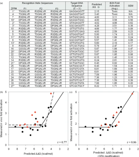
Gvalues for 27 novel modularly assembled ZFPs con-structed using Barbas GNN modules (Figure 2). Predicted
G values ranged from 2.1 kcal/mol for ZFP #1, con-taining three strong modules to 8.2 kcal/mol for ZFP #27 containing three weak modules.
In vivoactivities of ZFPs are highly correlated with predicted binding energies
preventing analysis of these constructs. Several models describing the relationship between predicted and mea-sured activity were evaluated, with segmental linear regression providing the best fit (r= 0.77; Figure 2b, dashed line). Inspection of the data revealed that the GTA-specific module (QSSSLVR) was present in most ZFPs that exhibited significantly greater activation than predicted (Figure 2b, red diamonds). Excluding ZFPs con-taining this module from the analysis increased the corre-lation coefficient to 0.86 (Figure 2b, solid line).
The predictions described above relied on published
in vitro binding affinities for ZFPs in which modules were evaluated in a fixed context (15) to estimate bind-ing contributions of individual modules. In an alternate approach, we predicted ZFP performance by solving indi-vidual module contributions as component variables of a system of linear equations. Briefly, in constructing the 27 different three-finger proteins, nine ZFDs were used approximately 8–10 times (approximately three times at each of the possible three positions, Table 1). Assuming that the energy contributions of individual ZFDs in a ZFP are additive, the B2H activity of each ZFP was considered to result from its particular combination of modules (Supplementary Figure 1). Individual module contribu-tions were calculated for each ZFP using a leave-one-out linear system solution. Expected lacZ activation in the B2H assay for each of the ZFPs was then predicted by summing individual module contributions. As shown in Figure 3, expected levels of activation computed in this manner were highly correlated with actual B2H activity measurements (r= 0.86).
The energy contributions computed using a system of linear equations to analyze in vivo activity data from
the B2H assay indicate that the GTA-specific QSSSLVR module binds with higher affinity than previous Kd
esti-mates. This is consistent with our conclusion based on inspection of energies computed fromin vitrobinding con-stants (Figure 2b). We estimated a new value for this module by calculating the Kd that optimizes the
correla-tion of predicted energies with the B2H data. This approach resulted in an estimated Kd of 2.5 nM for this
module, 10-fold lower than the previously reported value of 25 nM (15). Incorporating this new estimate improved correlation between thein vitroenergy model andin vivo
fold activation data (r= 0.86, Figure 2c).
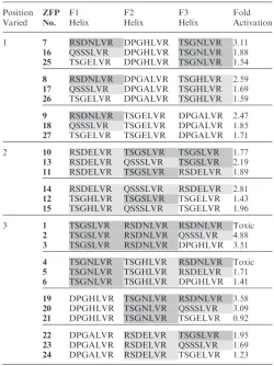
[image:6.612.307.558.84.419.2]To directly evaluate the effects of individual module affinities on in vivo performance, sets of related ZFPs designed to vary at a single module position were analyzed for differences in B2H activity (Table 2). For all three sets of ZFPs in which the F1 position was varied (while F2 and F3 were fixed), the greatest in vivo activity was observed when the F1 position contained a high affinity module; the least activity was observed with a low affinity module in this position. The same trend was observed for all four
Table 2. Single module substitutions in ZFPs alter target affinity
Position Varied ZFP No. F1 Helix F2 Helix F3 Helix Fold Activation
1 7 RSDNLVR DPGHLVR TSGNLVR 3.11
16 QSSSLVR DPGHLVR TSGNLVR 1.88
25 TSGELVR DPGHLVR TSGNLVR 1.54
8 RSDNLVR DPGALVR TSGHLVR 2.59
17 QSSSLVR DPGALVR TSGHLVR 1.69
26 TSGELVR DPGALVR TSGHLVR 1.59
9 RSDNLVR TSGELVR DPGALVR 2.47
18 QSSSLVR TSGELVR DPGALVR 1.85
27 TSGELVR TSGELVR DPGALVR 1.71
2 10 RSDELVR TSGSLVR TSGSLVR 1.77
13 RSDELVR QSSSLVR TSGSLVR 2.19
11 RSDELVR TSGSLVR RSDELVR 1.89
14 RSDELVR QSSSLVR RSDELVR 2.81
12 TSGHLVR TSGSLVR TSGELVR 1.43
15 TSGHLVR QSSSLVR TSGELVR 1.96
3 1 TSGSLVR RSDNLVR RSDNLVR Toxic
2 TSGSLVR RSDNLVR QSSSLVR 4.88
3 TSGSLVR RSDNLVR DPGHLVR 3.51
4 TSGNLVR TSGHLVR RSDNLVR Toxic
5 TSGNLVR TSGHLVR RSDELVR 1.71
6 TSGNLVR TSGHLVR DPGHLVR 1.41
19 DPGHLVR TSGNLVR RSDNLVR 3.58
20 DPGHLVR TSGNLVR QSSSLVR 3.09
21 DPGHLVR TSGNLVR TSGELVR 0.92
22 DPGALVR RSDELVR TSGSLVR 1.95
23 DPGALVR RSDELVR QSSSLVR 1.69
24 DPGALVR RSDELVR TSGELVR 1.23
[image:6.612.41.290.93.288.2]Individual ZFDs, represented by their DNA recognition helix, are shaded to indicate their affinity class (Strong, Moderate, Weak, see Table 1). For each subgroup (demarcated by horizontal lines), modules in two positions were held constant while the position indicated in the leftmost column was varied. Fold activation denotes performance of the ZFP in the B2H assay. Toxic refers to the poor growth ofE. coli cultures observed when cells expressed certain ZFPs.
Table 1. ZFP variants that differ in the middle (F2) position bind targets with variable affinities
Helix N- to -C
Target 50–30
Affinity (Kd, nM)
Strong QSSNLVR GAA 0.5
RSDHLTT TGG 0.5
RSDNLVR GAG 1
QSGDLRR GCA 2
TSGNLVR GAT 3
DPGNLVR GAC 3
QRAHLER GGA 3
TSGSLVR GTT 5
RSDKLTR GGG 6
RSDDLVR GCG 9
Moderate TSGHLVR GGT 15
RSDELVR GTG 15
QSSSLVR GTA 25
Weak DPGHLVR GGC 40
DPGALVR GTC 40
TSGELVR GCT 65
DCRDLAR GCC 80
The data in this table were reported by Segalet al. (15). Each ZFD in the F2 position was selected to bind a particular target triplet. The F1 and F3 modules (derived from Zif268) were the same in each construct. A binding affinity constant for each ZFP variant was determined using an EMSA. Here, modules are classified based on the affinity of the ZFPs for their cognate target sites (Strong, Moderate, Weak). Three modules from each class were chosen to construct 27 diverse three-module ZFPs (Table 2).
groups in which the F3 position was varied while the F1 and F2 fingers were fixed. For sets in which the F2 posi-tion was varied, only one strong module (TSGSLVR) and one moderate affinity module (QSSSLVR) were tested. In these cases, the moderate affinity module outperformed the high affinity module. These results suggest that the effect of single module substitutions on relative binding affinity can be predicted reliably in most cases.
In summary, three lines of analysis: (i) predictions based on in vitrobinding constants for modules in a fixed con-text, (ii) predictions derived from a system of linear equa-tions based onin vivoperformance and (iii) analysis of the effects of various single finger substitutionsin vivo, demon-strate thatin vivoperformance for ZFPs can be predicted based on DNA-binding affinities of individual ZFDs.
In vitroDNA binding affinities of ZFPs are highly correlated with predicted binding energies
Our success in estimating the activities of ZFPs in the B2H assay suggested that our scoring scheme could be applied more generally to predict in vitro ZFP affinities. To test whether activation measured in the B2H assay directly reflects DNA binding affinity for the desired target site, 9 of the 27 engineered proteins, along with a control Zif268 protein, were chosen for in vitro binding affinity measurements (Figure 4). Kd values were determined
using fluorescence anisotropy (FA), a rapid and reprodu-cible solution-based DNA binding assay that allows com-putation of the bound fraction of a fluorescently labeled ligand, based on the decrease in its rotational velocity due to binding (33,34).
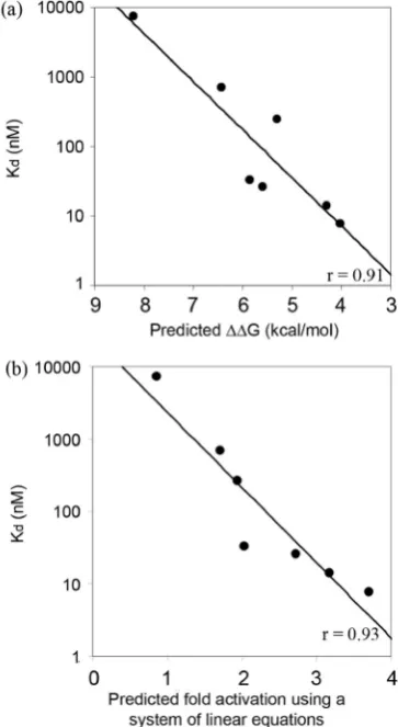
As shown in Figure 5, binding affinity constants deter-mined by FA were highly correlated with predicted ener-gies. The two ZF proteins with highest predicted affinities were toxic to bacterial cells, prohibiting purification of sufficient quantities of protein for in vitro analysis. Energies computed based on previously published
in vitroaffinity measurements for modules in a fixed con-text (15) were proportional to the log ofKd’s measured in
our experiments (r= 0.91) (Figure 5a). As before, assum-ing aKdof 2.5 nM for the QSSSLVR module significantly
improved the correlation (r= 0.97). Predictedin vivo acti-vation levels generated by the leave-one-out linear system method were also highly correlated with experimentally determined binding constants (r= 0.93; Figure 5b). Thus, results obtained using a rapid and reliable spectro-scopic method suggest that ZFP binding affinities mea-sured in vitro generally correspond to results obtained
in vivo using the B2H system. This demonstrates that our rule-based strategy can be used to predict ZFP DNA binding affinity.
A binding energy threshold for ZFP functionin vivo
To evaluate the generality of this rule-based approach, we calculated predicted energies for another set of modularly assembled ZFPs that had been previously evaluated using the B2H system (25). From 168 modularly assembled ZFPs, we selected all ZFPs comprising GNN or TGG modules for which publishedin vitroDNA binding affinity constants are available [measured in the F2 position of the standard Zif268 variant backbone (15)]. As shown in Figure 6a, based on a segmental linear regression model, binding energies for 24 of these 26 ZFPs are highly corre-lated (r= 0.80) with reported B2H activity measurements. These results are also in excellent agreement with the results described above and shown in Figure 2b, although slightly higher activation levels were uniformly observed in the latter experiments. Notably, both sets of experi-ments identify a G of 5 kcal/mol (corresponding to aKdof100 nM) as the threshold for zinc-finger function
in vivo (in bacterial cells). We also used the scoring func-tion generated from the B2H experiments performed by us (and shown in Figure 2c) to predict B2H activity for the 24 ZFPs evaluated by Ramirezet al. (25). Again, the pre-dicted and measured fold-activation scores were in close agreement, with a correlation coefficient of 0.79 (Figure 6b). Taken together, these results suggest that the scoring function developed and evaluated may be gen-erally applicable to ZFPs assembled using the Barbas lab GNN modules.
DISCUSSION
[image:7.612.59.298.70.305.2]Using a rule-based strategy that combines experimentally determined binding energies of individual ZFDs, we were able to compute binding energies for ZFPs made from a particular set of well-characterized GNN modules (15). We also showed that these predicted binding energies are in excellent agreement (r= 0.91; Figure 5a) with binding affinity constants measured directlyin vitro.Furthermore, we showed a strong correlation between these computed
binding energies and ZFP activities in a B2H system for two different sets of modularly assembled three-finger ZFPs. This is an important advance because a ZFP that lacks activity in the B2H system will also have a high
[image:8.612.349.516.279.604.2]Figure 6. Predicted ZFP performance agrees within vivoactivity for an independently generated set of ZFPs. Data shown are for 24 of 26 ZFPs containing characterized GNN or TGG modules, constructed and eval-uated by Ramirez et al. (25) (a) A segmental linear regression model provides an excellent fit ofin vivoZF-induced fold activation measured in the B2H assay with predicted binding energies (r = 0.80). (b) ZF-induced fold activation values measured in the B2H assay for 24 ZFPs from Ramirezet al. (25) are also highly correlated (r= 0.79) with pre-dicted fold-activation levels calculated based on a scoring function derived from the segmental linear regression model fit for the 25 ZFPs shown in Figure 2a (see text for details). Note: predictions for 2 of 26 ZFPs containing characterized GNN or TGG modules from Ramirez et al. (25) were considered outliers (values were outside the range included in these graphs); they were not included in the regression analysis.
Figure 4.Determining binding affinity constants using fluorescence anisotropy. (a) A representativein vitrobinding isotherm obtained using FA. Data points for each ZFP were collected using three separate purified protein preparations, each assayed for binding activity on a different day. Curve fitting was performed usingPrism. (b)Kdvalues for seven modularly assembled ZFPs, determined in FA experiments. Note that two ZFPs
were toxic to host cells, preventing purification of proteins in quantities required forin vitroanalysis.
[image:8.612.74.256.282.614.2]probability of failing to function as a ZFN in human cells (24–26). Thus, using only our scoring method, researchers can now identify target sites that will have a high probability of failing to yield functional zinc-finger arrays by the method of modular assembly. Our rule-based strategy will thus allow researchers to focus their modular assembly efforts on a smaller number of target sites with a higher probability of success.
We believe that our results also provide one potential explanation for the discrepancy between the overwhelm-ing success rates for a previousin vitroreport (35) and the lowin vivosuccess rates observed for ZFPs in the recent study of Ramirez et al. (25): many of the modules used to perform modular assembly likely possess low affinities. Our data suggest, in fact, that 30–50% of poten-tial three-finger ZFPs made wholly from the Barbas GNN modules will fail to function in the B2H system, a result in agreement with the recently published results of Ramirezet al. (25).
Although our results demonstrate that the energy con-tributions of individual ZFDs in a ZFP array are additive, we also believe they lend additional support to the notion that context is an important parameter that should be accounted for when engineering multi-finger ZFPs (i.e. that one single ZFD module will not always be optimal or adequate for recognition of its cognate 3-bp subsite in different multi-finger ZFP contexts). For example, our data show that although a weak finger will sometimes be found in a nonfunctional ZFP array (if it is joined together with other weak affinity ZFDs), it will also sometimes be found in functional arrays when paired with stronger affi-nity ZFDs. In addition, our data show that although strong fingers will sometimes be found in functional ZFPs, they can be found in nonfunctional ZFPs. Furthermore, the use of three strong fingers in a ZFP can lead to toxicity inE. coli cells. Although the precise mechanism of this toxicity is unclear, a reasonable hypoth-esis is that excessively high affinity leads to binding to related but off-target sequences with sufficient affinity to cause biological consequences (essentially, excessive affi-nity leading to problems of specificity). Thus, our data further re-enforce the ideas that individual ZFDs do not function completely independently and that the specific attributes of neighboring fingers do matter in the context of engineering a multi-finger ZFP.
The importance of context-dependent effects also sug-gests that identification of additional ZFDs with variable affinities for GNN triplets may be needed if the efficiency of modular assembly is to be improved. If such ZFDs were available, it might be possible to achieve higher success rates for modular assembly by creating several ZFPs for a given target site so as to identify a combination that balances affinities (and presumably, specificities) of its component ZFDs. A related point is that our findings also suggest one possible reason why more complex selec-tion-based methods that account for context-dependent effects [e.g. the OPEN method recently described by Joung and colleagues (26,32)] may be more successful than modular assembly: these methods are able to balance the overall affinity and specificity of the final ZFP array by identifying optimal combinations from various ZFDs with
a range of affinities and specificities for their target 3-bp subsites.
The strong correlations among predicted binding ener-gies, in vivo activities, and in vitro binding affinity con-stants for the ZFPs analyzed in this work suggests that our rule-based approach might be extended to evaluate arrays assembled using GNN modules from other sources (17,19) and non-GNN modules. We have not yet evalu-ated such modules, but our work demonstrates two ways this could be achieved: (i) by directly measuring in vitro
binding constants for modules in the F2 position of a standardized ZFP framework and (ii) by computing indi-vidual module contributions to ZFP binding as compo-nent variables of a system of linear equations that describe their activities (measuredin vivoin this work, butin vitro
binding constants could also be used). The energy scoring scheme proposed here will allow researchers to determine whether a modular assembly strategy is likely to be feasible for specific targets of interest, based on cur-rently available well-characterized modules, or whether an alternative selection-based engineering strategy should be considered.
A recent study on the use of ZFNs for homologous recombination cited lack of specificity as a primary deter-minant of ZFN-mediated toxicity in human cells (24). A likely mechanism for ZFN-induced toxicity is through binding to genomic sequences similar to the desired target sequence. As noted above, we observed toxicity in bacter-ial cells for several ZFPs, even in the absence of a fused nuclease domain, suggesting that ZFP binding to certain sites in genomic DNA can be toxic, particularly for high affinity ZFPs. Although this is the first published report of such toxicity in bacterial cells that we are aware of, it has been observed previously for several other sites (Joung,J.K. unpublished data). However, bacterial expres-sion of ZFPs with affinities in the pM range, with no toxic effects, has also been reported (19,36,37). High-through-put chip or microfluidics-based DNA binding experiments (38–41) could be used to obtain affinity and specificity data for virtually every possible target site for a given ZFP, providing additional insight into ZFP-induced toxi-city and into the fundamental rules that govern the affinity and specificity of DNA recognition by zinc-finger DNA binding proteins.
A correlation between ZFP binding constants measured
in vitro and functional activity measured in vivo has also been observed by others using different reporter sys-tems (37). A similar degree of correlation was observed using the B2H system in our study (Supplementary Figure 2). Our results further demonstrate that measur-able ZFP activity in an in vitro binding assay does not necessarily translate into adequate function in vivo, in agreement with Beerli et al. (42). However, the energy threshold we determined for ZFP activity in vivo, using B2H assays, corresponds to a Kd of 100 nM, and thus
differs from the estimated threshold Kd of 10 nM
different laboratories using different assays and with ZFPs containing different numbers of fingers.
Stormo and colleagues (43–47) have shown that the DNA-binding specificity of ZFPs can be effectively pre-dicted from additive energy contributions of individual residues that make base-specific contacts with target site nucleotides. Our results complement this idea by demonstrating that the affinity of ZFPs also can be pre-dicted, using affinity data for component modules. Our application of the binding energy additivity concept differs somewhat from that used by Stormo to predict specificity in that it assumes additivity of energy contributions at the individual finger rather than individual residue level. Also, our approach implicitly includes energetic contributions of residues that are not directly involved in base contacts (e.g. phosphate contacts), as well as energetic contribu-tions resulting from context-dependent effects that pre-sumably occur among recognition helix residues within each finger.
The apparent simplicity of modular assembly has contributed to the current focus on C2H2 ZFDs as the
domains of choice for designing custom DNA binding proteins. Our results make it possible, for the first time, to reliably identify prospective binding sites that are unli-kely to yield functional ZFDs by modular assembly using a set of GNN-specific finger modules. The rule-based strategy presented here can provide accurate guidance for both in vitro binding affinities and in vivo
functionality for engineered ZFPs by computing energy contributions of individual ZFDs. We have updated the Zinc Finger Targeter (ZiFiT) web server (http://bindr.gd cb.iastate.edu/ZiFiT) (48) so that it now provides users with a list of potential ZFP-target site pairs for a desired genomic sequence, scored according to the procedures developed and validated in this work.
SUPPLEMENTARY DATA
Supplementary Data are available at NAR Online.
ACKNOWLEDGEMENTS
We thank members of our groups and colleagues, espe-cially Fengli Fu, Deepak Reyon, David Wright, Ronnie Winfrey, Ben Lewis, Bob Farnham, Abd Elhamid Azzaz, Les Miller, Gaya Amarasinghe and Vasant Honavar and the referees for their helpful suggestions and valuable feedback. We also thank Guru Rao for the use of his spectrophotometer.
FUNDING
National Institutes of Health (GM066387 to D.D.); National Science Foundation (DBI0501678 to D.F.V.); National Institutes of Health (GM069906 and GM078369 to J.K.J.); and graduate research assistant-ships provided by United States Department of Agriculture (MGET 2001-52100-11506, NSF IGERT0504304 and ISU’s Center for Integrated Animal
Genomics (CIAG). Funding for open access charge: National Science Foundation (DBI0501678).
Conflict of interest statement. None declared.
REFERENCES
1. Durai,S., Mani,M., Kandavelou,K., Wu,J., Porteus,M.H. and Chandrasegaran,S. (2005) Zinc finger nucleases: custom-designed molecular scissors for genome engineering of plant and mammalian cells.Nucleic Acids Res.,33, 5978–5990.
2. Klug,A. (2005) Towards therapeutic applications of engineered zinc finger proteins. FEBS Lett.,579, 892–894.
3. Porteus,M.H. and Carroll,D. (2005) Gene targeting using zinc finger nucleases.Nat. Biotechnol.,23, 967–973.
4. Wu,J., Kandavelou,K. and Chandrasegaran,S. (2007) Custom-designed zinc finger nucleases: what is next?Cell. Mol. Life Sci.,64, 2933–2944.
5. Cathomen,T. and Joung,J.K. (2008) Zinc-finger nucleases: the next generation emerges.Mol. Ther.,16, 1200–1207.
6. Desjarlais,J.R. and Berg,J.M. (1993) Use of a zinc-finger consensus sequence framework and specificity rules to design specific DNA binding proteins. Proc. Natl Acad. Sci. USA,90, 2256–2260. 7. Jamieson,A.C., Wang,H. and Kim,S.H. (1996) A zinc finger
direc-tory for high-affinity DNA recognition.Proc. Natl Acad. Sci. USA,
93, 12834–12839.
8. Beerli,R.R., Segal,D.J., Dreier,B. and Barbas,C.F. 3rd. (1998) Toward controlling gene expression at will: specific regulation of the erbB-2/HER-2 promoter by using polydactyl zinc finger proteins constructed from modular building blocks.Proc. Natl Acad. Sci. USA,95, 14628–14633.
9. Wolfe,S.A., Nekludova,L. and Pabo,C.O. (2000) DNA recognition by Cys2His2 zinc finger proteins. Annu. Rev. Biophys. Biomol. Struct.,29, 183–212.
10. Pabo,C.O., Peisach,E. and Grant,R.A. (2001) Design and selection of novel Cys2His2 zinc finger proteins.Annu. Rev. Biochem.,70, 313–340.
11. Segal,D.J. (2002) The use of zinc finger peptides to study the role of specific factor binding sites in the chromatin environment.Methods,
26, 76–83.
12. Pavletich,N.P. and Pabo,C.O. (1991) Zinc finger-DNA recognition: crystal structure of a Zif268-DNA complex at 2.1 A. Science,252, 809–817.
13. Elrod-Erickson,M., Rould,M.A., Nekludova,L. and Pabo,C.O. (1996) Zif268 protein-DNA complex refined at 1.6 A: a model system for understanding zinc finger-DNA interactions.Structure,
4, 1171–1180.
14. Miller,J.C. and Pabo,C.O. (2001) Rearrangement of side-chains in a Zif268 mutant highlights the complexities of zinc finger-DNA recognition.J. Mol. Biol.,313, 309–315.
15. Segal,D.J., Dreier,B., Beerli,R.R. and Barbas,C.F. 3rd. (1999) Toward controlling gene expression at will: selection and design of zinc finger domains recognizing each of the 5’-GNN-3’ DNA target sequences.Proc. Natl Acad. Sci. USA,96, 2758–2763.
16. Wolfe,S.A., Greisman,H.A., Ramm,E.I. and Pabo,C.O. (1999) Analysis of zinc fingers optimized via phage display: evaluating the utility of a recognition code.J. Mol. Biol.,285, 1917–1934. 17. Liu,Q., Xia,Z., Zhong,X. and Case,C.C. (2002) Validated zinc finger
protein designs for all 16 GNN DNA triplet targets.J. Biol. Chem.,
277, 3850–3856.
18. Dreier,B., Beerli,R.R., Segal,D.J., Flippin,J.D. and Barbas,C.F. 3rd. (2001) Development of zinc finger domains for recognition of the 5’-ANN-3’ family of DNA sequences and their use in the construction of artificial transcription factors.J. Biol. Chem.,276, 29466–29478. 19. Bae,K.H., Kwon,Y.D., Shin,H.C., Hwang,M.S., Ryu,E.H.,
Park,K.S., Yang,H.Y., Lee,D.K., Lee,Y., Park,J.et al. (2003) Human zinc fingers as building blocks in the construction of arti-ficial transcription factors.Nat. Biotechnol.,21, 275–280. 20. Dreier,B., Fuller,R.P., Segal,D.J., Lund,C.V., Blancafort,P.,
Huber,A., Koksch,B. and Barbas,C.F. 3rd. (2005) Development of zinc finger domains for recognition of the 50-CNN-30 family DNA
21. Alwin,S., Gere,M.B., Guhl,E., Effertz,K., Barbas,C.F. 3rd., Segal,D.J., Weitzman,M.D. and Cathomen,T. (2005) Custom zinc-finger nucleases for use in human cells.Mol. Ther.,12, 610–617.
22. Beumer,K., Bhattacharyya,G., Bibikova,M., Trautman,J.K. and Carroll,D. (2006) Efficient gene targeting in Drosophila with zinc-finger nucleases.Genetics,172, 2391–2403.
23. Segal,D.J., Crotty,J.W., Bhakta,M.S., Barbas,C.F. 3rd. and Horton,N.C. (2006) Structure of Aart, a designed six-finger zinc finger peptide, bound to DNA.J. Mol. Biol.,363, 405–421. 24. Cornu,T.I., Thibodeau-Beganny,S., Guhl,E., Alwin,S.,
Eichtinger,M., Joung,J.K. and Cathomen,T. (2008) DNA-binding specificity is a major determinant of the activity and toxicity of zinc-finger nucleases.Mol. Ther.,16, 352–358.
25. Ramirez,C.L., Foley,J.E., Wright,D.A., Muller-Lerch,F., Rahman,S.H., Cornu,T.I., Winfrey,R.J., Sander,J.D., Fu,F., Townsend,J.A.et al. (2008) Unexpected failure rates for modular assembly of engineered zinc fingers.Nat. Methods,5, 374–375. 26. Maeder,M.L., Thibodeau-Beganny,S., Osiak,A., Wright,D.A.,
Anthony,R.M., Eichtinger,M., Jiang,T., Foley,J.E., Winfrey,R.J., Townsend,J.A.et al. (2008) Rapid ‘‘open-source’’ engineering of customized zinc-finger nucleases for highly efficient gene modifica-tion.Mol. Cell,31, 294–301.
27. Wright,D.A., Thibodeau-Beganny,S., Sander,J.D., Winfrey,R.J., Hirsh,A.S., Eichtinger,M., Fu,F., Porteus,M.H., Dobbs,D., Voytas,D.F.et al. (2006) Standardized reagents and protocols for engineering zinc finger nucleases by modular assembly.Nat. Protoc.,
1, 1637–1652.
28. Ryder,S.P., Frater,L.A., Abramovitz,D.L., Goodwin,E.B. and Williamson,J.R. (2004) RNA target specificity of the STAR/GSG domain post-transcriptional regulatory protein GLD-1.Nat. Struct. Mol. Biol.,11, 20–28.
29. Seidman, C.E. (1997) Transformation using calcium chloride. In Ausubel, F.M. (ed),Current Protocols in Molecular Biology, Vol. I Unit 1.8. John Wiley & Son, Inc., New York.
30. Lundblad,J.R., Laurance,M. and Goodman,R.H. (1996) Fluorescence polarization analysis of DNA and protein-protein interactions.Mol. Endocrinol.,10, 607–612.
31. LiCata,V.J. and Wowor,A.J. (2008) Applications of fluorescence anisotropy to the study of protein-DNA interactions.Methods Cell Biol.,84, 243–262.
32. Hurt,J.A., Thibodeau,S.A., Hirsh,A.S., Pabo,C.O. and Joung,J.K. (2003) Highly specific zinc finger proteins obtained by directed domain shuffling and cell-based selection.Proc. Natl Acad. Sci. USA,100, 12271–12276.
33. Veprintsev,D.B. and Fersht,A.R. (2008) Algorithm for prediction of tumour suppressor p53 affinity for binding sites in DNA.Nucleic Acids Res.,36, 1589–1598.
34. Hayouka,Z., Rosenbluh,J., Levin,A., Maes,M., Loyter,A. and Friedler,A. (2008) Peptides derived from HIV-1 Rev inhibit HIV-1 integrase in a shiftide mechanism. Biopolymers,90, 481–487. 35. Segal,D.J., Beerli,R.R., Blancafort,P., Dreier,B., Effertz,K.,
Huber,A., Koksch,B., Lund,C.V., Magnenat,L., Valente,D.et al. (2003) Evaluation of a modular strategy for the construction of novel polydactyl zinc finger DNA-binding proteins.Biochemistry,
42, 2137–2148.
36. Yang,W.P., Wu,H. and Barbas,C.F. 3rd. (1995) Surface plasmon resonance based kinetic studies of zinc finger-DNA interactions. J. Immunol. Methods,183, 175–182.
37. Kang,J.S. (2007) Correlation between functional and binding activities of designer zinc-finger proteins.Biochem. J.,403, 177–182. 38. Bulyk,M.L. (2006) DNA microarray technologies for measuring
protein-DNA interactions.Curr. Opin. Biotechnol.,17, 422–430. 39. Berger,M.F., Philippakis,A.A., Qureshi,A.M., He,F.S., Estep,P.W.
3rd and Bulyk,M.L. (2006) Compact, universal DNA microarrays to comprehensively determine transcription-factor binding site specificities.Nat. Biotechnol.,24, 1429–1435.
40. Maerkl,S.J. and Quake,S.R. (2007) A systems approach to mea-suring the binding energy landscapes of transcription factors. Science,315, 233–237.
41. Berger,M.F., Badis,G., Gehrke,A.R., Talukder,S., Philippakis,A.A., Pena-Castillo,L., Alleyne,T.M., Mnaimneh,S., Botvinnik,O.B., Chan,E.T. et al. (2008) Variation in homeodomain DNA binding revealed by high-resolution analysis of sequence preferences.Cell,
133, 1266–1276.
42. Beerli,R.R., Dreier,B. and Barbas,C.F. 3rd. (2000) Positive and negative regulation of endogenous genes by designed transcription factors.Proc. Natl Acad. Sci. USA,97, 1495–1500.
43. Stormo,G.D. and Fields,D.S. (1998) Specificity, free energy and information content in protein-DNA interactions.Trends Biochem. Sci.,23, 109–113.
44. Benos,P.V., Bulyk,M.L. and Stormo,G.D. (2002) Additivity in protein-DNA interactions: how good an approximation is it? Nucleic Acids Res.,30, 4442–4451.
45. Benos,P.V., Lapedes,A.S. and Stormo,G.D. (2002) Probabilistic code for DNA recognition by proteins of the EGR family.J. Mol. Biol.,323, 701–727.
46. Liu,J. and Stormo,G.D. (2005) Quantitative analysis of EGR pro-teins binding to DNA: assessing additivity in both the binding site and the protein.BMC Bioinformatics,6, 176.
47. Liu,J. and Stormo,G.D. (2008) Context-dependent DNA recogni-tion code for C2H2 zinc-finger transcriprecogni-tion factors.Bioinformatics,
24, 1850–1857.