R E S E A R C H A R T I C L E
Open Access
The role of
Enterococcus
spp. and
multidrug-resistant bacteria causing
pyogenic liver abscesses
Marcus M. Mücke
1,2, Johanna Kessel
2,3, Victoria T. Mücke
1,2, Katharina Schwarzkopf
1,2, Michael Hogardt
2,4,
Christoph Stephan
2,3, Stefan Zeuzem
1,2, Volkhard A. J. Kempf
2,4and Christian M. Lange
1,2*Abstract
Background:Pyogenic liver abscesses (PLA) remain a significant clinical problem. Unfortunately, little is known about current bacterial susceptibility profiles and the incidence of multidrug resistant organisms (MDROs) causing PLA in Western countries. Yet, this crucial information is pivotal to guide empirical antibiotic therapy. Aim of this study was to provide detailed characteristics of PLA with a special focus on underlying bacterial pathogens and their susceptibility to antibiotics.
Methods:A retrospective study of patients diagnosed with PLA from 2009 to 2015 in a large tertiary reference center in Germany was performed in order to characterize PLA and antimicrobial susceptibility profiles of causative bacterial species.
Results:Overall, 86 patients were included. The most common causes of PLA were bile duct stenosis/obstruction (31.4%) and leakage of biliary anastomosis (15.1%). Frequent predisposing diseases were malignancies (34.9%), diabetes (24.4%) and the presence of liver cirrhosis (16.3%). Of note,Enterococcusspp. were the most frequently cultured bacterial isolates (28.9%), and in 1/3 of cases vancomycin resistance was observed. In addition, a relevant frequency of gram-negative MDROs was identified. In particular, an alarming 10% and 20% of gram-negative bacteria were resistant to carbapenems and tigecycline, respectively. Of note, MDRO status did not predict ICU stay or survival in multivariate regression analysis. The mortality rate in our series was 16.3%.
Conclusion:Our study demonstrates an as yet underreported role ofEnterococcusspp., often associated with vancomycin resistance, as well as of gram-negative MDROs causing PLA.
Keywords:Pyogenic liver abscess, Bacterial pathogens, Susceptibility profiles, Multidrug-resistant organisms, Vancomycin-resistant enterococci
Background
With an annual incidence of 1.1 to 2.3 per 100,000 and mortality rates of up to 12% in developed countries, pyo-genic liver abscesses (PLAs) remain a significant clinical problem in the Western World [1–4]. Even higher inci-dence rates have been reported in Asian countries, e.g. in Taiwan (17.06 cases per 100,000) [5]. Due to various se-vere predisposing diseases (e.g. biliary strictures or
cancer), the frequent need of external and internal drain-age, and a plethora of potential causative microorganisms, medical management of PLA can be highly complex.
In general, multidrug-resistant organisms (MDROs), including vancomycin-resistant enterococci (VRE), methicillin-resistant Staphylococcus aureus (MRSA) or multidrug-resistant gram-negative bacteria (MRGN), are increasingly being observed worldwide [6, 7]. Growing resistance in particular among certain gram-positive and gram-negative pathogens – so-called “ESKAPE” patho-gens (Enterococcus faecium,Staphylococcus aureus, Klebsi-ella pneumoniae, Acinetobacter baumannii, Pseudomonas aeruginosa, and Enterobacter species [8]) – causing * Correspondence:[email protected]
1
Department of Internal Medicine 1, University Hospital Frankfurt, Frankfurt am Main, Germany
2University Center for Infectious Diseases, University Hospital Frankfurt,
Frankfurt am Main, Germany
Full list of author information is available at the end of the article
infections in hospitals and in the community are worry-some. Of special concern are reports portraying a growing number of organisms resistant to all available antibiotics, including polymyxin [7, 9–11]. Recently, several case reports of PLA caused by MDROs have been published [12, 13]. In addition, Lo et al. noted in a series of Asian pa-tients with PLAs an increase of MDR isolates (Klebsiella pneumoniae) from 1.6% to 14.3% within 10 years in Singapore [14]. In contrast, current data from Western countries are largely lacking.
Early retrospective studies have revealed remarkable differences between PLA characteristics in Asian and Western countries [1–4, 14–16]. For example,Klebsiella pneumoniae has been identified as the predominant cause of PLA in Asia [5, 17–19], whereas other Entero-bacteriaceae such as E. coli, as well as Staphylococcus spp., Streptococcusspp., Enterococcusspp., or anaerobes were predominantly isolated in the Western World [1–4]. Unfortunately, little is known about prevailing susceptibility profiles and the incidence of MDROs causing PLA in the Western countries. However, this crucial information is pivotal to guide antibiotic ther-apy, one of the fundaments of PLA treatment.
In this retrospective study we therefore aimed to further describe PLA characteristics including a detailed analysis of current bacterial and fungal isolates causing PLA in a large tertiary reference center in Germany. Our study re-veals a so far underestimated role ofEnterococcusspp. and MDRO in secondary PLA and thereby helps to guide em-pirical antibiotic therapy of PLA.
Methods
Study population
All adult patients admitted to the University Hospital Frankfurt, Germany, between January 2009 and December 2015 with the discharge diagnosis of PLA were eligible for inclusion. For identification of possible patients, the pa-tient chart database of the University Hospital Frankfurt was systematically searched for code K75.0 or K83.0 of the International Classification of Diseases, Tenth Revision, German Modification. Cases were included if (1) one or more discrete hepatic abscess cavities were confirmed by at least one imaging modality – ultrasound (US), com-puted tomography (CT) or magnetic resonance imaging (MRI)–as well as (2) by either positive culture results re-trieved from the abscess or resolution of symptoms after antibiotic therapy. Patients were excluded if they were younger than 18 years old, if parasitic/amoebic abscesses were diagnosed or if available data were incomplete. The local ethics committee approved this study.
Clinical data collection, definitions
Charts were systematically reviewed and information ob-tained was gathered in a data collection form. Information
recorded included sex, age, date of admission/discharge, underlying medical condition, initial symptoms and the intake of immunosuppressant agents, antibiotics, and proton-pump inhibitors. Additionally, initial laboratory values were documented. Laboratory results were consid-ered to be the first values obtained upon hospital admis-sion due to PLA or within 24 h upon presentation of PLA when PLA was not the initial cause of hospitalization. Im-aging reports (CT, MRI, US) were analyzed and number and size of PLAs were documented.
For conventional microbiological culture procedures, aerobic and anaerobic conditions including the use of thioglycolate enrichment medium were applied. Species identification of recovered microorganisms was performed by matrix-assisted laser desorption/ionization time-of-flight (MALDI–TOF) mass spectrometry (VITEK MS, bio-Mérieux, Nürtingen, Germany) and VITEK2 (biobio-Mérieux, Nürtingen, Germany). Antibiotic susceptibility testing (AST) was done by VITEK2 (bioMérieux, Nürtingen, Germany) according to Clinical and Laboratory Standards Institute (CLSI) guidelines and/or antibiotic gradient tests (Etest), where necessary. All laboratory tests were per-formed under strict quality-controlled criteria (laboratory accreditation according to ISO 15189:2007 standards; certificate number D–ML–13102–01–00, valid through January 25th, 2021).
In the vast majority of cases, positive microbiological results of abscess cavity cultures were obtained. A bacterial isolate was considered to be an MDRO if it belonged to either category VRE, MRSA or MRGN. MRGN status was defined according to the German KRINKO guideline [20]. When microbiological results were available the initial empiric antibiotic treatment was assessed and considered to be adequate if the re-trieved isolates were testes to be susceptible. Addition-ally, mycotic coinfections were documented.
The responsible physician defined the assumed cause of PLA. The therapeutic modality was classified as either surgery, percutaneous drainage (either CT- or US-guided), biliary drainage by endoscopic retrograde cholangiography (ERCP) or percutaneous transhepatic cholangiography and drainage (PTCD), or solitary med-ically managed. Complications and outcome, e.g. treat-ment on an intensive care unit (ICU), recurrence of abscesses, mortality and duration of hospitalization, were also recorded.
Finally, local hospital surveillance data was analyzed to compare rates of Enterococcus spp. and VRE causing PLA with the overall documented infection rates of En-terococcus spp. and VRE in our
gastroenterology/hepa-tology wards between 2010 and 2015. In brief,
7.1.3 (Roche Diagnostics IT solutions, Germany). For the calculation of the relative VRE rate,E. faeciumand/orE. faecium-VRE positive cultures of all microbiological specimens submitted during 2010–2015 to the labora-tory were counted. Patients with an infection where both VRE and Enterococcus were isolated were counted as patients with a VRE infection only to avoid duplicates.
Statistical analysis
For statistical analysis BiAS, Version 11.03, was applied. Group differences were calculated using the nonpara-metric Mann-Whitney U test (continuous variables) or Fischer’s exact (categorical variables), as appropriate. After bivariate/univariate analysis, multivariate analysis was per-formed by using backward selection and aP value ≥0.10 for removal from the model. Only patients with complete data for the remaining covariates were included in multi-variate analysis. Sex and age were forced into the model. Odds ratios (OR) and respective 95% confidence intervals (CI) were calculated for each variable. All statistical tests were two-sided and P values <0.05 were considered to be significant.
Results
Patient characteristics
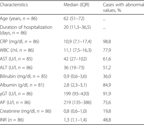
From 130 identified charts, 86 patients (55 men and 31 women, median age 62 years [IQR 51–72 years]) match-ing the described criteria were included in this study (Fig. 1). Detailed clinical characteristics and laboratory results as well as symptoms of patients, underlying dis-eases, direct cause and abscess’ characteristics are dis-played in Tables 1 and 2, respectively.
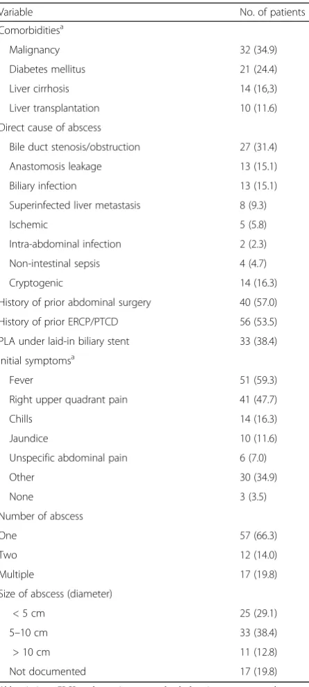
As expected from a hospital with a major hepatobiliary surgery and liver transplant center, most of the PLA were of secondary nature originating in bile duct sten-osis/obstruction (31.4%), anastomosis leakage and biliary infection (both 15.1%) as well as a superinfected liver metastasis (9.3%). In 14 patients (16.3%) the cause of liver abscess remained cryptogenic.
The most frequent comorbidities included malignan-cies in 34.9%, diabetes in 24.4%, the presence of liver cir-rhosis in 16.3%, and prior liver transplantation in 16.3% of cases. Of note, 74.4% and 17.4% of patients received proton-pump inhibitors and immunosuppressive therapy prior to PLA formation, respectively.
Microbiological investigations
Microbiological cultures (blood and/or abscess cavity cultures) were set up in all 86 patients and were positive in 77 out of 86 (89.5%). Mycotic coinfections were docu-mented in 21 cases (24.4%), mostly caused by Candida albicans(76.2% of all mycotic coinfections). The number of recovered bacterial/mycotic species per patient is rep-resented in Fig. 2. Overall, 135 bacterial isolates were
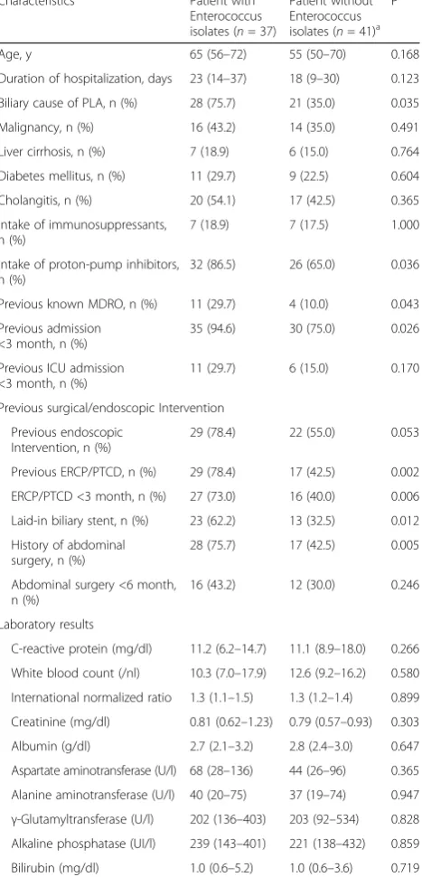
identified; a detailed overview is displayed in Table 3. In short, both gram-negative (48.9%) and gram-positive (46.7%) aerobic bacteria were frequently cultured, whereas anaerobic bacteria were identified relatively rarely (4.4%). Of note, the most common isolated bacter-ial species detected were Enterococcus spp. (28.9%, in total:E. faecium26;E. faecalis13). Patients’ characteris-tics with cultures positive for Enterococcus spp. and bi-variate analysis are displayed in Table 4. In multibi-variate analysis, there was a trend to Enterococcus infections in patients taking proton-pump inhibitors (P = 0.057, OR 3.73, 95% CI 0.96–14.41). ERCP/PTCD in the last three
[image:3.595.305.539.87.331.2]Fig. 1Study overview
Table 1Age, duration of hospitalization and laboratory abnormalities among patients with pyogenic liver abscess
Characteristics Median (IQR) Cases with abnormal values, %
Age (years,n= 86) 62 (51–72) ...
Duration of hospitalization (days,n= 86)
20 (11,3–36,5) ...
CRP (mg/dl,n= 86) 10,9 (7,1–17,4) 98,8
WBC (/nl,n= 86) 11,1 (7,5–16,3) 77,9
AST (U/l,n= 85) 42 (27–102) 61,6
ALT (U/l,n= 86) 36 (19–73) 51,2
Bilirubin (mg/dl,n= 85) 0,9 (0,6–3,6) 36,0
Albumin (g/dl,n= 81) 2,8 (2,3–3,1) 84,9
γGT (U/l,n= 86) 199 (93–420) 91,9 AP (U/l,n= 86) 219 (135–386) 75,6
Creatinine (mg/dl,n= 86) 0,8 (0,6–1,0) 19,8
[image:3.595.305.538.531.732.2]months (P = 0.010, OR 4.23, 95% CI 1.41–12.77) and history of prior abdominal surgery (P= 0.036, OR 3.36, 95% CI 1.08–10.44) were independently associated with Enterococcusspp. infection in patients with PLA.
Summaries of antimicrobial susceptibility and resistance profiles of enterococci, gram-positive and gram-negative aerobic bacteria are displayed in Table 5. Detailed resist-ance profiles of individual bacterial isolates are shown in
[image:4.595.305.541.87.224.2]Additional file 1: Table S1, susceptibility and resistance profiles of gram-positive aerobic bacteria excluding en-terococci in Additional file 2: Table S3 and those of anaer-obic bacteria in Additional file 3: Table S4. Overall, 25 MDROs were identified. Most interestingly, 35.9% of all Enterococcus spp. were classified as VRE (E. faecium 46.2%,E faecalis7.7%) including cases of teicoplanin, dap-tomycin-, and linezolid- (intermediate) resistance (Table 5, Additional file 1: Table S1). Importantly, although the PLA patient populations were comparable over the study period, the relative VRE rate (VRE among all Enterococci) in patients with PLA increased over time (Additional file 4: Figure S1A and B), while in other patients without PLA it did not change significantly (Additional file 5: Figure
Table 2Clinical characteristics of patients, underlying diseases and abscess’characteristics
Variable No. of patients (%)
Comorbiditiesa
Malignancy 32 (34.9)
Diabetes mellitus 21 (24.4)
Liver cirrhosis 14 (16,3)
Liver transplantation 10 (11.6)
Direct cause of abscess
Bile duct stenosis/obstruction 27 (31.4)
Anastomosis leakage 13 (15.1)
Biliary infection 13 (15.1)
Superinfected liver metastasis 8 (9.3)
Ischemic 5 (5.8)
Intra-abdominal infection 2 (2.3)
Non-intestinal sepsis 4 (4.7)
Cryptogenic 14 (16.3)
History of prior abdominal surgery 40 (57.0)
History of prior ERCP/PTCD 56 (53.5)
PLA under laid-in biliary stent 33 (38.4)
Initial symptomsa
Fever 51 (59.3)
Right upper quadrant pain 41 (47.7)
Chills 14 (16.3)
Jaundice 10 (11.6)
Unspecific abdominal pain 6 (7.0)
Other 30 (34.9)
None 3 (3.5)
Number of abscess
One 57 (66.3)
Two 12 (14.0)
Multiple 17 (19.8)
Size of abscess (diameter)
< 5 cm 25 (29.1)
5–10 cm 33 (38.4)
> 10 cm 11 (12.8)
Not documented 17 (19.8)
Abbreviations:ERCPendoscopic retrograde cholangiopancreatography, PTCDpercutaneous transhepatic cholangiography and drainage a
Patients fit to plural categories were counted in each category
[image:4.595.58.281.108.603.2]Fig. 2Number of bacterial isolates recovered per case in patients with pyogenic liver abscess
Table 3Bacterial isolates from abscess cavity cultures
Bacterial isolatesa Number of isolates
Gram-positive aerobes
Staphylococcus aureus 2
Coagulase neg. Staphylococci 13
Viridans streptococci 7
Group A Streptococci 1
Enterococcusspp. 39
other gram-positive species 1
Gram-negative aerobes
Escherichia coli 23
Klebsiellaspp. 15
other Enterobacteriaceae 14
Pseudomonasspp. 6
Stenotrophomonasspp. 3
Acinetobacterspp. 1
other gram-negative species 4
Anaerobes 6
No bacterial growth 9
a
[image:4.595.305.538.454.706.2]S2A and B). In addition, 12 cases of MRGN were identi-fied (16,7% of gram-negative bacteria). Of note, approxi-mately 10% and 20% of tested gram-negative aerobe bacteria were resistant to carbapenems and to tigecycline, respectively. Resistance rates to fluoroquinolones, broad-spectrum penicillin/β-lactamase inhibitor combinations (BSP/βLI) and cephalosporins were relatively high (20–
[image:5.595.276.534.99.645.2]57%, Table 5). Results of fungal isolates analysis are dis-played in Additional file 6: Table S2. Next, we analyzed both patient populations with and without MDRO causing
Table 4Patients’characteristics and baseline parameters with and without cultivatedEnterococcusspp.
Characteristics Patient with Enterococcus isolates (n= 37)
Patient without Enterococcus isolates (n= 41)a
P
Age, y 65 (56–72) 55 (50–70) 0.168
Duration of hospitalization, days 23 (14–37) 18 (9–30) 0.123
Biliary cause of PLA, n (%) 28 (75.7) 21 (35.0) 0.035
Malignancy, n (%) 16 (43.2) 14 (35.0) 0.491
Liver cirrhosis, n (%) 7 (18.9) 6 (15.0) 0.764
Diabetes mellitus, n (%) 11 (29.7) 9 (22.5) 0.604
Cholangitis, n (%) 20 (54.1) 17 (42.5) 0.365
Intake of immunosuppressants, n (%)
7 (18.9) 7 (17.5) 1.000
Intake of proton-pump inhibitors, n (%)
32 (86.5) 26 (65.0) 0.036
Previous known MDRO, n (%) 11 (29.7) 4 (10.0) 0.043
Previous admission <3 month, n (%)
35 (94.6) 30 (75.0) 0.026
Previous ICU admission <3 month, n (%)
11 (29.7) 6 (15.0) 0.170
Previous surgical/endoscopic Intervention
Previous endoscopic Intervention, n (%)
29 (78.4) 22 (55.0) 0.053
Previous ERCP/PTCD, n (%) 29 (78.4) 17 (42.5) 0.002
ERCP/PTCD <3 month, n (%) 27 (73.0) 16 (40.0) 0.006
Laid-in biliary stent, n (%) 23 (62.2) 13 (32.5) 0.012
History of abdominal surgery, n (%)
28 (75.7) 17 (42.5) 0.005
Abdominal surgery <6 month, n (%)
16 (43.2) 12 (30.0) 0.246
Laboratory results
C-reactive protein (mg/dl) 11.2 (6.2–14.7) 11.1 (8.9–18.0) 0.266
White blood count (/nl) 10.3 (7.0–17.9) 12.6 (9.2–16.2) 0.580
International normalized ratio 1.3 (1.1–1.5) 1.3 (1.2–1.4) 0.899
Creatinine (mg/dl) 0.81 (0.62–1.23) 0.79 (0.57–0.93) 0.303
Albumin (g/dl) 2.7 (2.1–3.2) 2.8 (2.4–3.0) 0.647
Aspartate aminotransferase (U/l) 68 (28–136) 44 (26–96) 0.365
Alanine aminotransferase (U/l) 40 (20–75) 37 (19–74) 0.947
γ-Glutamyltransferase (U/l) 202 (136–403) 203 (92–534) 0.828 Alkaline phosphatase (UI/l) 239 (143–401) 221 (138–432) 0.859
Bilirubin (mg/dl) 1.0 (0.6–5.2) 1.0 (0.6–3.6) 0.719
Data are presented as media (IQR) unless otherwise indicated
Abbreviations:ERCPendoscopic retrograde cholangiopancreatography,
PTCDpercutaneous transhepatic cholangiography and drainage
aPatients without bacterial isolates were excluded
Table 5Summary of susceptibility profiles of aerobic bacteria All gram-positive aerobes includingEnterococcus spp.
Antibiotics Susceptible (%) Intermediate (%) Resistant (%) Total:
Ampicillin 21 (44.7) 0 (0) 26 (55.3) 47
Amox/Clav. 21 (44.7) 0 (0) 26 (55.3) 47
Pip/Taz. 21 (47.7) 0 (0) 23 (52.3) 44
Cefuroxime 12 (19.0) 0 (0) 51 (81.0)a 63
Cefotaxime 10 (20.4) 0 (0) 39 (79.6)a 49
Gentamycin 27 (51.9) 0 (0) 25 (48.1) 52
Tigecycline 42 (100) 0 (0) 0 (0) 42
Levofloxacin 21 (35.0) 2 (3.3) 37 (61.7) 60
Vancomycin 43 (76.8) 0 (0) 13 (23.2) 56
Imipenem 13 (34.2) 0 (0) 25 (65.8) 38
Linezolid 44 (97.8) 1 (2.2) 0 (0) 45
Enterococcusspp. only
Antibiotics Susceptible (%) Intermediate (%) Resistant (%) Total:
Ampicillin 12 (31.6) 0 (0) 26 (68.4) 38
Erythromycin 9 (25.0) 3 (8.3) 24 (66.7) 36
Imipenem 13 (34.2) 0 (0) 25 (65.8) 38
Tigecycline 32 (100) 0 (0) 0 (0) 32
Vancomycin 21 (60.0) 0 (0) 14 (40.0) 35
Teicoplanin 35 (92.1) 1 (2.6) 2 (5.3) 38
Levofloxacin 10 (26.3) 1 (2.6) 27 (71.1) 38
Linezolid 33 (97.1) 1 (2.9) 0 (0) 34
Daptomycin 7 (87.5) 0 (0) 1 (12.5) 8
All gram-negative aerobes
Antibiotics Susceptible (%) Intermediate (%) Resistant (%) Total:
Ampicillin 8 (15.1) 1 (1.9) 44 (83.0) 53
Amox/Clav 17 (33.3) 5 (9.8) 29 (56.9) 51
Pip/Taz 30 (52.6) 6 (10.5) 21 (36.8) 57
Cefuroxime 17 (33.3) 8 (15.7) 26 (51.0) 51
Cefotaxime 30 (60.0) 0 (0) 20 (40.0) 50
Ceftazidim 10 (66.7) (52.2) 2 (13.3) 3 (20.0) 15
Imipenem 53 (84.1) 3 (4.8) 7 11.1) 63
Meropenem 55 (90.2) 1 (1.6) 5 (8.2) 61
Gentamicin 52 (83.9) 0 (0) 10 (16.1) 62
Tigecycline 35 (71.4) 4 (8.2) 10 (20.4) 49
TMP/SMX 42 (73.7) 0 (0) 15 (26.3) 57
Levofloxacin 42 (68.9) 2 (3.3) 17 (27.9) 61
Ciprofloxacin 41 (68.3) 1 (1.7) 18 (30.0) 60
Amox/Clavamoxicillin/clavulanic acid,Pip/Tazpiperacillin/tazobactam, TMP/SMXtrimethoprim/sulfamethoxazole
a
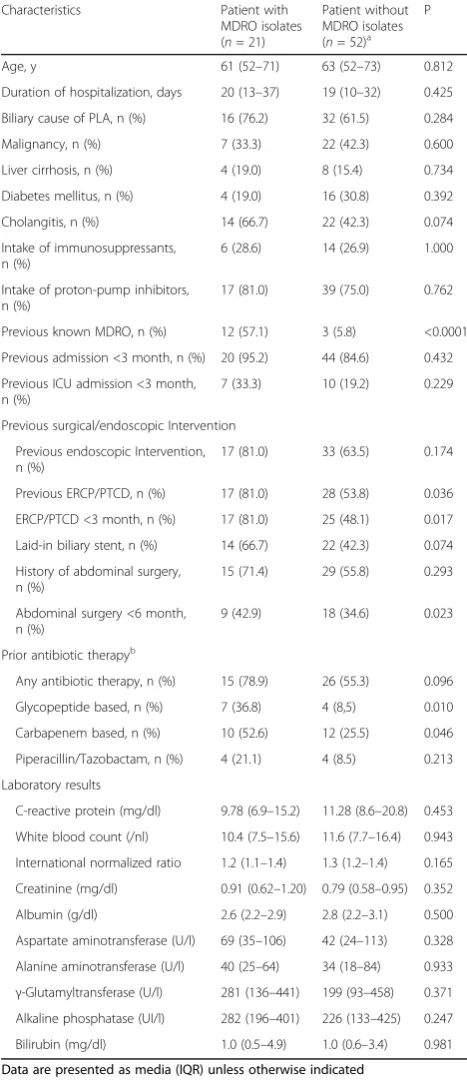
[image:5.595.58.292.116.602.2]PLA. Results of bivariate analyses are depicted in Table 6. Of note, in multivariate analysis prior known MDRO (P= 0.0002, OR 30.51, 95% CI 5.15–180.78) and the use of glycopeptide antibiotics prior to culture collection (P= 0.030, OR 6.46, 95% CI 1.20–34.87) were independ-ently associated with MDRO causing PLA in our patients.
Treatment of PLA
As initial empiric antibiotic therapy, 48.8% of included patients received a carbapenem-based regimen, 22.4% of patients BSP/βLI, and 17.6% of patients a third gener-ation cephalosporin. In 32.6% and 8.1% of patients, a glycopeptide antibiotic or tigecycline was added to initial empirical treatment, respectively. 12 patients (14.0%) were treated with an additional antimycotic agent upon diagnosis. Of note, 35.7% of patients had received an inappropriate initial empirical antibiotic treatment, as evidenced by subsequent microbiological culture results.
Only 7 patients (8.1%) were solitary medically man-aged. Almost all patients received a percutaneous drainage of the abscess cavity (43.0% CT-guided, 47.7% US-guided). Additional interventional ERCP/ PTCD was performed in 25.6% of cases. Surgery was necessary in 9.3% of cases.
Complications & outcome
The median duration of hospitalization was 20 days. Recurrent hepatic abscesses after discharge were re-ported in 18 patients (20.9%).
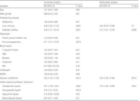
Twenty-four patients (27.9%) required intensive care therapy. Results of uni- and multivariate analysis are depicted in Table 7. Of note, only diabetes mellitus (P = 0.048, OR 3.72, 95% CI 1.01–13.70) and mycotic coinfection (P = 0.012, OR 5.54, 95% CI 1.46–21.06) as well a carbapenem based initial empirical antibiotic ther-apy (P = 0.037, OR 3.73, 95% CI 1.09–12.89) independ-ently predicted an ICU stay during hospitalization.
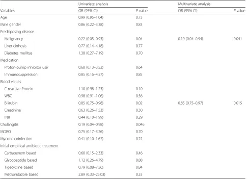
Fourteen patients died during their hospital stay, resulting in an overall mortality rate of 16.3%. In univari-ate and multivariunivari-ate analysis (Table 8), increased biliru-bin levels (multivariate P = 0.015, OR 0.85, 95% CI 0.75–0.97) and presence of malignancy (multivariate P= 0.041, OR 0.19, 95% CI 0.04–0.94) were significantly associated with mortality of PLA. Of note, neither the MDRO status nor the correct initial empiric therapy was related to ICU stay or survival.
Discussion
[image:6.595.304.538.111.652.2]In this study, we provide detailed characteristics of PLAs with a special focus on bacterial pathogens causing PLA in a large German tertiary reference center. We observed a so far underreported role of Enterococcus spp. and
Table 6Patients’characteristics and baseline parameters with and without cultivated multidrug-resistant organisms
Characteristics Patient with
MDRO isolates (n= 21)
Patient without MDRO isolates (n= 52)a
P
Age, y 61 (52–71) 63 (52–73) 0.812
Duration of hospitalization, days 20 (13–37) 19 (10–32) 0.425
Biliary cause of PLA, n (%) 16 (76.2) 32 (61.5) 0.284
Malignancy, n (%) 7 (33.3) 22 (42.3) 0.600
Liver cirrhosis, n (%) 4 (19.0) 8 (15.4) 0.734
Diabetes mellitus, n (%) 4 (19.0) 16 (30.8) 0.392
Cholangitis, n (%) 14 (66.7) 22 (42.3) 0.074
Intake of immunosuppressants, n (%)
6 (28.6) 14 (26.9) 1.000
Intake of proton-pump inhibitors, n (%)
17 (81.0) 39 (75.0) 0.762
Previous known MDRO, n (%) 12 (57.1) 3 (5.8) <0.0001
Previous admission <3 month, n (%) 20 (95.2) 44 (84.6) 0.432
Previous ICU admission <3 month, n (%)
7 (33.3) 10 (19.2) 0.229
Previous surgical/endoscopic Intervention
Previous endoscopic Intervention, n (%)
17 (81.0) 33 (63.5) 0.174
Previous ERCP/PTCD, n (%) 17 (81.0) 28 (53.8) 0.036
ERCP/PTCD <3 month, n (%) 17 (81.0) 25 (48.1) 0.017
Laid-in biliary stent, n (%) 14 (66.7) 22 (42.3) 0.074
History of abdominal surgery, n (%)
15 (71.4) 29 (55.8) 0.293
Abdominal surgery <6 month, n (%)
9 (42.9) 18 (34.6) 0.023
Prior antibiotic therapyb
Any antibiotic therapy, n (%) 15 (78.9) 26 (55.3) 0.096
Glycopeptide based, n (%) 7 (36.8) 4 (8,5) 0.010
Carbapenem based, n (%) 10 (52.6) 12 (25.5) 0.046
Piperacillin/Tazobactam, n (%) 4 (21.1) 4 (8.5) 0.213
Laboratory results
C-reactive protein (mg/dl) 9.78 (6.9–15.2) 11.28 (8.6–20.8) 0.453
White blood count (/nl) 10.4 (7.5–15.6) 11.6 (7.7–16.4) 0.943
International normalized ratio 1.2 (1.1–1.4) 1.3 (1.2–1.4) 0.165
Creatinine (mg/dl) 0.91 (0.62–1.20) 0.79 (0.58–0.95) 0.352
Albumin (g/dl) 2.6 (2.2–2.9) 2.8 (2.2–3.1) 0.500
Aspartate aminotransferase (U/l) 69 (35–106) 42 (24–113) 0.328
Alanine aminotransferase (U/l) 40 (25–64) 34 (18–84) 0.933
γ-Glutamyltransferase (U/l) 281 (136–441) 199 (93–458) 0.371 Alkaline phosphatase (UI/l) 282 (196–401) 226 (133–425) 0.247
Bilirubin (mg/dl) 1.0 (0.5–4.9) 1.0 (0.6–3.4) 0.981
Data are presented as media (IQR) unless otherwise indicated Abbreviations:ERCPendoscopic retrograde cholangiopancreatography, PTCDpercutaneous transhepatic cholangiography and drainage a
Patients without resistance profile of bacterial isolates or without bacterial isolates were excluded
b
other MDRO in the pathogenesis of secondary PLA, and present–to the best of our knowledge –a unique ana-lysis of current bacterial susceptibility profiles from a large tertiary reference center in a Western country, which may be utilized to guide empirical antibiotic treat-ment of secondary PLA.
Of note, marked differences between PLA with pa-tients in Western and Asia countries have been un-covered [16]. In our study, as in earlier reports from Western countries [1–3, 16], malignant and non-malignant biliary disease was the most identifiable direct cause of PLA. In Asian countries, the causes of PLAs often remain cryptogenic and the most commonly iso-lated bacterium is Klebsiella pneumoniae [5, 17–19]. Similarly to other Western reports [1–4], we have ob-served a divergent bacterial spectrum, characterized by a plethora of gram-positive and gram-negative bacteria, which have to be taken into account for choosing optimal antibiotic regimens. Most interestingly, Enterococcus spp. were most frequently isolated (in 28.9% of cases) in our study. So far,Enterococcuswas considered to play a negli-gible role in PLA in Asia [5, 17–19] as well as the Western
World: In the latter, rates were accounted for less than 7.2% of patients [2, 4, 14], only one study documented rates of 13.9% [21]. Two Western reports summarized Streptococcusspp. andEnterococcus spp. as one group of isolates (no exact percentage ofEnterococcusalone was in-dicated). By doing so higher rates were documented (22.6% and 29.5%) [3, 22].
Enterococcusspp. has been observed as one of the pre-dominant bacterial pathogens in cholangitis, especially in the setting of therapeutic endoscopy or presence of biliary endoprosthesis [23–25]. In line with these findings, many PLA patients included in this study had already received a therapeutic endoscopy prior to PLA manifestation. More-over, we observed that ERCP/PTCD three months prior to PLA occurrence was independently associated with posi-tive cultures forEnterococcusspp.
[image:7.595.57.545.98.450.2]Although enterococci belong to the physiological flora of the alimentary tract and are traditionally considered to be of low virulence, in more seriously ill patients en-terococcal infections have been associated with higher risk of treatment failure and mortality and antimicrobial ther-apy is warranted [26]. Of note, most cultured enterococci
Table 7Predictors of intensive care unit stay during hospitalization
Univariate analysis Multivariate analysis
Variables OR (95% CI) Pvalue OR (95% CI) Pvalue
Age 1.01 (0.97–1.04) 0.69
Male gender 0.88 (0.32–2.43) 0.81
Predisposing disease
Malignancy 1.06 (0.39–2.88) 0.91
Liver cirrhosis 3.38 (1.02–11.22) 0.047 3.45 (0.79–14.99) 0.1
Diabetes mellitus 3.50 (1.21–10.14) 0.021 3.72 (1.01–13.70) 0.048
Medication
Proton-pump inhibitor use 1.39 (0.44–4.39) 0.57
Immunosuppression 4.11 (1.27–13.37) 0.019
Blood values
C-reactive Protein 1.02 (0.97–1.07) 0.37
WBC 1.03 (0.97–1.09) 0.29
Bilirubin 1.06 (0.95–1.18) 0.29
Creatinine 1.82 (0.87–3.82) 0.11
INR 6.74 (0.67–67.24) 0.10
Cholangitis 1.64 (0.62–4.36) 0.32
MDRO 1.08 (0.36–3.29) 0.89
Mycotic coinfection 3.92 (1.33–11.55) 0.013 5.54 (1.46–21.06) 0.012
Initial empirical antibiotic treatment
Carbapenem based 4.37 (1.49–12.81) 0.007 3.73 (1.09–12.89) 0.037
Glycopeptide based 3.35 (1.21–9.25) 0.019
Tigecycline based 2.14 (0.43–10.58) 0.35
in our study were resistant to commonly administered broad-spectrum antibiotics (e.g. piperacillin/tazobactam or imipenem). With an intrinsic resistance to cephalosporins and BSP as typical in case ofE. faecium, therapy options are largely limited to glycopeptide antibiotics (vancomycin or teicoplanin), linezolid or daptomycin.
Over the past decades MDRO are increasingly being reported worldwide. VRE rates in Europe vary signifi-cantly. In a large survey of patients with healthcare asso-ciated infections, in approximately 10,1% of patients VRE was documented [27]. Comparable results were ob-served in a survey of 126 ICUs in the United States [28].
Of note, vancomycin resistance was detected in more than 1/3 ofEnterococcusspp.in our PLA study (VRE-rate inE. faeciumup to 46%) and we observed an increase of VRE over the study period. In addition, our study provides evidence for an increasing risk of resistant gram-negative bacteria in PLA. 16,7% of all gram-negative bacteria were classified as MRGN. Even more worrisome, approximately 11% of all gram-negative bacteria were resistant to carba-penems. In line with these observations, Lo et al. noted an increase of MDRGN from 1.64 in 2001 to 14.29% in 2011
in Singapore [14]. Finally, 50% of all anaerobes isolated in our study were resistant to metronidazole. Yet, the rele-vance of this finding remains unclear because these iso-lates (Propionibacterium acnes) may be contaminants rather than causative bacteria.
Interestingly, MDRO cultivated in PLA patients were strongly associated with prior history of MDRO and more often found after prior antibiotic therapy, espe-cially after the use of glycopeptide antibiotics. Moreover, neither the MDRO status nor the correct initial empiric therapy was associated with ICU stay or survival. In view of this data, MDRO may reflect severity of illness rather than being a predictor of mortality.
[image:8.595.56.544.98.449.2]However, in a setting with a high prevalence of Entero-coccusand VRE infection, as described, it appears neces-sary to treat critically ill patients with PLA with a combination of a carbapenem and an antibiotic targeting Enterococcus such as teicoplanin until microbiological test results are available. To avoid further spreads of re-sistance, rigorous de-escalation strategies appear to be warranted and stable patients may be empirically treated with a third-generation cephalosporin in combination
Table 8Predictors of survival in patients suffering from pyogenic liver abscess
Univariate analysis Multivariate analysis
Variables OR (95% CI) Pvalue OR (95% CI) Pvalue
Age 0.99 (0.95–1.04) 0.73
Male gender 0.86 (0.22–3.38) 0.83
Predisposing disease
Malignancy 0.22 (0.05–0.93) 0.04 0.19 (0.04–0.94) 0.041
Liver cirrhosis 0.77 (0.14–4.18) 0.77
Diabetes mellitus 1.38 (0.27–7.19) 0.70
Medication
Proton-pump inhibitor use 0.68 (0.13–3.52) 0.64
Immunosuppression 0.85 (0.16–4.57) 0.85
Blood values
C-reactive Protein 1.10 (0.98–1.23) 0.10
WBC 0.98 (0.91–1.06) 0.56
Bilirubin 0.85 (0.75–0.98) 0.02 0.85 (0.75–0.97) 0.015
Creatinine 0.63 (0.26–1.53) 0.30
INR 0.44 (0.10–1.99) 0.29
Cholangitis 0.19 (0.04–0.98) 0.046
MDRO 0.75 (0.17–3.26) 0.70
Mycotic coinfection 0.41 (0.10–1.67) 0.22
Initial empirical antibiotic treatment
Carbapenem based 0.60 (0.15–2.33) 0.46
Glycopeptide based 1.12 (0.26–4.79) 0.88
Tigecycline based 0.79 (0.08–7.56) 0.84
with an agent against anaerobes. Careful screening for Enterococcus spp. and MRDO infections would be re-quired in that scenario.
Limitations of our study remain in its retrospective de-sign, based on a single diagnosis code with a relatively small study population with 86 patients in total. Further-more, it was performed in a single major hepatobiliary surgery and liver transplant center. Thus, the spectrum of disease may reflect more the specific patient popula-tion and hence, not all observapopula-tions and conclusions may be generalizable. However, it provides valuable information in a setting of growing numbers in biliary/ abdominal surgery and endoscopic interventions.
Conclusion
Our study demonstrates a so far underreported role of Enterococcus spp. in secondary. A worrisome number of VRE and other MRGN such as Escherichia coli and Klebsiella pneumoniae have been observed. Patients on PPI, or with prior ERCP/PTCD, or history of abdominal surgery appear to be at higher risk for VRE, and those with a prior history of MDRO infection at considerably higher risk for MDRO as a cause of PLA. Thus, thorough microbiological diagnostics is pivotal to tailor individual treatment regimens in order to prevent further selection of bacterial resistance in PLA, a diagnosis in which long durations of antibiotic therapy are often required.
Additional files
Additional file 1: Table S1.Overview of bacterial isolates and their susceptibility profile. (XLSX 68 kb)
Additional file 2: Table S3.Susceptibility profiles of gram-positive aerobes, excludingEnterococcussupp. (DOCX 50 kb)
Additional file 3: Table S4.Susceptibility profiles of anaerobic bacteria. (DOCX 39 kb)
Additional file 4: Figure S1.MDRO rate (MDRO among all PLA patient) and relative VRE rate (VRE among all Enterococci) in patients with PLA. A marked increase of VRE causing PLA was observed. Data is represented per year (A) and with retrospect to defined study periods (B). (TIFF 12962 kb)
Additional file 5: Figure S2.Relative VRE rate (VRE among all Enterococci) of all reported Enterococcus infections among patients without PLA. Local hospital surveillance data in our gastroenterology/ hepatology wards revealed no increase in the relative VRE rate among all non-PLA patients. Results are represented per year (A), and with retro-spect to defined study periods (B). (TIFF 11180 kb)
Additional file 6: Table S2.Overview of fungal isolates and their susceptibility profile. (XLSX 47 kb)
Abbreviations
βLI:β-lactamase inhibitor; BSP: Broad-spectrum penicillin; CI: Confidence interval; CT: Computed tomography; ERCP: Endoscopic retrograde cholangiography; ICU: Intensive care unit; MDROs: Multidrug-resistant organisms; MRGN: Multidrug-resistant gram-negative bacteria; MRI: Magnetic resonance imaging; MRSA: Methicillin-resistantStaphylococcus aureus; OR: Odds ratio; PLA: Pyogenic liver abscess; PTCD: Percutaneous transhepatic cholangiography and drainage; US: Ultrasound; VRE: Vancomycin-resistant enterococci
Funding
CML is supported by the Deutsche Forschungsgemeinschaft (LA 2806/5–1). VAJK was supported by a grant from the Deutsche Forschungsgemeinschaft (DFG- research unit 2251).
Availability of data and materials
All data generated or analysed during this study are either included in this published article and its supplementary information files or are available from the corresponding author on reasonable request.
Authors’contributions
The authors have contributed to the manuscript by planning the study (MMM, JK, VAJK, CML), collecting the data (MMM, JK, VTM, KS, MH, VAJK, CML), analysis and interpretation of the data (all authors), and preparation and revision of the manuscript (all authors). All authors approved of the final version of the manuscript and the authorship list.
Authors’information
Johanna Kessel, for the Antibiotic Stewardship Team of the University Hospital Frankfurt.
Competing interests
The authors declare that they have no competing interests.
Consent for publication
Not applicable.
Ethics approval and consent to participate
The local ethics committee (University Hospital Frankfurt) approved this study.
Publisher’s Note
Springer Nature remains neutral with regard to jurisdictional claims in published maps and institutional affiliations.
Author details
1
Department of Internal Medicine 1, University Hospital Frankfurt, Frankfurt am Main, Germany.2University Center for Infectious Diseases, University Hospital Frankfurt, Frankfurt am Main, Germany.3Department of Internal Medicine 2, University Hospital Frankfurt, Frankfurt am Main, Germany. 4
Institute of Medical Microbiology and Infection Control, University Hospital Frankfurt, Frankfurt am Main, Germany.
Received: 11 March 2017 Accepted: 9 June 2017
References
1. Hansen PS, Schonheyder HC. Pyogenic hepatic abscess. A 10-year population-based retrospective study. APMIS. 1998;106(3):396–402. 2. Kaplan GG, Gregson DB, Laupland KB. Population-based study of the
epidemiology of and the risk factors for pyogenic liver abscess. Clin Gastroenterol Hepatol. 2004;2(11):1032–8.
3. Meddings L, Myers RP, Hubbard J, et al. A population-based study of pyogenic liver abscesses in the United States: incidence, mortality, and temporal trends. Am J Gastroenterol. 2010;105(1):117–24.
4. Mohsen AH, Green ST, Read RC, McKendrick MW. Liver abscess in adults: ten years experience in a UK Centre. QJM. 2002;95(12):797–802.
5. Tsai FC, Huang YT, Chang LY, Wang JT. Pyogenic liver abscess as endemic disease. Taiwan Emerg Infect Dis. 2008;14(10):1592–600.
6. Peleg AY, Hooper DC. Hospital-acquired infections due to gram-negative bacteria. N Engl J Med. 2010;362(19):1804–13.
7. Weist K, Diaz Hogberg L. ECDC publishes 2013 surveillance data on antimicrobial resistance and antimicrobial consumption in Europe. Euro Surveill. 2014:19(46).
8. Boucher HW, Talbot GH, Bradley JS, et al. Bad bugs, no drugs: no ESKAPE! An update from the Infectious Diseases Society of America. Clin Infect Dis. 2009;48(1):1–12.
9. Souli M, Galani I, Giamarellou H. Emergence of extensively drug-resistant and pandrug-resistant gram-negative bacilli in Europe. Euro Surveill. 2008:13(47).
11. Valencia R, Arroyo LA, Conde M, et al. Nosocomial outbreak of infection with pan-drug-resistant Acinetobacter Baumannii in a tertiary care university hospital. Infect Control Hosp Epidemiol. 2009;30(3):257–63.
12. Di Carlo P, Pantuso G, Cusimano A, et al. Two cases of monomicrobial intraabdominal abscesses due to KPC–3 Klebsiella Pneumoniae ST258 clone. BMC Gastroenterol. 2011;11:103.
13. Kumar P, Ghosh S, Rath D, Gadpayle AK. Multidrug resistant citrobacter: an unusual cause of liver abscess. BMJ Case Rep. 2013;2013
14. Lo JZ, Leow JJ, Ng PL, et al. Predictors of therapy failure in a series of 741 adult pyogenic liver abscesses. J Hepatobiliary Pancreat Sci. 2015;22(2):156–65. 15. Huang CJ, Pitt HA, Lipsett PA, et al. Pyogenic hepatic abscess. Changing
trends over 42 years. Ann Surg. 1996;223(5):600–7. discussion 7-9 16. Cerwenka H. Pyogenic liver abscess: differences in etiology and treatment in
Southeast Asia and Central Europe. World J Gastroenterol. 2010;16(20):2458–62. 17. Tian LT, Yao K, Zhang XY, et al. Liver abscesses in adult patients with and
without diabetes mellitus: an analysis of the clinical characteristics, features of the causative pathogens, outcomes and predictors of fatality: a report based on a large population, retrospective study in China. Clin Microbiol Infect. 2012;18(9):E314–30.
18. Kim JK, Chung DR, Wie SH, Yoo JH, Park SW. Korean study Group for Liver a. Risk factor analysis of invasive liver abscess caused by the K1 serotype Klebsiella Pneumoniae. Eur J Clin Microbiol Infect Dis. 2009;28(1):109–11. 19. Okano H, Shiraki K, Inoue H, et al. Clinicopathological analysis of liver
abscess in Japan. Int J Mol Med. 2002;10(5):627–30.
20. Wendt C, von Baum H, Kaase M, Meyer E, Suger-Wiedeck H, Ruscher C. Hygiene measures for infection or colonization with multidrug-resistant gram-negative bacilli. Commission recommendation for hospital hygiene and infection prevention (KRINKO) at the Robert Koch institute (RKI). Bundesgesundheitsblatt Gesundheitsforschung Gesundheitsschutz. 2012;55(10):1311–54.
21. Rahimian J, Wilson T, Oram V, Holzman RS. Pyogenic liver abscess: recent trends in etiology and mortality. Clin Infect Dis. 2004;39(11):1654–9. 22. Ruiz-Hernandez JJ, Leon-Mazorra M, Conde-Martel A, Marchena-Gomez J,
Hemmersbach-Miller M, Betancor-Leon P. Pyogenic liver abscesses: mortality-related factors. Eur J Gastroenterol Hepatol. 2007;19(10):853–8. 23. Gargouri D, Ouakaa-Kchaou A, Kochlef A, et al. Microbiological study and
antimicrobial susceptibility of bile in biliary therapeutic endoscopy. Tunis Med. 2015;93(10):602–5.
24. Weber A, Schneider J, Wagenpfeil S, et al. Spectrum of pathogens in acute cholangitis in patients with and without biliary endoprosthesis. J Inf Secur. 2013;67(2):111–21.
25. Anderson DJ, Shimpi RA, McDonald JR, et al. Infectious complications following endoscopic retrograde cholangiopancreatography: an automated surveillance system for detecting postprocedure bacteremia. Am J Infect Control. 2008;36(8):592–4.
26. Chatterjee I, Iredell JR, Woods M, Lipman J. The implications of enterococci for the intensive care unit. Crit Care Resusc. 2007;9(1):69–75.
27. Jones RN, Flonta M, Gurler N, Cepparulo M, Mendes RE, Castanheira M. Resistance surveillance program report for selected European nations (2011). Diagn Microbiol Infect Dis. 2014;78(4):429–36.
28. Fridkin SK, Edwards JR, Courval JM, et al. The effect of vancomycin and third-generation cephalosporins on prevalence of vancomycin-resistant enterococci in 126 U.S. adult intensive care units. Ann Intern Med. 2001;135(3):175–83.
• We accept pre-submission inquiries
• Our selector tool helps you to find the most relevant journal • We provide round the clock customer support
• Convenient online submission • Thorough peer review
• Inclusion in PubMed and all major indexing services • Maximum visibility for your research
Submit your manuscript at www.biomedcentral.com/submit