R E V I E W
Open Access
Human pathogens associated with the
blacklegged tick
Ixodes scapularis
: a
systematic review
Mark P. Nelder
1*, Curtis B. Russell
1, Nina Jain Sheehan
1, Beate Sander
1,2,3, Stephen Moore
1, Ye Li
4, Steven Johnson
4,
Samir N. Patel
5,6and Doug Sider
1,7Abstract
Background:
The blacklegged tick
Ixodes scapularis
transmits
Borrelia burgdorferi
(sensu stricto) in eastern North
America; however, the agent of Lyme disease is not the sole pathogen harbored by the blacklegged tick. The
blacklegged tick is expanding its range into areas of southern Canada such as Ontario, an area where exposure to
blacklegged tick bites and tick-borne pathogens is increasing. We performed a systematic review to evaluate the
public health risks posed by expanding blacklegged tick populations and their associated pathogens.
Methods:
We followed PRISMA (Preferred Reporting Items for Systematic Reviews and Meta-Analyses) guidelines
for conducting our systematic review. We searched Ovid MEDLINE, Embase, BIOSIS, Scopus and Environment
Complete databases for studies published from 2000 through 2015, using subject headings and keywords that
included
“
Ixodes scapularis
”
,
“
Rickettsia
”
,
“
Borrelia
”
,
“
Anaplasma
”
,
“
Babesia
”
and
“
pathogen.
”
Two reviewers screened
titles and abstracts against eligibility criteria (i.e. studies that included field-collected blacklegged ticks and studies
that did not focus solely on
B. burgdorferi) and performed quality assessments on eligible studies.
Results:
Seventy-eight studies were included in the final review, 72 were from the US and eight were from Canada
(two studies included blacklegged ticks from both countries). Sixty-four (82 %) studies met
≥
75 % of the quality
assessment criteria. Blacklegged ticks harbored 91 distinct taxa, 16 of these are tick-transmitted human pathogens,
including species of
Anaplasma,
Babesia,
Bartonella,
Borrelia,
Ehrlichia,
Rickettsia,
Theileria
and
Flavivirus. Organism
richness was highest in the Northeast (Connecticut, New York) and Upper Midwest US (Wisconsin); however,
organism richness was dependent on sampling effort. The primary tick-borne pathogens of public health concern
in Ontario, due to the geographic proximity or historical detection in Ontario, are
Anaplasma phagocytophilum,
Babesia microti,
B. burgdorferi,
Borrelia miyamotoi, deer tick virus and
Ehrlichia muris-like sp. Aside from
B. burgdorferi
and to a much lesser concern
A. phagocytophilum, these pathogens are not immediate concerns to public health in
Ontario; rather they represent future threats as the distribution of vectors and pathogens continue to proliferate.
Conclusions:
Our review is the first systematic assessment of the literature on the human pathogens associated
with the blacklegged tick. As Lyme disease awareness continues to increase, it is an opportune time to document
the full spectrum of human pathogens transmittable by blacklegged ticks.
Keywords:
Bacteria, Blacklegged ticks, Infectious disease,
Ixodes, Parasites, Pathogens, Public health, Symbionts,
Vector-borne, Viruses, Zoonoses
* Correspondence:[email protected]
1Enteric, Zoonotic and Vector-borne Diseases; Communicable Diseases, Emergency Preparedness and Response; Public Health Ontario, Toronto, Ontario, Canada
Full list of author information is available at the end of the article
Background
The blacklegged tick
Ixodes scapularis
is the vector of
Borrelia burgdorferi
(
sensu stricto
) (agent of Lyme
dis-ease) in eastern North America. Blacklegged ticks are
three-host, non-nidiculous ticks with larvae and nymphs
that feed on small rodents (e.g. white-footed mouse) and
passerine birds, and adults that feed on large mammals
(white-tailed deer, humans). The blacklegged tick’s range
has been expanding northward from its precinctive
habi-tats in the Northeast and Upper Midwest US over the
last several decades [1–3]. In Ontario, this range
expan-sion has not been uniform; spreading primarily into
sub-urban and rural areas with mixed deciduous forests,
where vertebrate hosts are abundant and local climate is
favourable to blacklegged tick survival [4]. Until the
mid-1990s,
B. burgdorferi
and
Babesia microti
(babesiosis)
were the only pathogens known to be transmitted by
black-legged ticks [5–7]. Blackblack-legged ticks were soon implicated
as vectors of
Anaplasma phagocytophilum
(anaplasmosis),
and more recently,
Borrelia miyamotoi
(
B. miyamotoi
dis-ease) and deer tick virus (DTV; DTV encephalitis) [8–10].
Uncommon in Ontario’
s blacklegged ticks, so far, these
pathogens are likely to become more prevalent in the
fu-ture, as has been the case in recently-invaded jurisdictions
such as Maine [11].
In order to assess the public health risks due to
blacklegged ticks, an understanding of blacklegged
tick-associated organisms is essential, particularly their
distribu-tion, prevalence and capacity to cause human disease. We
performed a systematic review of the scientific literature to
identify organisms (targeting human pathogens) associated
with blacklegged ticks in eastern North America and to
assess which organisms pose a threat to the health of
Ontarians.
Methods
Search strategy
We followed PRISMA (Preferred Reporting Items for
Systematic Reviews and Meta-Analyses) guidelines for
conducting our systematic review [12]. With the assistance
of Public Health Ontario’s Library Services, we developed
our primary search strategy in Medline and customized it
for other databases to account for database-specific
vocabu-lary and functionality. Our search used subject headings
and keywords that included
“North America”,
“
Ixodes
sca-pularis
”,
“
Rickettsia
”,
“
Bartonella
”,
“
Borrelia
”,
“
Anaplasma
”,
“
Babesia
”,
“Powassan”,
“pathogen”
and
“blacklegged tick.”
We conducted a systematic review of English-language
studies using five electronic databases: Ovid MEDLINE(R)
In-Process & Other Non-Indexed Citations and Ovid
MEDLINE(R) (Ovid platform: 1 January 1995 to 20 April
2015); Embase (Ovid platform: 1 January 1996 to Week 16,
2015); Scopus (1 January 1999 to 20 April 2015);
Environ-ment Complete (EBSCOhost Research Databases: 1 January
1995 to 20 April 2015); and BIOSIS Previews (Ovid
plat-form: 1 January 2002 to Week 20, 2015). All searches are
current as of 20 April 2015 (full search strategy for Ovid
Medline, Additional file 1).
Study selection
Studies included in the review were required to meet the
following eligibility criteria: (i) included field-collected
I.
scapularis
from Canada or US; (ii) published on or after
1 January 1995; and (iii) did not test solely for
B.
burg-dorferi
. We limited studies to those that tested for at
least one organism besides
B. burgdorferi
as our
em-phasis was on the incidence and prevalence of organisms
other than
B. burgdorferi
. Studies that focused on
experi-mental models–in the absence of field-collected
black-legged ticks–were excluded (e.g. modeling studies,
studies with experimental infections of hosts or ticks), as
were studies concentrating on human case reports, patient
treatment or blood-donor screening (as these did not
spe-cifically link an organism to field-collected blacklegged
ticks). We limited the final number of studies to those
published on or after 1 January 2000; we did not identify
any additional organisms from studies published from 1
January 1995 through 31 December 1999. Two reviewers
independently screened titles and abstracts against
eligibil-ity criteria and differences were resolved by consensus
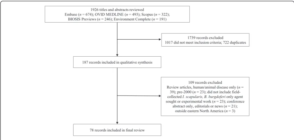
(Mark P Nelder, Nina Jain Sheehan) (Fig. 1). We excluded
one study because it did not identify the state(s) where the
ticks were collected [13].
Data extraction and quality assessment
We populated a data extraction table with the study’s
first author, year(s) of study, location of study, tick or host
collection methods, organisms tested for (gene targets),
key organism detection methods, prevalence of organisms
(by tick stage), explicit mention of positive and negative
pathogen controls, acknowledgement of sequence
submis-sions to GenBank (where the organism identification was
uncertain) (Additional file 2).
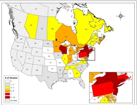
Data synthesis and analysis
We structured study outcomes by organism detected,
jur-isdiction (province or state) where detected and crude
prevalence (Table 1; Figs. 2 and 3). Pathogen prevalence is
reported as a crude prevalence, indicating the prevalence
reported includes pooled tick stages (larvae, nymphs and
adults), along with pooled engorgement status, collection
methods and pathogen detection methods. To determine
what tick-borne diseases are reportable to public health
officials, we reviewed reportable disease lists for
Ana-plasma
,
Babesia
,
Bartonella
,
Borrelia
, DTV,
Ehrlichia
and
Rickettsia
in Canada (Manitoba, Ontario and Quebec) and
the US (Illinois, Indiana, Michigan, Minnesota, New York,
Ohio, Pennsylvania and Wisconsin) (Table 2). We created
maps presenting crude prevalence (total positive ticks/
total ticks tested in a province or state) for the pathogens
of concern (Figs. 2 and 3) using the Environmental
Sys-tems Research Institute (ESRI) ArcMap Geographic
Infor-mation System software (v10.3.1), manually choosing data
classes, cut-off values and map colours for each of the
generated maps. We calculated Pearson product-moment
correlation coefficients (significance determined by linear
regressions) to test the linear association between the
number of organisms detected and the number of (i)
stud-ies conducted in a jurisdiction, (ii) ticks tested in a
juris-diction and (iii) organisms sought in a jurisjuris-diction.
Results
Study characteristics
Seventy-eight studies were included in our final review
(Additional file 2) [4, 8, 15–90]. Eight studies included
blacklegged ticks from Canada and 72 studies included
samples from the US (two studies included Canadian
and US blacklegged ticks). In Canada, Ontario (
n
= 6)
was sampled the most frequently, followed by Nova Scotia
(
n
= 5) (Fig. 2). In the US, New York (
n
= 18) was sampled
the most frequently, followed by Wisconsin (
n
= 12), New
Jersey (
n
= 10), Connecticut (
n
= 9), Massachusetts (
n
= 7),
Pennsylvania (
n
= 7) and Rhode Island (
n
= 7) (Fig. 2).
Most Canadian studies did not provide details on the
sub-provincial regions where tick collection occurred; however,
the areas sampled most frequently were southern regions
of Ontario and Nova Scotia. In the US, counties that were
sampled the most were Dutchess (New York) (
n
= 11),
Westchester (New York) (
n
= 9), Monmouth (New Jersey)
(
n
= 6) and New London (Connecticut) (
n
= 6). No studies
included blacklegged ticks from Alabama, Kansas,
Missouri, Ohio, Oklahoma, South Dakota, Vermont or
West Virginia. From 2000 through 2007, three studies
were published per year (range = 2–4); from 2009 through
2015, 6.8 studies were published per year (range = 1–19).
Quality assessment
Twenty studies (26 % or 20 out of 78) met 100 % of
quality assessment criteria and an additional 44 studies
(56 %) met 75 % of criteria (Additional file 3).
Thirty-three studies (42 %) collected blacklegged ticks by
drag-ging or flagdrag-ging; 21 (27 %) collected ticks from mammal
or bird hosts; 15 (19 %) used a combination of dragging
and animal collections; and nine (12 %) did not report
collection methods or employed other techniques.
Thirty-three studies (42 %) explicitly reported positive and negative
controls; four (5 %) reported negative controls only; 10
(13 %) reported positive controls only; and 31 (40 %)
[image:3.595.56.538.87.317.2]did not report any controls (Additional file 2). Six
stud-ies (38 % or six out of 16) included GenBank accession
numbers for gene sequenced PCR products where
spe-cies identification was indeterminate.
Descriptive analysis
Blacklegged ticks harbored 91 distinct taxa. Sixteen of
these organisms are tick-transmitted human pathogens;
the remainder is primarily gut or surface microbes of
blacklegged ticks and not transmittable to humans or
symbionts of blacklegged ticks (Table 1). The highest
number of organisms detected in the Canadian-sampled
blacklegged ticks were from Nova Scotia (
n
= 4; i.e. at
least four separate identifications of the organisms
sought in the ticks sampled), followed by three each in
Alberta, Manitoba, New Brunswick, Ontario and Prince
Edward Island (Table 2). US-sampled blacklegged ticks
harbored 91 organisms. The highest number of
organ-isms were detected in Connecticut (
n
= 42), followed by
[image:4.595.59.541.98.485.2]New York (
n
= 36), Wisconsin (
n
= 9), Tennessee (
n
= 9),
Massachusetts (
n
= 8) and Pennsylvania (
n
= 8). Studies in
different jurisdictions sought varying numbers of pathogens
and often used various detection methods. The mean ± SE
number of studies per jurisdiction (4.2 ± 0.60; median = 3;
n
= 78) was positively correlated (
r
= 0.77,
n
= 37,
P
<
0.0001) with the mean ± SE number of organisms
de-tected per jurisdiction (5.2 ± 1.09; median = 4;
n
= 78).
Similarly, the number of blacklegged ticks tested in a
jurisdiction (mean = 2,797 ± 1,022.6; median = 696) was
positively correlated with the number of organisms
de-tected per jurisdiction (
r
= 0.72,
n
= 37,
P
< 0.0001). In
the studies reviewed, researchers performed 149 unique
sampling events, where a sampling event is the testing
of blacklegged ticks for a specific organism in a specific
jurisdiction. The number of organisms sought per
sam-pling event (mean = 2.8 ± 0.29; median = 2;
n
= 149) was
positively correlated (
r
= 0.95,
n
= 149,
P
< 0.0001) with
the number of organisms detected per sampling event
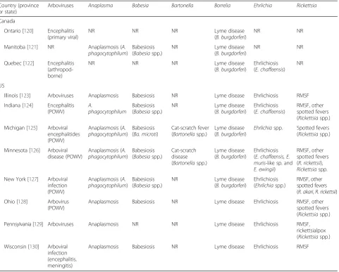
Table 1
Summary of tick-borne, reportable pathogens and diseases for select provinces and states
aCountry (province or state)
Arboviruses Anaplasma Babesia Bartonella Borrelia Ehrlichia Rickettsia
Canada
Ontario [120] Encephalitis (primary viral)
NR NR NR Lyme disease
(B. burgdorferi)
NR NR
Manitoba [121] NR Anaplasmosis (A. phagocytophilum)
Babesiosis (Babesiaspp.)
NR Lyme disease
(B. burgdorferi)
NR NR
Quebec [122] Encephalitis (arthropod-borne)
NR NR NR Lyme disease
(B. burgdorferi)
Ehrlichiosis (E. chaffeensis)
NR
US
Illinois [123] Arboviruses Anaplasmosis Babesiosis NR Lyme disease Ehrlichiosis RMSF
Indiana [124] Encephalitis (POWV)
A.
phagocytophilum
Babesiosis (Babesiaspp.)
NR Lyme disease
(B. burgdorferi)
Ehrlichiosis (E. chaffeensis)
RMSF, other spotted fevers (Rickettsiaspp.)
Michigan [125] Arboviral encephalitides (POWV)
Anaplasmosis (A. phagocytophilum)
Babesiosis (Ba. microti)
Cat-scratch fever (Bartonellaspp.)
Lyme disease (B. burgdorferi)
Ehrlichiaspp. Spotted fevers (Rickettsiaspp.)
Minnesota [126] Arboviral disease (POWV)
Anaplasmosis (A. phagocytophilum)
Babesiosis (Babesiaspp.)
Cat-scratch disease (Bartonellaspp.)
Lyme disease (B. burgdorferi)
Ehrlichiosis (E. chaffeensis, E. muris-like sp. and
E. ewingii)
RMSF, other spotted fevers (R. rickettsii),
Rickettsiaspp.
New York [127] Arboviral infection (POWV)
Anaplasmosis (A. phagocytophilum)
Babesiosis (Babesiaspp.)
NR Lyme disease
(B. burgdorferi)
Ehrlichiosis (Ehrlichiaspp.)
RMSF, other spotted fevers (R. akari,R. rickettsii)
Ohio [128] Arbovirus (POWV)
Anaplasmosis Babesiosis NR Lyme disease Ehrlichiosis RMSF, other spotted fevers (Rickettsiaspp.)
Pennsylvania [129] Arboviruses Anaplasmosis NR NR Lyme disease Ehrlichiosis RMSF, rickettsialpox (Rickettsiaspp.)
Wisconsin [130] Arboviral infection (encephalitis, meningitis)
Anaplasmosis Babesiosis NR Lyme disease Ehrlichiosis RMSF
a
(mean = 2.3 ± 0.28; median = 2;
n
= 149). We performed
multiple tests on the same dataset, however, since these
P
-values are very small, adjusting for multiple testing
would not alter the statistical significance of the results.
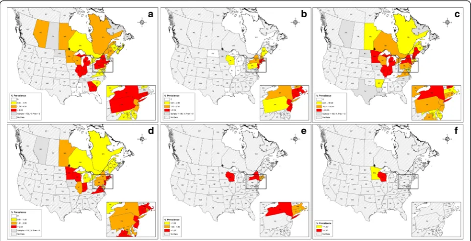
Thirty-two distinct organisms transmitted by ticks were
reported in the studies reviewed (Table 1). Five species of
Anaplasmataceae were detected;
A. phagocytophilum
was
most prevalent in Rhode Island (21.3 %;
n
= 684), Georgia
(17.3 %;
n
= 910), Connecticut (13.0 %;
n
= 454), Indiana
(10.8;
n
= 712), New York (8.9 %;
n
= 25,098), Michigan
(7.9 %;
n
= 444), Pennsylvania (6.5 %;
n
= 1,559) and
Illi-nois (6.5 %;
n
= 278) (Fig. 3; Table 1).
Ehrlichia muris
-like
sp. was restricted to Wisconsin (5.4 %;
n
= 4,066) and
Minnesota (3.0 %;
n
= 534) (Fig. 3). Blacklegged ticks
har-bored one Bartonellaceae species,
Bartonella henselae
,
which was only found in ticks from New Jersey (100 %;
n
= 1), New York (2.3 %;
n
= 88) and Pennsylvania (3.1 %;
n
= 544). Blacklegged ticks contained 12
Rickettsia
taxa;
Rickettsia
endosymbiont of
Ixodes scapularis
, a
black-legged tick obligate endosymbiont (non-pathogenic),
was the most prevalent taxa, particularly in Pennsylvania
(64.9 %;
n
= 94), Indiana (63.0 %;
n
= 100), Tennessee
(51.1 %;
n
= 47) and Maine (46.0 %;
n
= 100). Seven out
of 12
Rickettsia
species were restricted geographically to
the Southern US and the known human pathogen
Rickett-sia parkeri
was limited to Louisiana (16.6 %;
n
= 18). Ticks
sampled across all 78 studies contained six species of
Bor-relia. Borrelia burgdorferi
was most prevalent in New
Hampshire (52.3 %;
n
= 509), Maine (49.1 %;
n
= 10,004),
Minnesota (47.2 %;
n
= 803), Indiana (45.3 %
n
= 506),
Michigan (39.5 %;
n
= 696), Illinois (33.9 %;
n
= 460) and
New York (31.6 %;
n
= 21,363).
Borrelia miyamotoi
was
most prevalent in Connecticut (4.7 %;
n
= 1,226), Indiana
(4.1 %;
n
= 487), Virginia (3.5 %;
n
= 173), Minnesota
(2.9 %;
n
=700), New Jersey (2.7 %;
n
= 765), Massachusetts
(2.5 %;
n
= 159) and Wisconsin (2.4 %;
n
= 3,151) (Fig. 3).
Blacklegged ticks harbored three Apicomplexan parasites;
Ba. microti
was most prevalent in Connecticut (6.7 %;
n
=
1,198), New Jersey (6.5 %;
n
= 1.195) and Massachusetts
(5.3 %;
n
= 851) (Fig. 3). Several viruses were detected in
[image:5.595.59.539.86.451.2]blacklegged ticks; however, only DTV and Powassan
virus (POWV) are of concern as human pathogens.
Deer tick virus/POWV was most common in New York
(3.4 %;
n
= 91) (Fig. 3). Pathogens sought for but not
detected in any samples included
Coxiella burnetii
(New York,
n
= 88),
Francisella tularensis
(New York,
n
= 374; Florida,
n
= 52; Georgia,
n
= 13) and
Rickettsia
rickettsii
(New York,
n
= 88) (Additional file 2).
Two of the studies reported 55 additional bacteria taxa,
common genera were
Acinetobacter
(Enterobacteriaceae),
Afipia
(Alphaproteobacteria),
Pseudomonas
(Pseudomona-daceae),
Sphingomonas
(Sphingomonadaceae) and
Stenotro-phomonas
(Xanthomonadaceae) [19, 64]. While most of
the bacteria identified are commonly found in the
environ-ment (i.e., soil, water), some are associated with mammals,
such as
E. coli
,
Shigella
sp. and
Streptococcus
sp. These
add-itional bacteria are likely a part of the microbial community
(as commensal organisms or environmental contaminants)
of the blacklegged tick’s gut or surface and not necessarily
transmitted or maintained by blacklegged ticks.
The mandatory reporting requirements of pathogens
varied among the jurisdictions reviewed. Deer tick virus
is typically reportable as an arboviral infection in most
of the jurisdictions reviewed except for Manitoba
(Table 2). In Canada, anaplasmosis is reportable only in
Manitoba, yet reportable in all US states reviewed. In
Canada, babesiosis is reportable only in Manitoba, but in
all US states reviewed except Pennsylvania. Bartonellosis
was not reportable in any Canadian jurisdiction and
re-portable only in Michigan and Minnesota in the US.
Lyme disease (as
B. burgdorferi
) is reportable in all
juris-dictions reviewed; however, infection by other
Borrelia
species is not. In Canada, only Quebec requires
report-ing of ehrlichiosis, but ehrlichiosis is reportable in all US
states reviewed. In Canada, rickettsial infections are not
reportable, but are reportable in all US states reviewed.
Discussion
Our systematic review of 78 North American studies
published since 2000 documented that blacklegged ticks
are associated with 91 distinct organisms. Sixteen of these
organisms are tick-transmitted human pathogens. The
remainder of the organisms are non-pathogenic gut or
surface biota (as commensal organisms or environmental
contaminants), intracellular symbionts of ticks or have
un-known pathologies in vertebrates. Blacklegged ticks are
the principal vectors of six of these human pathogens:
A.
phagocytophilum
,
Ba. microti
,
B. burgdorferi
,
B.
miyamo-toi
, DTV and
Ehrlichia muris
-like sp. Due to studies
docu-menting their proximity to, or detection in, Ontario, the
six pathogens represent the primary risks in the province,
with two presenting more immediate threats than others
due to their recent detection in parts of Ontario (i.e.
A.
phagocytophilum
,
B. burgdorferi
). Similar to our review,
20 human pathogens were reported from Europe’s sister
taxa to the blacklegged tick,
Ixodes ricinus
[91, 92]. Given
the blacklegged tick’s liberal feeding behavior and
propen-sity to bite humans, continued identification of
black-legged tick-pathogen relationships and assessing their
public health implications is justified.
Fig. 3Distribution and crude prevalence for human pathogens transmitted byIxodes scapularisin eastern North America (2000–2015).
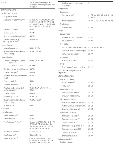
[image:6.595.57.539.88.334.2]Table 2
Distribution of
Ixodes scapularis
organisms in eastern
North America, from 78 studies reviewed (2000
–
2015)
Organism Jurisdiction where organism
detected (Canadian provinces and US states)a
Tick-borne organisms
Alphaproteobacteria
Anaplasmataceae
Anaplasma phagocytophilumb,c Canada: AB, MB, NB, NL, NS, ON, PE, QC, SK; US: CT, DE, GA, IL, IN, MA, MD, ME, MI, MN, NC, ND, NH, NJ, NY, PA, RI, SC, TN, VA, WI
Ehrlichia chaffeensisb US: NY
Ehrlichia ewingiib US: TN
EhrlichiaPanola Mountain sp.b US: TN
Ehrlichia muris-like sp.
(orE.sp. nr.muris)b US: MN, WI
Bartonellaceae
Bartonella henselaeb US: NJ, NY, PA
Unidentified/uncharacterized
Bartonellaspp.
US: MD, NJ, PA
Rickettsiaceae
CandidatusRickettsia cooleyi
(or cooleyi-like)d US: FL, GA, TN, TX
CandidatusRickettsia IRS4 US: MA
CandidatusRickettsia amblyommii US: TN
Rickettsia buchnerid US: MN
Rickettsia massiliae/Rickettsiasp. Bar 29
US: NC
Rickettsia parkerib US: LA
Rickettsia peacockiid US: TX
Rickettsiaendosymbiont of
Ixodes scapularisd US: CT, IN, LA, MA, ME, NY, PA,TN, WI
Rickettsiasp. Is-I US: GA
Rickettsiasp. TR-39 US: FL, GA
Unidentified/uncharacterized
Rickettsiaspp.
US: MD, NC, TN
Wolbachiasp. US: CT
Spirochaetes
Spirochaetaceae
Borrelia andersoniib US: MI
Borrelia bissettiib US: LA
Borrelia burgdorferis.s.b Canada: AB, MB, NB, NS, NL, ON, PE, QC; US: AR, CT, DE, GA, IA, IL, IN, LA, MA, MD, ME, MI, MN, NC, ND, NH, NJ, NY, PA, RI, SC, TX, VA, WI
Borrelia kurtenbachiib Canada: NS; US: NY
Borrelia lonestarib US: AR, MA, NY
Borrelia miyamotoib Canada: AB, MB, NB, NS, ON, PE,
QC; US: CT, IA, IL, IN, MA, MD, ME, MI, MN, NJ, NY, PA, RI, VA, WI
Table 2
Distribution of
Ixodes scapularis
organisms in eastern
North America, from 78 studies reviewed (2000
–
2015)
(Continued)
Unidentified/uncharacterized
Borreliasp.
US: NY
Piroplasmida
Babesiidae
Babesia microtib US: CT, MA, MD, ME, MN, NH, NJ,
NY, PA, WI
Babesia odocoilei US: IN, IL, ME, MI, MS, PA, TN, WI
Theileriidae
Theileria cervi US: TN
Viruses
Bunyaviridae
Blacklegged tick phlebovirus US: NY
South Bay virus US: NY
Flaviviridae
Deer tick virus (POWV lineage II)b US: CT, MA, NY, RI, WI Powassan virus (POWV lineage I)b US: NY
POWV (not subtyped)b US: NY Reoviridae
St. Croix River viruse US: WI
Other
Ixodes scapularismononegaviralese US: NY
Non-vector-borne, gut/surface orgnaisms
Alphaproteobacteria
Bradyrhizobiaceae
Afipia broomeaef US: NY
Afipia felisf US: CT
Caulobacteraceae
Uncultured bacterium 1 US: CT
Uncultured bacterium 2 US: CT
Methylobacteriaceae
Methylobacterium mesophilicumf US: CT
Methylobacteriumsp. strain G296-5 US: CT
Uncultured bacterium 3 US: CT
Sphingomonadaceae
Sphingomonas elodeaf US: CT
Sphingomonas melonisf US: CT
Sphingomonassp. US: CT
Sphingomonassp. strain BF2 US: CT
Sphingomonassp. AV069 US: CT
Sphingopyxis alaskensis US: CT
Sphingobacteriumsp. US: NY
Betaproteobacteria
Blacklegged ticks transmit two of the Anaplasmataceae
bacteria reported in the review (i.e.
A. phagocytophilum
and
Ehrlichia muris
-like sp.). The number of human
granulocytic anaplasmosis cases has increased in the US
from less than 200 cases in 1997 to over 2,700 cases in
2013, with most cases reported from Massachusetts,
Minnesota, New York and Wisconsin [93, 94]. Patients
infected with
A. phagocytophilum
display (in order of
decreasing incidence) malaise, fever, myalgia, headache,
arthralgia and nausea [95].
Anaplasma phagocytophilum
is relatively rare (< 0.5 %) in Ontario blacklegged ticks
and we are not aware of any anaplasmosis cases diagnosed
from the province [4].
Ehrlichia muris
-like sp. is restricted
to Wisconsin and Minnesota; however, this pathogen has
not been widely tested for elsewhere. Little is known about
this pathogen; nevertheless, it has been identified in 73
pa-tients from Indiana, Michigan, Minnesota, North Dakota
and Wisconsin (all cases reported tick-bite exposures from
Minnesota or Wisconsin) displaying fever, malaise,
head-ache and myalgia [96]. Considering
A. phagocytophilum
is present in Ontario and the distribution of
Ehrlichia
muris
-like sp. is not well-characterized, awareness of
[image:8.595.308.538.112.280.2] [image:8.595.59.248.125.701.2]these pathogens by public health officials is warranted.
We expected a relatively high number of rickettsial
agents to be reported from blacklegged ticks, given the
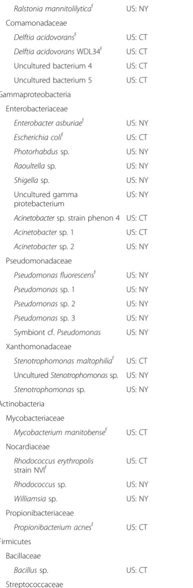
Table 2
Distribution of
Ixodes scapularis
organisms in eastern
North America, from 78 studies reviewed (2000
–
2015)
(Continued)
Burkholderiasp. US: CT
Ralstonia mannitolilyticaf US: NY
Comamonadaceae
Delftia acidovoransf US: CT
Delftia acidovoransWDL34f US: CT
Uncultured bacterium 4 US: CT
Uncultured bacterium 5 US: CT
Gammaproteobacteria
Enterobacteriaceae
Enterobacter asburiaef US: NY
Escherichia colif US: CT
Photorhabdussp. US: NY
Raoultellasp. US: NY
Shigellasp. US: NY
Uncultured gamma protebacterium
US: NY
Acinetobactersp. strain phenon 4 US: CT
Acinetobactersp. 1 US: CT
Acinetobactersp. 2 US: NY
Pseudomonadaceae
Pseudomonas fluorescensf US: NY
Pseudomonassp. 1 US: NY
Pseudomonassp. 2 US: NY
Pseudomonassp. 3 US: NY
Symbiont cf.Pseudomonas US: NY
Xanthomonadaceae
Stenotrophomonas maltophiliaf US: CT
UnculturedStenotrophomonassp. US: NY
Stenotrophomonassp. US: NY
Actinobacteria
Mycobacteriaceae
Mycobacterium manitobensef US: CT
Nocardiaceae
Rhodococcus erythropolis
strain NVIf
US: CT
Rhodococcussp. US: NY
Williamsiasp. US: NY
Propionibacteriaceae
Propionibacterium acnesf US: CT
Firmicutes
Bacillaceae
Bacillussp. US: CT
Streptococcaceae
UnculturedStreptococcussp. US: CT
Table 2
Distribution of
Ixodes scapularis
organisms in eastern
North America, from 78 studies reviewed (2000
–
2015)
(Continued)
Unclassified Bacteria
Uncultured bacterium 6 US: CT
Uncultured bacterium 7 US: CT
Uncultured bacterium 8 US: CT
Uncultured bacterium 9 US: CT
Uncultured bacterium 10 US: CT
Uncultured bacterium 11 US: CT
Uncultured bacterium 12 US: CT
Uncultured bacterium 13 US: CT
Plastid clone US: CT
Uncultured bacterium 14 US: NY
Uncultured/unidentified bacterium US: NY
a
Canada: AB, Alberta; MB, Manitoba; NB, New Brunswick; NL, Newfoundland; NS, Nova Scotia; ON, Ontario; PE, Prince Edward Island; SK, Saskatchewan; QC, Quebec. US: AR, Arkansas; CT, Connecticut; DE, Delaware; FL, Florida; GA, Georgia; IL, Illinois; IN, Indiana; IA, Iowa; LA, Louisiana; ME, Maine; MD, Maryland; MA, Massachusetts; MI, Michigan; MN, Minnesota; MS, Mississippi; NH, New Hampshire; NJ, New Jersey; NY, New York; NC, North Carolina; ND, North Dakota; PA, Pennsylvania; RI, Rhode Island; SC, South Carolina; TN, Tennessee; TX, Texas; VT, Vermont; VA, Virginia; WI, Wisconsin
b
Known or putative human pathogen,B. andersoni[131];B. bissettii[132];
B. kurtenbachii[133];B. lonestari[134];R. parkeri[91]
c
Both theAp-hastrain (human disease strain) and theAp-variant-1strain were pooled during data extraction, since not all studies distinguished between the two
d
Known or putative endosymbiont ofIx. scapularis
e
Likely a viral genome integrated intoI. scapularisgenome [135,136]
f
extensive association of
Rickettsia
with hematophagous
invertebrates such as fleas (Siphonaptera) and other ticks
(Ixodidae, Argasidae) [91]. Human disease has not been
associated with the majority of rickettsial organisms
identified in this review; however, careful clinical and
epidemiological studies can lead to some of these being
identified as human pathogens (e.g.
R. parkeri
) [97].
Cur-rently, there is no evidence demonstrating blacklegged
ticks as effective vectors of
Rickettsia
; however, further
investigation is justified where unexplained cases of
spotted fever occur with tick-bite histories.
The reviewed studies reported that blacklegged ticks
harbor six species of
Borrelia
, two of which are primarily
reported from blacklegged ticks (i.e.
B. burgdorferi
and
B.
miyamotoi
).
Borrelia burgdorferi
was widely distributed
throughout the range of the blacklegged tick (at least in
the ticks examined in these studies), including Ontario
(prevalence = 16 % of blacklegged ticks), with the highest
prevalence (> 30 %) in the Northeast and Midwest US.
Studies focusing solely on
B. burgdorferi
were not
in-cluded in review; therefore, caution must be used when
interpreting the prevalence of
B. burgdorferi
. Patients with
acute
B. burgdorferi
infection usually display erythema
migrans (bulls-eye rash), fever, myalgia, headache,
arthral-gia, neck stiffness and arthritis [98]. In Ontario, the Lyme
disease incidence rate has increased from 0.2 (2002) to 1.6
cases per 100,000 population (2014) [99].
Borrelia
miyamo-toi
is a newly recognized human pathogen, which based on
the reviewed studies has a widespread distribution in North
America (Ontario prevalence < 0.5 %). Recently, 53 patients
with Lyme disease-like symptoms from Connecticut,
Massachusetts, New Jersey, New York and Rhode Island
showed serological evidence for
B. miyamotoi
infection
[100, 101]. Patients with
B. miyamotoi
disease display
fever, fatigue, headache, myalgia, chills and nausea [10,
102]. To our knowledge,
B. miyamotoi
disease has not
been diagnosed in Ontario. Since our literature search was
performed, a new blacklegged tick-transmitted
Borrelia
has been associated with human disease in Wisconsin,
Candidatus
Borrelia mayonii [103, 104].
Borrelia
burgdor-feri
has rightfully been the
Borrelia
of concern to public
health; however, other
Borrelia
species are beginning to
emerge as additional threats to public health.
While the focus of this systematic review was organisms
of public health significance, it is important to note the
superficially benign microbes and symbionts associated
with blacklegged ticks. While outside the scope of this
review, the gut microorganisms of blacklegged ticks
can moderate the colonization of human pathogens in
blacklegged ticks, such as with
B. burgdorferi
[105,
106]. Several of the gut or surface bacteria (e.g.
Steno-trophomonas maltophilia
and
Rhodococcus erythropolis
)
associated with opportunistic infections in humans,
es-pecially in immunocompromised individuals [107–109].
Our understanding of the role that symbionts play in
the biology of blacklegged ticks and the blacklegged
tick’s ability to transmit pathogens is poorly understood
and represents an opportunity for future research.
Symbi-onts, such as
Rickettsia
endosymbiont of
Ixodes scapularis
and
Wolbachia
sp. (closely related to
W. pipientis
supergroup A) were reported from the studies reviewed.
Wolbachia
symbionts are involved in the manipulation of
arthropod reproduction (e.g. cytoplasmic incompatibility
and parthenogenesis) in other arthropods such as
Dros-ophila
and parasitoid wasps [110]. Understanding how the
entire tick microbiome regulates pathogen acquisition and
transmission is a burgeoning field, undoubtedly providing
insights into potential blacklegged tick management
op-tions. In addition, a better understanding of
Ix. scapularis
symbionts could lead to novel management tools.
In the reviewed studies,
Ba. microti
was most common in
blacklegged ticks collected from the Coastal Atlantic states
of the Northeast US. In the US, the number of babesiosis
cases has increased from approximately 1,100 (2011) to
1,800 (2013), with most cases reported from Connecticut,
Massachusetts, New Jersey and New York [94, 111].
Pa-tients with babesiosis display fever, fatigue, headache, chills
and arthralgia [112]. While
Ba. microti
could eventually
oc-cupy the same distribution of
B. burgdorferi
, it will do so at
a slower rate due to several factors. For example,
black-legged tick transmission of
Ba. microti
to reservoir hosts is
not as efficient when compared to
B. burgdorferi
[113, 114].
In addition, the survival of
Ba. microti
is low in
overwinter-ing blacklegged tick nymphs when compared to
B.
burgdor-feri
[115].
Babesia microti
has not been detected in Ontario
blacklegged ticks (in the published literature) or identified
as the cause of locally acquired disease, to our knowledge.
Deer tick virus (POWV lineage II) was the most
common viral agent detected in the studies reviewed,
a viral species serologically indistinguishable from POWV
(POWV lineage I) but with unique nucleotide and amino
acid sequences. In 2009, DTV was the cause of a fatal case
of encephalitis from New York, the first report implicating
DTV as an agent of human disease [116]. Soon after this
initial case report, additional cases were reported in New
York that displayed fever, malaise, confusion, seizure,
headache, rash and vomiting [117]. There is no evidence
for the presence of DTV in the Ontario’s blacklegged ticks;
however, field research is underway to determine its
pres-ence (Curtis B Russell, unpublished data).
disease in vertebrates. While symbionts and non-human
pathogens are relatively benign, it is important to
under-stand their ecology as well, because they likely play roles in
determining the distribution and prevalence of the human
pathogens discussed through competitive exclusion [118].
In Ontario blacklegged ticks, only a handful of pathogens
have been sought after and there is an opportunity to better
understand their ecology (e.g.
Babesia
,
Ehrlichia
,
Rickettsia
,
DTV). Understanding the complex epidemiology of the
hu-man pathogens transmittable by blacklegged ticks will
require longitudinal, ecological (host and vector
com-petence) and epidemiological studies in endemic and
emerging areas.
While comprehensive, our systematic review does have
several limitations. We did not perform a search of the grey
literature (publications on testing results from public health
or government institutions); therefore, our results might be
biased towards primary research with positive results due
to publication bias. In Canada, blacklegged ticks collected
by passive surveillance were likely used in multiple studies
for pathogen detection; however, an examination of the
studies shows that overlap (same ticks used in separate
studies) involved only two studies each from 2007 through
2012 and was limited to the detection of
A.
phagocytophi-lum
and
B. burgdorferi
. Another limitation is that studies
were undertaken where pathogen prevalence or disease
burden are high, meaning prevalence does not represent a
uniform value across a province or state. The heterogeneity
among studies in terms of collection methods, blacklegged
tick stages assayed, engorgement levels of ticks, molecular
methods used or gene targets undoubtedly influenced the
crude prevalences we reported. For example, we expect that
ticks collected from reservoir hosts (pathogen acquisition
can also vary by host type and tick stage) would have a
higher pathogen load, compared to host-seeking ticks
collected by dragging. The number of organisms
de-tected in a jurisdiction is dependent upon sampling
ef-fort and testing methods; therefore, little-studied areas
may indeed hold more blacklegged tick- organisms.
The presence of pathogens in neighboring jurisdictions
or in Ontario itself provides the basis for assessing the
risks of blacklegged tick-transmitted infections in Ontario,
but this method does not allow for an accurate estimate of
when these pathogens will be a threat to Ontario. As far
as we know, blacklegged ticks transmit and maintain all
the underscored pathogens within a rodent-mammal-tick
cycle in temperate, mixed deciduous forests. Given the
pathogens share a common transmission cycle, these
path-ogens should eventually occur throughout the blacklegged
tick’s range. The review identified that most research has
occurred in relatively small geographic areas, representing
an opportunity to determine pathogen incidence and
prevalence outside of highly sampled regions. As research
continues to detect pathogens in blacklegged ticks and
reservoir hosts, we expect the number of pathogens to
increase. In addition, comprehensive studies, seeking
all possible pathogens using standardized methods, are
needed for better comparison of pathogen prevalence
across northeastern North America. Our review is the
first systematic assessment of literature, identifying and
bringing together the scattered knowledge of human
pathogens associated with the blacklegged tick.
Conclusions
Viewed as a mere pest until the mid-1970s, the blacklegged
tick was not known to transmit pathogens to humans or
other animals [119]. In the following 40 years, the
black-legged tick has turned into the most important vector in
North America. Our review has identified several human
pathogens besides
B. burgdorferi
that are transmittable by
blacklegged ticks, yet we have not demonstrated disease in
Ontario at this time for
A. phagocytophilum
,
Ba. microti
,
B.
miyamotoi
, DTV or
Ehrlichia muris
-like sp. While most of
these pathogens do not represent immediate public health
threats in Ontario, there is an opportunity to ensure
mitiga-tion efforts are in place prior to their arrival. The growing
public and physician awareness concerning Lyme disease
provides a catalyst upon which to synergize awareness of
other tick-borne diseases. In addition, surveillance of
patho-gens in field-collected blacklegged ticks will establish
base-line data and inform local risk assessments. Public health in
Ontario needs to remain vigilant because Ontario (i) has a
relatively higher population of at-risk people in Canada, (ii)
has blacklegged tick populations that continue to expand
and (iii) is positioned next to jurisdictions where these
pathogens occur.
Additional files
Additional file 1:Search strategy example using Ovid MEDLINE(R) In-Process & Other Non-Indexed Citations and Ovid MEDLINE(R) 1 January 1995 to 20 April 2015. (DOCX 26 kb)
Additional file 2:Summary of organisms detected inIxodes scapularis collected throughout eastern North America, from 78 studies reviewed (2000–2015). (DOCX 117 kb)
Additional file 3:Critical appraisal of 78 studies in review. (DOCX 61 kb)
Abbreviations
ESRI:Environmental Systems Research Institute; DTV: deer tick virus; PCR: polymerase chain reaction; POWV: Powassan encephalitis virus; PRISMA: Preferred Reporting Items for Systematic Reviews; NR: not reportable; RMSF: Rocky Mountain spotted fever; STROBE: Strengthening the Reporting of OBservational Studies in Epidemiology.
Competing interests
The authors declare that they have no competing interests.
Authors’contributions
screened titles and abstracts against eligibility criteria. YL performed statistical analyses. SJ prepared the maps. BS, SM, SNP, LF and DS
participated in the study design and helped draft the manuscript. All authors read and approved the final manuscript.
Acknowledgements
We thank PHO’s Library Services (Susan Massarella, Beata Pach) for developing search strategies. We thank Rajitha Liyanage and Lauren Ramsey (PHO) for bibliographical support. Additionally, we thank three anonymous reviewers that provided valuable comments to improve the manuscript.
Author details
1Enteric, Zoonotic and Vector-borne Diseases; Communicable Diseases, Emergency Preparedness and Response; Public Health Ontario, Toronto, Ontario, Canada.2Institute of Health Policy, Management and Evaluation, University of Toronto, Toronto, ON, Canada.3Institute for Clinical Evaluative Sciences, Toronto, ON, Canada.4Analytic Services, Knowledge Services, Public Health Ontario, Toronto, ON, Canada.5Public Health Ontario Laboratories, Public Health Ontario, Toronto, ON, Canada.6Department of Laboratory Medicine and Pathobiology, University of Toronto, Toronto, ON, Canada. 7Department of Clinical Epidemiology and Biostatistics, McMaster University, Hamilton, ON, Canada.
Received: 24 February 2016 Accepted: 21 April 2016
References
1. Ogden NH, St-Onge L, Barker IK, Brazeau S, Bigras-Poulin M, Charron DF, et al. Risk maps for range expansion of the Lyme disease vector,Ixodes scapularis, in Canada now and with climate change.Int J Health Geogr. 2008, doi: 10.1186/1476-072X-7-24.
2. Spielman A. The emergence of Lyme disease and human babesiosis in a changing environment. Ann N Y Acad Sci. 1994;740:146–56.
3. Leighton PA, Koffi JK, Pelcat Y, Lindsay LR, Ogden NH. Predicting the speed of tick invasion: an empirical model of range expansion for the Lyme disease vectorIxodes scapularisin Canada. J Appl Ecol. 2012;49:457–64. 4. Nelder MP, Russell C, Lindsay LR, Dhar B, Patel SN, Johnson S, et al.
Population-based passive tick surveillance and detection of expanding foci of blacklegged ticksIxodes scapularisand the Lyme disease agentBorrelia burgdorferiin Ontario, Canada.PLoS One. 2014;9:e105358.
5. Pancholi P, Kolbert CP, Mitchell PD, Reed KD, Dumler JS, Bakken JS, et al. Ixodes damminias a potential vector of human granulocytic ehrlichiosis. J Infect Dis. 1995;172:1007–12.
6. Burgdorfer W, Barbour AG, Hayes SF, Benach JL, Grunwaldt E, Davis JP. Lyme disease-a tick-borne spirochetosis? Science. 1982;216:1317–9. 7. Steere AC, Grodzicki RL, Kornblatt AN, Craft JE, Barbour AG, Burgdorfer W, et al.
The spirochetal etiology of Lyme disease. N Engl J Med. 1983;308:733–40. 8. Dupuis AP, Peters RJ, Prusinski MA, Falco RC, Ostfeld RS, Kramer LD. Isolation
of deer tick virus (Powassan virus, lineage II) fromIxodes scapularisand detection of antibody in vertebrate hosts sampled in the Hudson Valley, New York State.Parasit Vectors. 2013, doi: 10.1186/1756-3305-6-185. 9. Telford SR, Dawson JE, Katavolos P, Warner CK, Kolbert CP, Persing DH.
Perpetuation of the agent of human granulocytic ehrlichiosis in a deer tick-rodent cycle. Proc Natl Acad Sci U S A. 1996;93:6209–14.
10. Molloy PJ, Telford SR, Chowdri HR, Lepore TJ, Gugliotta JL, Weeks KE, et al. Borrelia miyamotoidisease in the Northeastern United States: a case series. Ann Intern Med. 2015;163:91–8.
11. Robinson S. Infectious disease epidemiology report: tick-borne diseases, Maine - 2013. 2014. http://www.maine.gov/dhhs/mecdc/infectious-disease/ epi/publications/TBD-report-2013.pdf. Accessed 2 Nov 2015.
12. Liberati A, Altman DG, Tetzlaff J, Mulrow C, Gøtzsche PC, Ioannidis JP, et al. The PRISMA statement for reporting systematic reviews and meta-analyses of studies that evaluate health care interventions: explanation and elaboration. Ann Intern Med. 2009;339:b2700.
13. Stromdahl EY, Evans SR, O'Brien JJ, Gutierrez AG. Prevalence of infection in ticks submitted to the human tick test kit program of the U.S. Army Center for Health Promotion and Preventive Medicine. J Med Entomol. 2001;38:67–74. 14. Ontario Agency for Health Protection and Promotion (Public Health
Ontario). Meta-tool for quality appraisal of public health evidence: PHO MetaQAT. Toronto: Queen’s Printer for Ontario; 2015.
15. Swanson KI, Norris DE. Co-circulating microorganisms in questingIxodes scapularisnymphs in Maryland. J Vector Ecol. 2007;32:243–51.
16. Aliota MT, Dupuis AP, Wilczek MP, Peters RJ, Ostfeld RS, Kramer LD. The prevalence of zoonotic tick-borne pathogens inIxodes scapularis collected in the Hudson Valley, New York State. Vector Borne Zoonotic Dis. 2014;14:245–50.
17. Anderson JF, Armstrong PM. Prevalence and genetic characterization of Powassan virus strains infectingIxodes scapularisin Connecticut. Am J Trop Med Hyg. 2012;87:754–9.
18. Barbour AG, Bunikis J, Travinsky B, Hoen AG, Diuk-Wasser MA, Fish D, et al. Niche partitioning ofBorrelia burgdorferiandBorrelia miyamotoiin the same tick vector and mammalian reservoir species. Am J Trop Med Hyg. 2009;81:1120–31.
19. Benson MJ, Gawronski JD, Eveleigh DE, Benson DR. Intracellular symbionts and other bacteria associated with deer ticks (Ixodes scapularis) from Nantucket and Wellfleet, Cape Cod, Massachusetts. Appl Environ Microbiol. 2004;70:616–20.
20. Bouchard C, Leighton PA, Beauchamp G, Nguon S, Trudel L, Milord F, et al. Harvested white-tailed deer as sentinel hosts for early establishingIxodes scapularispopulations and risk from vector-borne zoonoses in southeastern Canada. J Med Entomol. 2013;50:384–93.
21. Brackney DE, Nofchissey RA, Fitzpatrick KA, Brown IK, Ebel GD. Stable prevalence of Powassan virus inIxodes scapularisin a northern Wisconsin focus. Am J Trop Med Hyg. 2008;79:971–3.
22. Cherepko J, Berry GJ, Keeler SP, Huffman JE. Prevalence ofBorrelia burgdorferi, Bartonellaspp.,Bartonella henselae,Babesia microtiandAnaplasma phagocytophilainIxodes scapularisticks collected in Monroe County, Pennsylvania, show a risk for co- and tri-infections. J Pa Acad Sci. 2010;84:74–8. 23. Cohen SB, Yabsley MJ, Freye JD, Dunlap BG, Rowland ME, Huang J, et al.
Prevalence ofEhrlichia chaffeensisandEhrlichia ewingiiin ticks from Tennessee. Vector Borne Zoonotic Dis. 2010;10:435–40.
24. Courtney JW, Dryden RL, Montgomery J, Schneider BS, Smith G, Massung RF. Molecular characterization ofAnaplasma phagocytophilumandBorrelia burgdorferiinIxodes scapularisticks from Pennsylvania. J Clin Microbiol. 2003;41:1569–73.
25. Crowder CD, Carolan HE, Rounds MA, Honig V, Mothes B, Haag H, et al. Prevalence ofBorrelia miyamotoiinIxodesticks in Europe and the United States. Emerg Infect Dis. 2014;20:1678–82.
26. Curran KL, Kidd JB, Vassallo J, Van Meter VL.Borrelia burgdorferiand the causative agent of human granulocytic ehrlichiosis in deer ticks. Delaware Emerg Infect Dis. 2000;6:408–11.
27. Dibernardo A, Cote T, Ogden NH, Lindsay LR. The prevalence ofBorrelia miyamotoiinfection, and co-infections with otherBorreliaspp. inIxodes scapularis ticks collected in Canada.Parasit Vectors. 2014;doi: 10.1186/1756-3305-7-183. 28. Diuk-Wasser MA, Liu Y, Steeves TK, Folsom-O'Keefe C, Dardick KR,
Lepore T, et al. Monitoring human babesiosis emergence through vector surveillance New England. USA Emerg Infect Dis. 2014;20:225–31. 29. Drebot MA, Lindsay R, Barker IK, Artsob H. Characterization of a human
granulocytic ehrlichiosis-like agent fromIxodes scapularis, Ontario. Canada Emerg Infect Dis. 2001;7:479–80.
30. Ebel GD, Campbell EN, Goethert HK, Spielman A, Telford SR. Enzootic transmission of deer tick virus in New England and Wisconsin sites. Am J Trop Med Hyg. 2000;63:36–42.
31. Eskow E, Rao RV, Mordechai E. Concurrent infection of the central nervous system byBorrelia burgdorferiandBartonella henselae: evidence for a novel tick-borne disease complex. Arch Neurol. 2001;58:1357–63.
32. Fang QQ, Mixson TR, Hughes M, Dunham B, Sapp J. Prevalence of the agent of human granulocytic ehrlichiosis inIxodes scapularis(Acari: Ixodidae) in the coastal southeastern United States. J Med Entomol. 2002;39:251–5.
33. Fritzen C, Mosites E, Applegate RD, Telford SR, Huang J, Yabsley MJ, et al. Environmental investigation following the first human case of babesiosis in Tennessee. J Parasitol. 2014;100:106–9.
34. Fryxell RT, Steelman CD, Szalanski AL, Kvamme KL, Billingsley PM, Williamson PC. Survey of Borreliae in ticks, canines, and white-tailed deer from Arkansas, U.S.A.Parasit Vectors. 2012; 5:doi: 10.1186/1756-3305-5-139. 35. Goltz L, Varela-Stokes A, Goddard J. Survey of adultIxodes scapularisSay for
disease agents in Mississippi. J Vector Ecol. 2013;38:401–3.
37. Hamer SA, Roy PL, Hickling GJ, Walker ED, Foster ES, Barber CC, et al. Zoonotic pathogens inIxodes scapularis,Michigan. Emerg Infect Dis. 2007;13:1131–3. 38. Hamer SA, Tsao JI, Walker ED, Mansfield LS, Foster ES, Hickling GJ. Use of
tick surveys and serosurveys to evaluate pet dogs as a sentinel species for emerging Lyme disease. Am J Vet Res. 2009;70:49–56.
39. Hamer SA, Hickling GJ, Keith R, Sidge JL, Walker ED, Tsao JI. Associations of passerine birds, rabbits, and ticks withBorrelia miyamotoiandBorrelia andersonii in Michigan, U.S.A. Parasit Vectors. 2012;5:doi: 10.1186/1756-3305-5-231. 40. Hamer SA, Goldberg TL, Kitron UD, Brawn JD, Anderson TK, Loss SR, et al.
Wild birds and urban ecology of ticks and tick-borne pathogens, Chicago, Illinois, USA, 2005-2010. Emerg Infect Dis. 2012;18:1589–95.
41. Hamer SA, Lehrer E, Magle SB. Wild birds as sentinels for multiple zoonotic pathogens along an urban to rural gradient in greater Chicago, Illinois. Zoonoses Public Health. 2012;59:355–64.
42. Hamer SA, Hickling GJ, Walker ED, Tsao JI. Increased diversity of zoonotic pathogens andBorrelia burgdorferistrains in established versus incipient Ixodes scapularispopulations across the Midwestern United States. Infect Genet Evol. 2014;27:531–42.
43. Han GS, Stromdahl EY, Wong D, Weltman AC. Exposure toBorrelia burgdorferi and other tick-borne pathogens in Gettysburg National Military Park, South-Central Pennsylvania, 2009. Vector Borne Zoonotic Dis. 2014;14:227–33. 44. Herrin BH, Zajac AM, Little SE. Confirmation ofBorrelia burgdorferisensu
stricto andAnaplasma phagocytophiluminIxodes scapularis. Southwestern Virginia Vector Borne Zoonotic Dis. 2014;14:821–3.
45. Hersh MH, Tibbetts M, Strauss M, Ostfeld RS, Keesing F. Reservoir competence of wildlife host species forBabesia microti. Emerg Infect Dis. 2012;18:1951–7. 46. Hersh MH, Ostfeld RS, McHenry DJ, Tibbetts M, Brunner JL, Killilea ME, et al.
Co-infection of blacklegged ticks withBabesia microtiandBorrelia burgdorferiis higher than expected and acquired from small mammal hosts. PLoS One. 2014;9:e99348.
47. Hoen AG, Rollend LG, Papero MA, Carroll JF, Daniels TJ, Mather TN, et al. Effects of tick control by acaricide self-treatment of white-tailed deer on host-seeking tick infection prevalence and entomologic risk forIxodes scapularis-borne pathogens. Vector Borne Zoonotic Dis. 2009;9:431–8. 48. Holman MS, Caporale DA, Goldberg J, Lacombe E, Lubelczyk C, Rand PW, et
al.Anaplasma phagocytophilum,Babesia microti, andBorrelia burgdorferiin Ixodes scapularis, southern coastal Maine. Emerg Infect Dis. 2004;10:744–6. 49. Keesing F, McHenry DJ, Hersh M, Tibbetts M, Brunner JL, Killilea M, et al.
Prevalence of human-active and variant 1 strains of the tick-borne pathogenAnaplasma phagocytophilumin hosts and forests of eastern North America. Am J Trop Med Hyg. 2014;91:302–9.
50. Kogut SJ, Thill CD, Prusinski MA, Lee JH, Backerson PB, Coleman JL, et al. Babesia microti, upstate New York. Emerg Infect Dis. 2005;11:476–8. 51. Krakowetz CN, Dibernardo A, Lindsay LR, Chilton NB. TwoAnaplasma
phagocytophilumstrains inIxodes scapularisticks, Canada. Emerg Infect Dis. 2014;20:2064–7.
52. Kurtti TJ, Felsheim RF, Burkhardt NY, Oliver JD, Heu CC, Munderloh UG. Rickettsia buchnerisp. nov., a rickettsial endosymbiont of the blacklegged tickIxodes scapularis. Int J Syst Evol Microbiol. 2015;65:965–70.
53. Layfield D, Guilfoile P. The prevalence ofBorrelia burgdorfieri(Spirochaetales: spirochaetaceae) and the agent of human granulocytic ehrlichiosis (Rickettsiaceae: Ehrlichieae) inIxodes scapularis(Acari: Ixodidae) collected during 1998 and 1999 from Minnesota. J Med Entomol. 2002;39:218–20. 54. Lee X, Coyle DR, Johnson DK, Murphy MW, McGeehin MA, Murphy RJ, et al.
Prevalence ofBorrelia burgdorferiandAnaplasma phagocytophiluminIxodes scapularis(Acari: Ixodidae) nymphs collected in managed red pine forests in Wisconsin. J Med Entomol. 2014;51:694–701.
55. Leydet BF, Liang FT. Detection of LymeBorreliain questingIxodes scapularis (Acari: Ixodidae) and small mammals in Louisiana. J Med Entomol. 2014;51:278–82. 56. Lovrich SD, Jobe DA, Kowalski TJ, Policepatil SM, Callister SM. Expansion of
the Midwestern focus for human granulocytic anaplasmosis into the region surrounding La Crosse, Wisconsin. J Clin Microbiol. 2011;49:3855–9. 57. Margos G, Hojgaard A, Lane RS, Cornet M, Fingerle V, Rudenko N, et al. Multilocus
sequence analysis ofBorrelia bissettiistrains from North America reveals a new Borreliaspecies,Borrelia kurtenbachii. Ticks Tick Borne Dis. 2010;1:151–8. 58. Massung RF, Mauel MJ, Owens JH, Allan N, Courtney JW, Stafford KC, et al.
Genetic variants ofEhrlichia phagocytophila, Rhode Island and Connecticut. Emerg Infect Dis. 2002;8:467–72.
59. Massung RF, Mather TN, Priestley RA, Levin ML. Transmission efficiency of the AP-variant 1 strain ofAnaplasma phagocytophila. Ann N Y Acad Sci. 2003;990:75–9.
60. Mays SE, Hendricks BM, Paulsen DJ, Houston AE, Trout Fryxell RT. Prevalence of five tick-borne bacterial genera in adultIxodes scapularis removed from white-tailed deer in western Tennessee.Parasit Vectors. 2014;7:doi: 10.1186/s13071-014-0473-y.
61. McCall JW, Baker CF, Mather TN, Chester ST, McCall SD, Irwin JP, et al. The ability of a topical novel combination of fipronil, amitraz and (S)-methoprene to protect dogs fromBorrelia burgdorferiandAnaplasma phagocytophilum infections transmitted byIxodes scapularis. Vet Parasitol. 2011;179:335–42. 62. Michalski M, Rosenfield C, Erickson M, Selle R, Bates K, Essar D, et al.
Anaplasma phagocytophilumin central and western Wisconsin: a molecular survey. Parasitol Res. 2006;99:694–9.
63. Moncayo AC, Cohen SB, Fritzen CM, Huang E, Yabsley MJ, Freye JD, et al. Absence ofRickettsia rickettsiiand occurrence of other spotted fever group rickettsiae in ticks from Tennessee. Am J Trop Med Hyg. 2010;83:653–7. 64. Moreno CX, Moy F, Daniels TJ, Godfrey HP, Cabello FC. Molecular analysis of
microbial communities identified in different developmental stages of Ixodes scapularisticks from Westchester and Dutchess Counties, New York. Environ Microbiol. 2006;8:761–72.
65. Ogden NH, Margos G, Aanensen DM, Drebot MA, Feil EJ, Hanincova K, et al. Investigation of genotypes ofBorrelia burgdorferiinIxodes scapularisticks collected during surveillance in Canada. Appl Environ Microbiol. 2011;77:3244–54. 66. Ogden NH, Lindsay LR, Hanincova K, Barker IK, Bigras-Poulin M, Charron DF, et al. Role of migratory birds in introduction and range expansion ofIxodes scapularisticks and ofBorrelia burgdorferiandAnaplasma phagocytophilum in Canada. Appl Environ Microbiol. 2008;74:1780–90.
67. Pritt BS, Sloan LM, Johnson DK, Munderloh UG, Paskewitz SM, McElroy KM, et al. Emergence of a new pathogenicEhrlichiaspecies, Wisconsin and Minnesota, 2009. N Engl J Med. 2011;365:422–9.
68. Prusinski MA, Kokas JE, Hukey KT, Kogut SJ, Lee J, Backenson PB. Prevalence ofBorrelia burgdorferi(Spirochaetales: Spirochaetaceae), Anaplasma phagocytophilum(Rickettsiales: Anaplasmataceae), and Babesia microti(Piroplasmida: Babesiidae) inIxodes scapularis(Acari: Ixodidae) collected from recreational lands in the Hudson Valley Region, New York State. J Med Entomol. 2014;51:226–36.
69. Roellig DM, Fang QQ. Detection ofAnaplasma phagocytophilumin ixodid ticks from equine-inhabited sites in the Southeastern United States. Vector Borne Zoonotic Dis. 2012;12:330–2.
70. Rollend L, Bent SJ, Krause PJ, Usmani-Brown S, Steeves TK, States SL, et al. Quantitative PCR for detection ofBabesia microtiinIxodes scapularisticks and in human blood. Vector Borne Zoonotic Dis. 2013;13:784–90. 71. Russart NM, Dougherty MW, Vaughan JA. Survey of ticks (Acari: Ixodidae) and
tick-borne pathogens in North Dakota. J Med Entomol. 2014;51:1087–90. 72. Schulze TL, Jordan RA, Healy SP, Roegner VE, Meddis M, Jahn MB, et al. Relative
abundance and prevalence of selectedBorreliainfections inIxodes scapularis andAmblyomma americanum(Acari: Ixodidae) from publicly owned lands in Monmouth County, New Jersey. J Med Entomol. 2006;43:1269–75. 73. Schulze TL, Jordan RA, Healy SP, Roegner VE. Detection ofBabesia microti
andBorrelia burgdorferiin host-seekingIxodes scapularis(Acari: Ixodidae) in Monmouth County, New Jersey. J Med Entomol. 2013;50:379–83. 74. Scoles GA, Papero M, Beati L, Fish D. A relapsing fever group spirochete
transmitted byIxodes scapularisticks. Vector Borne Zoonotic Dis. 2001;1:21–34. 75. Shukla SK, Vandermause MF, Belongia EA, Reed KD, Paskewitz SM,
Kazmierczak J. Importance of primer specificity for PCR detection of Anaplasma phagocytophilaamongIxodes scapularisticks from Wisconsin. J Clin Microbiol. 2003;41:4006.
76. Smith MP, Ponnusamy L, Jiang J, Ayyash LA, Richards AL, Apperson CS. Bacterial pathogens in ixodid ticks from a Piedmont County in North Carolina: prevalence of rickettsial organisms. Vector Borne Zoonotic Dis. 2010;10:939–52.
77. Smith Jr RP, Elias SP, Borelli TJ, Missaghi B, York BJ, Kessler RA, et al. Human babesiosis, Maine, USA, 1995-2011. Emerg Infect Dis. 2014;20:1727–30. 78. Steiner FE, Pinger RR, Vann CN, Abley MJ, Sullivan B, Grindle N, et al. Detection
ofAnaplasma phagocytophilumandBabesia odocoileiDNA inIxodes scapularis (Acari: Ixodidae) collected in Indiana. J Med Entomol. 2006;43:437–42. 79. Steiner FE, Pinger RR, Vann CN, Grindle N, Civitello D, Clay K, et al. Infection
and co-infection rates ofAnaplasma phagocytophilumvariants,Babesiaspp., Borrelia burgdorferi, and the rickettsial endosymbiont inIxodes scapularis (Acari: Ixodidae) from sites in Indiana, Maine, Pennsylvania, and Wisconsin. J Med Entomol. 2008;45:289–97.