M E E T I N G A B S T R A C T S
Open Access
Sepsis 2016 Paris
Paris, France. 6-8 December 2016
Published: 6 December 2016
P1
Reduction in time to first dose antibiotics in one Australian Emergency Department
Marcia Ingles, Gary Crowfoot
Belmont Hospital Emergency Department, Hunter New England Local Health District, New South Wales Health, New South Wales, Australia Correspondence:Marcia Ingles ([email protected])
Critical Care2016,20(Suppl 3):P1
Background
Sepsis affects over 26 million people worldwide each year result-ing in a death every 3 to 4 seconds [1]. For every hour that an-tibiotics are delayed after the first episode of hypotension, there is a 7.6 % increase in the risk of mortality [2]. Thus, international
sepsis guidelines recommend the administration of broad
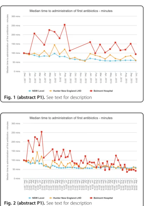
spectrum antimicrobial therapy within 1 hour of recognition [3]. In 2011, the New South Wales Clinical Excellence Commission (CEC) developed the Sepsis Kills program including the Adult Emergency Sepsis Pathway [4]. This pathway was introduced to Emergency Departments (ED) and auditing of time to first antibi-otics commenced. Belmont Hospital Emergency Department has approximately 25000 presentations per year. In 2012, 163 pa-tients were diagnosed with sepsis. Time to first antibiotics for sepsis patients peaked at 254 minutes (Fig. 1). Discussion of these results highlighted the need to develop education strat-egies in order to reduce time to first administration of antibiotics.
Materials and methods
Audits of the CEC Sepsis database [5] over 4 years included a total of 769 patients. Collected data entered included: age, triage time and category, clinical observations, time and amount of intravenous fluid, time to first antibiotics, and diagnosis. The ta-bles provided reflect median time to antibiotics. In 2012 a Clinical Nurse Specialist from the ED was designated Sepsis Lead. Over 4 years, the Sepsis Lead worked collaboratively with clinical staff to develop and implement several strategies to decrease time to first antibiotics
Results
A multimodal approach strategy was adopted, which included: regu-lar audits; targeted education programs for triage nurses and nursing team leaders; the introduction of Sepsis September - a month dedi-cated to sepsis awareness and education; and the Sepsis Road show. These interventions were well received by Emergency staff. As a dir-ect result time to first antibiotics was reduced to a median of 41 mi-nutes (Fig.2).
Conclusions
The collaborative effort between the Sepsis Lead and clinical staff has produced a significant reduction in time to first antibiotics from 254 minutes to 41 minutes at last audit. The success achieved at Bel-mont Hospital due to this multimodal approach strategy has the po-tential to be translated globally. It also serves to highlight the importance of the Emergency Nurse in early recognition and initi-ation of treatment for sepsis.
References
1. Gigliotti E, Steele J, Cassidy D, Bell-Gordon CR: The development and implementation of a nurse practitioner sepsis screening team: Impact on transfer mortality. Journal of Nursing Education and Practice 2014, 4(6):77–83
2. Clinical Excellence Commission: Sepsis Kills Program [http://www.
cec.health.nsw.gov.au/programs/sepsis]
3. Dellinger RP, Levy MM, Rhodes A, Annane D, Gerlach H, Opal SM,
Sevransky J, Sprung CL, Douglas IS, Jaeschke J, et al.: Surviving Sepsis Campaign: International Guidelines for Management of Severe Sepsis and Septic Shock, 2012. Intensive Care Med 2013, 39:165–228
4. Clinical Excellence Commission: Sepsis Tools [http://www.cec.health.
nsw.gov.au/programs/sepsis/sepsis-tools#Pathways]
5. Clinical Excellence Commission: Sepsis Database [http://www.cec.
health.nsw.gov.au/]
Fig. 1 (abstract P1).See text for description
Fig. 2 (abstract P1).See text for description
[image:1.595.304.540.352.689.2]P2
The search for genetic markers in the development of acute respiratory failure
Tamara V Smelaya1,2, Artem N Kuzovlev2, Lubov E Salnikova2,3
1
Main Military Clinical Hospital of Internal Troops of Russia, Balashikha, Moscow region, Russia;2V. A. Negovsky Research Institute of General
Reanimatology, Russian Academy of Sciences, Moscow, Russia;3
N.I. Vavilov Institute of General Genetics, Russian Academy of Sciences, Moscow, Russia
Correspondence:Artem N Kuzovlev ([email protected])
Critical Care2016,20(Suppl 3):P2
Background
Despite advances in diagnostic methods and antibiotic therapy, community-acquired pneumonia (CAP) is still a major cause of mor-bidity and mortality worldwide. Risk of CAP has been attributed to pathogen virulence, host susceptibility and epidemiologic factors. A significant number of patients with CAP develop severe complica-tions, such as sepsis, acute respiratory distress syndrome (ARDS), multiple organ dysfunction syndrome (MODS) and less fatal condi-tions (pleuritis, empyema) and syndromes (acute respiratory failure (ARF)). The variable clinical presentation of CAP suggests a genetic predisposition.
Materials and methods
This study was conducted to establish the possible contribution of functional gene polymorphisms in the oxidative stress related genes to the development of community-acquired pneumonia (CAP) complications. CAP subjects (n = 350) were genotyped for 16 polymorphic variants in the genes of xenobiotics detoxification CYP1A1, AhR, GSTM1, GSTT1, ABCB1, redox-status SOD2, CAT, GCLC, and vascular homeostasis ACE, AGT, AGTR1, NOS3, MTHFR, VEGFa.
Results
The multilocus model which included six or more risk alleles in the CYP1A1, GCLC, AGT and AGTR1 genes was associated with pleuritis, empyema, acute respiratory distress syndrome (ARDS), all pulmonary complications together and acute respiratory fail-ure. Genetically mediated correlation between clinical conditions in CAP patients is shown in Table 1. In silico analysis with Set Distiller mode identified N-acetylcysteine (P = 1.08E-08) and oxy-gen (P = 1.92E-06) as the best descriptors for the considered gene set. Acute infections of the airways are associated with oxidative stress, which enhances viscosity of bronchial mucus, reduces the mucociliary clearance rate and expedites lung dis-ease aggravation and progression. N-acetylcysteine is a well-known mucolytic and antioxidant drug, an indirect precursor of glutathione.
Conclusions
The results of the study indicate that pneumonia aggravation up to destructive intrapulmonary complications and ARDS is mediated by polymorphisms in oxidative stress-related genes.
P3
A retrospective study evaluating the efficacy of identification and management of sepsis at a Western Cape Province district level hospital internal medicine department, in comparison to the guidelines stipulated in the Surviving Sepsis Campaign 2012
Raisa Bhikoo
Department of Internal Medicine, Tygerberg Hospital, Cape Town, South Africa
Correspondence:Raisa Bhikoo ([email protected])
Critical Care2016,20(Suppl 3):P3
Background
Currently there is little data on identification, management and out-comes of patients with sepsis in developing countries. In low income countries major concerns regarding accessibility to healthcare, limita-tions due to costs, lack of resources and delayed presentalimita-tions of patients with sepsis make implementing protocols, based on de-veloped countries patients profile difficult. Thus in Sub-Saharan Africa there has been a widespread shift towards protocol devel-opment that is cost effective and specific for the epidemiologic and ecologic data.
Materials and methods
The aim of our study is to assess the efficacy of clinicians at a district level hospital in the Western Cape at identifying and managing sep-sis. Furthermore, we will assess the outcome of patients in terms of in-hospital mortality and length of hospital stay given the above management. A retrospective study design was applied when analyz-ing data from the routine burden of disease audit done on a 3 monthly basis at Karl Bremer Hospital.
Results
The total sample size obtained was 70 patients. A total of 18/70 (26 %) patients had an initial triage blood pressure indicative of sepsis induced hypotension however only 1/18 (5.5 %) of these patients received an initial crystalloid fluid bolus of 30 ml/kg. The median time for antibiotic administration in septic shock was 4.65 hours. Further a positive delay in antibiotic administra-tion (p value = 0.0039) was demonstrated. A total of 7/70 pa-tients received no antibiotics in the first 24 hours of hospital admission. Of these 7, 3 of these patients could be classified as septic shock at presentation. All 3 patients died within 36 hours of hospital admission. The data showed 8/12 (66 %) of patients with septic shock received inappropriate amounts of fluids. The in-hospital mortality for sepsis was found to be 4/24 (17 %), for severe sepsis 11/34 (32 %) and a staggering 9/12(75 %) for sep-tic shock. A positive association between in-hospital mortality and the following was found:
Time to first dose antibiotic administration (OR = 1.07, P value = 0.027, 95%CI = 1.008-1.14). For every 1-hour delay in antibiotic ad-ministration the chance of death increased by 7 %.
Source appropriate antibiotics (OR = 0.17, P value = 0.005, 95 % CI = 0.048-0.59). The chance of death amongst patients that received source appropriate antibiotics is 83 % less than those who did not.
Early appropriate intravenous fluid administration (OR = 0.33, P value = 0.040,95 % CI = 0.11-0.95). Appropriate intravenous fluids was associated with a 67 % reduction in in-hospital mortality. Conclusion
The outcomes of the study concluded that there is room for improve-ment by our clinicians with regards to appropriate identification and management of patients with sepsis. Flawed management has inevit-ably shown to have an impact on in-hospital mortality. Simple, cost ef-fective measures that can implemented regardless of resources include early appropriate antibiotics and early aggressive fluid therapy; with the potential for an inordinate impact on mortality.
Acknowledgements
Dr Sarah Versfeld: MBChb (Stell). Medical officer in the Department of Internal Medicine, Karl Bremer Hospital.
Dr MMDe V Basson: MBChb (Stell), Hons BSc in Epidemiology (Stell), MMed in Internal Medicine (Stell).
Table 1 (abstract P2).Correlation between clinical conditions in stratified analysis based on the cumulative gene risk score
Number of the risk alleles in the genes,
0-5 risk alleles 6-9 risk alleles
CYP1A1(rs2606345 - T, rs4646903 - T,
rs1048943 - A),
GCLC(rs17883901 - T),
AGT(rs699 - C),
AGTR1(rs5186 - C)
PC - ARF r = 0.43 (P= 2.4E-07) r = 0.57 (P= 2.1E-18)
PSI - ARF r = 0.60 (P = 1.6E-06) r = 0.67 (P = 3.0E-16)
[image:2.595.57.288.585.707.2]Head of Internal Medicine and senior consultant at Karl Bremer Hospital. [email protected]
Dr Almero H Oosthuizen: MBChb (Stell) (cum laude), Dip.PEC (SA), MMed EM (UCT) FCEM (SA). Senior consultant in the Department of Emergency Medicine, Karl Bremer Hospital.
P4
Validation of the Sepsis Severity Score compared with standard severity scores in predicting hospital mortality in severe sepsis and septic shock patients
Bodin Khwannimit, Rungsun Bhurayanontachai, Veerapong Vattanavanit Division of Critical Care Medicine, Department of Internal Medicine, Faculty of Medicine, Prince of Songkla University, Hat Yai, Songkhla, 90110 Thailand
Correspondence:Bodin Khwannimit ([email protected])
Critical Care2016,20(Suppl 3):P4
Background
Recently, the Sepsis Severity Score (SSS) [1] was constructed to esti-mate the probability of hospital mortality in severe sepsis and septic shock patients. The aim of this study was to validate and compare the performance of the SSS with the Acute Physiology and Chronic Health Evaluation (APACHE) II-IV [2], Simplified Acute Physiology Score (SAPS) II and SAPS 3 scores in predicting hospital outcome in sepsis patients.
Materials and methods
A retroprospective analysis was conducted of prospectively collected data from all consecutive sepsis patients admitted to the medical in-tensive care unit of a tertiary university teaching hospital. The per-formance of the severity scores was evaluated by discrimination, calibration and overall performance.
Results
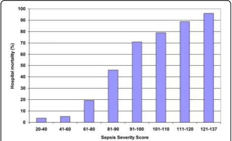
From a total of 931 patients, 802 patients (87.8 %) who had sep-tic shock were enrolled. The hospital mortality rate was 43.9 % and the mean age was 59.3 ± 21.5 years. The most common sources of ICU admission were from the emergency room (47.5 %), general wards (43.9 %), and transfers from other hospi-tals (8.6 %). Mechanical ventilation was used in 808 patients (88.5 %). Community-acquired infections accounted for 70.2 %. The most common sources of infection were respiratory (49.1 %), gastrointestinal (14.8 %) and primary bloodstream infections (9.9 %). The median SSS was 80 (range 20–137). Hospital mortal-ity by the SSS is demonstrated in Fig.3.
The standard mortality ratios of observed mortality to predicted mor-tality were between 0.81 and 1.1. The SSS presented good discrimin-ation with an area under the receiver operating characteristic curve (AUC) of 0.892. The performances of each severity scoring system are shown in Table2.
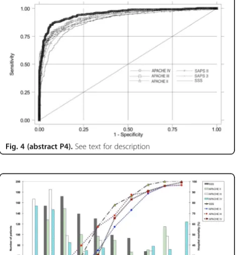
The discriminative performance of the SSS was not statistically significant from the APACHE II (P = 0.07), SAPS II (P = 0.06) or SAPS 3 (P = 0.11). However, the APACHE IV score showed the best discrimination with an AUC of 0.948 and the overall per-formance by a Brier score of 0.096. The AUC of the APACHE IV score was statistically greater than the SSS (P < 0.0001), APACHE II (P < 0.0001), APACHE III (P = 0.0002), SAPS II (P < 0.0001) and SAPS 3 (P < 0.0001) (Fig. 4). The calibration of all scores was poor (Hosmer-Lemeshow goodness-of-fit H test <0.05). The cali-bration curves for the SSS and APACHE scores are presented in Fig.5.
Conclusions
The SSS provided good discrimination as well as the APACHE II, SAPS II and SAPS 3 scores. However, the APACHE IV score had the best dis-crimination and overall performance in our severe sepsis and septic shock patients. The SSS needs to be adapted and modified with new parameters to improve the performance.
References
1. Osborn TM, Phillips G, Lemeshow S, Townsend S, Schorr CA, Levy
MM, et al. Sepsis severity score: an internationally derived scoring system from the surviving sepsis campaign database. Crit Care Med. 2014:1969–1976.
2. Zimmerman JE, Kramer AA, McNair DS, Malila FM. Acute Physiology
and Chronic Health Evaluation (APACHE) IV: hospital mortality assessment for today's critically ill patients. Crit Care Med. 2006:1297–1310.
Fig. 3 (abstract P4).See text for description
Table 2 (abstract P4).Performance of the SSS, APACHE II, III, IV, SAPS II and SAPS 3 scores
Score Predicted AUC SMR H C Brier
score (mean ±
SD)
mortality (95 % CI) (95 % CI)
SSS 82.7 ± 22.9 39.9 ± 20.8 0.892 1.09 95.3* 36.3* 0.183
(0.871-0.913)
(0.99-1.21)
APACHE II 22.8 ± 9.7 47.8 ± 27.1 0.913 0.92 67.9* 10.8*** 0.125
(0.895-0.931)
(0.83-1.01)
APACHE III 88.2 ± 40.1 39.7 ± 30 0.931 1.1 43.6* 11.5*** 0.120
(0.914-0.948)
(1.00-1.22)
APACHE IV - 45 ± 32 0.948 0.97 68.1* 6.2*** 0.096
(0.934-0.962)
(0.88-1.07)
SAPS II 54.2 ± 22.8 51 ± 32.8 0.913 0.86 57.8* 22.3** 0.124
(0.894-0.932)
(0.78-0.95)
SAPS 3 71.6 ± 19.3 54 ± 28.1 0.909 0.81 114.2* 49.2* 0.139
(0.889-0.929)
(0.73-0.9)
*P < 0.0001, **P < 0.05, ***P > 0.05
[image:3.595.304.540.191.335.2] [image:3.595.305.539.395.632.2]P5
What did hospital physicians know about Sepsis before sepsis 3.0? A monocentric survey amongst 132 physicians
Emilie Tourteau1, Amel Filali2, Nicolas van Grunderbeeck1,4, Olivier
Nigeon1,3, Hélène Bazus4, Juliette Masse3, Jihad Mallat1, Didier Thevenin1
1Service de Réanimation Polyvalente & USC, Centre Hospitalier de Lens,
Lens, France;2Service de Maladies Infectieuses, CHRU Lille, Lille, France;
3Service d'Accueil des Urgences, Centre Hospitalier de Lens, Lens,
France;4Service de Maladies Infectieuses, Centre Hospitalier de Lens, Lens, France
Correspondence:Nicolas van Grunderbeeck ([email protected])
Critical Care2016,20(Suppl 3):P5
Background
Sepsis in a public health problem and septic patients suffer high mortality[1,2]. Few data are available about physicians' knowledge and capacity to manage sepsis outside the Intensive Care Unit (ICU) [3]. The physicians' knowledge about sepsis could explain discrepancies in management between Emergency Department (ED) and ward septic patients, who suffer a poorer quality of care and a poorer outcome[4]. We aimed to assess knowledge and ability to handle sepsis management through a questionnaire.
Materials and methods
Observational, prospective, monocentric study during 6 months (May to December 2015). A 26 questions sheet was distributed to all physi-cians. Questionnaires were anonymous, included age, experience and specialty. Questions involved definitions, epidemiology, outcome, diagnosis and management of severe sepsis/septic shock. Eight ques-tions out of 25 were core quesques-tions, and a 26th evaluated physicians' self-appreciation of ability. Evaluation included levels of good, wrong answers (WA) or "no knowledge" (NK) with specialties and self-estimated ability. Physicians were compared as fellows or senior, and specialties between "referent" (ED or Anesthesiologists/Intensivists vs
others). Statistical analysis was performed though Chi2, Fisher exact, or Mann–Whitney tests.
Results
A hundred and thirty-two physicians (48.9 %), answered to the ques-tionnaire: 34 ED or ICU/anesthesia physicians (26 %), 82 medical specialists (62 %), 16 surgeons (12 %). Forty-five percent were fel-lows. Median age was 29 years, median experience was 1 year after diploma. Referent physicians expressed higher self-confidence (97 vs 52 %, p < 0.001). Nearly all of them knew about existence of inter-national guidelines (97 %), whereas a third of others did not (68 %, p = 0.001). Surgeons had poorer results and regarded themselves as unable to handle sepsis. Fellows and senior physicians' knowledge were equal (11 vs 10,8 % of wrong answers). Core questions revealed flaws about definitions (22 % WA), sepsis outside the ICU (29 % WA/ NK), possible severity without hypotension (40.5 % WA/NK), import-ance of lactatemia (24 % WA/NK), existence of guidelines (24 % WA/ NK), delay of antibiotic treatment (78 % agreed for up to a 6 h delay). Knowledge about fluid expansion without hypotension was 88,5 %, but 32 % failed about fluid choice.
Conclusions
Our study presents limits: it's monocentric with a small sample, with no differences between intensivists or anesthesiologists, and some questions may have been difficult to interpret. Moreover, it was "medical-limited" and did not evaluate the nurses’teams, a key fac-tor. Nevertheless, it provides arguments to think that management of sepsis could be improved through better knowledge outside the ICU, that could trigger prompt recognition and management, or call to specialized teams.
References
1. Angus DC, van der Poll T. Severe sepsis and septic shock. N Engl J Med. 2013 Nov 21;369(21):2063.
2. Gaieski DF, Edwards JM, Kallan MJ, Carr BG. Benchmarking the incidence and mortality of severe sepsis in the United States. Crit Care Med 2013;41 (5):1167–74.
3. Djurkovic S, Baracaldo JC, Guerra JA, Sartorius J, Haaupt MT. A survey of clinicians addressing the approach to the management of severe sepsis and septic shock in the United States. J Crit Care 2010 Dec; 25(4):658.e1-6.
4. Esteban A, Frutos-Vivar F, Ferguson ND, Peñuelas O, Lorente JA, Gordo F, Honrubia T, Algora A, Bustos A, García G. Sepsis incidence and outcome: contrasting the intensive care unit with the hospital ward. Crit Care Med. 2007 May;35(5):1284–9.
P6
CD14 of human polymorphonuclear leukocytes in endotoxin priming for respiratory burst
Isabella Prokhorenko1, Dmitry Kabanov1, Svetlana Zubova1, Sergey
Grachev1,2
1FSBSI Institute of Basic Biological Problems RAS, Pushchino, Moscow
region, Russia;2SBEI HPE I.M. Sechenov’s First Moscow State Medical University of Russian’s Ministry of Healthcare, Moscow, Russia Correspondence:Isabella Prokhorenko ([email protected])
Critical Care2016,20(Suppl 3):P6
Background
The induction of inflammatory responses by lipopolysaccharides (LPSs, endotoxins) is achieved by the coordinate and sequential action of four principal LPS-binding proteins: LPS-binding protein (LBP), CD14, Toll-like receptor 4 (TLR4) and Myeloid differentiation protein –MD-2 [1]. CD14, the cell surface receptor of monocytes and neutrophils, is required for LPS activation of phospholipases, protein tyrosine kinases, protein kinases A and C as well as MAP kinases [2]. TLR4 independently of CD14 cannot mobilize all of the adapter proteins that it requires for full signaling activity [3]. CD14 functional multiplicity provides the reason for their implica-tion in target therapy of inflammatory response to LPS by devel-opment of blocking monoclonal antibodies (mAbs) specific to human CD14 [4] or TLR4 [5]. In the present study we have examined CD14 involvement in endotoxin priming of human Fig. 4 (abstract P4).See text for description
[image:4.595.57.292.94.348.2]polymorphonuclear leukocytes (PML) for respiratory burst trig-gered by N-formyl-methionyl-leucyl-phenylalanine (fMLP). Materials and methods
Human PML were isolated from heparinized blood of healthy volun-teers by standard procedure and incubated with or without anti-CD14 mAbs UCHM-1 or isotype-matched IgG2a for 30 min before priming by LPS. Control cells and cells that had been preexposed to mAbs or IgG2a were placed into chemiluminometer chambers con-taining solution for luminol-enhanced chemiluminescence (CL) meas-urement supplemented with autologous serum (2 %) and 0.01 mM CaCl2. PML priming was achieved by addition of 100 ng/ml S- or Re-LPS from Escherichia coli O55:B5 or JM103 followed by continuous gentle shaking for 30 min at 37 °C. CL had been triggered by adding of 1μM fMLP and the light emission was recorded continuously for 20 min. Statistical significance was determined by applying the Wilcoxon’s signed-rank date analysis. Differences were considered to be significant when p < 0.05.
Results
LPS is known to be engaged by CD14 and then transferred to TLR4 [2, 6, 7]. To assess CD14 involvement in endotoxin priming of human PML for fMLP-triggered respiratory burst we used the full anti-CD14 mAbs containing Fc-regions. We have therefore examined the effect of anti-CD14 mAbs as well as IgG2a on fMLP-triggered respiratory burst of control LPS-unprimed PML. As described in Fig. 6aincubation of PML with anti-CD14 mAbs decreased the level of fMLP-triggered CL caused by reactive oxygen species (ROS) production (p < 0.05). The same pattern was recorded when isotype-matched IgG2a had been employed (p < 0.05) (Fig.6a). It is not yet known whether this inhibitory effect of the IgG2a’s Fc-regions on fMLP-triggered ROS generation will be still maintained during PML priming by endotoxins. When PML had been exposed to anti-CD14 mAbs as well as to IgG2a and then primed by Re-LPS near the same suppression of fMLP-triggered CL was detected (p < 0.05) (Fig. 6b). Compared to the control cells (75.29 ± 23.4 RU) the average value of fMLP-triggered CL in Re-LPS primed PML was increased up to 161.77 ± 49.4 RU (p < 0.05). However, after exposition of PML to anti-CD14 mAbs or IgG2a followed by Re-LPS priming the fMLP-triggered CL was reduced up to 112.55 ± 34.3 RU (by 30 %) or up to 126.90 ± 17.6 RU (by 22 %), respect-ively (Fig.6b). Since“quenching”effects of Fc-regions of full anti-CD14 mAbs as well as isotype-matched IgG2a on fMLP-triggered respiratory burst have been almost identical it is not possible at the moment to make decision what is the real impact of F(ab')2-regions of used anti-CD14 mAbs in PML priming by LPS. Some indication on the impact of F(ab')2-regions in this process may came from the comparison of anti-CD14 mAbs effect on PML priming by different LPS-glycoforms, namely S- or Re-LPS. As fol-lows from Fig.7a and bthe most pronounced inhibitory effect of anti-CD14 mAbs on PML priming by S-LPS from E. coli has been achieved (by 40 %). Especially role of CD14 in delivery of S-LPS to TLR4 is well described [8].
Conclusions
LPS is known to be engaged by CD14 and then transferred to TLR4 [2, 6, 7]. To assess CD14 involvement in endotoxin priming of human PML for fMLP-triggered respiratory burst we used the full anti-CD14 mAbs containing Fc-regions. We have therefore examined the effect of anti-CD14 mAbs as well as IgG2a on fMLP-triggered respiratory burst of control LPS-unprimed PML. As described in Fig.6a incuba-tion of PML with anti-CD14 mAbs decreased the level of fMLP-triggered CL caused by reactive oxygen species (ROS) production (p < 0.05). The same pattern was recorded when isotype-matched IgG2a had been employed (p < 0.05) (Fig.6a). It is not yet known whether this inhibitory effect of the IgG2a’s Fc-regions on fMLP-triggered ROS generation will be still maintained during PML priming by endo-toxins. When PML had been exposed to anti-CD14 mAbs as well as to IgG2a and then primed by Re-LPS near the same suppression of fMLP-triggered CL was detected (p < 0.05) (Fig.6b). Compared to the control cells (75.29 ± 23.4 RU) the average value of fMLP-triggered CL in Re-LPS primed PML was increased up to 161.77 ± 49.4 RU (p < 0.05). However, after exposition of PML to anti-CD14 mAbs or IgG2a followed by Re-LPS priming the fMLP-triggered CL was reduced up
to 112.55 ± 34.3 RU (by 30 %) or up to 126.90 ± 17.6 RU (by 22 %), re-spectively (Fig. 6b). Since “quenching” effects of Fc-regions of full anti-CD14 mAbs as well as isotype-matched IgG2a on fMLP-triggered respiratory burst have been almost identical it is not possible at the moment to make decision what is the real impact of F(ab')2-regions of used anti-CD14 mAbs in PML priming by LPS. Some indication on the impact of F(ab')2-regions in this process may came from the comparison of anti-CD14 mAbs effect on PML priming by different LPS-glycoforms, namely S- or Re-LPS. As follows from Fig.7a and b
the most pronounced inhibitory effect of anti-CD14 mAbs on PML priming by S-LPS from E. coli has been achieved (by 40 %). Especially role of CD14 in delivery of S-LPS to TLR4 is well described [8].
References
1. Peri F, Piazza M: Therapeutic targeting of innate immunity with Toll-like receptor 4 (TLR4) antagonist. Biotechnology Advances 2012, 30: 251–260. 2. Yan S, Al-Hertani W, Byers D, Bortolussi R: Lipopolysaccharide-binding
protein- and CD14-dependent activation of mitogen-activated protein kinase p38 by lipopolysaccharide in human neutrophils is associated with priming of respiratory burst. Infect Immun 2002, 70: 4068–4074. 3. Beutler B, Jiang Z, Georgel P, Crozat K, Croker B, et al.: Genetic analysis of
host resistance: Toll-like receptor signaling and immunity at Large. Annu Rev Immunol 2006, 24: 353–389.
4. Verbon A, Dekkers P, Tessa ten Hove, Hack C, Pribble J, et al.: IC14,
an anti-CD14 antibody, inhibits endotoxin-mediated symptoms and
inflammatory responses in humans. J Immunol 2001, 166: 3599–3605.
5. Dunn-Siegrist I, Leger O, Daubeuf B, Poitevin Y, Depis F, et al.: Pivotal involvement of Fcγreceptor IIA in the neutralization of lipopolysaccharide signaling via a potent novel anti-TLR4 monoclonal antibody 15C1. J Biol Chem 2007, 282: 34817–34827.
6. Golenbock D, Liu Y, Millham F, Freeman M, Zoeller R: Surface expression of human CD14 in Chinese hamster ovary fibroblasts imparts
macrophage-like responsiveness to bacterial endotoxin. J Biol Chem 1993, 268: 22055–22059.
7. Plociennikowska A, Hromada-Judycka A, Borzecka K, Kwiatkowska K:
Co-operation of TLR4 and raft proteins in LPS-induced pro-inflammatory signaling. Cell Mol Life Sci 2015, 72: 557–581.
8. Triantafilou M, Triantafilou K, Fernandez N: Rough and smooth forms of fluorescein-labelled bacterial endotoxin exhibit CD14/LBP dependent and independent binding that is influenced by endotoxin concentration. Eur J Biochem 2000, 267: 2218–2226.
Fig. 6 (abstract P6).Effect of anti-CD14 mAbs or IgG2a on fMLP-triggered respiratory burst of control unprimed (a) and Re-LPS
primed human PML (b)
Fig. 7 (abstract P6).Comparison of the anti-CD14 mAbs effects on
fMLP-triggered respiratory burst of human PML primed by Re-LPS (a)
[image:5.595.305.540.479.566.2] [image:5.595.305.539.621.698.2]P7 Withdrawn
P8
Clarithromycin exerts protective immunomodulatory action during Klebsiella pneumoniaeB5055 induced pneumonia in BALB/c mice
Vijay Kumar1,2, Sanjay Chhibber1
1Department of Microbiology, Panjab University, Chandigarh, India;2
Department of Paediatrics and Child Health, Mater Research, School of Medicine, University of Queensland, Brisbane, Queensland, Australia Correspondence:Vijay Kumar ([email protected])
Critical Care2016,20(Suppl 3):P8
Background
Bacterial pneumonia is a life threatening condition causing acute lung injury (ALI) leading to development of sepsis depending on the inva-siveness of the bacteria and host immune status. This present study was designed to find out the protective immunomodulatory action of macrolide antibiotic (i.e. clarithromycin) in mouse model of pneumonia induced byK. pneumoniaeB5055.
Materials and Methods
Pneumonia was induced by intranasal instillation ofKlebsiella pneumoniae B5055 (104cfu/ml) by holding the mice in upright position without any an-aesthesia. Experimental animals were divided in two groups (i.e. Group A; control (oral saline), Group B; clarithromycin (30 mg/kg/day/os). At desig-nated period of time (1, 2, 3, 5 and 7 days) mice were eutha-nized by cervical dislocation and lungs were harvested and homogenized in 1 ml of sterile normal saline under sterile condi-tions. The lung homogenate was used for measuring bacterial load, malondialdehyde (MDA), nitric oxide (NO) and myeloperoxi-dase (MPO) and pro-inflammatory (i.e. IL-1α, IL-6, TNF-α) anti-inflammatory (i.e. IL-10) cytokine levels in lungs. Histopathological examination of the lungs was also carried out along with lung al-veolar macrophage function (i.e. macrophage spreading and phagocytosis) assays.
Results
Clarithromycin treatment led to the significant (p < 0.05) reduction in pulmonary neutrophil infiltration along with significant (p < 0.05) decrease in other markers of inflammation (TNF-α, IL-α, IL-6, MDA, NO and MPO) associated with acute tissue injury or inflam-mation. The increase in bacterial clearance in lungs of mice treated with clarithromycin was associated with significant (p < 0.05) increase in macrophage spreading as well as their phago-cytic potential (i.e. phagophago-cytic uptake and intracellular killing) of alveolar macrophages.
Conclusions
Clarithromycin has a great potential as an immunomodulatory anti-biotic duringK. pneumoniae B5055 induced pneumonia associated acute lung inflammation or injury (ALI).
P9
Histone levels determined by an in-house immunoglobulin Y assay are associated to clinical outcomes in sepsis
Jerico R. Santos1, Jesus Emmanuel A. D. Sevillejal2,3, Jose B.
Nevado, Jr2,4
1College of Medicine, University of the Philippines-Manila, Manila,
Philippines;2Institute of Molecular Biology and Biotechnology, National Institutes of Health, University of the Philippines-Manila, Manila, Philippines;3Institute of Human Genetics, National Institutes of Health,
University of the Philippines-Manila, Manila, Philippines;4Department of
Biochemistry and Molecular Biology, College of Medicine, University of the Philippines-Manila, Manila, Philippines
Correspondence:Jerico R. Santos ([email protected])
Critical Care2016,20(Suppl 3):P9
Background
Experimental models have shown that histones are involved in the pro-gression of sepsis and may be candidate prognostic biomarkers. There are limited prospective cohort studies of suspected septic patients aimed at determining the risk associated with increased level of histones to their progression to severe sepsis. To address this, we (1) developed an IgY-based immunoassay that can detect and quantify blood histone levels; (2) tested whether there is a difference in histone levels among septic and fe-brile patients that present in the emergency room; and (3) tested whether there is an association between histone levels in serum and the progres-sion of patient to developing hypotenprogres-sion (sBP < 90 mmHg), azotemia, (creatinine > 2.0 mg/dL (176.8 μmol/L)) or thrombocytopenia (platelet count < 100,000/μL) using the developed IgY-based immunoassay. Materials and methods
An indirect ELISA was developed for detecting human histones that used polyclonal immunoglobulin Y (IgY) extracted from eggs laid by calf histone-challenged chickens as a primary antibody. Chickens were inocu-lated with calf histones and the IgY from eggs were extracted by means of a delipidation, salt precipitation and affinity chromatography. To determine its clinical utility as a prognostic kit, a prospective cohort study of 4 febrile patients and 21 septic patients seeking consult at the emergency room were enrolled based on the inclusion criteria. Higher total histone levels measured by immunoglobulin Y was then determined if it was related to the development of hypotension, azotemia or thrombocytopenia. Results
The limit of detection of this IgY-based immunoassay was determined to be 2036 pg/mL. Comparison of histone levels between 4 febrile pa-tients, and 21 septic patients within 3 days of admission in the hospital showed that septic patients have significantly higher (p = 0.0314) levels of histones as compared to febrile patients (Fig.8).
In this cohort, those that developed hypotension, azotemia or thrombocytopenia during their hospital stay have higher levels of histones upon admission (p = 0.0257) compared to those that did not (Fig.9). Receiver operator curve analysis showed that at 2101 pg/uL (Fig.10), likelihood ratio was 3.545 which had an estimated sensitiv-ity (76.9 %), specificsensitiv-ity (81.81 %), PPV (83.33), and NPV (75 %) (p = 012, Chi square test). At a cutoff of <2081 pg/uL, likelihood ratio was 5.909 of sensitivity (45.45 %) and specificity (92.31 %).
Conclusions
Histone levels may be predictive to the progression of defined clinical parameters related to worse outcomes in septic patients.
Fig. 8 (abstract P9).Histone Levels of Febrile and Septic Patients in
the Emergency Room. Septic patients (Mean = 2106 pg/μL, 95 % CI,
[image:6.595.307.540.479.623.2]P10
Inflammatory changes in renal tissues following Gram-positive pulmonary challenge
Helena M. Linge, Kanta Ochani, Ke Lin, Ji Young Lee, Ping Wang, Manoj Tembhre, Shu Fang Liu, Pravin C. Singhal, Edmund J. Miller
Heart and Lung Research Unit, The Feinstein Institute for Medical Research, Northwell Health, Manhasset, NY USA
Correspondence:Edmund J. Miller ([email protected])
Critical Care2016,20(Suppl 3):P10
Background
Acute lung injury (ALI) is an important cause of morbidity and mortality in critically ill patients. Although the condition has many etiologies, pneumonia and sepsis are leading triggers. Age is a major determinant of clinical outcome, and there are age-dependent alterations in the re-sponses to pulmonary challenge that can trigger kidney inflammation (a precursor of chronic kidney disease) and higher mortality rates.
Staphylococcus aureus infection is a frequent cause of pneumonia, es-pecially in elderly patients. To develop a better understanding of the pathophysiology, and assist in defining future targets, we developed a lung model of staphylococcal pneumonia in rodents. Using this model we have examined inflammatory responses, hemodynamics, and car-diac proteasome activation, neutrophil trafficking in pulmonary inflam-mation, and age-related changes [1–3]. Here we examined renal inflammation following pulmonary challenge.
Materials and methods
C57Bl6 Mice (8–10wks, n = 6-8/group) were challenged intratra-cheally with staphylococcal cell wall components, lipoteichoic acid (LTA), and peptidoglycan (PGN): (0.2 μg + 0.66 μg); (30 mg + 100 mg); (150 mg + 500 mg); or saline alone. Pulse oximetry was performed on awake animals before challenge and immedi-ately prior to euthanasia. At 6, 24 or 72 hrs post challenge, ani-mals were euthanized, bronchoalveolar lavage (BAL) performed, and tissues harvested. Concentrations of plasma and BAL cyto-kines were assessed by ELISA. The left kidney was fixed for histological analysis, and the right kidney was assessed for cyto-kine mRNA by qPCR, cytocyto-kine protein using ELISA, and activity of specific proteasome subunits by ProCISE [4].
Results
BAL from LTA/PGN challenged mice showed significant increases in neutrophils, total protein, IL-6, KC, MCP-1, TNFa and MIF as in previ-ous studies. Additionally, while there was no LTA or PGN detected in the plasma, LTA/PGN challenged mice had increased plasma concen-trations of IL-6, IL-1b, KC and TNFa. In renal tissue, there were time and concentration dependent changes in IL1b, IL-6, TNFa, IP10, and MCP-1 and an increase in proteasome b5 subunit activity.
Conclusions
Even in these young animals pulmonary challenge induced signifi-cant inflammatory changes within the lung, blood and kidney. Not-ably, in the kidney there was increased activity of proteasome b5, a regulator of IκB/NFκB activation and signaling pathways. Our recent study uncovered a time dependent, NF-kB switch that regulates tran-sition from endothelial barrier injury to repair [5]. If we can define changes in the injury-to-repair phase in the kidney, we may reveal specific targets for therapeutic manipulation in a clinically relevant time frame to improve outcome from ALI associated renal involve-ment, particularly in the older individual.
References
1. Linge HM, Lee JY, Ochani K, Koga K, Kohn N, Ojamaa K, Powell SR, Miller EJ: Age influences inflammatory responses, hemodynamics, and cardiac proteasome activation during acute lung injury. Exp Lung Res 2015, 41(4):216–227.
2. Palestro C, Linge HM, Nichols KJ, Ochani K, Bhargava KK, Miller EJ:
Neutrophil Trafficking in Pulmonary Inflammation: Monitoring Migration and Blockade with 111In-Labeled Leukocytes Journal of Pulmonary & Respiratory Medicine 2015, 5:289–297.
3. Lee JY, Linge HM, Ochani K, Lin K, Miller EJ: N-Ethylmaleimide
Sensi-tive Factor (NSF) Inhibition Prevents Vascular Instability following Gram-Positive Pulmonary Challenge. PLoS One 2016, 11(6):e0157837.
4. Kirk CJ, Powell SR, Miller EJ: Assessment of cytokine-modulated
proteasome activity. Methods Mol Biol 2014, 1172:147–162.
5. Liu G, Ye X, Miller EJ, Liu SF: NF-kappaB-to-AP-1 switch: a mechanism
regulating transition from endothelial barrier injury to repair in endotoxemic mice. Sci Rep 2014, 4:5543.
P11
Cathelicidin protects against intestinal barrier dysfunction in polymicrobial sepsis
Jeffery HO1, Xiaodong Liu1, Thomas Kwong2, Lin Zhang1, Hung Chan1, Sunny H Wong2, Gordon Choi1, Tony Gin1, Matthew TV Chan1, William
KK Wu1
1Department of Anaesthesia and Intensive Care, Prince of Wales
Hospital, The Chinese University of Hong Kong, Shatin, Hong Kong;
2Department of Medicine and Therapeutics, Prince of Wales Hospital,
The Chinese University of Hong Kong, Shatin, Hong Kong Correspondence:Jeffery HO ([email protected])
Critical Care2016,20(Suppl 3):P11
Fig. 9 (abstract P9).Patients that progressed into having hypotension, azotemia or thrombocytopenia have significantly higher histone levels than those that did not. In this cohort, those that progressed to severe sepsis (Mean = 2110, 95 % CI 2095–2125) have significantly higher levels of histones in their blood (P < 0.05, two-tailed t-test), as compared to those who did not progress (Mean = 2084, 95 % CI 2073–2096)
[image:7.595.57.292.88.227.2] [image:7.595.57.291.301.479.2]Background
Accumulating evidence suggests that the intestinal barrier func-tion is impaired during systemic inflammafunc-tion as in sepsis. Animal models of sepsis revealed several ileal pathological changes. These include epithelial apoptosis, disruption of tight junctions and increased intestinal permeability [1,2]. The impaired gut bar-rier function may increase the risk of bacterial translocation from the gut lumen to the bloodstream, aggravating systemic inflam-mation. The molecular mechanism associated with this phenotype remains largely unknown. Cathelicidin represents one of the most important classes of antimicrobial peptides in mammals. In addition to bactericidal property, this peptide inhibits endotoxinduced pyroptosis of leukocytes, suppresses the release of in-flammatory mediators and protects endothelial cells from apop-tosis [3–5]. In this study, we aimed to investigate the role of murine cathelicidin-related antimicrobial peptide (mCRAM), a ro-dent antimicrobial peptide analogous to human LL-37, in main-taining gut barrier function in sepsis.
Materials and methods
129X1/SvJ mice that were wild type (cnlp+/+) or deficient for cathelicidin (cnlp−/−) were used. Polymicrobial sepsis was induced by cecal-ligation and puncture (CLP) [6]. Animals were divided into four groups: cathelicidin wild type and knockout mice receiving either CLP or sham surgery. At 24 h, blood and ileal tissues were harvested for total bacterial DNA quantifica-tion, immunoblotting and histological staining. The survival rates and septic severity were recorded every 12 hours until seven days after the surgery. Sepsis morbidity was evaluated by Murine Sepsis Severity (MSS) score [7]. At 20 h after CLP or
sham surgery, the animals were orally fed with 4 kDa
fluorescein-dextran. Three hours later, serum was harvested and fluorescence was measured.
Results
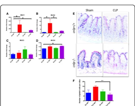
Knocking out cnlp gene has significantly increased septic severity and reduced 7-day survival of 129X1/SvJ mice. Real time PCR target-ing bacterial 16S rDNA revealed that total bacterial DNA load in-creased more than threefold (Fig.11).
Without cathelicidin, the expression of mucus has also reduced considerably, as revealed by Alcian blue periodic acid Schiff reac-tion and RT-PCR targeting MUC1, MUC2, MUC3 and MUC4 genes (Fig. 12). Immunoblotting revealed cleavage of caspase 3 and PARP involving in apoptosis (Fig. 13). Consistently, confocal mi-croscopy using TUNEL staining identified greater number of puncta per villus. Deletion of cnlp gene increased ileal permeabil-ity to 4kD Fluorescein-labelled dextran, accompanied with re-duced expression of tight junction proteins claudin-1 and occludin (Fig.14).
Conclusions
This study demonstrated the critical role of cathelicidin in maintain-ing gut barrier integrity in polymicrobial sepsis. This may partially ex-plain the association between lower LL-37 and higher mortality in septic patients [8].
References
[1] Li Q, Zhang Q, Wang C et al. Disruption of tight junctions during polymicrobial sepsis in vivo. J Pathol. 2008; 218: 210–221.
[2] Wu W, Jiang RL, Wang JC et al. Effect of Shenfu injection on intestinal mucosal barrier in a rat model of sepsis. Am J Emerg Med. 2015; 33: 1237–1243.
[3] Hu Z, Murakami T, Suzuki K et al. Antimicrobial cathelicidin peptide LL-37 inhibits the LPS-ATP-induced pyroptosis of macrophages by dual mechanism. PLoS One. 2014; 9:e85765.
[4] Murakami T, Obata T, Kuwahara-Arai K et al. Human antimicrobial cathelicidin peptide LL-37 suppresses the production and release of septic mediators in D-galactosamine-sensitized endotoxin shock mice. Int Immunol. 2009; 21:905–912.
[5] Suzuki K, Murakami T, Kuwahara-Arai K et al. Human antimicrobial catheli-cidin peptide LL-37 suppresses the LPD-induced apoptosis of endothelial cells. Int Immunol. 2011; 23:185–93.
[6] Rittirsch D, Huber-Lang MS, Flierl MA et al. Immunodesign of experimental sepsis by cecal ligation and puncture. Nat Prot. 2009; 4: 31–36.
[7] Shrum B, Anantha RV, Xu SX et al. A robust scoring system to evaluate sepsis severity in an animal model. BMC Res Notes. 2014; 7: 233. [8] Leaf DE, Croy HE, Abrahams SJ et al. Cathelicidin antimicrobial protein,
vitamin D, and risk of death in critically ill patients. Crit Care 2015; 19:80.
Fig. 11 (abstract P11).Cathelicidin is upregulated after cecal-ligation and puncture induced sepsis in 129X1/SvJ (cnlp+/+) mice from which total RNA and protein were collected from distal ileums over a period of time for (a) real time PCR targeting clnp for mCRAMP. Genetic knockout of cnlp led to (b) reduced survival, (c) higher sepsis severity score and (d) increased bacterial DNAemia upon experimental sepsis. Error bars represents standard error of the mean. P < 0.05 denotes statistical significance, Mann–Whitney U-test
[image:8.595.305.540.186.339.2] [image:8.595.304.539.428.612.2]P12 Withdrawn
P13
Proline-specific peptidases in the plasma of patients with septic shock: their potential as biomarker and associations with established parameters
Gwendolyn Vliegen1, Kaat Kehoe1, Robert Verkerk1, Erik Fransen2, Esther Peters3,4, Anne-Marie Lambeir1, Peter Pickkers3, Philippe G. Jorens5, Ingrid
De Meester1
1Laboratory of Medical Biochemistry, University of Antwerp,
Universiteitsplein 1, 2610 Wilrijk, Belgium;2StatUa Center for Statistics,
University of Antwerp, Prins Boudewijnlaan 43, 2650 Edegem, Belgium;3
Department of Intensive Care Medicine, Radboud university medical
center, 6500HB Nijmegen, The Netherlands;4Department of
Pharmacology and Toxicology, Radboud university medical center,
6500HB Nijmegen, The Netherlands;5Department of Critical Care
Medicine, Antwerp University Hospital and Laboratory of Experimental Medicine and Pediatrics, University of Antwerp, Wilrijkstraat 10, 2650 Edegem, Belgium
Correspondence:Gwendolyn Vliegen ([email protected])
Critical Care2016,20(Suppl 3):P13
Background
To date, there is no standard diagnostic test to identify septic patients, although early identification is necessary to improve their outcome [1]. We sought to identify novel biomarkers by studying the enzymatic activity of the proline-specific peptidases dipeptidyl pep-tidase 4 (DPP4), prolyl carboxypeppep-tidase (PRCP), fibroblast activation proteinα(FAP) and prolyl oligopeptidase (PREP) in the plasma of 32 controls and 40 patients with septic shock on days 1, 3, 5 and 7. Materials and methods
Enzymatic activity measurements were done using in-house developed specific assays [2–4]. On the same days, inflammatory (e.g. TNF-α, IL-6) , hemodynamic (e.g. mean arterial pressure, pulse) and metabolic param-eters (e.g. bilirubin, lactate) were measured as well. The data was used to generate receiver operating characteristic (ROC) curves and to iden-tify associations. A survival analysis (up to 90 days) was also performed. Results
Significant differences between the control group and the patients with septic shock could be found for all four enzymes. PREP and PRCP showed a higher enzymatic activity on day 1 compared to the controls (p≤0.05 and p≤0.01, respectively). FAP and DPP4, on the other hand, were both much lower in the patients group on all studied time points (p≤0.001). PREP, FAP and DPP4 showed to be very good in discriminating cases from controls with area under the curve (AUC) values of 0.91 (CI: 0.85-0.99), 0.96 (CI: 0.92-1) and 0.94 (CI: 0.89-0.85-0.99), respectively. PRCP had a lower predicting value with an AUC of 0.69 (CI: 0.58-0.82). A nominally sig-nificant association was observed between survival and the DPP4 enzym-atic activity at day 1 (p≤0.05), with a higher DPP4 enzymatic activity being associated with an increase in survival. Several interesting positive associations, such as PRCP with lactate, PREP with IL-1 receptor antagonist and PREP with intestinal fatty-acid binding protein, were identified. PRCP and PREP were also associated with the dosage of noradrenalin adminis-tered to maintain an acceptable mean arterial pressure.
Conclusions
DPP4, FAP and PREP showed to be good in discriminating between con-trols and cases and should be further explored as possible candidate bio-markers for sepsis. The associations found also warrant further research. 1. Dellinger RP, Levy MM, Rhodes A, Annane D, Gerlach H, Opal SM, et al. Surviving Sepsis Campaign. Crit. Care Med. 2013;41:580–637. 2. Matheeussen V, Lambeir A-M, Jungraithmayr W, Gomez N, Mc Entee K, Van der Veken P, et al. Method comparison of dipeptidyl peptidase IV activity assays and their application in biological samples containing reversible inhibitors. Clin. Chim. Acta. 2012;413:456–62.
3. Kehoe K, Verkerk R, Sim Y, Waumans Y, Van der Veken P, Lambeir A-M, et al. Validation of a specific prolylcarboxypeptidase activity assay and its suitability for plasma and serum measurements. Anal. Biochem. 2013;443:232–9.
4. Goossens F, De Meester I, Vanhoof G, Scharpé S. A sensitive method for the assay of serum prolyl endopeptidase. Eur. J. Clin. Chem. Clin. Biochem. 1992;30:235–8.
P14 Withdrawn
P15
Dexamethasone prevents epithelial barrier dysfunction in endotoxemic rats
Aline Barbosa Ribeiro, Ana Paula Trevelin Souza, Humberto Giusti, Celso Rodrigues Franci, Rafael Simone Saia
Department of Physiology, Ribeirão Preto Medical School, University of São Paulo, Ribeirão Preto, São Paulo, Brazil
Correspondence:Rafael Simone Saia ([email protected])
Critical Care2016,20(Suppl 3):P15
Background
The systemic inflammation represents a primary injury to the intestinal epithelium structure. In acute phase, the massive production of inflam-matory mediators (cytokines and nitric oxide) contributes to the intestinal permeability, bacterial translocation and tight junction re-arrangement. During the past decades, the clinical administration of glucocorticoids has been widely discussed and the Surviving Sepsis Campaign’s recommendation is the low-dose treatment for those Fig. 13 (abstract P11).Increased apoptosis was detected in
cathelicidin-knockout (cnlp−/−) mice compared to -wild type mice
(cnlp+/+) after CL-induced sepsis as demonstrated by (a)
immuno-blotting for cleaved caspase-3 and poly ADP ribose polymerase (PARP), and (b) TUNEL staining. Quantification was shown, for TUNEL, as (c) number of punta per villus. All specimens were collected at 24 hr after CL or Sham surgery. Error bars denote standard error of the mean. P < 0.05 denotes statistical significance, Kruskal-Wallis test
[image:9.595.57.291.88.233.2] [image:9.595.57.292.331.462.2]patients refractory to vasopressor therapy. Our aim was to investigate the possible role of dexamethasone on lipopolysaccharide (LPS)-in-duced intestinal epithelial barrier dysfunction. Furthermore, we evalu-ated its ability to modulate the inflammatory response and also the expression of proteins of the intestinal tight junctions.
Materials and methods
To conduct these experiments, male rats had their jugular vein cannu-lated for endotoxin administration, one day before the experiment. Rats were pre-treated with dexamethasone (synthetic glucocorticoid; 0.1 or 1 mg/kg, intraperitoneal) before LPS administration (1.5 mg/kg, intra-venous). At 6 h after endotoxemia induction, the intestinal permeability was evaluated by injecting FITC-dextran 4 kDa in the ileum, mesenteric lymph nodes were collected for microbiological analysis and also cyto-kines were quantified in the plasma and intestinal mucosa by ELISA technique. Additionally, the integrity of the tight junctions was deter-mined by transmission electron microscopy, histological analysis, the expression of tight junction proteins (occludin, claudin-1, claudin-2, junctional adhesion molecule-A) as well as their localization.
Results
Our results demonstrated that dexamethasone administration reduces the LPS-induced permeability in the ileum and prevented the bacterial translocation to the mesenteric lymph nodes. The plasma and mucosa concentrations of TNF-α, IL-1β, IL-6, IL-10 and IFN-γwere significantly reduced in dexamethasone-treated rats. Furthermore, treatment with dexamethasone reverted the LPS-induced epithelial barrier dysfunction, increasing the expression of occludin and claudin-1, reducing the claudin-2 cleavage and also the localization of these proteins at the ap-ical portion. Moreover, the histologap-ical damages and the morphology of the tight junctions were preserved by the dexamethasone adminis-tration, therefore reducing their opening induced by endotoxemia. Conclusions
Together these results suggest a protective role for dexamethasone preventing the intestinal barrier dysfunction induced by systemic in-flammation, possibly modulating the inflammatory response. Further-more, these experimental findings reinforce the Surviving Sepsis Campaign’s recommendation for the administration of low gluco-corticoid doses in septic shock patients.
Acknowledgements
CNPq and FAPESP
P16
Cytokine response in adult severe sepsis patients with features of macrophage activation syndrome
Renee R. Anderko1, Vanessa M. Jackson1, Octavia M. Peck Palmer1,2,
Derek C. Angus1, John A. Kellum1,3, Joseph A. Carcillo1,4
1Department of Critical Care Medicine, University of Pittsburgh School of
Medicine, Pittsburgh, PA, USA;2University of Pittsburgh Medical Center, Pittsburgh, PA, USA; Department of Pathology, University of Pittsburgh School of Medicine, Pittsburgh, PA, USA;3Center for Critical Care Nephrology, University of Pittsburgh, Pittsburgh, PA, USA;4Department
of Pediatrics, University of Pittsburgh School of Medicine, Pittsburgh, PA, USA; Children’s Hospital of Pittsburgh of University of Pittsburgh Medical Center, Pittsburgh, PA, USA
Correspondence:Renee R. Anderko ([email protected])
Critical Care2016,20(Suppl 3):P16
Background
Shakoory and colleagues performed a post hoc analysis of a ran-domized clinical trial of anakinra, an interleukin-1 receptor antag-onist [1], and reported that severe sepsis patients with features of macrophage activation syndrome (MAS), defined by the com-bined presence of hepatobiliary dysfunction and disseminated intravascular coagulation, represented 5.6 % of the severe sepsis population. Additionally, anakinra, in comparison with placebo, reduced 28-day mortality (34.6 % vs 64.7 %; p < 0.05) in a subset of patients with features of MAS [1]. Based on these findings, we hypothesized that the Protocolized Care for Early Septic Shock (ProCESS) trial [2] would contain a similar proportion of septic patients with features of MAS and a similar mortality rate. We further hypothesized that MAS patients would have elevated
circulating biomarkers related to macrophage activation, including ferritin, interleukin-18 (IL-18), interleukin-1β(IL-1β), and
interferon-γ (IFN-γ).
Materials and methods
We selected all patients with features of MAS and age-matched con-trols without features of MAS from the ProCESS trial. Concentrations of circulating ferritin, IL-18, IL-1β, and IFN-γ were measured in plasma samples collected on day 1, and mortality was assessed at hospital dis-charge and at 90 days. The MAS patients were further stratified accord-ing to their circulataccord-ing ferritin levels, and a threshold of 1200 ng/ml was established based on the upper limit of the 95 % confidence inter-val for the mean ferritin concentration in MAS survivors.
Results
Of the 1341 ProCESS subjects, 82 exhibited features of MAS (6.1 %). In-hospital and 90-day mortality rates were higher in the MAS patients than the non-MAS patients (42.7 % vs 9.8 % and 61.0 % vs 28.1 %; p < 0.05). As outlined in Table3, MAS subjects showed higher circulating ferritin, IL-18, and IL-1βconcentrations (p < 0.05). Approximately 27 % of septic patients with features of MAS had ferritin concentrations greater than 1200 ng/ml. Interestingly, these patients had higher in-hospital mortality (63.6 %) and 90-day mortality (72.7 %), as well as higher IL-18, IL-1β, and IFN-γconcentrations compared to the septic pa-tients without MAS (p < 0.05) (Table4).
Conclusions
Post hoc analysis of ProCESS patients illustrated that features of MAS are relatively uncommon but lethal in severe sepsis. Septic patients with features of MAS and ferritin levels greater than 1200 ng/ml had the highest mortality and increased IL-18, IL-1β, and IFN-γ concentra-tions compared to non-MAS septic patients, making this an attractive subset to target in future anti-inflammatory agent trials.
References
1. Shakoory B, Carcillo JA, Chatham WW, Amdur RL, Zhao H, Dinarello CA, Cron RQ, Opal SM: Interleukin-1 receptor blockade is associated with reduced mor-tality in sepsis patients with features of macrophage activation syndrome: re-analysis of a prior phase III trial. Critical Care Medicine 2016, 44:275–281.
2. ProCESS Investigators, Yealy DM, Kellum JA, Huang DT, Barnato AE,
Weissfeld LA, Pike F, Terndrup T, Wang HE, Hou PC, et al.: A randomized trial of protocol-based care for early septic shock. The New England Journal of Medicine 2014, 370:1683–1693.
Table 3 (abstract P16).Clinical characteristics of septic patients with and without features of MAS
Biochemical Profile
Septic Patients with MAS (n = 82)
Septic Patients without MAS (n = 82)
P value*
Ferritin, ng/ml, 0 hr mean (sd, median, range) [95 % CI]
1834.49 (3316.41, 482.00, 14.30-17,502.00) [1095.30, 2573.70]
444.65 (846.45, 213.90, 5.70-6536.00) [257.17, 632.12]
<0.0001
IL-18, pg/ml, 0 hr mean (sd, median, range) [95 % CI]
1582.68 (2273.23, 847.04, 123.61-15,214.01) [1079.20, 2086.20]
558.13 (499.23, 408.62, 159.84-3107.64) [446.86, 669.41]
<0.0001
IFN-γ, pg/ml, 0 hr mean (sd, median, range) [95 % CI]
115.29 (534.76, 12.28, 0.10-4704.60) [−3.15, 233.73]
27.62 (71.38, 0.10, 0.10-590.33) [11.71, 43.53]
0.1413
IL-1β, pg/ml, 0 hr mean (sd, median, range) [95 % CI]
69.21 (176.85, 8.92, 0.08-928.70) [26.99, 111.42]
12.96 (29.25, 4.74, 0.08-183.43) [5.53, 20.38]
0.0001
Mortality
In-hospital mortality, %
42.7 9.8 <0.0001
90-day mortality, % 61.0 28.1 <0.0001
[image:10.595.305.539.484.698.2]P17
Evaluation of a novel molecular host response assay to diagnose infection in patients after high-risk gastro-intestinal surgery: a pilot study
D.M. Verboom1,2, M.E. Koster-Brouwer1,2, K. van de Groep1,2, J.F.
Frencken1,2, B. Scicluna3, S.S. Gisbertz4, M.I. van Berge Henegouwen4, J.P. Ruurda5, R. van Hillegersberg5, T. van der Poll3,6,7, M.J.M. Bonten1,8,
O.L. Cremer2
1Julius Center for health sciences and primary care, University Medical
Center Utrecht, Utrecht, the Netherlands;2Department of Intensive Care, University Medical Center Utrecht, Utrecht, the Netherlands;3Center for
Experimental and Molecular Medicine, Academic Medical Center,
University of Amsterdam, Amsterdam, the Netherlands;4Department of
Surgery, Academic Medical Center, Amsterdam, the Netherlands;
5Department of Surgery, University Medical Center Utrecht, Utrecht, the
Netherlands.;6Center for Infection and Immunity, Academic Medical Center, Amsterdam, the Netherlands;7Division of Infectious Diseases,
Academic Medical Center, Amsterdam, the Netherlands|;8Department of
Medical Microbiology, University Medical Center Utrecht, Utrecht, the Netherlands
Correspondence:D.M. Verboom ([email protected])
Critical Care2016,20(Suppl 3):P17
Conflicts of interest
The authors declare that they do not have a conflict of interest related to the subject matter.
Background
Patients after esophagectomy are prone to develop sepsis due to pneumonia, anastomotic leakage, or wound infection. A new diag-nostic test (SeptiCyte LAB) that measures the expression of four
blood gene biomarkers has diagnostic utility in distinguishing sepsis from SIRS [1]. However, test performance in the immediate postoper-ative setting has not been investigated.
Materials and methods
We studied patients undergoing esophagectomy in two Dutch uni-versity hospitals from 2011 to 2013 who had developed a complica-tion requiring ICU (re)admission within 30 days. Blood was collected postoperatively and when a complication occurred. After RNA extrac-tion, samples were analyzed on an Applied Biosystems® 7500 fast Dx Real-Time PCR instrument. SeptiCyte LAB results (a score, range 0– 10) were categorized into four probability bands according to the manufacturer's specification.
Results
Among 384 patients 113 (29 %) subjects had developed at least one complication and were thus eligible for study inclusion; paired sam-ples (i.e., both a direct postoperative and follow-up sample) were available for 71 (63 %) of these. Sample preparation or processing is-sues resulted in exclusion of 4 pairs, leaving 67 (59 %) patients for analysis (Table5).
Five (7 %) patients had active infection during postoperative sampling, and 55 (82 %) on the day of their first complication. SeptiCyte results were highly variable immediately after surgery and at follow-up (Fig. 15), but scores increased significantly as complications evolved (median 2.7 (IQR 1.6-3.4) versus 7.3 (5.7-8.7); p < 0.001). Importantly, samples taken during infection yielded higher scores then specimens taken at onset of a non-infectious complication (median 7.3 (5.7-8.7) versus 5.2 (4–6.3); p < 0.001).
Considering all 134 samples separately, 45 were classified as 'sep-sis not likely' based on a SeptiCyte score <3.1; among these, we observed 2 cases of confirmed infection (false negative rate 4 %). Likewise, among 89 events with a score >3.1, infection was ruled-out or never clinically suspected in 33 cases (false positive rate 37 %). The incorporation of SeptiCyte LAB results markedly im-proved the performance of SIRS criteria in classifying confirmed infections (c-statistic 0.86 (95%CI 0.80-0.93) versus 0.97 (95%CI 0.94-0.99); p < 0.001). However, a final diagnosis of infection could not be made with certainty on 21 occasions (including two post-operative samples), which precluded formal estimation of test characteristics.
Conclusion
SeptiCyte LAB is a novel sepsis biomarker which may have clical utility in monitoring patients at high risk for postoperative in-fections, but its diagnostic value in this setting has to be further evaluated.
Acknowledgements
We thank ImmunExpress for kindly providing lab kits and technical assistance.
References
1. McHugh L, Seldon TA, Brandon RA, Kirk JT, Rapisarda A, Sutherland
AJ, Presneill JJ, Venter DJ, Lipman J, Thomas MR, et al. A Molecular Host Response Assay to Discriminate Between Sepsis and Infection-Negative Systemic Inflammation in Critically Ill Patients: Discovery and Validation in Independent Cohorts. PLoS Med, 2015.12(12): p. e1001916.
2. Klein Klouwenberg PM, Ong DS, Bos LD, de Beer FM, van Hooijdonk
RT, Huson MA, Straat M, van Vught LA, Wieske L, Horn J, et al., Interobserver agreement of Centers for Disease Control and Prevention criteria for classifying infections in critically ill patients. Crit Care Med, 2013.41(10): p. 2373–2378.
Table 4 (abstract P16).Clinical characteristics of septic patients with features of MAS and ferritin concentrations >1200 ng/ml vs septic patients without features of MAS
Biochemical Profile
Septic Patients without MAS (n = 82)
Septic Patients with MAS and Ferritin >1200 (n = 22)
P value*
Ferritin, ng/ml, 0 hr mean (sd, median, range) [95 % CI]
444.65 (846.45, 213.90, 5.70-6536.00) [257.17, 632.12]
5584.60 (4561.20, 3535.00, 1345.30-17,502.00) [3561.90, 7607.20]
<0.0001
IL-18, pg/ml, 0 hr mean (sd, median, range) [95 % CI]
558.13 (499.23, 408.62, 159.84-3107.64) [446.86, 669.41]
2625.30 (3513.40, 1252.40, 305.85-15,214.01) [1067.30, 4183.40]
<0.0001
IFN-γ, pg/ml, 0 hr mean (sd, median, range) [95 % CI]
27.62 (71.38, 0.10, 0.10-590.33) [11.71, 43.53]
287.61 (997.26, 34.09, 0.10-4704.60) [−154.63, 729.85]
0.0287
IL-1β, pg/ml, 0 hr mean (sd, median, range) [95 % CI]
12.96 (29.25, 4.74, 0.08-183.43) [5.53, 20.38]
45.32 (94.76, 8.55, 0.08-395.15) [2.19, 88.46]
0.0074
Mortality
In-hospital mortality, %
9.8 63.6 <0.0001
90-day mortality, % 28.1 72.7 0.0003
[image:11.595.56.291.117.339.2]P18
Validation of sepsis-3 diagnostic criteria in intensive care unit patients
D.M. Verboom1,2, J.F. Frencken1,2, T. van der Poll3,4, M.J.M. Bonten1,5, O.L. Cremer2, P.M.C. Klein Klouwenberg5
1Julius Center for Health Sciences and Primary Care, University Medical
Center Utrecht, Utrecht, the Netherlands;2Department of Intensive
Care, University Medical Center Utrecht, Utrecht, the Netherlands;3 Center for Infection and Immunity, Academic Medical Center,
Amsterdam, the Netherlands;4Division of Infectious Diseases, Academic
Medical Center, Amsterdam, the Netherlands;5Department of Medical
Microbiology, University Medical Center Utrecht, Utrecht, the Netherlands
Correspondence:D.M. Verboom ([email protected])
Critical Care2016,20(Suppl 3):P18
Conflicts of interest
The authors declare that they do not have a conflict of interest related to the subject matter.
Background
The Third International Consensus definitions for sepsis aimed to improve the discrimination between sepsis and uncomplicated infec-tions [1]. Our goal was to assess the construct and criterion validity of the new definitions in a cohort of ICU patients with infection. Methods
We analyzed ICU patients with suspected infection who were enrolled as part of a prospective cohort study in the Netherlands between June 2011 and April 2015 [2]. Infections were classified as either present at admission or newly-acquired in the ICU (onset >48 hours). Sepsis and septic shock were defined according to both old and updated criteria [1,3]. The apparent incidences of sepsis and septic shock according to both definitions, and associated hospital mortality rates were com-pared. In addition, we explored minor variations in the timing of diag-nostic criteria relative to infection onset, and inclusion of lactate for the definition of septic shock. Sepsis status was added to a baseline logistic model (including age, gender, race and chronic comorbidity) to evalu-ate the value of the sepsis-3 criteria in predicting mortality.
Results
Among 1582 included patients, 1081 had suspected infection at admis-sion; 1020 (94 %) and 391 (36 %) of these fulfilled sepsis and shock cri-teria according to the sepsis-3 definitions, compared to 1044 (97 %) and 290 (27 %) for the old definitions (agreement 92 % and 81 %, re-spectively). Mortality was 28 % (95%CI 25–31) in patients meeting sepsis-3 criteria compared to 27 % (95%CI 25–30) for new definitions, this was 42 % (95%CI 37–47) and 50 % (95%CI 45–56) for septic shock (Figure 1). Among 501 patients with ICU-acquired infection, 298 (59 %) and 141 (28 %) patients as compared to 497 (98 %) and 96 (19 %) pa-tients fulfilled the new and old definitions, respectively (agreement 60 % and 73 %). Mortality in sepsis was 38 % (95%CI 33–44) and 35 % (95%CI 31–39) and was 50 % (95%CI 41–58) versus 69 % (95%CI 59–78) for septic shock. Although the observed incidence of sepsis and septic shock varied widely by both variations in the time window used for de-fining sepsis and inclusion of lactate for dede-fining shock, neither modifi-cation impacted mortality (Figure 1). Sepsis-3 had small, but statistically significant incremental value in predicting mortality in patients with in-fection at admission (AUC 0.62 versus 0.60;p = 0.0002), nearly equal the old sepsis definition in this respect (AUC 0.61 versus 0.60;p = 0.0338). Conclusion
Agreement between new and old sepsis definitions was higher in infec-tions at admission than in ICU-acquired infecinfec-tions. Mortality was similar using the various sepsis definitions. Sepsis-3 had no clinical relevant additive value in predicting mortality infections at admission.
References
1. Singer M, Deutschman CS, Seymour CW, Shankar-Hari M, Annane D,
Bauer M, Bellomo R, Bernard GR, Chiche JD, Coopersmith CMet al.: The Third International Consensus Definitions for Sepsis and Septic Shock (Sepsis-3).Jama2016, 315(8):801–810.
2. Klein Klouwenberg PM, Ong DS, Bos LD, de Beer FM, van Hooijdonk RT,
Huson MA, Straat M, van Vught LA, Wieske L, Horn Jet al.: Interobserver agreement of Centers for Disease Control and Prevention criteria for
Table 5 (abstract P17).Patient characteristics by infection status at the time of a first postoperative complication (n = 67)
Post-hoc likelihood of infection P -Value No
infection*
Undetermined Confirmed infection (N = 12) (N = 19) (N = 36) Age, years 65.5 (61.0,
72.0)
67.0 (53.0, 73.0)
65.0 (59.0, 69.0)
0.90
Charlson Comorbidity Index 5.5 (0.0, 6.9)
6.4 (0.0, 6.4) 6.4 (5.1, 9.5) 0.15
SOFA score 5.0 (2.0, 6.0)
5.0 (3.0, 6.0) 6.0 (4.0, 8.0) 0.24
Complication onset, days after surgery 3.0 (2.0, 4.0)
5.0 (3.0, 11.0) 6.5 (3.0, 8.0) 0.09
ICU length of stay following onset complication, days
1.5 (1.0, 5.0)
2.0 (1.0, 4.0) 8.5 (4.5, 16.5) <0.001
Hospital mortality 1 (8 %) 1 (5 %) 12 (33 %) 0.03
SIRS criteria
T > 38 °C or T < 36 °C 4 (33 %) 7 (37 %) 13 (36 %) 0.98 WBC <4 or >12 (109
/ml) 4 (33 %) 10 (53 %) 23 (64 %) 0.18 Respiratory rate >20/min, PCO2 < 32 mmHg or
mechanical ventilation
5 (42 %) 11 (58 %) 31 (86 %) 0.01
Heart rate >90/min 6 (50 %) 13 (68 %) 28 (78 %) 0.19
The likelihood of infection was based on post hoc physician assessment using validated definitions [2]. Patients considered to have 'possible' infection were classified as undetermined; patients considered to have 'probable' or 'definite' infection were classified as confirmed. *These include 5 patients initially treated with antibiotics in whom infection was later ruled out, and 7 patients in whom infection was never clinically suspected
Data show medians (Q1–Q3) or n (%). SOFA = Sequential Organ Failure Assessment, SIRS = Systemic Inflammatory Response Syndrome, T = Temperature, WBC = White blood cell count
A
B
0 1 2 3 4 5 6 7 8 9 10
0 5 10 15 20 25
Post-operative Samples
Septicyte Score
Frequency
No infection Undetermined infection Confirmed infection
0 1 2 3 4 5 6 7 8 9 10
0 5 10 15 20 25
Follow Up Samples
SeptiCyte Score
Frequency
[image:12.595.56.290.107.310.2] [image:12.595.58.291.406.697.2]