Open Access
R E S E A R C H A R T I C L E
© 2010 Chau et al; licensee BioMed Central Ltd. This is an Open Access article distributed under the terms of the Creative Commons Attribution License (http://creativecommons.org/licenses/by/2.0), which permits unrestricted use, distribution, and reproduction in any medium, provided the original work is properly cited.
Research article
A prospective descriptive study of cryptococcal
meningitis in HIV uninfected patients in Vietnam -
high prevalence of
Cryptococcus neoformans var
grubii
in the absence of underlying disease
Tran TH Chau
1, Nguyen H Mai
1, Nguyen H Phu
1, Ho D Nghia
1, Ly V Chuong
1, Dinh X Sinh
1, Van A Duong
2,
Pham T Diep
2, James I Campbell
2,3, Stephen Baker
2,3, Tran T Hien
1, David G Lalloo
4, Jeremy J Farrar
2,3and
Jeremy N Day*
2,3Abstract
Background: Most cases of cryptococcal meningitis occur in patients with HIV infection: the course and outcome of disease in the apparently immunocompetent is much more poorly understood. We describe a cohort of HIV uninfected Vietnamese patients with cryptococcal meningitis in whom underlying disease is uncommon, and relate presenting features of patients and the characteristics of the infecting species to outcome.
Methods: A prospective descriptive study of HIV negative patients with cryptococcal meningitis based at the Hospital for Tropical Diseases, Ho Chi Minh City. All patients had comprehensive clinical assessment at baseline, were cared for by a dedicated study team, and were followed up for 2 years. Clinical presentation was compared by infecting isolate and outcome.
Results: 57 patients were studied. Cryptococcus neoformans var grubii molecular type VN1 caused 70% of infections; C. gattii accounted for the rest. Most patients did not have underlying disease (81%), and the rate of underlying disease did not differ by infecting species. 11 patients died while in-patients (19.3%). Independent predictors of death were age ≥ 60 years and a history of convulsions (odds ratios and 95% confidence intervals 8.7 (1 - 76), and 16.1 (1.6 - 161) respectively). Residual visual impairment was common, affecting 25 of 46 survivors (54.3%). Infecting species did not influence clinical phenotype or outcome. The minimum inhibitory concentrations of flucytosine and amphotericin B were significantly higher for C. neoformans var grubii compared with C. gattii (p < 0.001 and p = 0.01 respectively). Conclusion: In HIV uninfected individuals in Vietnam, cryptococcal meningitis occurs predominantly in people with no clear predisposing factor and is most commonly due to C. neoformans var grubii. The rates of mortality and visual loss are high and independent of infecting species. There are detectable differences in susceptibility to commonly used antifungal drugs between species, but the clinical significance of this is not clear.
Background
Cryptococcus neoformans is the most important cause of invasive fungal disease worldwide. In the absence of HIV, neurological disease occurs most commonly in patients with some other cause of immunosuppression, such as malignancy or organ transplantation. Disease in these
patients, and in HIV patients, is most usually due to infection with either Cryptococcus neoformans var grubii (C. grubii), or Cryptococcus neoformans var neoformans (C. neoformans) [1]. In the tropics and subtropics,
Cryp-tococcus gattii (C. gattii )is a recognized cause of menin-goencephalitis in the immunocompetent, and it is an emerging cause of disease in humans and animals in Brit-ish Columbia [2-7].
Cryptococcal meningitis is rare in immunocompetent patients - treatment guidelines for these patients are * Correspondence: [email protected]
2 Oxford University Clinical Research Unit, Hospital for Tropical Diseases, 190
Ben Ham Tu, Quan 5, Ho Chi Minh City, Vietnam
based largely upon evidence from trials in HIV patients, although data suggest there are significant differences in clinical presentation and prognosis between the two groups[8-20]. We carried out a prospective descriptive study to determine the clinical presentation and outcome in HIV negative patients presenting to our hospital with cryptococcal meningitis.
Methods Study design
Prospective observational descriptive study.
Setting and participants
The study was conducted at the Hospital for Tropical Diseases, Ho Chi Minh City, the tertiary referral centre for infectious diseases in tropical southern Vietnam. The hospital serves a population of 40 million people and the well defined patterns of inter-hospital referral in Vietnam mean all patients in the catchment area with cryptococcal meningitis are likely be referred for treatment. All patients received initial treatment and follow-up on the dedicated central nervous systems (CNS) infection ward. Ethical approval for the study was from the Hospital for Tropical Diseases, Viet Nam and the Oxford University Tropical Ethics Committee, UK.
Entry Criteria
The study was open to adult patients (age≥15 years) in whom HIV infection was excluded through 2 negative HIV antibody tests. Cryptococcal meningitis was defined as follows:
Either: Positive cerebrospinal fluid (CSF) India ink examination or positive CSF cryptococcal antigen test or positive CSF cryptococcal culture.
Or: a syndrome consistent with meningoencephalitis (1 or more of headache, neck stiffness, confusion, coma, convulsions, focal neurology) and culture or antigen posi-tive blood.
Evidence of other central nervous system infection was an exclusion criterion.
Patients were recruited between November 1998 and July 2007. Written informed consent was obtained prior to study entry.
Patient assessment
All patients were assessed daily by the dedicated study team. CSF was examined using standard methods includ-ing Gram's, Ziehl-Neelsen and India ink stains, and cul-ture on blood, Sabouraud's and Lowenstein-Jensen media. HIV infection was excluded using Rapid Test and 2 HIV antibody tests (Determine HIV1/2, Abbott, Maid-enhead, UK).
Microbiology
C. neoformans was identified by the demonstration of budding encapsulated yeasts on India ink stain of cere-brospinal fluid (CSF). CSF pellets were cultured at 35°C on chocolate and blood agar, brain-heart infusion broth, and at 30°C and 35°C on Sabouraud's agar (Oxoid, Bas-ingstoke, UK). Identification was confirmed through demonstration of the growth of characteristic colonies on bird seed agar and API 32C sugar assimilation tests (BioMerieux SA, Marcy l'Etoile, France). Isolates were stored at -30°C on Microbank plastic beads (Pro-Lab Diagnostics, Neston, Cheshire, UK) and later revived for speciation. C. gattii was identified by biotyping with Canavanine Glycine Bromothymol Blue agar, and isolates were further divided into eight molecular groups using Polymerase Chain Reaction- Restriction Fragment Length Polymorphism (PCR-RFLP) analysis of the URA5 gene[21]. Controls were provided by Dr Wieland Meyer, Westmead Millennium Institute for Medical Research, Sydney, Australia.
Semi-quantitative Cryptococcus antigen testing was performed using the Murex Cryptococcus Test (Remel, Lenexa, USA) according to the manufacturer's instruc-tions. Sensitivity testing was performed using YeastOne Sensititre plates (Trek Diagnostic Systems, East Grin-stead, UK) according to the manufacturer's instructions on the yeast isolated at presentation, prior to treatment. All plates were examined at 48 and 72 hours after incuba-tion at 30°C.
Treatment
Patients were treated according to Vietnamese national guidelines with intravenous amphotericin B 0.7 to 1mg/ kg/day (Cipla, Mumbai, India) with oral flucytosine 100mg/kg/day (Valeant Pharmaceuticals, Versailles, France) at the physician's discretion, followed by flucon-azole 400mg/day (Ranbaxy, Gurgaon, India). Treatment duration was at the discretion of the attending physician, guided by the clinical response of the patient. Raised intracranial pressure was managed by repeated lumbar puncture.
Follow-up
Patients were followed for 2 years after hospital dis-charge.
Statistical analysis
exact test were used. For continuous parameters the Mann-Whitney U-test was used. Forward step-wise logistic regression was performed to determine signifi-cant variables independently associated with outcome.
Results Clinical Findings
Fifty-seven patients were recruited. Forty patients were transferred from other hospitals, and the median dura-tion in these hospitals before transfer was 5 days (inter-quartile range 1 - 12 days). All had clinical syndromes consistent with cryptococcal meningitis and all under-went lumbar puncture. All had positive CSF India ink examination and positive CSF culture. History and exam-ination findings on presentation are shown in Additional file 1: table S1. The duration of symptoms was unclear in one patient, due to a history of chronic headache (3 years) prior to diagnosis. The most frequent presenting symp-toms were headache, fever and neck stiffness. Oculomo-tor nerve palsies and visual disturbance (decreased acuity or diplopia) were common on admission, affecting 18 (31.6%) and 27 (47%) patients respectively. Papilloedema was common, present in 22 of 55 patients (40%, data missing in 2 patients). Eleven patients (19%) had poten-tially immunosuppressive pre-existing health problems. Seven had taken more than three months of corticoster-oid (nephrotic syndrome (2), Evan's syndrome (2), chronic arthritis (2) and systemic lupus erythematosus (1)). Two patients had cirrhosis, one had cirrhosis and renal impairment and one had diabetes.
Clinical investigations results are shown in Additional file 1: table S1. The baseline CSF opening pressure was frequently raised, greater than 30cmCSF in 33 of 54 patients (61%). Generally, CSF showed a predominance of lymphocytes with low blood glucose to CSF ratio. The CSF lactate was usually raised. CD4 counts were mea-sured on a single occasion in 20 patients. 9 patients had CD4 counts of less than 400 cells/uL, of whom 3 were receiving treatment with corticosteroids, 2 had renal impairment and one had lupus.
Imaging Results
56 of 57 patients had chest X-rays on admission. 12 were abnormal - six showed infiltrative shadowing, five were consistent with old resolved pulmonary TB, and one showed bilateral pleural effusions. 39 scans were per-formed on 36 patients (26 CT brain scans (Toshiba XVI-sion, Toshiba, Japan), 13 Magnetic Resonance (General Electric, UK, 1.5T), (MR) brain scans. Abnormalities included ventricular dilatation, infarction, focal abnor-malities consistent with cryptococcomas and meningeal enhancement.
Microbiological Findings
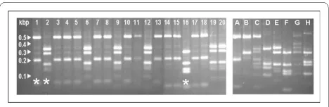
All patients had characteristic encapsulated yeasts seen on cerebrospinal fluid India ink examination, and positive CSF culture. 52 patients had CSF cryptococcal antigen testing - all were positive (median titre 1/256, range 1/4 to 1/32768). Ten of 56 patients (18%) had blood cultured and were fungaemic, and 44 of 53 patients tested had cryptococcal antigen detectable in blood, giving this sim-ple test a sensitivity of 83%. 35 isolates were revivable for PCR-RFLP typing and antifungal sensitivity testing. 10 were C. gattii, and 25 were C. neoformans var grubii. All the C. neoformans var grubii fell into molecular group VN1. One C. gattii isolate was molecular group VG2, the rest were VG1 (figure 1). One of 10 (10%) patients with C.
gattii infection had underlying disease, versus 7 of 25 (28%) patients with C. neoformans var grubii infection (p = 0.45, Fisher's exact test).
Susceptibility Testing
Antifungal susceptibility results (Table 1) were interpre-table for 34 of the 35 isolates (1 isolate grew too slowly despite incubation at a range of temperatures). Break-points for Cryptococcus neoformans are not defined; we present the minimum inhibitory concentrations (MIC) of drug that inhibited 90% (MIC90) and geometric mean MICs (GMICs) of the isolates according to species (Table 1). The GMICs of flucytosine and amphotericin were sig-nificantly higher for C. neoformans var grubii isolates than for C. gattii (8.23 vs 1.85 μg/ml, (95% CI 7.22 - 9.37 and 1.18 - 2.90, p < 0.001)) and 0.714 μg/ml vs 0.451 μg/ ml, (95% CI 0.617 - 0.826 and 0.341 - 0.598 p = 0.01) respectively. There were no differences in the MICs of any other azole by species. We could not detect any asso-ciation between antifungal susceptibility and clinical out-come.
Outcome
11/57 (19%) patients died during admission, and the median time to death was 14 days (4 -156 days). Three patients withdrew from the study (one after 12 and two after 37 days of treatment). The median duration of
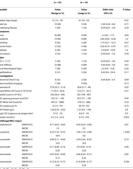
[image:3.595.305.539.598.674.2]Table 1: Features of 35 cryptococcal meningitis cases according to infecting species.
C. neoformans var grubii C. neoformans var gattii
(n = 25) (n = 10)
Variable Value Value Odds ratio P Value
(Range or %) (range or %) (95% CI)
Median Age (range) 31 (15 - 74) 41 (18 - 53) 0.47
Male Sex 10 (40) 5 (50) 1.48 (0.26 - 8.4) 0.71
Underlying Disease 7 (28) 1 (10) 0.29 (0.01 - 2.9) 0.39
Symptoms
Fever 20 (80) 9 (90) 2.2 (0.2 - 117) 0.64
Headache 19 (90) 8 (89) 0.85 (0.04 - 55.8) 1.0
Vomiting 9 (36) 5 (50) 1.75 (0.31 - 10.1) 0.47
Blurred vision 12 (52) 4 (40) 0.62 (0.10 - 3.47) 0.71
Diplopia 4 (20) 2 (20) 1.0 (0.95 - 10.9) 1.0
Convulsions 4 (16) 1 (10) 0.59 (0.01 - 7.2) 0.59
Signs
GCS < 11/15 7 (28) 1 (10) 0.29 (0.01 - 2.9) 0.39
Neck Stiffness 22 (88) 8 (80) 0.56 (0.05 - 7.8) 0.61
Focal Neurological Signs 7 (28) 5 (50) 2.5 (0.4 - 15.0) 0.26
Papilloedema 5 (21) 5 (50) 3.63 (0.6 - 24.3) 0.11
Investigations
Abnormal Chest X-ray 8 (32) 2 (20) 0.54 (0.05 - 3.7) 0.69
Abnormal Brain Imaginga 7 (58) 7(100) - 0.13
Haematocrit 37.8 (25.3 - 51.4) 38.8 (31.1 - 46) 0.65
Blood White Cell Count (×10^9/ml) 11.5(3.3 - 26.6) 13.6 (7.1 - 26.1) 0.37
Platelet count (×10^9/L) 256 (56.9 - 356) 305 (140 - 487)
CSF opening pressure (cmCSF)b 34.2 (2 - > 40) 30.5 (15 - > 40) 0.44
CSF White Cell Count/ml 287 (2 - 1080) 279 (12 - 480) 0.18
CSF Lymphocyte %c 53 (12 - 97) 56 (19 - 93) 0.74
CSF Protein (g/L) 1.28 (0.35 - 2.63) 1.25 (0.4 - 1.94) 0.9
Log2 CSF Cryptococcal antigen titre5 7.4 (3 - 15) 8.6 (2 - 14) 0.5
Lactate (mmol/L) 6.2 (1.6 - 22.5) 4.2 (2 - 8.4) 0.052
Antifungal MICs (mg/L)
Amphotericin GM(95%CI) 0.71 (0.62 - 0.83) 0.45 (0.34 - 0.60) 0.01
MIC90 1.0 0.5
Flucytosine GM(95%CI) 8.23 (7.22 - 9.37) 1.85 (1.18 - 2.90) < 0.001
MIC90 16/0 4.0
Fluconazole GM(95%CI) 6.96 (5.11 - 9.49) 4.54 (2.44 - 8.42) 0.19
MIC90 16.0 16.0
Itraconazole GM(95%CI) 0.11 (0.08 - 0.14) 0.9 (0.05 - 0.14) 0.48
MIC90 0.26 0.26
Voriconazole GM(95%CI) 0.07 (0.05 - 0.09) 0.06 (0.04 - 0.51) 0.85
MIC90 0.13 0.26
Posaconazole GM(95%CI) 0.13 (0.10 - 0.17) 0.10 (0.06 - 0.17) 0.336
MIC90 0.26 0.26
aN = 26, bN = 33, cN = 32, dN = 31
GM = Geometric Mean
ment was 131 days (range: 51- 426 days). One (10%) of 10 patients with C. gattii died compared with 6 (24%) of 25 patients with C. neoformans var grubii infection (p = 0.65). Complications at discharge included four patients with bilateral blindness (no light perception), 4 with uni-ocular blindness and one with complete visual and hear-ing loss. All cases of blindness occurred in patients with opening CSF pressures of ≥ 30cm CSF - 9 of 28 patients with opening pressure ≥ 30cm CSF developed blindness compared with none of 17 patients with opening pressure < 30 cm CSF, p = 0.008). Five of 9 patients with blindness (56%) had papilloedema at baseline compared with 14 (39%) of 36 patients with no visual loss (odds ratio 1.85, 95% confidence interval 0.33 - 11.09, p = 0.47). There were no differences in the rates of papilloedema, blind-ness or opening CSF pressure > 30cmCSF by infecting species. Fourteen patients had other neurological deficits including blurred vision and/or cranial nerve palsies.
On univariate analysis the admission findings associ-ated with death were presence of underlying disease, his-tory of convulsions, Glasgow Coma Score ≤ 11, age ≥ 60 years, lower platelet count, CSF cryptococcal antigen titre ≥ 1/512 and CSF yeast cell count > 400 cells/ml. Six of 21 (29%) patients with CSF opening pressure < 30cm CSF died, compared with 7 of 33 (21%) patients with opening pressures ≥ 30cm CSF (odds ratio 0.68, 95% confidence interval 0.16 - 2.94, p = 0.745). Two of 9 patients with CD4 lymphopenia died.
Multivariate analysis was limited by the small number of deaths. We tested the interaction between easily mea-sured clinical parameters (age ≥ 60 years, underlying dis-ease, Glasgow coma score < 11 and convulsions) with outcome. Only age ≥ 60 years and a history of convulsions were independently associated with death (Additional file 1: table S1). Length of history had no impact on outcome. We could detect no differences in clinical phenotype or outcome according to infecting species.
Two years after diagnosis a further 4 patients had died (total deaths 15 of 57, 26.3%). 14 patients (24.6%) had made complete recoveries with no residual deficit. One died due to multi-drug resistant tuberculosis, compli-cated by relapse of cryptococcal disease. Three patients with underlying disease continued on long term flucon-azole prophylaxis. It was not possible to determine the 2 year outcome in 9 of the 57 patients.
Discussion
Cryptococcal meningitis in HIV uninfected patients in tropical Viet Nam is most commonly due to Cryptococcus
neoformans var grubii. Most patients (81%) have no con-comitant immunosuppressive disease, although CD4 lymphopenia was detected in 9 patients, 3 of whom had no other underlying conditions.
C. neoformans var grubii, the commonest cause of cryptococcal meningitis since the HIV epidemic, usually occurs in patients with an underlying immunosuppres-sive condition and has rarely been reported in the absence of underlying disease, in southeast Asia or else-where [2,12,14,22-27]. Classical teaching would suggest that, in our tropical location, more cases would have been due to infection with Cryptococcus gattii[28,29]. Against expectation, in our series C. neoformans var grubii infec-tion outweighed C. gattii infection 2.5-fold. We could only revive isolates for speciation from 35 patients, but since the study has finished we continue to isolate C.
neo-formans var grubii and C. gattii from HIV uninfected patients in the ratio 3:1. We could not detect differences in the rate of underlying disease according to infecting species, although our study lacked power since there were only 10 cases of C. gattii infection.
All patients in our study had positive CSF India ink examination and culture - a rate higher than generally reported in the literature [1]. This may be a reflection of the large volumes of CSF (5 - 10 mls) routinely taken from patients with suspected meningitis in our hospital in order to exclude tuberculous meningitis.
In the tropical Northern Territories of Australia the incidences of C. gattii and C. neoformans infection are equal[30]. In Queensland the experience is more like our own - in a 3 year period C. neoformans infections out-weighed C. gattii 5 fold, although the total number of cases is not clear from the report[30]. However, it is not clear whether these cases were meningoencephalitis or extra-neural disease, and the varietal form of C.
neofor-mans was not described for the majority of isolates. All the C. neoformans var grubii strains from our series were of URA5-RFLP molecular type VN1, as also recently reported from China [31]. In that series, there was remarkable genetic homogeneity between the VN1 iso-lates, perhaps representing a strain in Asia with increased ability to cause infection in the immunocompetent. The significance of CD4 lymphopenia in our patients is uncertain. Only 20 patients had the investigation, and the test was not performed serially. Thus it is not clear whether it represents a transient phenomenon as a conse-quence of infection, or whether it represents a genuine pre-existing immune deficit, such as idiopathic CD4 lym-phopenia, known to be associated with an increased risk of cryptococcosis [32]. HIV infection is unlikely since each patient received at least 2 HIV antibody tests.
although we did not find length of history to be associ-ated with death. The independent variables associassoci-ated with death were age and convulsions. We did not find the presence of underlying disease to be independently asso-ciated with outcome, although analysis was limited by the low number of deaths. CSF opening pressure at baseline was highly associated with development of blindness, but there was no association with papilloedema, which prob-ably reflects the fact that papilloedema is a late indicator of raised intracranial pressure. However, it could be a function of the size of our study. Only 25% of patients made a complete recovery.
Historically, the disease phenotypes due to infection with C. neoformans var grubii and C. gattii are considered to be different. We found no difference in clinical pheno-type between the 2 infections, and the pattern of disease seen in our cases mimics that seen in case series of C.
gat-tii infection published from Papua New Guinea[17,27]. Like us, Seaton et al found presence of convulsions to be a predictor of death in patients with C. gattii meningitis. In addition, the rate of blindness in our patients is similar to that reported for C. gattii infection in immunocompe-tent adults, and higher than in HIV infected patients. The lack of difference in the clinical phenotypes of disease according to infecting species and the difference in rates of visual loss between HIV positive and negative patients support the hypothesis that host immune status is key in determining clinical phenotype in cryptococcal dis-ease[30]. There are retrospective data suggesting that immune mediating drugs (corticosteroids) may reduce the risk of blindness in C. gattii infection [34,35]. The similar clinical phenotype seen in our predominantly C.
neoformans var grubii infected patients suggests that this may be a worthwhile hypothesis to test in our patients.
There are no established breakpoints for anti-fungal drugs and C. neoformans, which makes the interpretation of anti-fungal MICs difficult [36-39]. The geometric mean MICs of flucytosine and amphotericin B were sta-tistically significantly higher for C. neoformans var grubii. This is consistent with the experience in Malaysia, but differs from reports from Brazil and Taiwan which found
C. gattii to be inherently less susceptible to amphotericin B and flucytosine [39-41]. Thompson found no difference in MICs by species, but was comparing strains from mul-tiple geographic regions[42]. Our data are reassuring, suggesting that it is safe to extrapolate treatment doses for our patients with C. gattii from trials in C. neoformans infection. However, the clinical significance of differences in MICs of flucytosine and amphotericin B remain unclear. MIC data were available for only 7 of the isolates from the 11 patients that died - we could not demonstrate any correlation between the MIC of any drug and patient outcome.
Recently, Cryptococcus gattii has been seen to establish new ecological niches, with subsequent successful human-mediated dispersal [43]. It is possible that disease incidence in the immunocompetent will increase [44,45]. There has been little change in the treatment of crypto-coccal meningitis over the past 10 years. Amphotericin B remains a key component of treatment, but therapy is protracted, expensive and difficult to administer. Com-bined with flucytosine, it has been associated with more rapid CSF sterilisation in HIV patients, and more recently slower rates of CSF sterilisation have been shown to be associated with an increased risk of death [46,47]. The identification of a reliable surrogate marker of outcome in cryptococcal meningitis enables anti-fungal therapeutic trials to be undertaken with smaller numbers of patients, and it should now be possible to answer clinical questions in groups where disease is rare, such as HIV uninfected patients. However, differences in complication rates, such as blindness, between HIV infected and uninfected patients may be due to differences in disease pathogene-sis (perhaps immune mediated) and clinical endpoint tri-als, although difficult, may still be necessary.
Conclusions
In HIV uninfected individuals in Vietnam, cryptococcal meningitis occurs predominantly in people with no clear predisposing factor and is most commonly due to C.
neo-formans var grubii. The pattern of disease is similar to that described in patients with C. gattii meningitis, sug-gesting underlying immune status is key in generating clinical phenotype. The rates of mortality and visual loss are high and independent of infecting species.
Funding
The study was funded by the Wellcome Trust (UK) and JND was supported by a British Infection Society Fellow-ship. The funding sources had no involvement in study design, collection, analysis or interpretation of the data, writing of the report, or the decision to submit for publi-cation.
Additional material
Competing interests
The authors declare that they have no competing interests.
Authors' contributions
JJF, TTHC, NHM, NHP, HDN, LVC, DXS, TTH, DGL & JND conceived the study, par-ticipated in its design, coordination, collected data, and helped to draft the manuscript. TTHC analysed the data. VAD, PTD, JIC, and JND performed the classical mycology and MIC testing. VAD, SB and JND performed the molecular typing. JND wrote the final manuscript. All authors read and approved the final manuscript.
Acknowledgements
With thanks to Tien Bui and Associate Professor Dee Carter, Head of Microbiol-ogy Biochemistry, School of Molecular and Microbial Biosciences University of Sydney for advice, and Dr Wieland Meyer, Associate Professor, Westmead Mille-nium Institute for Medical Research, Sydney, Australia for providing control strains.
Author Details
1Hospital for Tropical Diseases, 190 Ben Ham Tu, Quan 5, Ho Chi Minh City,
Vietnam, 2Oxford University Clinical Research Unit, Hospital for Tropical
Diseases, 190 Ben Ham Tu, Quan 5, Ho Chi Minh City, Vietnam, 3Centre for
Tropical Medicine, Nuffield Department of Clinical Medicine, Oxford University, Oxford, OX3 7LJ, UK and 4Liverpool School of Tropical Medicine, Liverpool, L3
5QA, UK
References
1. Casadevall A, Perfect JR: Cryptococcus neoformans. 1st edition. Washington: American Society for Microbiology Press; 1998. 2. Kwon-Chung KJ, Bennett JE: Epidemiologic differences between the
two varieties of Cryptococcus neoformans. Am J Epidemiol 1984,
120(1):123-130.
3. Kwon-Chung KJ, Sorrell TC, Dromer F, Fung E, Levitz SM: Cryptococcosis: clinical and biological aspects. Med Mycol 2000, 38(Suppl 1):205-213. 4. Duncan C, Schwantje H, Stephen C, Campbell J, Bartlett K: Cryptococcus
gattii in wildlife of Vancouver Island British Columbia, Canada. J Wildl Dis 2006, 42(1):175-178.
5. Hoang LM, Maguire JA, Doyle P, Fyfe M, Roscoe DL: Cryptococcus neoformans infections at Vancouver Hospital and Health Sciences Centre (1997-2002): epidemiology microbiology and histopathology. J Med Microbiol 2004, 53(Pt 9):935-940.
6. Kidd SE, Hagen F, Tscharke RL, Huynh M, Bartlett KH, Fyfe M, Macdougall L, Boekhout T, Kwon-Chung KJ, Meyer W: A rare genotype of Cryptococcus
gattii caused the cryptococcosis outbreak on Vancouver Island (British Columbia, Canada). Proc Natl Acad Sci USA 2004, 101(49):17258-17263. 7. Stephen C, Lester S, Black W, Fyfe M, Raverty S: Multispecies outbreak of
cryptococcosis on southern Vancouver Island British Columbia. Can Vet J 2002, 43(10):792-794.
8. Bennett JE, Kwon-Chung KJ, Howard DH: Epidemiologic differences among serotypes of Cryptococcus neoformans. Am J Epidemiol 1977,
105(6):582-586.
9. Diamond RD, Bennett JE: Prognostic factors in cryptococcal meningitis. A study in 111 cases. Ann Intern Med 1974, 80(2):176-181.
10. Dismukes WE, Cloud G, Gallis HA, Kerkering TM, Medoff G, Craven PC, Kaplowitz LG, Fisher JF, Gregg CR, Bowles CA, et al.: Treatment of cryptococcal meningitis with combination amphotericin B and flucytosine for four as compared with six weeks. N Engl J Med 1987,
317(6):334-341.
11. Dromer F, Mathoulin S, Dupont B, Brugiere O, Letenneur L: Comparison of the efficacy of amphotericin B and fluconazole in the treatment of cryptococcosis in human immunodeficiency virus-negative patients: retrospective analysis of 83 cases. French Cryptococcosis Study Group. Clin Infect Dis 1996, 22(Suppl 2):S154-S160.
12. Dromer F, Mathoulin S, Dupont B, Laporte A: Epidemiology of cryptococcosis in France: a 9-year survey (1985-1993). French Cryptococcosis Study Group. Clin Infect Dis 1996, 23(1):82-90. 13. Dromer F, Mathoulin S, Dupont B, Letenneur L, Ronin O: Individual and
environmental factors associated with infection due to Cryptococcus neoformans serotype D. French Cryptococcosis Study Group. Clin Infect Dis 1996, 23(1):91-96.
14. Mitchell DH, Sorrell TC, Allworth AM, Heath CH, McGregor AR, Papanaoum K, Richards MJ, Gottlieb T: Cryptococcal disease of the CNS in immunocompetent hosts: influence of cryptococcal variety on clinical manifestations and outcome. Clin Infect Dis 1995, 20(3):611-616. 15. Moosa MY, Coovadia YM: Cryptococcal meningitis in Durban South
Africa: a comparison of clinical features laboratory findings and outcome for human immunodeficiency virus (HIV)-positive and HIV-negative patients. Clin Infect Dis 1997, 24(2):131-134.
16. Rozenbaum R, Goncalves AJ: Clinical epidemiological study of 171 cases of cryptococcosis. Clin Infect Dis 1994, 18(3):369-380.
17. Seaton RA, Verma N, Naraqi S, Wembri JP, Warrell DA: Visual loss in immunocompetent patients with Cryptococcus neoformans var. gattii
meningitis. Trans R Soc Trop Med Hyg 1997, 91(1):44-49.
18. Stevens DA, Denning DW, Shatsky S, Armstrong RW, Adler JD, Lewis BH:
Cryptococcal meningitis in the immunocompromised host: intracranial hypertension and other complications. Mycopathologia 1999, 146(1):1-8.
19. van der Horst CM, Saag MS, Cloud GA, Hamill RJ, Graybill JR, Sobel JD, Johnson PC, Tuazon CU, Kerkering T, Moskovitz BL, et al.: Treatment of cryptococcal meningitis associated with the acquired
immunodeficiency syndrome. National Institute of Allergy and Infectious Diseases Mycoses Study Group and AIDS Clinical Trials Group. N Engl J Med 1997, 337(1):15-21.
20. Perfect JR, Dismukes WE, Dromer F, Goldman DL, Graybill JR, Hamill RJ, Harrison TS, Larsen RA, Lortholary O, Nguyen MH, et al.: Clinical Practice Guidelines for the Management of Cryptococcal Disease: 2010 Update by the Infectious Diseases Society of America. Clin Infect Dis 2010,
50(3):291-322.
21. Meyer W, Castaneda A, Jackson S, Huynh M, Castaneda E: Molecular typing of IberoAmerican Cryptococcus neoformans isolates. Emerg Infect Dis 2003, 9(2):189-195.
22. Chuang YM, Ho YC, Chang HT, Yu CJ, Yang PC, Hsueh PR: Disseminated cryptococcosis in HIV-uninfected patients. Eur J Clin Microbiol Infect Dis 2008, 27(4):307-310.
23. Chuang YM, Ku SC, Liaw SJ, Wu SC, Ho YC, Yu CJ, Hsueh PR: Disseminated
Cryptococcus neoformans var. grubii infections in intensive care units. Epidemiol Infect 2009:1-8.
24. Jongwutiwes U, Sungkanuparph S, Kiertiburanakul S: Comparison of clinical features and survival between cryptococcosis in human immunodeficiency virus (HIV)-positive and HIV-negative patients. Jpn J Infect Dis 2008, 61(2):111-115.
25. Kiertiburanakul S, Wirojtananugoon S, Pracharktam R, Sungkanuparph S:
Cryptococcosis in human immunodeficiency virus-negative patients. Int J Infect Dis 2006, 10(1):72-78.
26. Shih CC, Chen YC, Chang SC, Luh KT, Hsieh WC: Cryptococcal meningitis in non-HIV-infected patients. QJM 2000, 93(4):245-251.
27. Seaton RA, Naraqi S, Wembri JP, Warrell DA: Predictors of outcome in
Cryptococcus neoformans var. gattii meningitis. QJM 1996,
89(6):423-428.
28. Fisher D, Burrow J, Lo D, Currie B: Cryptococcus neoformans in tropical northern Australia: predominantly variant gattii with good outcomes. Aust N Z J Med 1993, 23(6):678-682.
29. Speed B, Dunt D: Clinical and host differences between infections with the two varieties of Cryptococcus neoformans. Clin Infect Dis 1995,
21(1):28-34. discussion 35-26
30. Chen S, Sorrell T, Nimmo G, Speed B, Currie B, Ellis D, Marriott D, Pfeiffer T, Parr D, Byth K: Epidemiology and host- and variety-dependent characteristics of infection due to Cryptococcus neoformans in Australia and New Zealand. Australasian Cryptococcal Study Group. Clin Infect Dis 2000, 31(2):499-508.
31. Chen J, Varma A, Diaz MR, Litvintseva AP, Wollenberg KK, Kwon-Chung KJ: Cryptococcus neoformans strains and infection in apparently immunocompetent patients China. Emerg Infect Dis 2008,
14(5):755-762.
32. Netea MG, Brouwer AE, Hoogendoorn EH, Van der Meer JW, Koolen M, Verweij PE, Kullberg BJ: Two patients with cryptococcal meningitis and idiopathic CD4 lymphopenia: defective cytokine production and reversal by recombinant interferon- gamma therapy. Clin Infect Dis 2004, 39(9):e83-87.
33. Pappas PG, Perfect JR, Cloud GA, Larsen RA, Pankey GA, Lancaster DJ, Henderson H, Kauffman CA, Haas DW, Saccente M, et al.: Cryptococcosis in human immunodeficiency virus-negative patients in the era of effective azole therapy. Clin Infect Dis 2001, 33(5):690-699. 34. Seaton RA, Verma N, Naraqi S, Wembri JP, Warrell DA: The effect of
corticosteroids on visual loss in Cryptococcus neoformans var. gattii meningitis. Trans R Soc Trop Med Hyg 1997, 91(1):50-52.
35. Phillips P, Chapman K, Sharp M, Harrison P, Vortel J, Steiner T, Bowie W:
Dexamethasone in Cryptococcus gattii central nervous system infection. Clin Infect Dis 2009, 49(4):591-595.
Received: 2 December 2009 Accepted: 9 July 2010 Published: 9 July 2010
This article is available from: http://www.biomedcentral.com/1471-2334/10/199 © 2010 Chau et al; licensee BioMed Central Ltd.
This is an Open Access article distributed under the terms of the Creative Commons Attribution License (http://creativecommons.org/licenses/by/2.0), which permits unrestricted use, distribution, and reproduction in any medium, provided the original work is properly cited.
36. Archibald LK, Tuohy MJ, Wilson DA, Nwanyanwu O, Kazembe PN, Tansuphasawadikul S, Eampokalap B, Chaovavanich A, Reller LB, Jarvis WR, et al.: Antifungal susceptibilities of Cryptococcus neoformans. Emerg Infect Dis 2004, 10(1):143-145.
37. Pfaller MA, Buschelman B, Bale MJ, Lancaster M, Espinel-Ingroff A, Rex JH, Rinaldi MG: Multicenter comparison of a colorimetric microdilution broth method with the reference macrodilution method for in vitro susceptibility testing of yeast isolates. Diagn Microbiol Infect Dis 1994,
19(1):9-13.
38. Pfaller MA, Messer SA, Boyken L, Rice C, Tendolkar S, Hollis RJ, Doern GV, Diekema DJ: Global trends in the antifungal susceptibility of
Cryptococcus neoformans (1990 to 2004). J Clin Microbiol 2005,
43(5):2163-2167.
39. Tay ST, Tanty Haryanty T, Ng KP, Rohani MY, Hamimah H: In vitro susceptibilities of Malaysian clinical isolates of Cryptococcus
neoformans var. grubii and Cryptococcus gattii to five antifungal drugs. Mycoses 2006, 49(4):324-330.
40. Chen YC, Chang SC, Shih CC, Hung CC, Luhbd KT, Pan YS, Hsieh WC:
Clinical features and in vitro susceptibilities of two varieties of
Cryptococcus neoformans in Taiwan. Diagn Microbiol Infect Dis 2000,
36(3):175-183.
41. Trilles L, Fernandez-Torres B, Lazera Mdos S, Wanke B, Guarro J: In vitro antifungal susceptibility of Cryptococcus gattii. J Clin Microbiol 2004,
42(10):4815-4817.
42. Thompson GR, Wiederhold NP, Fothergill AW, Vallor AC, Wickes BL, Patterson TF: Antifungal susceptibilities among different serotypes of
Cryptococcus gattii and Cryptococcus neoformans. Antimicrob Agents Chemother 2009, 53(1):309-311.
43. Kidd SE, Bach PJ, Hingston AO, Mak S, Chow Y, MacDougall L, Kronstad JW, Bartlett KH: Cryptococcus gattii dispersal mechanisms British Columbia, Canada. Emerg Infect Dis 2007, 13(1):51-57.
44. Bartlett KH, Kidd SE, Kronstad JW: The Emergence of Cryptococcus gattii
in British Columbia and the Pacific Northwest. Curr Infect Dis Rep 2008,
10(1):58-65.
45. Upton A, Fraser JA, Kidd SE, Bretz C, Bartlett KH, Heitman J, Marr KA: First contemporary case of human infection with Cryptococcus gattii in Puget Sound: evidence for spread of the Vancouver Island outbreak. J Clin Microbiol 2007, 45(9):3086-3088.
46. Bicanic T, Muzoora C, Brouwer AE, Meintjes G, Longley N, Taseera K, Rebe K, Loyse A, Jarvis J, Bekker LG, et al.: Independent association between rate of clearance of infection and clinical outcome of HIV-associated cryptococcal meningitis: analysis of a combined cohort of 262 patients. Clin Infect Dis 2009, 49(5):702-709.
47. Brouwer AE, Rajanuwong A, Chierakul W, Griffin GE, Larsen RA, White NJ, Harrison TS: Combination antifungal therapies for HIV-associated cryptococcal meningitis: a randomised trial. Lancet 2004,
363(9423):1764-1767.
Pre-publication history
The pre-publication history for this paper can be accessed here: http://www.biomedcentral.com/1471-2334/10/199/prepub
doi: 10.1186/1471-2334-10-199
Cite this article as: Chau et al., A prospective descriptive study of cryptococ-cal meningitis in HIV uninfected patients in Vietnam - high prevalence of
Cryptococcus neoformans var grubii in the absence of underlying disease BMC