0022-538X/91/105647-06$02.00/0
Copyright
©
1991, American Society for MicrobiologyThe T/t Common
Region of Simian
Virus
40 Large
T
Antigen
Contains
a
Distinct Transformation-Governing
Sequence
ERIKA
MARSILIO,1
SENG H. CHENG,2 BRIAN SCHAFFHAUSEN,3 EVA PAUCHA,1tAND
DAVID
M.LIVINGSTON'*
Dana-Farber
Cancer Institute andDepartmentsof Medicine and Pathology, Harvard Medical School, Boston,Massachusetts 021151;
Laboratory ofCellular Regulation, Genzyme Corporation, Framingham, Massachusetts017012;
and Department
of Biochemistry,
HealthScience Campus, Tufts University, Boston, Massachusetts 02J13
Received 12 December 1990/Accepted26 June 1991
Simian virus 40 large T antigen (T)cantransformcultured cells, but the mechanismsby which it functions
arenotentirely understood. Several lines of evidencehave suggested that the amino-terminal 130 residuesof Tmay be sufficient toconfer the transforming capability. Oligonucleotide-directed mutagenesis wasusedto generateaseries of deletion and substitutionmutantswithin the amino-terminal82 residues of T,thesegment which is shared with simian virus 40 smalltantigen(t). Resultsofstability and transformationassaysofthese
mutantsstronglysuggestthat the 1-to-82 region of T containssequenceswhichgovernTtransformingactivity
and affect in vivo stability.
Instability
andadefect in transformingactivity could be separated fromoneanothergenetically. Thus, the1-to-82 regionappearstocontainaspecific region that contributestothe transforming
function of the protein. Thissegmentoperates bymeans other than the simple binding of pRband/orp107.
Simian
virus 40 large T antigen (T) has been shown by
many
laboratories to elicit the neoplastic transformation of
cultured rodent cells (45). However, although there is new
information
showing that its transforming function likely
depends
on
its
ability to modulate the activities of at least
two tumor
suppressor gene
products, pRb and
p53, and a
third
protein,
p107,
the detailed mechanism by which T
transforms is still
largely unknown (7, 8, 9, 25, 46, 47). What
seems
clear is
that T must perform more than one
biochem-ical
act to
elicit the appearance of neoplastic properties. In
this
regard,
it
appears
that at
least some T transforming
functions
are
the
products
of blocks
of sequences located in
the
amino-terminal half of the molecule
(1,
3,
4, 5, 37,
38).
Multiple
reports
indicate that various amino-terminal T
fragments extending from residues
1to
127 to 300 can
establish
early-passage
ratcells in culture, although
atfre-quencies lower
than
those of
wild type
(1-5, 37,
38).
More-over,
these truncated
species
caninduce certain
primary
strains and
established
cell lines
tomanifest
anchorage-independent growth (3, 5, 37, 38). Therefore,
the
amino-terminal third of
the
protein
canperform biochemical
func-tions,
which,
taken
together,
aresufficient for
the
transformation of
somecells.
Other functions
which have
been shown
genetically
todepend
onsequences
within the
amino-terminal
region
of T
arebinding
tothe
viral
replica-tion
origin (32)
and nuclear
localization
(19, 20). However,
the
abilities
tobind
the
origin
and
tolocalize
in the
nucleus
are
dispensable
for
transformation
(3, 12, 18, 21,
26, 32, 34).
Another
activity associated with
the
amino-terminal
half of T
is the
ability
tostimulate DNA
synthesis
in
quiescent
cells
(14,
31,
39).
The role
of this
activity
in the
T-dependent
transformation mechanism
remains
tobe
fully determined,
although,
in the
caseof
twoother DNA
tumorvirus
species,
adenoviruses
and
bovine
papillomavirus,
transformation
function
cannotbe
separated
from
the
ability
toinitiate DNA
synthesis
in
quiescent
cells
(24, 29, 30, 48).
*
Corresponding
author.t
Deceased.Recent reports
indicate that mutations within the
C-termi-nal
-60%
of large T, which disrupt the
p53
binding
region,
render the molecule
incapable
of transforming REF52 but
not
C3H1Ot1/2
cells (33, 40). The
former is a continuous line
of
ratcells,
and the latter is
acontinuous
line
of
murine
cells.
Some of
the
aforementioned
mutations also suppressed the
immortalization of primary C57BL/6
mouse
cells
by
T(43,
44).
Therefore, although
T-p53 complex formation
seems
notto
be
involved
in
the
transformation of
C3H1Ot1/2,
it does
appear
tobe
required for
the
transformation of
REF52
cells
(40). The
formation of
T-p53 complexes
may
also
play
asignificant
role in
supporting
the
immortalizing
function
of
T
in
certain
primary murine
cells
(33, 43).
Given
the
additional
fact
that the
amino-terminal
-130
residues
of
T arecapable
of
immortalizing
other rodent cell
species
(1), the
protein
may
be able
to exertits
immortalizing function by
atleast
two
independent
biochemical routes,
oneof
which involves
p53 binding
and the
other which does
not.Moreover, there
may
be
significant
host range limitations
onthe
expression
of
these
functions.
A
large
number
of amino-terminal
T
mutantswhich
areeither
partly
orfully defective for
transforming activity
have
been identified
(6, 12, 21, 34,
35, 37,
38, 42).
For
example,
Kalderon
and Smith
(21)
have
shown that
numerousmutantswith lesions
involving
the sequence
extending
from
residues
105
to115
aretransformation defective.
Recently,
it
wasshown that these
mutantsfail
totransform cells
toanchor-age-independent growth (3).
Taken
together,
the
results
suggest
that the
105-to-115
segment
constitutes
afunctional
domain devoted
toT
transforming
function. This
region
bears strong
similarity
in both
primary
sequence and
pre-dicted
secondary
structure toatransforming
domain
of
the
adenovirus
ElA
product
(10,
41).
In
adenovirus
ElA,
this
region
has
been
implicated
in the
stimulation of host cell
DNA
synthesis
and the maintenance
of
viral
transforming
activity (24, 30, 36,
48, 50,
51).
This segment is
nowknown
to
operate
in
both
Tand
ElA
by
serving
as abinding
site for
at
least
twocell-encoded
proteins,
pRb
and
p107 (7-9,
46,
47).
The
former
is
aknown
tumorsuppressor gene
product
(22,
23).
Although
there
is
presently
nodirect evidence
to5647
on November 10, 2019 by guest
http://jvi.asm.org/
D2C2 a PvB-Q 55-70 59-64 46-51 40-45 34-3 dl135
A B A B A B AB A B A B A B
.-_.
__^-_o
N58 48
36.5
B
EMdI59-64 EMsb59-64 Pvu-O
0 2 4 8 11 23 0 2 4 8 11 23 0 2 4 8 11 230 2 4 8 11 23
1debes
_T
FIG. 1. Western blot andpulse-chase analysis of amino-terminal deletionmutantsof T.(A) Western blotanalysis of T deletionmutants. Allcellsweregrownat37°C inahumidified, 10% C02-containing atmosphereinDulbecco's modifiedEagle's medium (GIBCO) supplemented with10% fetal calfserum(Flow Laboratories). Dishes (100 mm) of nearly confluent CV-1P cellsweretransfectedinduplicate (lanes labelled A andB)with20 p.gofplasmid DNA by the calcium phosphate precipitationmethod, and cell lysateswereprepared24 hlater.Equalamounts of totalproteinextract(800 ,ug)wereimmunoprecipitatedwithpAB423 (7). The immune complexeswereseparated by sodiumdodecyl sulfate
gel electrophoresis, transferred to nitrocellulose, and assayed in a Western blot by using pAB423 and anti-mouse immunoglobulin G
conjugated to alkaline phosphatase. The lane marked D2C2 contains a pAB423 immunoprecipitate of a clonal CV-1P cell line which constitutively synthesizes T. pBR328 DNAwasusedas anegative control, while pPVU-O,aplasmid which encodes wild-type T,wasused as apositive control. The designations 65-70,59-64, 46-51, 40-45, and 34-39representthenewly generated internal deletionmutantsdescribed inthe text, andthe numbersrepresentthespecific residues whichhavebeen removed in eachcase.d11135 isaTmutant(generouslydonated by J. Pipas) in which residues 17 to 27 have been deleted. The new deletion mutants described were generated by using the gapped heteroduplex method essentiallyasdescribedby Kalderonetal.(19) and Harveyetal.(16). The synthetic oligonucleotides usedtogenerate the variant Tspecieswereeach 30nucleotides long. Following annealing of the mutagenic oligonucleotidestothegappedheteroduplexes, Escherichia coliDH5a was transformed by each DNA heteroduplex. Colonies bearing themutant DNAs were first identifiedby colony hybridization and then verifiedbyDNAsequencing. (B) Pulse-chase analysis of deletion and substitution mutantsof T. Dishes (60 mm)
A
TAg
180
116
_ 84
pBR328
'-willoo'111",w ;.
Ori.-WIM,::::: lO'lItqHW*$ftw-- -!. 7is! ,,illlllllf
on November 10, 2019 by guest
http://jvi.asm.org/
[image:2.612.133.477.85.588.2]support the
suspicion, the latter is suspected of having
analogous
properties. Moreover, genetic
analysis strongly
suggests
that T and
ElA
binding to pRb and/or
p107
con-tributes
to
their
transforming properties (7, 8, 46, 47).
T
and
the other
simian virus 40 early region product, small
t
antigen
(t), share an 82-residue
amino-terminal segment.
This region
lies
upstream of the pRb-p107 binding segment
and
has
not
been extensively studied for potential functions.
In
support of the possibility that this region might contribute
to
T
transforming
function, we recently
found that its
complete elimination
from T was
linked to the failure of T to
induce anchorage-independent
growth (28). Given that the
1-to-82 sequence
represents more than 50% of the mass of
the
smallest
truncated T species with overt
'rI.nsforming
activity,
we
chose
to evaluate it further as a potential
contributor
to
the
transforming function of the protein.
The
initial
strategy was to
generate a set of T mutants
containing
serial, colinear
deletions of six amino acids
lo-cated
within the
1-to-82 region. Specifically, the deletions
extended from
residues 34 to 70. Mutagenesis was
per-formed
by
aspecific oligonucleotide-directed
method, with
an
intact simian virus
40 early
region-containing
plasmid,
pPVU-O,
as
template.
pPVU-O
contains the entire early
region
of
simian virus
40
and the viral early promoter cloned
in
pBR328
(21). To check
for
expression of these various
mutants
and
of another
deletion
mutant,
d11135
from the
laboratory of
J.
Pipas
(34),
which
lacks residues 17 to 27, an
equivalent
amount
of
each mutant DNA and the wild-type
plasmid
wastransfected
into
CV-1P
cells. The
transfections
were
carried
out in
duplicate.
The resulting viral proteins
produced
in
these transient
assays were
immunoprecipitated
with
pAB423,
a
monoclonal
antibody which
recognizes an
epitope
in the
carboxy
terminus
of
T
(15).
The
various
mutant
T
species
wereanalyzed by
Western
immunoblotting
(7).
All
immunoprecipitations
wereperformed
in
antibody
excess
with
astandard
quantity
of cell
extract.The
results
are
shown in
Fig.
1A.
In
duplicate
setsof transfections
(labelled
A
and
B),
each
mutantplasmid
gave rise
tothe
synthesis
of
animmunoreactive
T
species
which
comigrated
with
the
wild-type
protein.
Similar
results
wereobtained
following
transfection
of
BALB/c/3T3
cI
A-31, Rat-1,
orCREF cells
(data
notshown).
Given
that
cells transfected
with all but the
d165-70
mutantyielded significantly
less
Tthan
the
wild-type control, pulse-chase
labelling analysis
of
these
various
proteins
wascarried
out overaperiod
of 24
hin
aneffort
to assessthe
stability
of
each
of the
mutantproteins.
All the
mutants, except
for
d165-70,
werecharac-terized
by
less than
normal half-lives.
Only
the results of the
pulse-chase
analysis
of
onedeletion
mutant areshown
here as anexample
(Fig. 1B).
As
shown in the left
panel
of
Fig.
1B,
the
half-life of
the
deletion
mutantd159-64
ranged
from 2
to
4
h,
compared
with
morethan 12 h for the
wild-type
counterpart.
Thus,
the
abnormally
low
steady-state
levels of
mutant
T
present in these
extracts aredue,
atleast in
part,
torapid protein
turnover.The
instability notwithstanding,
each
[image:3.612.322.563.93.182.2]of
these unstable
mutants,
asshown
earlier,
retained the
ability
tobind
pRb,
p107,
and
p53
readily (9), suggesting
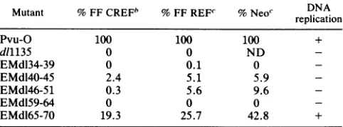
noTABLE 1. Focus-forming activity ofvarious T deletionmutants' Mutant % FF CREFb % FF REFc % Neoc DNA
replication
Pvu-O 100 100 100 +
d11135 0 0 ND
-EMdl34-39 0 0.1 0
-EMdl40-45 2.4 5.1 5.9
-EMdl46-51 0.3 5.6 9.6
-EMdl59-64 0 0 0
-EMdl65-70 19.3 25.7 42.8 +
aSparse60-mmdishes of eitherREF(Whittaker, M.A. Bioproducts)or CREFcells (11)were transfectedwith 10pLgof plasmid DNA. Cells were maintainedin culture for aperiodof 3 to 4weeks,after whichtimethey were fixed and the foci were stainedwith anti-T pAB423 and goat anti-mouse immunoglobulinG conjugated to alkalinephosphatase(Promega). Values are shown as percentages of wild-type activity. Neo, percent Neo-resistant colonies obtained after transfection of REF cells with equal amounts of wild-typeor mutant plasmid DNA (10p.g)and plasmid DNA containing a Neo-resistantgene (10 ,ug). FF,focus formation on the indicated cell lines. Cellsweresubsequently maintainedin cultureandfed with G418 (400 p.g/ml) foranequalamountof time.Thereplicationassay was performed with CV-1P cells(33). Typically,1to 2 ,ug of DNA wastransfectedonto a60%confluent dish.Three tofour daysfollowingtransfection, the DNA wasisolatedby the Hirt method (17),digested with DpnI,separated by electrophoresis in agarose gels,andthen blottedonto nitrocellulose (34). Random-primed, 32P-labelled wild-type plasmidDNA wasusedas a probe(Boehringer Mannheim random-primedDNAlabelling kitwasused essentiallyasdescribedby the manufac-turer).ND,notdetermined.
bValuesrepresentaverages for 12experiments. cValuesrepresentaverages for sixexperiments.
major
defect in these functions. Therefore, they do
notappear
tobe
wholly inert.
Given that all
mutantproteins could
bind
pRb, p107, and
p53,
weasked whether any
of
the
plasmids
encoding them
could
transform
anestablished
ratcell
line,
CREF (11),
asmeasured
in
afocus
formation
assay.
The
results of these
focus
formation
assays
aresummarized in Table 1 and
areexpressed
aspercentages
of
the
wild-type
effects. In these
experiments,
all mutants, with the
exception
of
d165-70,
failed to
transform CREF cells
toany
degree.
Similarly, they
failed
totransform
primary
ratembryo
fibroblasts
(REF).
In
this case, the
identity of
agiven
focus
as anauthentic T
transformant
waschecked
by
in situ
immunoperoxidase
staining with pAB423. Only
those foci which testedpositive
by
staining
in this assay
werescored here.
An
activated
rasallele has
been shown
topartially
com-plement
transformation-defective
T
mutants(27).
In view of
this,
wealso
cotransfected
anactivated
ras-containing
plas-mid,
PM1
(generously
donated
by
Michael
Corbly), along
with the
different
mutantDNAs
into REF and CREF cells.
The
activated
ras,
however,
failed
to rescue ourdeletion
mutants
in this
transformation
assay
(data
notshown).
Similarly,
when each
of the
mutantplasmids
wascotrans-fected
with
aplasmid containing
aG418
resistancegene
intoREF in search
of
T-containing
immortal
colonies,
none ofthe
mutantsfunctioned
normally
in
comparison
with the wild
type
(Table
1).
However,
asin the
previously
described
ofCV-1P cellsweretransfected with10 ,ugofDNA.
Twenty-four
hours laterthe cellswerelabelledfor 3 h with 200
,uCi
of
[35S]methionine
per ml in serum-free medium.
They
were then refed withcomplete
mediumlacking
radioactivity.
Cell
extracts wereprepared and
immunoprecipitated
with themonoclonalantibody pAB423
(7)
attheindicatedtimes.Thedesignations
0
to23 indicate the time in hours after
removal of
radioactivity,
when the relevant celllysates
wereprepared.
Theorigin
of the Tdoublet in all of the relevant lanes is unclear,
although,
given
the knownsusceptibility
ofTtoproteolytic
degradation
after
extraction, there is the
suspicion
that the lower band
maybe
adegradation
product
ofthe upperband.on November 10, 2019 by guest
http://jvi.asm.org/
180
- 116
- 84
- 58
- 48
- 36.5
FIG. 2. Immunoprecipitation of the mutants EMdl59-64 and EMsb59-64. Cells were maintained in culture as described in the legendtoFig. 1.CV-1P cellsweretransfectedwith 20,ugofplasmid DNA. Twenty-four hours posttransfection, thecells werelabelled for 3 hwith 200,uCiof[35S]methionineperml inserum-freemedium. Cellextractswerepreparedandimmunoprecipitatedwith the
mono-clonalantibody pAB423 (7). Lanes 1 and 2 containtwoindependent immunoprecipitates obtained fromcells transfected with the substi-tutionmutant EMsb59-64; lane 3contains the immunoprecipitates obtained fromcells transfected withEMdl59-64;lane 4contains the immunoproducts from cells transfected with pPVU-O (encodes wild-type T); andlane 5 contains the immunoproducts from cells which have beenmock transfected. Arrows (from topto bottom) indicatetherespective migrationpositionsofp107, pRb,andp53.
assays,d165-70functionedbetterthan therestof themutants tested.
Finally,
we alsoinvestigated
whetherthe mutant Tspecies
retained DNAreplicating activity by assaying
themutantgenomesfor their
ability
toreplicate
inCV-1P cells.
Withtheexception
ofd165-70,
allthe mutantswerejudged
to bedefective intheability
toreplicate
DNAasmeasured inablottingassayafter DNAtransfection. Thisfailure
could
bearesultofinsufficientfunctional
T,
the shorthalf-lifeof
therelevant mutant
proteins,
or adefect intheir intrinsic
DNA
replication
function.To determinewhether the
inability of these deletion
mu-tants totransform cells was dueto the short half-life
of the
protein
withinthecellandnot tothedisruption of
apotential
transforming domain,
wesought
toengineer
amutantwithin
the 1-to-82region
which was as stable aswild-type
T.The
sequence NAAIRS has been found to assumedifferent
secondary
structural motifs in differentproteins (49).
As aresult,
otherssuggested
thatthis
sequenceis inherently
flexible and thatthis characteristic
allows it the freedom
toassume different conformations in different
environments;
hence,
itcouldbe used inmutagenesis experiments in which
the aim is to minimizeconformational
disruption following
grosschanges
inprimary protein
structure(17a,
49). Since [image:4.612.116.240.77.323.2]deleting
sequences between residues 17 and64 led
toclear
instability,
wesuspected
thatlossofsequence inthisregion
TABLE 2. Focus formation activity ofsubstitution anddeletionmutants'
Mutant
~~~Expt
1 Expt2Mutant (%FFCREF) Ep
(%FFCREF) % FF REF % Neo
Pvu-O 100 100 100
EMdl59-64 0 0 0
EMsb59-64 10.8 10.4 13
a Two separateexperiments
measuring
the focusformationactivityofthe substitutionmutant, EMsb59-64, and the deletion mutant,EMdl59-64,were performed. Eachwasperformed under thesameconditions described for the experiment whose resultsareshown in Table 1. In experiment 1, CREFcells werestudied.Inexperiment 2,ananalogousexperimentwasperformed with primaryREF. Allcellsweremaintained under thesameconditions described inthelegendtoFig.1.Values represent averagesfor threeexperiments. For abbreviations,seeTable1, footnotea.was
responsible
for
this defect. To
testthis
hypothesis,
anoligonucleotide
encoding
the
NAAIRS sequence
wasin-serted in frame
atthe
site of the
dl59-64
mutation. The
resulting
substitution
mutantprotein,
sb59-64, comigrated
with both wild-type and
d159-64 T, but, unlike the latter,
which was the
least stable
of
all the
mutantstested,
this
product exhibited
nearly
wild-type stability
in
pulse-chase
analysis
(Fig.
1B) and accumulated
towild-type
levels in
atransient
expression
assay
(Fig.
2), unlike its
progenitor,
dl59-64. In
this
experiment,
wesuspect
that the latter
waspresent
in
anamountbelow that
needed
to
detect
coprecip-itating proteins.
Indeed,
in
the past
it
was
readily
shown
tobind
topRb, p107,
and
p53 (9).
Furthermore, the sb59-64
protein retained
the
ability
tobind
pRb and p107 and bound
p53
like wild-type T
(Fig. 2). This notwithstanding, like the
d159-64
parental
mutant,
it
wasclearly defective in
focus-forming
activity
when
assayed
onCREF cells and REF
cells,
as
shown in
Table
2.Therefore, the
introduction
of NAAIRS at the site of the
former
deletion restored
wild-type
stability
but
failed
torestore
wild-type transforming activity. Indeed, the product
is
clearly still defective in both focus-forming and REF
colony-forming activity.
From
these
findings,
one
canargue
that
elements within the 1-to-82 sequence
affect
two aspects
of T
behavior, stability
and
transforming activity.
Indeed,
from
the
data presented here, these two functions appear to
be
separable.
Given these and
prior observations (9), it
seems
reasonable
tosuggest
that the
1-to-82 region may
constitute
atransformation-controlling
segment, at least
in
part,
distinct
from
that which gives rise to pRb, p107, and
p53
binding activity (9). Conceivably, it plays a role in the
T-pRb and/or T-p107
interaction, not so much by affecting
the
affinity
of either
protein for T but rather by altering the
functional effect(s) of T binding on one or more of these
elements. Another
possibility, among several, is suggested
by
the
existence of some elements of sequence homology
within
this region and transforming domain 1 of ElA.
Harlow and
coworkers have shown that elements of this
transformation-controlling region of ElA are responsible for
the
p300 binding
activity of
ElA
(47, 48). From the genetics
of the
ElA-p300
binding reaction, one can argue that p300
binding
contributes to
ElA
transforming activity. Although
there
is no direct supporting evidence at the moment, it
remains
possible
that
this region of T also binds
p300
or an
analogous protein which recognizes the segment in T
homol-ogous
tothat
in
ElA.
Experiments
aimed at testing this
possibility
arein progress.
0. 'f Al'".
-AL
T-
ANkAWL.4
01,111.k.
on November 10, 2019 by guest
http://jvi.asm.org/
[image:4.612.314.555.98.160.2]We are grateful to Margaret Bradley, Beth Weiner, and John Ludlow forhelpful suggestions; Janet Merrill for synthesis of the
oligonucleotides; and Ann Desai for expert help in preparing the
manuscript.
The work was supported by grants from the National Cancer Institute to D.M.L., S.H.C., and E.P. E.M. was supported by a researchfellowshipfrom theNational CancerInstitute.
REFERENCES
1. Asselin, C., and M. Bastin. 1985. Sequences frompolyomavirus and simian virus 40 large T genes capable of immortalizing
primary rat embryofibroblasts.J. Virol.56:958-968.
2. Chang, L. S., S. Pan, M. M. Pater, and G. Di Mayorca. 1985.
Differential requirementforSV40early genes inimmortalization
andtransformationofprimaryrat and human embryonic cells. Virology146:246-261.
3. Chen, S., and E. Paucha. 1990. Identification of a region of
simianvirus 40 large Tantigen requiredfor celltransformation.
J.Virol. 64:3350-3357.
4. Clayton, C. E., D.Murphy,M. Lovett, and P. W. J. Rigby.1983. AfragmentoftheSV40large Tantigen genetransforms.Nature
(London)299:59-61.
5. Colby, W. W., and T. Shenk. 1982. Fragments of the simian virus 40 transforming genefacilitate transformation ofrat
em-bryocells. Proc. Natl. Acad. Sci. USA79:5189-5193.
6. Cole, C.N., J.Tornow, R. Clark, and R. Tjian. 1986. Properties of the simianvirus 40(SV40)large Tantigens encodedbySV40
mutants withdeletions ingene A. J. Virol.57:539-546.
7. DeCaprio, J. A., J. W. Ludlow, J. Figge, J.-Y. Shew, C.-M. Huang, W.-H. Lee, E. Marsilio, E.Paucha, and D. M. Living-ston. 1988.SV40largetumor antigenforms a complex with the
productoftheretinoblastoma susceptibility gene. Cell
54:275-283.
8. Dyson, N., K.Buchkovich,P.Whyte,and E. Harlow. 1989. The
cellular107Kproteinthat binds toadenovirus ElA also associ-ates with the large T antigens of SV40 and JC virus. Cell
58:249-255.
9. Ewen, M. E., J. W.Ludlow,E.Marsilio,J. A.DeCaprio, R. C. Millikan,S.-H. Cheng,E.Paucha, and D. M. Livingston. 1989. An N-terminal transformation-governing sequence of SV40 largeTantigen contributestothebinding ofboth pllO Rb and a
second cellularprotein, p120. Cell 58:257-267.
10. Figge, J., T. Webster, T. F. Smith, and E. Paucha. 1988.
Prediction of similar transforming regions in simian virus 40
large T, adenovirus ElA, and myc oncoproteins. J. Virol. 62:1814-1818.
11. Fischer, P. B., L. E.Babiss,B.I. Weinstein,and H. S.Ginsberg. 1982. Analysis of type 5 adenovirus transformation with a clonedratembryo cell line(CREF).Proc.Natl. Acad.Sci.USA 79:3527-3531.
12. Fischer-Fantuzzi, L.,and C. Vesco. 1985. Deletionof43amino acids in the NH2-terminal half of the large tumor antigen of simian virus 40 results in a non-karyotic protein capable of transforming established cells. Proc. Natl. Acad. Sci. USA 82:1891-1895.
13. Gluzman, Y., and B. Ahrens. 1982. SV40earlymutantsthatare defective forviral DNA synthesis butcompetentfor
transfor-mation of culturedratand simiancells. Virology 123:78-92.
14. Graessmann, M., and A. Graessmann. 1976.Early SV40
specific
RNAcontains information for T-antigenformation andchroma-tin
replication.
Proc. Natl.Acad. Sci. USA 73:366-370. 15. Harlow,E.,L.V.Crawford,D.C.Pim,and N. M.Williamson.1981. Monoclonal antibodies specificforsimian virus 40tumor antigens.J. Virol. 39:861-869.
16. Harvey,R., K. M.Hehir, A. E.Smith,and S. H. Cheng. 1989.
pp60c-src
variantscontaininglesions that affectphosphorylation
attyrosines416and527. Mol. Cell.Biol. 9:3647-3659.17. Hirt, B. 1967. Selective extraction of polyoma DNA from
infected mousecell cultures. J. Mol.Biol. 26:365-369.
17a.Holman,P., and B. Schaffhausen.Unpublisheddata.
18. Jat, P. S.,and P. A. Sharp. 1986. Large Tantigensofsimian virus 40 and
polyomavirus efficiently
establishprimary
fibro-blasts. J.Virol. 59:746-750.
19.
Kalderon,
D., W. D. Richardson, A. F. Markham, and A. E. Smith. 1984. Sequence requirements for nuclear location ofsimian virus40large Tantigen. Nature (London)311:33-38.
20.
Kalderon,
D., B. L.Roberts, W. D.Richardson,and A. E. Smith. 1984. A short amino acid sequence able to specify nuclear location. Cell39:499-509.21.
Kalderon,
D.,and A. E.Smith. 1984. In vitromutagenesisof aputative DNAbindingdomain ofSV40 large T. Virology139: 109-137.
22. Klein, G. 1987. The approaching era of thetumor suppressor
gene. Science238:1539-1545.
23. Knudson, A. G., Jr. 1971.Mutation and cancer:statisticalstudy ofretinoblastoma.Proc. Natl. Acad. Sci. USA
68:820-823.
24. Lillie, J. W., P. M. Loewenstein, M. R. Green,and M. Green.1987. Functional domains of adenovirus type 5 Ela proteins. Cell50:1091-1100.
25. Livingston, D. M., and M. K.Bradley.1987. The simian virus 40 large T antigen: a lot packed into a little. Mol. Biol. Med.
4:63-80.
26. Manos, M. M., and Y.Gluzman. 1985. Geneticandbiochemical analysis of transformation-competent, replication-defective simianvirus 40large T antigenmutants.J.Virol.
53:120-127.
27. Michalovitz, D., L.Fischer-Fantuzzi,C.Vesco, J. M. Pipas, andM.Oren. 1987. Activated Ha-rascancooperatewithdefective simian virus 40 in the transformation of nonestablished rat
embryo fibroblasts. J. Virol.61:2648-2654.
28. Montano, X., R. C. Millikan,J. M.
Milhaven,
D. A. Newsome,J. W. Ludlow,A. K. Arthur, E. Fanning,
I.
Bikel, and D. M.Livingston. 1990.SV40smalltandanaminoterminal domainof
large T share a common transforming function. Proc. Natl. Acad. Sci. USA 87:7448-7452.
29. Moran, E., and M. B. Mathews. 1987. Multiple functional domains intheadenovirusElagene. Cell 48:177-178.
30. Moran, E.,B.
Zerler,
T. M.Harrison, and M. B. Mathews. 1986.Identification ofseparatedomains intheadenovirus ElAgene
for immortalization activity and the activation of virus early
genes. Mol. Cell. Biol.
6:3470-3480.
31. Mueller, C., A. Graessmann, and M.Graessmann. 1978. Map-pingofearly
SV40-specific
functions by microinjection of dif-ferentearly viralDNAfragments. Cell 15:579-585.32. Paucha, E., D.
Kalderon,
R.W.Harvey, and A. E. Smith. 1986. Simianvirus 40origin DNA-binding domainonlargeTantigen.J. Virol. 57:50-64.
33. Peden, K. W. C., A. Srinivasan, J. M. Farber, and J. M.Pipas. 1989. Mutants withchangeswithin or near ahydrophobic region ofsimian virus 40largetumorantigenaredefective forbinding cellular
protein
p53. Virology 168:13-21.34. Pipas, J.M., K.W.C.Peden, and D. Nathans.1983.Mutational analysis of simianvirus 40 Tantigen: isolation and characteri-zation of mutants with deletions in the
T-antigen
gene. Mol. Cell. Biol. 3:203-213.35. Rutila, J. E., M. J. Imperiale, and W. W. Brockman. 1986. Replication andtransformation functions of in
vitro-generated
simianvirus 40largeTantigenmutants.J. Virol.58:526-535.
36. Schneider, J. F., F. Fischer, C. R. Goding, and N. C. Jones. 1987. Mutational
analysis
oftheadenovirusElagene: the role oftranscriptional
regulation
in transformation. EMBO J.6:2053-2060.
37. Sompayrac, L. M., and K. J. Danna. 1982. Isolation and
characterization of simian virus 40
early
region
deletion mu-tants.J. Virol. 43:328-331.38. Sompayrac,L.M.,and K.J.Danna. 1985. Thesimian virus40
sequencesbetween 0.169 and 0.423 map unitsare notessential toimmortalizeearly-passage rat
embryo
cells. Mol. Cell. Biol.5:1191-1194.
39. Soprano, K.J., N.
Galanti,
G. J.Jonak,
S.McKercher,
J.M. Pipas, K. W. C. Peden, and R. Baserga. 1983. Mutationalanalysis
of simian virus 40 Tantigen:
stimulation of cellularDNA
synthesis
andactivation ofrRNAgenesby
mutantswithdeletionsin the
T-antigen
gene. Mol. Cell. Biol. 3:214-219.40. Srinivasan,A.,K.W. C.Peden,andJ.M.
Pipas.
1989.Thelarge
tumor
antigen
of simian virus40encodes at leasttwo distinct transforming functions. J.Virol. 63:5459-5463.on November 10, 2019 by guest
http://jvi.asm.org/
41. Stabel, S., P. Argos, and L. Phillipson. 1985. The release of growth arrest by microinjection of adenovirus Ela DNA. EMBO J. 4:2329-2336.
42. Sugano, S.,and N. Yamaguchi. 1984. Two classesof transfor-mation-deficient, immortalization-positive simian virus 40
mu-tants constructed by making three-base insertions in the T antigengene.J. Virol. 52:884-891.
43. Tevethia, M. J., J. M. Pipas, T. Kierstead, and C. Cole. 1988. Requirements for immortalization of primary mouse embryo
fibroblasts probed with mutantsbearing deletionsin the 3' end ofSV40geneA. Virology 162:76-89.
44. Thompson,D. L., D. Kalderon,A. E. Smith, and M. J. Tevethia. 1990. Dissociation of Rb-binding and anchorage-independent growth from immortalization and tumorigenicity using SV40 mutants producing N-terminally truncated large T antigens. Virology 178:15-34.
45. Tooze,J.(ed.).1980.DNAtumorviruses, 2nd ed.,partII.Cold
Spring Harbor Laboratory, Cold Spring Harbor, N.Y. 46. White,P.,K. J. Buchkovich, J. M. Horowitz, S.H.Friend, M.
Raybuck, R. A. Weinberg, and E. Harlow. 1988. Association betweenanoncogeneandananti-oncogene: theadenovirus Ela
proteinsbindtothe retinoblastomageneproduct.Nature (Lon-don)334:124-129.
47. White, P., N. M. Williamson, and E. Harlow. 1989. Cellular targetsfor transformationbytheadenovirus Elaproteins.Cell 56:67-75.
48. Whyte, P., H. E. Ruley, and E. Harlow.1988. Two regions of the adenovirus early region 1A proteins are required for transfor-mation. J. Virol. 62:257-265.
49. Wilson, I. A., D. H. Haft, E. D. Getzoff,J. A. Tainer, R. A. Lerner, and S. Brenner. 1985.Identical shortpeptidesequences in unrelated proteinscanhave different conformations:atesting
ground for theories of immune recognition. Proc. Natl. Acad. Sci. USA 82:5255-5259.
50. Zerler, B., E. Moran, K. Maruyama, J. Moomaw, T. Grod-zicker, and H. E. Ruley. 1986. Adenovirus ElA coding
se-quences that enable rasand pmtoncogenesto transform
cul-tured primary cells. Mol. Cell. Biol. 6:887-899.
51. Zerler, B., R. J. Roberts, M. B. Mathews, and E. Moran. 1987. Different functional domains of the adenovirus ElA gene are
involved in regulation of host cell cycle products. Mol. Cell. Biol. 7:821-829.