0022-538X/93/010557-05$02.00/0
Copyright © 1993, American SocietyforMicrobiology
The
Cytoplasmic Domain of the Human T-Cell Leukemia Virus
Type I
Envelope Can Modulate Envelope Functions
in
a
Cell
Type-Dependent Manner
CLAUDINE PIQUE, DANIELEPHAM, THOMAS TURSZ,ANDMARIE-CHRISTINE DOKHELAR*
CentreNational de laRechercheScientifique UA 1156, InstitutGustaveRoussy,
rueC. Desmoulins, 94805Villejuif, France Received 3August 1992/Accepted 29 September 1992
C-terminal truncations of the human T-cell leukemia virus type I envelope affected the intracellular maturationand syncytiumformationinacelltype-dependent manner.Theintracytoplasmic domain appears
dispensableforsyncytiumformation, but its truncation canmodulate theenvelopefunctionalityinsomecell
types.
The human T-cell leukemia virus type I (HTLV-I)
enve-lope proteins are synthesized as a precursor protein of61
kDa(15), which is cleaved into agp45 surface protein (SU)
and a gp2O transmembrane protein (TM). Little is known about the functional domains of the SUgp45 and TMgp2O proteins of the HTLV-I envelope. In other retroviruses, the SU proteinisimplicatedinreceptorbinding (6, 18, 19, 28),as
wellasinpostbindingevents, aswasshown for the V3 loop
in human immunodeficiency virus type 1 (HIV-1) (1, 22). With HTLV-I, we have shown that the SUgp45 envelope protein bearsaregion located between amino acids 195and
205 which is important in envelope-dependent syncytium formation (25). This region is also recognized by a
mono-clonal antibodyinhibiting HTLV-Iinfectivity (30).
Thecontributionof the TMproteinsofretrovirusestothe envelope functionalityhasbeen studiedmostlywith
lentivi-ruses. TheTM bearsaC-termninal fusionpeptide (9, 18), as
wellas regions implicatedin theSU andTMassociationin
HIV-1 (18), and a transmembrane anchorage domain that
alsoplaysarole in the fusioncapacityof the HIV-1envelope
(13). The role of theintracytoplasmic (IC) domain remains unclear. ThisIC domain is dispensable for normal intracel-lular maturation of the envelope glycoprotein in HIV-1 (7, 12, 18), although its presence can stabilize the envelope
proteins in certain celltypes (8, 11). The exact role of this region in envelope-mediated functions is not clearly estab-lished yet. In the absence of the IC domain, the CD4 receptor-binding capacity of the HIV-1 envelope is not affected (7, 12, 18), whereas in HIV-2 deletion of the IC domain increases theCD4-binding capacityoftheenvelope, which leads to an augmentation of the envelope-mediated cytopathic effect (14). Envelope-mediated fusion is not af-fected by deletion of the IC domain in HIV-i, since the mutants with deletions are capable of normal syncytium formation (2, 12, 18). Despite a normal syncytium-forming capacity, however, viral transmission is highly affected when theIC domain is absent in HIV-1(12). Altogether, the results suggest a complex role of the IC domain of the envelopeTMproteinof lentiviruses.
The IC domain of the TM envelope glycoproteinisvery
longinlentiviruses(166aminoacids inHIV-1)andshorter in typeC andtypeDretroviruses,includingHTLV-I(24amino
*Correspondingauthor.
acids) and Roussarcomavirus(RSV) (22 aminoacids). As
opposedto the situation described abovefor primate lenti-viruses,in RSVno differenceininfectivity betweenvirions bearing wild-type envelopes and thosebearing IC domain-deletedenvelopes couldbe observed (23). Thus,the impor-tanceof the IC domain seemstovaryamong retroviruses.
Toobtain furtherinsights into therole of the TMICdomain of retroviruses and define the functional domains of the HTLV-I envelope, we have performed truncations in the
HTLV-Ienvelope IC domain.
Several deletion mutants were constructed by introduc-tion of early terminaintroduc-tion codonsinto the HTLV-I envelope
expressor plasmid HTE-1, previously described (5).
HTE-438(Fig. 1A)containsastopcodonlocatedbeforeaputative transmembrane region, composed ofahydrophobic stretch
between amino acids 446 and 465 (amino acid 1 being the methionine initiation codon). HTE-438 was constructedby introducinganNheI 12-bp linkeratposition6495 (numbered
as in reference 27) by using anXhoI restriction site. The
HTE-438 mutant was transfected into COS-1 cells (simian
virus 40-transformed African green monkey kidney cells)
(10) bytheproceduredescribedbyCullen(4), using DEAE-dextran andchloroquine (Sigma, LaVerpillere, France).At 48 h posttransfection, the cellswere labeled and lysed for
immunoprecipitationwithserumfromapatientwithtropical
spasticparaparesis (TSP) as describedpreviously (25).
Im-munoprecipitations were performed with both the
trans-fected-cell lysate and the transfected-cell supernatant. As shown inFig. 1, transfection ofHTE-438 resulted in
secre-tion of theSUgp45andthe truncated TM in thesupernatant (Fig. 1B, lane 4), confirming an anchorage functionfor the domain located after amino acid 438. As a result of the deletion in the mutant protein, the apparent molecular
massesof theprecursorand the TMarereducedto55 and 15 kDa, respectively (Fig. 1B, lane 3). TMgpl5 is hardly detected in the HTE-438 mutant compared with the wild-type TMgp2O, because the deletion removes a cysteine
residue,the amino acid used for labelingtheprotein. The HTE-466 and HTE-476mutants(Fig. 2A)containan
early termination codon in the IC domain. HTE-466 was
constructedbyintroducinganNheI 12-bplinkerat position 6576 by using anAluI restriction site. HTE-476 was
con-structedby changingthetyrosinecodonatposition6607 for a nonsense codon by using site-directed mutagenesis as
describedpreviously (24).
557
on November 9, 2019 by guest
http://jvi.asm.org/
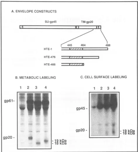
FIG. 1. Schematic representation of the HTE-438 mutant (A)
and results ofimmunoprecipitation of the envelope proteins
pro-ducedby the wild-typeexpressorand the HTE-438mutant(B).The putative anchorage domain, located between amino acids445 and
464,is indicated (hatched region). In HTE-438, a nonsense codon
was introduced at position 438. Lanes 1 and 3, transfected-cell lysates; lanes2and4, transfected-cell supernatants;lanes 1 and2, wild-type expressor;lanes 3 and 4, HTE-438.
The intracellular maturation of the HTLV-I envelope mutants(Fig. 2) havingprematuretermination codons in the IC domain was analyzed after transfection of the
corre-sponding vectors into COS-1 cells (10), K562 cells (20) (derived fromapatientwith chronic myeloidleukemia with
a Philadelphia chromosome), HSB2 cells(16), or 1301 cells
(31) (derived from a patient with T-cell leukemia). K562, 1301, and HSB2 cells were transfected by electroporation
with10 ,ugofplasmids addedto107cells in250,ul of culture medium. Thepulsewasperformed at250 V and 960,F. At 48 hposttransfection, cellswereused for
immunoprecipita-tion with aTSPpatient's serumafterradiolabeling orfora
syncytium assayasdescribedpreviously (25).
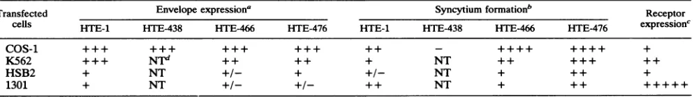
Figure 2Bshows that inCOS-1cells, the cleavage of the
precursorproteinwas asefficientfor the mutantsasfor the
wild-type envelopeexpressor.Asaresultofthe introduction
ofpremature stop codons, the molecular sizes ofboth the
precursor protein and the TMprotein in the mutantswere
lower than those for thewild type (Fig. 2B, lanes 2 to 4). Pulse-chase experiments showed that the kinetics of
enve-lope synthesis and of precursor cleavage are comparable
among envelopes with a mutated IC domain and the
wild-typeprotein(datanotshown). SU and TMproteintransport to theplasma membranewas also identicalamongmutated
andwild-type envelopes, as shown bycell surface labeling
[image:2.612.78.283.80.371.2]experiments usingiodine and lactoperoxidaseaspreviously
FIG. 2. Schematicrepresentationof the HTE-466 andHTE-476
mutants(A)and intracellular maturation(B)andtransporttothe cell surface (C) of the envelope proteins produced by the mutants. Proteinswereobtained fromCOS-1cells transfected withanegative
control (lanes 1), HTE-1wild-type envelope expressor (lanes 2),
HTE-466(lanes 3),andHTE-476(lanes 4).Theputativeanchorage
domain is shown(hatched region).
described (24) (Fig. 2C, lanes 2 to 4). This result also confirms that theregionbetween amino acids 438 and 466 is sufficient to allow normal membrane integration of the envelope proteins.
Since in lentiviruses the IC domain modulates the
enve-lope maturation in acell type-dependent manner, the func-tion of the HTLV-I IC domainwas analyzed in apanel of differentcell lines. In thesecells,thetransfectabilityisvery
low,and HTLV-Ienvelope expressioncould be detectedby immunoprecipitation only in K562 cells (data not shown). Western blot (immunoblot) analysis (using a TSPpatient's
serum)hadtobeperformedforcorrectdetection of thegp6l envelopeprecursorin K562, HSB2, and 1301 cells(Fig. 3). Equal amounts of proteins (as measured by a Bio-Rad protein assay) were loaded in the lanes of the sodium dodecyl sulfate-polyacrylamide gel.Twoindependent
exper-imentswere performed. These experiments show that the amounts of precursor detected in two of these cell lines (K562and 1301) arelower for themutant proteinsthan for the wild type(compare,ineachcase,lanes 3 and4with lane 2 [Fig. 3]), as opposed to what was observed with COS-1
cells(Fig. 2B).
The syncytium formation capacities of the mutant and wild-type envelopes werecompared after transfection into
the different cell lines described above. COS-1-transfected cellswerecocultivated withRSV-transformed XC cells(29),
which form large numbers of syncytia and are used as
indicator cells aspreviouslydescribed(25). For cells
grow-inginsuspension, anotherindicator cell linewasused, since syncytiaare moreeasilymonitored when both thetested and A. ENVELOPE CONSTRUCTS
SU-gp45 TM-gp20
445 464 488
HTE-1
438 HTE-438
B.CELL-ASSOCIATEDAND SECRETED ENVELOPE PROTEINS
1 2 3 4
gp6l-9 ._
gp45- _
gp2l
--gp15
A.ENVELOPE CONSTRUCTS
SU-gp45 TM-gp2O
445 464 488
HTE-1
HTE-476
HTE-466
B.METABOLIC LABELING C. CELLSURFACELABELING
1 2 3 4 1 2 3 4
gp6l-gp45
-gp20- * ]
gp20-.. l B
on November 9, 2019 by guest
http://jvi.asm.org/
[image:2.612.320.556.81.341.2]HSB2 1301 K562
1 2 3 4 1 2 3 4 1 2 3 4
gp61-
*FIG. 3. Expression of envelope proteins after transfection of human cell lines with the HTE-466andHTE-476 mutants.Western blot analysiswasperformedwith aTSP patient'sserum.Cellswere transfected with a negative control (lanes 1), HTE-1 wild-type expressor(lanes 2),HTE-476(lanes 3),orHTE-466(lanes4).
theindicatorcells grow insuspension. The assayconsisted ofacoculture of 2 x 10⁵ transfected cells (1301, HSB2, or K562) together with106indicator cells(1301). Syncytia were scored after 24 h ofcoculture, and four independent
exper-imentswereperformed in each case. As expected, HTE-438, the anchorage-negative mutant, was unable to elicit syncy-tium formation when transfected into COS-1 cells(Table 1). A marked increase in syncytium formation capacity was observed in the two mutants with a deleted IC region,
HTE-466and HTE-476, compared with the wild type when
COS-1orK562cellsweretransfectedand,to alesser extent, when HSB2 cells were transfected(Table1). Transfection of
1301cells with the mutants,however,resulted insyncytium
formation comparable to that observed with thewild-type envelope (HTE-476)orslightly diminished syncytium forma-tion(HTE-466).The addition ofaTSPpatient'sserum tothe coculture resulted in the inhibition ofsyncytium formation with both thewild-typeand mutant envelopes, whereas the addition of normalserum tothe coculture hadnoeffect(data notshown). Takentogether, these results show that theIC domain truncation results in unchangedordiminished (1301
transfected cells) or augmented (COS-1, K562, and HSB2
cells) syncytium formation capacity, with the phenotype
conferred by the mutation being cell type dependent. The
level of syncytium formation did not correlate with the amountofdetectableenvelopein the differentcells, since in K562 cells, for example, the mutants expressed smaller amountsofenvelopethan the wild type but formedsyncytia moreefficiently (Fig. 3 and Table1).The level ofsyncytium
formation might be inversely correlated to the level of receptorexpressionreflected in the abilities of the
nontrans-fected cell linestoform syncytia with infected cells (Table 1).
We have analyzed the role of the HTLV-I envelope IC domain in the envelope functions and localized the
anchor-age domain of the envelope atthe plasma membrane. Our results show that when a nonsense codon is introduced at
position 438 of the envelope protein, both SUgp45 and truncated TM are released in the supematant, whereas a nonsensecodon introducedatposition 466 allows anchorage at the cell surface. This confirms the sequence analysis
which predicts that the anchorage region corresponds to a
cluster ofhydrophobic amino acidslocated between amino acids 446 and 465. As expected,nosyncytiumformationwas
observedwith the secreted envelope encoded bythe HTE-438mutant.
In COS-1 cells, in the absence of the IC domain, no
modificationinthe kinetics of the envelopeprecursor
cleav-ageorthetransportof the HTLV-Ienvelope productsatthe cell surfacewasobserved. InK562 and 1301 cells,however, apartialorcompletetruncation ofthe IC domain(12or21of
22 amino acids in the case of the wild-type IC domain) resultsin markedly diminished envelope expression. These results show the cell typedependency of the IC domain in envelopeintracellular maturation. Similarresultshavebeen obtainedfor HIV-1(8, 11). For RSV, the IC domain is totally dispensablefornormal maturation of theenvelope proteins, at least in the cells testedsofar(23). Whether thecelltype dependencyof the ICdomain'seffectuponenvelope matu-ration is a general feature of retroviruses remains to be established.
We have examined the role ofthe HTLV-I envelope IC domain in the envelope functions by testingthe abilities of cells withmutant proteins toform syncytia. Depending on
the transfected cell type, truncation of the IC resulted in unchanged (transfected 1301 cells) or augmented (COS-1, HSB2, and K562cells) syncytiumformationcapacity ofthe envelope. The IC domain is thusdispensable for the
enve-lope-mediated bindingand fusioncapacity.However,the IC domain can modulate theenvelope functions inacell type-dependent manner. The augmented syncytium formation
observed with the truncated envelopes was not due to differences in the envelope transport to the cell surface, at least not in the COS-1 system, in which the intracellular maturation could befullystudied.
The observed augmented syncytium formation could be due toanincreased affinityof theenvelopefor its receptor,
aswas previously shown with HIV-2 (14). Inour HTLV-I
envelope mutants, the binding capacity of the envelope cannotbe examineddirectly,since there isnobindingassay
available yet. One indirect argument would suggest
in-TABLE 1. Envelope expression andsyncytiumformation in different celllines
Transfected Envelopeexpressiona Syncytiumformationb Receptor
cells HTE-1 HTE-438 HTE-466 HTE-476 HTE-1 HTE-438 HTE-466 HTE-476 expression'
COS-1 +++ +++ +++ +++ + ++++ +
K562 +++ Nt ++ ++ + NT ++ +++ ++
HSB2 + NT +/- + +/- NT + + + +
1301 + NT +/- +/- ++ NT + ++ +++++
a Results fromimmunoprecipitationandWesternblotexperiments(Fig.1to3).
bNumbersofsyncytiaperwellwerescored as follows: + + + ++, >80;+ + ++,>60;+++, >40;++,>20; +,>10;+/-, >5;-, 0.
cEvaluatedbythesyncytiumformationcapacityof the cell linewith HTLV-1-infectedcells.Numbersofsyncytiaperopticalfield of the invertedmicroscope
arescored asfollows: +++++,>80; ++,>20;+, >10.
dNT,nottested.
on November 9, 2019 by guest
http://jvi.asm.org/
[image:3.612.64.300.83.208.2] [image:3.612.63.560.618.688.2]creased receptoraffinity of the truncated HTLV-Ienvelope.
As stated above, augmented syncytium formation is ob-served only when the constructs are transfected in cells expressing small amounts of receptor butnotwhentheyare transfected in a cell line (1301 cells) expressing very large
amounts of receptor. The syncytiaform betweenan indica-torcell line (expressing theputative HTLV-Ireceptor) and an envelope-transfected test cell line. However, the enve-lope-transfected cells also endogenously express the puta-tive receptor at various levels.
Envelope-transfected
cell lines which express high levels of receptor(like 1301cells)
will have thecapabilityofsequestering largeamountsof the transfectedenvelope.Incontrast,cells
expressing
lowlevels of receptor(COS-1, K562, andHSB2)willbe, as testcells,very sensitive totheaffinityofthe envelope. Inthese cells, an increase in affinity will result in increased
syncytium
formation, such as that observed with the truncation of the ICdomain.
An alternative explanation for the observed augmented syncytium formation indistinct cells would be anincreased fusion capacityof the IC mutants, asobservedwith
HIV-2,
inwhichcytoplasmic-domaintruncation enhances the fusion
activityin a cell type-dependentmanner
(21).
Arole ofthe IC domain in the viralcell-to-celland cell-freetransmissionshas also been proposed (3, 17); these are affected when the IC domain is absent in HIV-1
(12).
These results were suggested to reflect the implication of the IC domain inpostfusionevents, since
syncytium
formationwas not mod-ified. Again, arole for the IC domain inparticle
release or viral transmission cannot be directly tested with HTLV-I because of the absence of an available infectiousproviral
clone.
Altogether, the results suggest a complex role for the IC domain of theenvelopeinretroviruses;the IC domain could be implicated in the following processes:
(i)
intracellular maturation in acelltype-dependent manner;(ii)
viral-parti-cle release, perhaps because of
required
interactions with thecore proteins, as shownrecentlywith HIV-1(32)
and a type D retrovirus(26); (iii) envelope-receptor
affinity
andmaybe, consequently, in the cytopathic effects; and
(iv)
postfusioneventsnecessaryfor viral entry. Incontrast tothe situationdescribed above, in RSV, however
(23),
no differ-ence in infectivity between virionsbearing
wild-type
enve-lopes and those bearingICdomain-deletedenvelopescould be observed. As discussed herein, our results showingincreasedsyncytium formation when the HTLV-I truncated envelope isexpressedin COS-1 orK562cells tendto favor the hypothesisofa functional role for this short domain in
HTLV-I,as waspreviouslyshown withprimatelentiviruses. This role remains to be specified for precise stages of the viral lifecycle.
Finally, in previous studies we have shown that the HTLV-I envelope is very sensitive to mutagenesis, since artificial introduction of mutations results inanonfunctional
protein in most cases. The IC domain, however, is an exception, since insertions of amino acids in this region (25) have no effect on syncytium formation. As shown here,
however,apartial truncationofthe IC domaincanmodulate
syncytium formation. This could explainwhy this domain,
althoughveryshort inHTLV-1, is very conserved andnever truncated in the natural strains.
We thankAnnick Harel-Bellan for criticalreadingof the
manu-scriptandhelpful suggestions.WethankAndrew Lever and J. Coste forprovidinguswithpatients'sera.
This workwassupported byagrant from theAgenceNationalede la RecherchesurleSIDA,Paris,France.
REFERENCES
1. Autiero, M.,P.Abrescia,M.Dettin,C. DiBello,andJ. Guardi-ola.1991.BindingtoCD4 ofsynthetic peptidespatternedonthe principal neutralizing domain of the HIV-1 envelope protein.
Virology185:820-828.
2. Berman, P.W.,W. M.Nunes,and0.K.Haffar.1988. Expres-sion ofmembrane-associated and secretedvariants ofgpl60of humanimmunodeficiencyvirustype 1 in vitroandincontinuous celllines. J. Virol.62:3135-3142.
3. Chakrabarti, L.,M.Emerman,P.Tiollais,and P.Sonigo.1989. The cytoplasmic domain of simian immunodeficiency virus transmembraneproteinmodulatesinfectivity.J.Virol. 63:4395-4403.
4. Cullen,B. R.1987. Useof
eukaryotic
expressiontechnology
in the functional expressionofclonedgenes. MethodsEnzymol.
152:684-704.
5. Dokhelar,M.C.,H.Pickford,J.Sodroski,and W. A. Haseltine. 1989. HTLV-Ip27rexregulatesgagandenvprotein
expression.
J. AcquiredImmuneDefic. Syndr.2:431-440.
6. Dorner,A.J.,andJ.M.Coffin.1986.Determinantsforreceptor interaction and cellkillingontheavianretrovirus
glycoprotein
gp85.Cell 45:365-374.
7. Earl, P. L., S. Koenig, and B. Moss. 1991.
Biological
and immunological properties of human immunodeficiency virus type 1envelopeglycoprotein:analysisofproteins
with trunca-tionsanddeletionsexpressed byrecombinantvacciniaviruses. J. Virol. 65:31-41.8. Gabuzda,D.H.,A.Lever,E.Terwilliger,andJ.Sodroski.1992. Effects of deletions in the cytoplasmic domain on
biological
functions of human immunodeficiencyvirus type 1
envelope
glycoproteins.J.Virol.66:3306-3315.
9. Gallaher,W.R.1987. Detectionofafusionpeptidesequence in the transmembraneproteinofhumanimmunodeficiencyvirus. Cell 80:327-328.
10. Gluzman,Y. 1981. SV40-transformed simian cellssupport the replicationofearlySV40mutants. Cell23:175-182.
11. Haffar, 0. K., D.J.Dowbenko, and P. W. Berman. 1991. The cytoplasmictailof HIV-1gpl60contains regionsthat associate withcellularmembranes.Virology180:439-441.
12. Helseth,E.,M.Kowalski,D.Gabuzda,U.Olshevsky,W. Hasel-tine, and J. Sodroski. 1990. Rapid
complementation
assays measuring replicative potential of human immunodeficiency virustype 1envelopeglycoproteinmutants.J. Virol. 64:2416-2420.13. Helseth,E., U.Olshevsky, D. Gabuzda,B.Ardman,W. Hasel-tine, and J. Sodroski. 1990. Changes in the transmembrane regionof the humanimmunodeficiencyvirustype1
gp4l
enve-lope glycoprotein affect membrane fusion. J. Virol. 64:6314-6318.
14. Hoxie,J.A., L. F.Brass,C. H.Pletcher, B.S.Haggarty,and B. H. Hahn.1991. Cytopathicvariantsofanattenuatedisolate of human immunodeficiency virus type 2 exhibit increased affinityforCD4.J. Virol. 65:5096-5101.
15. Hunter, E., and R. Swanstrom. 1990. Retrovirus envelope
glycoproteins. Curr.Top.Microbiol. Immunol.157:187-253. 16. Kasahara, T.,N.Mukaida,N.Hatake,K.Motoyoshi,T.Kawai,
and K. Shiori-Nakano. 1985. Interleukin 1 (IL1) dependent
lymphokine production by human leukemic T cell line HSB.2 subclones. J. Immunol. 134:1682-1689.
17. Kodama, T., D. P. Wooley,Y. M. Naidu, H. W. Kestler III, M. D. Daniel,Y.Li,and R.C. Desrosiers. 1989. Significanceof premature stop codons in env of simian immunodeficiency virus. J. Virol. 63:4709-4714.
18. Kowalski,M., J.Potz,L.Basiripour,T.Dorfman,W. C.Goh,E.
Terwilliger,A.Dayton,C.Rosen,W.Haseltine,andJ.Sodroski. 1987.Functionalregionsof theenvelope glycoproteinof human immunodeficiencyvirus type 1. Science 237:1351-1355. 19. Lasky, L. A., G. Nakamura, D. H. Smith, C. Fennie, C.
Shimasaki,E.Patzer,P.Berman,T.Gregory,andD.J.Capon. 1987.Delineation ofaregion of the humanimmunodeficiency
on November 9, 2019 by guest
http://jvi.asm.org/
virus type 1 gpl20glycoproteincriticalforinteraction with the CD4 receptor. Cell50:975-985.
20. Lozzio, C. B., and B. B. Lozzio.1975.Human chronic myelog-enous leukemia cell line with positive Philadelphia chromo-some. Blood 45:321-334.
21. Mulligan, M. J., G. V.Yamshchikov, G. D. Ritter, F. Gao, M. J. Jin, C. D.Nail, C. P. Spies, B. H. Hahn, and R. W. Compans. 1992. Cytoplasmicdomaintruncationenhances fusion activity by the exterior glycoprotein complex of human immunodefi-ciency virus type 2 in selected cell types. J. Virol. 66:3971-3975. 22. Page, K. A., S. M. Stearns, and D. R. Littman. 1992.Analysis of mutations in the V3 domain of gpl60 that affect fusion and infectivity.J.Virol. 66:524-533.
23. Perez, L. G., G. L. Davis, and E. Hunter. 1987. Mutantsofthe Rous sarcoma virusenvelopeglycoprotein thatlackthe trans-membrane anchor and cytoplasmic domains: analysisof intra-cellulartransportandassemblyintovirions.J. Virol. 61:2981-2988.
24. Pique, C., D. Pham, T. Tursz, and M. C. Dokhelar. 1992. HumanT-cellleukemiavirus type Ienvelope proteinmaturation process: requirements for syncytium formation. J. Virol. 66: 6524-6534.
25. Pique, C., T. Tursz, and M. C. Dokhelar. 1990. Mutations introduced along the HTLV-I envelope gene result in a non-functionalprotein:abasisforenvelopeconservation? EMBO J. 9:4243-4248.
26. Rhee,S. S., and E. Hunter.1990. Asingle amino acid
substitu-tion within the matrixproteinofatypeDretrovirusconvertsits morphogenesis tothatofaC retrovirus.Cell63:77-86. 27. Seiki, M., S. Hattori, Y. Hirayama, and M. Yoshida. 1983.
Human adult T-cell leukemia virus: complete nucleotide se-quenceof theprovirusgenomeintegratedin leukemia cell DNA. Proc. Natl. Acad. Sci. USA80:3618-3622.
28. Stoye, J. P., and J. M. Coffin. 1987. The four classes of endogenousmurine leukemia virus: structuralrelationshipsand potential for recombination. J. Virol.61:2659-2669.
29. Svoboda, J., P. Chyle, D. Simkovic, and I. Hilgert. 1963. Demonstration of the absence of infectious Rous virus in rat
tumor XC, whose structurally intact cells produce Rous
sar-comawhen transfectedtochicks.Folia Biol. (Warsaw) 9:77-81. 30. Tanaka, Y., L. Zeng, H. Shiraki, H. Shida, and H. Tozawa. 1991. Identification ofaneutralizationepitope on the envelope gp46 antigen of human T cell leukemia virus type I and induction of neutralizing antibody by peptide immunization. J. Immunol. 147:354-360.
31. Wiels, J., G. M. Lenoir, M. Fellous, M.Lipinski, J. C. Salomon, C.Tetaud, and T. Tursz. 1982. A monoclonal antibody with anti-Burkitt lymphoma specificity. I. Analysis of human hae-matopoietic andlymphoid cell lines. Int. J. Cancer 29:653-658. 32. Yu, X., X. Yuan, Z. Matsuda, T.-H. Lee, and M. Essex. 1992. The matrix protein of humanimmunodeficiency virus type I is required for incorporation of viral envelope protein intomature
virions.J. Virol. 66:4966-4971.