• No results found

Analysis of the cleavage site of the human immunodeficiency virus type 1 glycoprotein: requirement of precursor cleavage for glycoprotein incorporation.

N/A
N/A
Protected

Academic year: 2019

Share "Analysis of the cleavage site of the human immunodeficiency virus type 1 glycoprotein: requirement of precursor cleavage for glycoprotein incorporation."

Copied!
8
0
0

Loading.... (view fulltext now)

Full text

(1)

0022-538X/95/$04.0010

Copyright 1995, American Society for Microbiology

Analysis of the Cleavage Site of the Human Immunodeficiency

Virus Type 1 Glycoprotein: Requirement of Precursor

Cleavage for Glycoprotein Incorporation

JOHN W. DUBAY, SUSAN R. DUBAY, HAE-JA SHIN,ANDERIC HUNTER*

Department of Microbiology, University of Alabama at Birmingham, Birmingham, Alabama 35294

Received 12 October 1994/Accepted 8 May 1995

Endoproteolytic cleavage of the glycoprotein precursor to the mature SU and TM proteins is an essential step in the maturation of retroviral glycoproteins. Cleavage of the precursor polyprotein occurs at a conserved, basic tetrapeptide sequence and is carried out by a cellular protease. The glycoprotein of the human immu-nodeficiency virus type 1 contains two potential cleavage sequences immediately preceding the N terminus of the TM protein. To determine the functional significance of these two potential cleavage sites, a series of mutations has been constructed in each site individually, as well as in combinations that altered both sites simultaneously. A majority of the mutations in either potential cleavage site continued to allow efficient cleavage when present alone but abrogated cleavage of the precursor when combined. Despite being trans-ported efficiently to the cell surface, these cleavage-defective glycoproteins were unable to initiate cell-cell fusion and viruses containing them were not infectious. Viruses that contained glycoproteins with a single mutation, and that retained the ability to be processed, were capable of mediating a productive infection, although infectivity was impaired in several of these mutants. Protein analyses indicated that uncleaved glycoprotein precursors were inefficiently incorporated into virions, suggesting that cleavage of the glycoprotein may be a prerequisite to incorporation into virions. The substitution of a glutamic acid residue for a highly conserved lysine residue in the primary cleavage site (residue 510) had no effect on glycoprotein cleavage or function, even though it removed the only dibasic amino acid pair in this site. Peptide sequencing of the N terminus of gp41 produced from this mutant glycoprotein demonstrated that cleavage continued to take place at this site. These results, demonstrating that normal cleavage of the human immunodeficiency virus type 1 glycoprotein can occur when no dibasic sequence is present at the cleavage site, raise questions about the specificity of the cellular protease that mediates this cleavage and suggest that cleavage of the glycoprotein is required for efficient incorporation of the glycoprotein into virions.

During transport of retroviral envelope glycoproteins through the secretory pathway, the envelope precursor polyprotein is cleaved by a cellular enzyme into the surface (SU) and transmembrane (TM) subunits. This cleavage of retroviral glycoproteins is an essential step in the maturation of the glycoprotein to a functional complex (for reviews, see ref-erences 18 and 23). It results in the activation of hydrophobic fusion sequences that allow the viral membrane to fuse with the cellular membrane upon binding of the appropriate recep-tor. In the absence of cleavage of the precursor polyprotein, the virus is unable to enter cells via the normal mechanism of interacting with its cellular receptor and fusing with the cell membrane (2, 22). Cleavage of the glycoprotein precursor thus represents a step in the viral life cycle that could serve as a target for the design of antiviral therapies. Consequently, it is important to fully understand the sequence elements required for the functional cleavage of the glycoprotein precursor.

The cleavage of retroviral glycoprotein precursor molecules occurs at a tetrapeptide sequence that consists predominantly of basic residues and which generally conforms to an Arg-X-Lys/Arg-Arg consensus sequence. While several mutational analyses have been performed on retroviral glycoproteins in order to understand the sequence requirements for cleavage of the precursor, only a limited amount of information is avail-able about the enzymes that catalyze this cleavage (2, 3, 22, 26). It is known that the cleavage is catalyzed by a cellular protease

located in the Golgi complex (32, 38) which is normally re-sponsible for processing cellular precursors containing basic residues at the cleavage sites. One well-characterized enzyme having such activity is the Kex2 protease of the yeast

Saccha-romyces cerevisiae, which cleaves at Lys-Arg and Arg-Arg sites.

Similar proteases have been identified in mouse and human cells (for a review, see reference 1). One of these, furin, has been implicated in the cleavage of influenza virus hemaggluti-nin at a cleavage site that resembles the retroviral envelope glycoprotein cleavage site consensus sequence (34). Furin is of particular interest because while it cleaves at a dibasic peptide it requires an arginine residue at the24 position (17). More-over, recent data from experiments in which furin was ex-pressed in trans from a vaccinia virus showed that this enzyme could cleave the human immunodeficiency virus type 1 (HIV-1) glycoprotein (15). On the other hand, recent experi-ments with cell lines with defects in the furin gene indicate that the HIV-1 glycoprotein precursor can still be processed in its absence (25). At least five additional mammalian furin-like proteases have been identified (28, 35); of these, PC1-PC3 (29, 30) and PC2 (31) have similar substrate specificities but don’t require an arginine residue at the 24 position for cleavage (17).

The requirements for the cleavage of the envelope glycop-rotein of HIV-1 are of particular interest because the HIV-1 glycoprotein possesses two potential cleavage sequences imme-diately upstream of the gp41 N terminus. We were interested, therefore, in determining the contributions of both potential cleavage sites to the cleavage of the precursor polyprotein.

* Corresponding author. Phone: (205) 4321. Fax: (205) 934-1640.

4675

on November 9, 2019 by guest

http://jvi.asm.org/

(2)

MATERIALS AND METHODS

Cell culture and transfections.COS-1 cells were obtained from the American Type Tissue Collection (Rockville, Md.). HeLa-T4 and H9 cells were obtained from the AIDS Reference and Reagent Program, Division of AIDS, National Institute of Allergy and Infectious Diseases, and were originally contributed by Richard Axel and Robert Gallo, respectively. COS-1 and HeLa-T4 cells were maintained in Dulbecco’s modified Eagle medium containing 10% fetal bovine serum, and H9 cells were maintained in RPMI 1640 containing 15% fetal bovine serum. Transfection of COS-1 and HeLa-T4 cells was performed with DEAE-dextran at a concentration of 1.0 or 0.5 mg/ml, respectively.

Oligonucleotide-directed mutagenesis and DNA manipulations.Mutagenesis was performed essentially as described by Kunkel (21). The SalI-to-BamHI fragment of the molecular HIV-1 clone pHXB2Dgpt was cloned into M13mp19, and uracil-containing single-strand DNA was obtained by using Escherichia coli CJ236. The annealing and synthesis reactions were performed essentially as described previously (21). For the construction of the CS3, CS5, and CS7 mu-tations, uracil-containing single-strand DNA was prepared from the CS1 mutant and used for mutagenesis. Enzymes for DNA manipulations were obtained from New England Biolabs (Beverly, Mass.) and used according to manufacturer’s instructions or standard techniques. All plasmid DNA used for transfections was purified on cesium chloride gradients.

Protein expression and metabolic labeling.The mutant env genes were cloned into the pSRHS expression vector which has been described elsewhere (5). This vector contains the HIV-1 env gene under control of the simian virus 40 late promoter. In addition, it contains the Mason-Pfizer monkey virus 39long termi-nal repeat to provide polyadenylation sigtermi-nals. For biological atermi-nalysis, COS-1 cells were transfected as described above, and 48 h later, the cells were starved for 15 min in methionine- and cysteine-deficient Dulbecco’s modified Eagle medium. The cells were then pulse-labeled for 30 min with 100mCi of [35

S]methionine-cysteine mix (EXPRE35S35S Protein Labeling Mix; Dupont NEN, Boston, Mass.) in the same medium and either lysed immediately (pulse samples) or then chased for 3 h in complete medium (chase samples) before the addition of lysis buffer (1% Nonidet P-40, 0.1% sodium dodecyl sulfate [SDS], and 0.5% sodium de-oxycholate in phosphate-buffered saline [PBS]). The cell lysates were incubated with serum from an HIV-1-infected individual, and then 30ml of a 10% (wt/vol) suspension of fixed Staphylococcus aureus was added and incubated for an ad-ditional 15 min. The complexes were washed extensively and boiled in SDS sample buffer before being loaded onto an 8% polyacrylamide gel.

Peptide sequencing.For peptide sequence analysis, COS-1 cells were trans-fected with either the wild-type or mutant env genes in the pSRHS vector. Two days after transfection, the cells were labeled with 500mCi of [3H]leucine and 100mCi of [35S]methionine for 30 min and then chased with complete medium for 3 h. The cells were lysed, and immune complexes were collected as described above. The samples were run on an SDS–8% polyacrylamide gel and transferred onto polyvinylidene difluoride membrane (Immobilon-P; Millipore Corp., Bed-ford, Mass.), and the bands were visualized by autoradiography. The bands corresponding to gp41 were excised, and N-terminal sequence analysis was per-formed with sequential Edman degradation in an ABI 470A automated se-quencer (Applied Biosystems, Foster City, Calif.). The sample from each cycle of the sequencer was dried and dissolved in scintillation fluid. The counts in each sample were determined with an LKB scintillation counter.

Immunoprecipitation with C-terminal antibody.COS-1 cells were transfected and labeled with [35

S]methionine-cysteine mix as described above. The cells were lysed as described above and incubated for 1 h with a sheep antibody made to a synthetic peptide corresponding to amino acids 497 to 511 of gp160 (D6205; International Enzymes, Fallbrook, Calif.) shown in Fig. 1A. A rabbit anti-sheep immunoglobulin G antibody was added and followed by fixed S. aureus to pre-cipitate the antibody. The prepre-cipitates were washed and analyzed on an SDS-polyacrylamide gel (8% SDS-polyacrylamide) and visualized by fluorography.

Surface immunofluorescence.To analyze the surface-expressed glycoprotein, COS-1 cells were seeded onto glass coverslips and transfected 24 h later. Two days after transfection, the cells were subjected to surface immunofluorescence as previously described (5) with a monoclonal antibody which is specific for the V3 loop region of gp120. The cells were examined and photographed with a Zeiss

was washed three times with PBS containing 0.1% Tween 20 and incubated with a 1:2,000 dilution of horseradish peroxidase-conjugated streptavidin (Southern Biotech, Birmingham, Ala.) for 1 h at room temperature. The membrane was then washed six times with PBS before chemiluminescence detection with the Renaissance system (Dupont NEN) as described by the manufacturer. The blot was then allowed to sit for 1 day to exhaust any chemiluminescence activity before being exposed to film for detection of the35

S-labeled protein. In addition, the35

S-labeled proteins on the membrane were quantitated with an AMBIS radioanalytic imaging system (AMBIS Systems Inc., San Diego, Calif.).

HeLa-T4 cell fusion assay.HeLa-T4 cells were transfected with the pSRHS vector containing either wild-type or mutant env genes. Two days after transfec-tion, the cells were stained by the May-Grunwald technique as described previ-ously (5).

Infectivity assay of mutant virions.For infectivity assays, the mutant env genes were cloned into the infectious molecular clone pHXB2Dgpt (8) (provided by Beatrice Hahn, University of Alabama at Birmingham) by using the unique SalI and BamHI restriction enzyme sites. The mutants were confirmed by sequencing, and the constructs were transfected into COS-1 cells. The culture medium was harvested from the cells 48 h after transfection and adjusted to equivalent reverse transcriptase (RT) values by dilution with normal medium. To infect cells, 1 ml of the RT-adjusted supernatant was added to 1 ml of medium containing 33106H9 cells. The cells were incubated for 2 h at 378C, after which time the cells were transferred to a 25-cm2flask and the medium was adjusted to 10 ml. The cultures were split at the times indicated in Fig. 6. Aliquots from each time point were frozen and analyzed for RT activity. RT activity was measured by a microassay described previously (5) and quantitated with an AMBIS radio-analytic imaging system (Ambis Systems Inc.).

Glycoprotein incorporation into virions.For the analysis of glycoprotein in-corporation into virions, a pol-defective genome derived from pHXB2Dgpt was used. This construct, pFN, has been described elsewhere (6). The mutant env genes were cloned into pFN by using SalI and BamHI sites, and the mutants were confirmed by sequencing. The pFN constructs were transfected into 100-mm plates of COS-1 cells, and at 36 h posttransfection, the cells were labeled with 750

mCi of [35S]methionine-cysteine mix for 90 min. The labeling mix was then removed and replaced with complete medium, and the incubation was continued for 12 h. The supernatant was collected and spun at low speed to remove debris. The clarified supernatant was layered over a 15% sucrose cushion and centri-fuged at 100,0003g for 2 h in an SW41 rotor. The pellets were collected and immunoprecipitated as described above. The immune complexes were analyzed by SDS-PAGE (10% acrylamide) and visualized by fluorography.

RESULTS

Construction of mutants and glycoprotein expression in

COS-1 cells. The sequence requirements for the cleavage of

retroviral glycoproteins have been well studied (2, 3, 11, 12, 22, 26). The human immunodeficiency viruses are particularly in-teresting in that they contain two potential sites for cleavage of the glycoprotein precursor to the functional SU and TM sub-units (Fig. 1A). For the purpose of identifying the two sites, we have designated the C-terminal site as the primary cleavage site and the upstream site as the secondary site as shown in Fig. 1B. In order to examine the contribution of each of the two potential cleavage sites, we have constructed a series of muta-tions that alter the primary sequence of each of the sites indi-vidually or both sites in combination (Fig. 1B). In each case, the mutations were designed to remove sequence-adjacent ba-sic residues (dibaba-sic pairs) without interfering with the propen-sity of the region to form a turn on the surface of the molecule. The mutant env genes were cloned into a simian virus 40-based expression vector described previously (5). To determine

on November 9, 2019 by guest

http://jvi.asm.org/

(3)

the effects of these mutations on the synthesis and proteolytic processing of the mutant glycoproteins, COS-1 cells were transfected with the wild-type and mutant constructs and ei-ther pulse-labeled for 30 min or pulse-labeled and then chased for an additional 3 h. Cell lysates were prepared and immuno-precipitated with serum from an HIV-1-infected individual. The immune complexes were analyzed by 8% acrylamide SDS-PAGE (Fig. 2). In the case of the wild-type protein, the pre-cursor molecule, gp160, can be seen in the pulse lane (Fig. 2, WT, P) and the gp160 and gp120 bands are evident in the sample that was chased for an additional 3 h, indicating cleav-age of the precursor glycoprotein (Fig. 2, WT, P/C). The effects of the mutations on glycoprotein processing are shown in Fig. 2. The CS1 mutation which disrupts the pair of basic amino acids at the primary site had no detectable effect on the cleav-age of the precursor in this assay, although in kinetic assays this mutation was shown to slow the rate of precursor cleavage (data not shown). Mutations at the secondary site alone also resulted in the synthesis of glycoproteins that were cleaved in a manner similar to that of the wild-type glycoprotein (Fig. 2, lanes CS2, CS4, and CS6). For each of these cleaved mutants, however, we did observe a small amount of terminally glyco-sylated gp160 in addition to normal levels of gp120 and gp41 after the chase period, suggesting that each of the mutations allowed a fraction of the precursor molecules to reach the cell surface uncleaved.

Mutants in which changes were made at both potential cleavage sites were either poorly cleaved (CS3, which retains a dibasic pair in the 28site) or were unable to be cleaved (CS5 and CS7) as shown in Fig. 2. The uncleaved glycoproteins that accumulated in the chase showed an increase in their apparent molecular weight because of the terminal glycosylation of the precursor as it is transported through the secretory pathway. Others had reported, in contrast to our results for CS1, that disrupting the pair of basic amino acids in the primary cleavage site by substituting serine or threonine for the most C-terminal arginine (residue 511) resulted in an uncleaved precursor (2, 12). Our results confirm that a mutation which changes argi-nine-511 to a serine does block cleavage of the HIV-1 glyco-protein precursor (Fig. 2, lanes CS8) and demonstrate that the roles of lysine-510 and arginine-511 are distinct in defining the cleavage site.

Surface expression of wild-type and mutant glycoproteins.

(i) Surface immunofluorescence.The lack of proteolytic

cleav-age of the CS3, CS5, CS7, and CS8 mutant precursor glycop-roteins raised the possibility that these glycopglycop-roteins were not being transported through the secretory pathway to compart-ments in the Golgi complex where the glycoprotein could in-teract with the cleavage enzymes. To rule out the possibility that these mutations were affecting glycoprotein transport, we analyzed by immunofluorescence the glycoprotein on the sur-face of unfixed COS-1 cells transfected with either the wild-type or mutant-containing pSRHS expression vector as de-scribed in Materials and Methods.

[image:3.612.319.553.72.310.2]

Expression of the wild-type glycoprotein resulted in low-intensity staining around the periphery of expressing cells (Fig. 3A), and a similar level of fluorescence was seen with the CS1 construct (data not shown). In contrast, an unusually high level of surface immunofluorescence was seen with cells expressing FIG. 1. Diagram of mutations in the cleavage region of the HIV-1

glycopro-tein. (A) The amino acid sequence of the region encompassing the two potential cleavage sites is shown. The site of cleavage is indicated by the arrow, and the numbers above the sequence represent the amino acid position relative to the cleavage site. The region indicated by the double underline represents the pep-tide to which the antiserum used for Fig. 8 was raised. (B) A schematic repre-sentation of the mutations and their designations. Indicated above the wild-type (WT) sequence is the location of the sites designated as the primary (18) and secondary (28) potential proteolytic cleavage sites. The mutated residues are shown in boldface.

FIG. 2. Expression of mutant glycoproteins in COS-1 cells. COS-1 cells were transfected with the pSRHS expression vector containing wild-type (WT) or mutant env genes and pulse-labeled with [35

S]methionine for 30 min (P lanes) or pulse-labeled and chased for 3 h (P/C lanes). Cell lysates were prepared and immunoprecipitated with serum from an HIV-1-infected individual. The immune complexes were analyzed by SDS-PAGE and visualized by fluorography. The mutant designation is shown above the lanes, and the positions of the gp160 precursor and gp120 cleavage product are indicated to the right.

FIG. 3. Surface immunofluorescence of COS-1 cells expressing wild-type and mutant glycoproteins. COS-1 cells were plated onto glass coverslips and trans-fected with the wild-type or mutant pSRHS expression vector. At 48 h after transfection, the cells were prepared for immunofluorescence with a monoclonal antibody specific for the V3 loop of gp120 and counterstained with rabbit anti-mouse antiserum coupled to Texas red. (A) Wild type; (B) CS5; (C) CS8; (D) mock infection.

on November 9, 2019 by guest

http://jvi.asm.org/

[image:3.612.87.265.72.208.2]
(4)

the cleavage-defective glycoproteins CS5 and CS8 (Fig. 3B and C), and similar levels were seen with the other cleavage-defec-tive mutants, CS3 and CS7 (data not shown). These results confirmed that the mutant glycoproteins were being trans-ported to the cell surface and argue against their not being exposed to compartments of the Golgi apparatus where cleav-age of the glycoprotein occurs (32). The greater intensity of the surface fluorescence of cells expressing the mutant glycopro-teins is consistent with shedding or displacement of the wild-type gp120 from the surface of cells.

(ii) Biotinylation of cell surface-expressed glycoproteins.To

further quantitate the level of surface expression of the mutant glycoproteins, surface biotinylation of COS-1 cells transfected with the pSRHS vector was performed with the membrane-impermeable biotinylating compound sulfo-NHS-biotin. This compound adds a biotin molecule to lysine residues on the surface of cells which can then be detected by using streptavi-din linked to horseradish peroxidase in an immunoassay. As an internal control for the level of HIV-1 glycoprotein expression, the cells were metabolically labeled with [35 S]methionine-cys-teine immediately prior to biotinylation. The biotinylated, [35S]methionine-cysteine-labeled cells were immunoprecipi-tated with serum from an HIV-infected individual, separated by SDS-PAGE, and transferred to nitrocellulose as described in Materials and Methods. Thus, we were able to analyze the rate of glycoprotein biosynthesis in transfected cells by the [35S]methionine-cysteine label and the amount of glycoprotein on the surface of the same cells by biotinylation.

In the case of the wild-type glycoprotein, surface biotinyla-tion resulted in the labeling of gp120 as well as the precursor glycoprotein gp160 (Fig. 4A), indicating that both of these glycoproteins were present on the surface of cells. The CS1 mutation had a pattern of surface expression similar to that of the wild-type glycoprotein (Fig. 4A). In contrast, the uncleaved CS5 and CS8 mutant glycoproteins accumulated three to five times more glycoprotein precursor on the surface (Fig. 4A), despite the fact that the rate of glycoprotein synthesis was very similar for both the wild-type and mutant proteins (Fig. 4B). Quantitation of the gel with an AMBIS radioanalytic imaging system indicated that both the CS5 and CS8 mutants accumu-lated approximately 70% more protein than the wild-type

dur-the secondary site. If this were dur-the case, dur-then several charged residues would be expected to remain at the N terminus of gp41 and might interfere with fusion by adding charged amino acids to the hydrophobic fusion peptide. The wild-type and mutant glycoproteins were therefore assayed for their ability to fuse HeLa-T4 cells by transfecting the cells with the pSRHS vector expressing wild-type or mutant env genes. Two days posttransfection, numerous large syncytia were seen in the cells transfected with the wild-type pSRHS (Fig. 5A). Similar syn-cytia were seen in the cells transfected with all of the mutant glycoproteins that were able to be cleaved (Fig. 5B, C, E, and G). In contrast, none of the noncleaved glycoproteins (CS3, CS5, CS7, and CS8) were able to induce any detectable fusion of the HeLa-T4 cells as typified by CS3 and CS5 (Fig. 5D and F). The fact that all of the cleaved glycoproteins could induce fusion is most easily explained if proteolytic cleavage occurred at the primary site in both the wild-type and mutant glycopro-teins.

Infectivity analysis of virions containing mutant

glycopro-teins.While the HeLa-T4 assay is a sensitive indicator of the

ability of the glycoprotein to fuse cells, there have been reports of glycoproteins that were functional in fusion but that were unable to mediate a productive infection (6, 14). To assay the function of the mutant glycoproteins in an infectivity assay, the mutant env genes were cloned into the infectious molecular clone pHXB2Dgpt. The wild-type and mutant pHXB2Dgpt constructs were transfected into COS-1 cells, and 48 h later, the supernatants were harvested. The supernatants were ad-justed to equal RT levels with medium and used to infect H9 cells as described in Materials and Methods. The cultures were assayed for RT activity at the times indicated in Fig. 6. Infec-tion of H9 cells with the wild-type virus resulted in a rapid increase in RT activity with a maximum value reached 9 days after infection. The virus produced from the CS1 mutant rep-licated with kinetics similar to those of the wild-type virus. While in this experiment the culture infected with the CS1 mutant virus reached a slightly higher level of RT activity, this has not been a consistent finding. All of the other mutant glycoproteins that retain the ability to be cleaved (CS2, CS4, and CS6) resulted in virions that were able to mediate a pro-ductive infection; however, it should be noted that for each of these mutants the kinetics of virus spread were reduced com-pared with those of the wild-type virus or the CS1 mutant. As expected, all of the mutant viruses that encoded glycoproteins which were unable to be cleaved were unable to infect H9 cells.

Microsequencing of the N terminus of wild-type and CS1

gp41. The results of the experiments described above were

[image:4.612.82.269.75.203.2]

generally consistent with cleavage occurring at both potential cleavage sites, such that when cleavage at one site was blocked it could still take place at the other. With the exception of CS8, cleavage was blocked only when both sites were mutated. To further test this, N-terminal peptide sequencing was performed on [3H]leucine-labeled gp41 from COS-1 cells transfected with FIG. 4. Biotinylation of cell surface glycoproteins. For analysis and

quanti-tation of surface-expressed HIV-1 glycoproteins, COS-1 cells were transfected with the pSRHS vector containing mutant or wild-type (WT) env genes. The cells were metabolically labeled with [35

S]methionine-cysteine, surface biotinylated with the membrane-impermeable biotinylation reagent sulfo-NHS-biotin, and then immunoprecipitated as described in Materials and Methods. (A) Chemilu-minescence of surface-biotinylated HIV-1 glycoproteins from transfected COS-1 cells. (B) Autoradiograph of the same blot as in panel A (after the chemilumi-nescence was exhausted), indicating the rate of glycoprotein biosynthesis in each of the plates of transfected cells.

on November 9, 2019 by guest

http://jvi.asm.org/

(5)

the wild-type and CS1-containing pSRHS expression vectors as described in Materials and Methods. The sequencing was per-formed for 20 cycles. The results of these experiments are shown in Fig. 7 with the sequence corresponding to the N terminus of gp41 shown below the graphs, assuming that the cleavage occurs between residues R-511 and A-512. The peaks of radioactivity correspond with the position of leucine resi-dues in the sequence, confirming that cleavage occurred be-tween residues R-511 and A-512 in the wild-type glycoprotein (Fig. 7, WT). In addition, because [35S]methionine was used to visualize the gp41 band, a peak can be seen in cycle 19 corre-sponding to a methionine at this position in the protein. Sur-prisingly, sequencing of the CS1 glycoprotein gave a pattern of radiolabeled leucine and methionine residues similar to that of the wild-type gp41, indicating that the cleavage of the CS1

precursor glycoprotein continued to occur between the R-511 and A-512 residues even though this arginine is not part of a dibasic pair. This experiment was repeated several times with various transfection and immunoprecipitation protocols, and similar results were obtained each time.

Immunoprecipitation with antibody to the C terminus of

gp120.The possibility remained that cleavage of the CS1

pre-cursor was occurring at the 28 site but that amino peptidases present in the secretory pathway or on the cell surface cleaved the N terminus down to A-512. To test this, we utilized an antibody made to a peptide corresponding to residues 497 to 511 of the glycoprotein (Fig. 1A). This antibody has previously been reported to bind a small percentage of gp41 molecules present in HIV-1 virions, and it was concluded that inefficient cleavage could occur at the upstream site (7). Thus, if cleavage were occurring only at the secondary site in the case of CS1, the gp120 produced in CS1-transfected cells should no longer be recognized by the antiserum. Immunoprecipitates of wild-type and mutant envelope-producing cells were prepared as described in Materials and Methods and analyzed by SDS-FIG. 5. HeLa-T4 fusion assay of wild-type and mutant glycoproteins. HeLa-T4 cells were transfected with the pSRHS expression vector containing the wild-type or mutant env genes and at 48 h posttransfection were stained as described in Materials and Methods. (A) Wild type; (B) CS1; (C) CS2; (D) CS3; (E) CS4; (F) CS5; (G) CS6; (H) Mock.

FIG. 6. Infectivity assay of wild-type (WT) and mutant virions. The mutant env genes were cloned into the infectious molecular clone pHXB2Dgpt and transfected into COS-1 cells. At 2 days posttransfection, the supernatants were harvested and adjusted to equivalent RT values, and 1 ml was used to infect 33 106

H9 cells. The cells were split at the times indicated, and an aliquot was assayed for RT activity as described in Materials and Methods.

FIG. 7. N-terminal peptide sequence of gp41. COS-1 cells were transfected with the pSRHS expression vector containing the wild-type (WT) or CS1 env gene. The cells were labeled with [3

H]leucine and [35

S]methionine, and immu-noprecipitates were prepared as described in Materials and Methods. The im-munoprecipitates were separated by SDS-PAGE and transferred to a polyvinyli-dene difluoride membrane. The gp41 band was excised and microsequenced. The fractions from the sequencer were counted by liquid scintillation. The peptide sequence of the region around the cleavage sites is shown below the graphs.

on November 9, 2019 by guest

http://jvi.asm.org/

(6)

PAGE. As shown in Fig. 8, the antibody clearly recognized both the precursor and gp120 of the wild-type glycoprotein (Fig. 8, WT). Moreover, the ratio of gp120 to gp160 was similar whether AIDS patient antiserum or the antipeptide antiserum is used, suggesting that the bulk of the gp120 molecules retain this peptide. When the CS1 mutant glycoprotein was assayed in this manner, the antipeptide antibody recognized both the precursor gp160 and gp120 with an efficiency similar to that observed with the wild type (Fig. 8, CS1). Since it was possible that the antibody recognized a short peptide sequence up-stream of the 28cleavage site, its ability to immunoprecipitate the CS2-CS8 mutant glycoproteins was determined. As can be seen from Fig. 8, both CS2 gp160 and gp120 proteins and the uncleaved CS3 glycoproteins were recognized by the antibody. In contrast, the CS4 and CS5 proteins were only marginally precipitated and those of CS6 and CS7 were precipitated not at all. This suggests that the lysine and arginine residues at posi-tions 502 and 503 are critical residues in the epitope. Thus, this makes it seem unlikely that cleavage at the 28site would leave the epitope intact and supports the interpretation that cleavage of the CS1 precursor protein occurred only at the primary site.

Glycoprotein incorporation into virions. Although in the

avian retrovirus system, uncleaved glycoprotein is efficiently incorporated into virions (24), it was of interest to determine if the mutant, uncleaved HIV-1 glycoproteins could also be effi-ciently incorporated into virions. To assay this, the mutant env genes were cloned into a pol-defective genome, pFN, that has been described previously (6). The mutant constructs were transfected into COS-1 cells, and 36 h later, the cells were labeled with [35S]methionine-cysteine mix for 90 min. At the end of the labeling period, the label was removed and replaced with growth medium and the cells were incubated for an ad-ditional 12 h. The virions were pelleted from the culture me-dium and immunoprecipitated as described in Materials and Methods. The labeled cells were also lysed and immunopre-cipitated in parallel.

Cells expressing the wild-type glycoprotein contained both cleaved gp120 and a small amount of uncleaved gp160 (Fig. 9, cell-associated, WT). In contrast, in cells expressing the cleav-age mutants CS3, CS5, and CS7, gp160 was the predominant precipitated protein, although trace levels of gp120 could be seen in the CS3-expressing cells as we described above. Wild-type virions contained predominantly gp120, although a small fraction of uncleaved gp160 could be seen associated with them (Fig. 9, viral pellet, WT). In contrast, the mutant virions examined (CS3, CS5, CS7) all contained significant but lower amounts of uncleaved glycoprotein. Incorporation of these un-cleaved precursors was clearly inefficient since only a small fraction of the glycoprotein present in cells was incorporated (Fig. 9, viral pellet, CS3, CS5, CS7). Surprisingly a small amount of gp120 was found associated with each of the mutant virion preparations. This was most noticeable for CS3 virions for which the levels of gp120 and gp160 were nearly equivalent.

DISCUSSION

The endoproteolytic cleavage of the glycoprotein precursor into the SU and TM subunits is an essential step in the life cycle of retroviruses (2, 3, 27), and it has been shown that disrupting the proteolytic cleavage site of retroviral glycopro-teins results in virions that are noninfectious (2, 3, 12, 22, 27). A consensus sequence of Arg-X-Arg/Lys-Arg is present with cleavage of the precursor occurring after the last basic amino acid of this sequence. A similar sequence is also found at the cleavage sites of a number of other cellular and viral glycop-roteins (1), most notably the influenza virus hemagglutinin glycoprotein, which has been well studied in this regard (19, 34). In the case of Rous sarcoma virus, disrupting the pair of basic amino acids by changing the22 position to a glutamic acid residue blocks cleavage of the precursor (26). However, the site can still be cleaved by exogenously added trypsin (26). Similarly, mutations in HIV-1 that change the last basic amino acid of the 18cleavage site to serine or threonine block pro-teolytic processing (2, 12). These results suggested that there was a requirement for a pair of basic amino acids at this position for proper cleavage (3). The fact that the arginine residue at position 24 is conserved in most retroviruses has suggested that this residue is also critical.

HIV-1 is unique in having two potential proteolytic cleavage sites (Fig. 1A) that are conserved in nearly all isolates (24), and it has been reported that a small percentage of the glycopro-tein molecules are cleaved at a site upstream of what is gen-erally considered the cleavage site (7). However, changing the arginine residue at 21 to serine or threonine completely blocked cleavage, which argued against cleavage occurring at the secondary site (2, 12).

[image:6.612.346.523.69.247.2]

Consistent with the results of others, changing the arginine-511 residue to a polar residue (CS8) abrogated the ability of the precursor to be cleaved. However, changing the lysine-510 residue (22 position) to a glutamic acid had little effect on the ability of the glycoprotein to be cleaved. While others have FIG. 9. Incorporation of mutant glycoproteins into virions. The mutant env genes were cloned into a pol-defective genome and transfected into COS-1 cells. The cells were metabolically labeled as described in Materials and Methods at 36 h posttransfection. The virus-containing supernatants were clarified and pelleted through a 15% sucrose cushion. The recovered pellets and the cell lysates from the metabolically labeled cells were immunoprecipitated with serum from an HIV-1-infected individual. The immune complexes were analyzed by SDS-PAGE. The cell-associated panel was overexposed to indicate the relative lack of gp120 in the cell lysates of cleavage-defective mutants. The positions of the viral glycoprotein bands as well as of the p41 and p24 Gag bands are indicated to the right. WT, wild type.

on November 9, 2019 by guest

http://jvi.asm.org/

[image:6.612.91.266.71.115.2]
(7)

reported that changing amino acid 510 to an asparagine resi-due resulted in a glycoprotein that retained some ability to be cleaved (2), it was surprising, as will be discussed further be-low, that insertion of a negatively charged residue had no major effect on cleavage. When mutations were made in the secondary cleavage site alone, very little change in the proteo-lytic processing was seen. However, when combined with the glutamic acid mutation at 510, these mutations drastically in-terfered with cleavage of the precursor. Since previous data have indicated a requirement for a pair of basic amino acids for cleavage of retroviral glycoproteins (3), it seemed reasonable that in the case of the CS1 mutant cleavage might still be occurring at the secondary site. In this case, the loss of cleavage in the double mutants could be attributed to disruption of the secondary cleavage site. However, N-terminal peptide se-quencing of the CS1 gp41 indicated that it had a wild-type N terminus consistent with cleavage of the CS1 mutant glycop-rotein precursor at the primary site.

Moreover, when we employed an antibody to the region between the cleavage sites there was no detectable difference between the reactivity of gp120 from the wild-type glycoprotein and that from CS1. Thus, we conclude that even in the absence of a dibasic sequence, cleavage of the CS1 gp160 occurs after residue 511. Interestingly, some isolates of HIV-2 contain gly-coproteins which lack a dibasic sequence at this position, con-taining instead a threonine at the22 position (11, 24). How-ever, in the case of HIV-2RODthere is a K-E-K-R sequence in the region that would correspond to the secondary site in HIV-1. These other viruses may provide additional insights into processing of these glycoproteins.

It is of interest that both CS1 and CS2 are efficiently cleaved and yet, when the two mutations are combined (CS3), cleavage was greatly reduced. Similar results were obtained with CS1, CS4, and the combination of the two, CS5. This suggests that the structural features that allow proper cleavage are complex and may extend beyond the tetrabasic cleavage site. It is pos-sible that the upstream positively charged residues are essen-tial for neutralizing the additional negative charge added when the CS1 mutation was introduced. Changes in these upstream residues which remove a basic residue might, in the presence of the negative charge of the added glutamic acid at residue 510, alter the association of the enzyme with the precursor such that cleavage is reduced. While each of the mutations which change a single amino acid within the secondary site retains the ability to be proteolytically processed, Bosch and Pawlita (2) reported that multiple mutations in the secondary site blocked cleavage. These mutations, however, changed four of the five amino acids in this region, drastically reducing its positive charge and likely altering the tertiary structure around the cleavage site.

It had been shown previously that completely inhibiting cleavage of the precursor resulted in a nonfunctional glyco-protein (2, 3, 12, 22), and in the current study, those mutations which blocked cleavage also abrogated the capacity of the glycoprotein to cause fusion of HeLa-T4 cells. In contrast, all of the mutant glycoproteins that were proteolytically processed were functional in a HeLa-T4 fusion assay. Many examples exist of single amino acid changes in gp160 affecting the ability of the glycoprotein to mediate infection of CD4-express-ing cells (5, 9, 16, 39), and while all of the mutants that had cleavable glycoproteins were capable of inducing a productive infection, the infection of H9 cells by the CS2, CS4, and CS6 mutant virions occurred more slowly than that of the wild type. The fact that the CS4 mutant glycoprotein has two nonconser-vative amino acid replacements in the secondary site but is still functional suggests that, despite a high degree of sequence

conservation, the secondary site is able to tolerate considerable change.

Because of the critical role of proteolytic cleavage of the glycoprotein in the viral life cycle, it seemed possible that a mechanism might exist by which only correctly processed gly-coproteins would be incorporated into virions. This was inves-tigated by analyzing metabolically labeled virus particles from cells expressing the cleavage-defective mutants. Despite high levels of surface-expressed uncleaved Env (as evidenced by surface immunofluorescence and surface biotinylation), only a small amount of glycoprotein was seen associated with the mutant virions, indicating that uncleaved glycoproteins were poorly incorporated into virus. More importantly, while only a small amount of cleaved glycoprotein was observed in the cells, an amount of cleaved glycoprotein nearly equivalent to the precursor was present in some of the mutant virions (CS3 and CS5). This indicates that the cleaved glycoprotein is in-corporated into virions more efficiently than the uncleaved precursor and suggests that uncleaved glycoprotein molecules were selectively excluded from virions. The small amount of gp160 present in virions may be the result of incorporation of glycoprotein oligomers that contain both cleaved and un-cleaved glycoprotein (mixed oligomers). This would be con-sistent with our previous observation that small amounts of wild-type gp160 can be found associated with infectious virions (6). Given recent observations that point to a required in-teraction between the matrix domain of the capsid precursor and the viral gp41 for glycoprotein incorporation (4, 6, 10, 42, 43), these results raise the possibility that cleavage of the glycoprotein is required for such an interaction to occur. Ex-periments are currently under way to clarify these observa-tions.

On the basis of the Arg-X-Lys/Arg-Arg consensus sequence, several cellular proteases have been identified that could be responsible for the cleavage of retroviral glycoproteins (1). The gene for a protease that performs a similar cleavage of precur-sor proteins at Arg-X-Lys/Arg-Arg sequences was cloned from human cells, the product of which is known as furin (13, 40). It appears to be a member of a family of proteases with func-tional similarity as well as sequence homology (1, 29–31, 33). Furin has been shown to be able to cleave the HIV-1 glycop-rotein when both pglycop-roteins are simultaneously expressed in CV-1 cells (15). Moreover, cleavage was blocked by inhibitors containing the wild-type cleavage site but not when the inhib-itor contained a mutant sequence (R-A-I-R). A region of ho-mology between the different furin-like enzymes lies in the active site where a conserved Asp-Asp-Gly motif is found (1, 13). This has been proposed to form an area of high negative charge within the substrate binding pocket that results in the specificity for basic amino acids at the cleavage site (36). Al-though furin has been reported to cleave substrates with an R-A-A-R cleavage sequence (41), it is surprising that the in-sertion of a negatively charged residue into the cleavage site, as in the CS1 mutation, has no effect on cleavage, raising the possibility that other enzymes are involved. Proteases other than furin itself that are capable of cleaving the HIV Env precursor clearly exist in the Golgi complex, since functional cleavage of the HIV-1 glycoprotein was observed in cells that lack a functional furin gene (25). Thus, it is possible that a second protease is involved in cleaving the CS1 mutant polyprotein. Further experiments are required to clearly define the cellular enzymes involved in cleavage of the HIV-1 glyco-protein.

on November 9, 2019 by guest

http://jvi.asm.org/

(8)

REFERENCES

1. Barr, P. 1991. Mammalian subtilisins: the long-sought dibasic processing enzymes. Cell 66:1–3.

2. Bosch, V., and M. Pawlita. 1990. Mutational analysis of the human immu-nodeficiency virus type 1 env gene product proteolytic cleavage site. J. Virol.

64:2337–2344.

3. Dong, J., J. W. Dubay, L. G. Perez, and E. Hunter. 1992. Mutations within the proteolytic cleavage site of the Rous sarcoma virus glycoprotein define a requirement for dibasic residues for intracellular cleavage. J. Virol. 66:865– 874.

4. Dorfman, T., F. Mammano, W. A. Haseltine, and H. G. Gottlinger. 1994. Role of the matrix protein in the virion association of the human immuno-deficiency virus type 1 envelope glycoprotein. J. Virol. 68:1689–1696. 5. Dubay, J. W., S. J. Roberts, B. Brody, and E. Hunter. 1992. Mutations in the

leucine zipper of the human immunodeficiency virus affect fusion and infec-tivity. J. Virol. 66:4748–4756.

6. Dubay, J. W., S. J. Roberts, B. H. Hahn, and E. Hunter. 1992. Truncation of the human immunodeficiency virus type 1 transmembrane glycoprotein cy-toplasmic domain blocks virus infectivity. J. Virol. 66:6616–6625. 7. Fenouillet, E., and J.-P. Gluckman. 1992. Immunologic analysis of human

immunodeficiency virus type 1 envelope glycoprotein proteolytic cleavage. Virology 187:825–828.

8. Fisher, A. G., L. Ratner, H. Mitsuya, L. M. Marselle, M. E. Harper, S.

Broder, R. C. Gallo, and F. Wong-Staal.1986. Infectious mutants of HTLV-III with changes in the 39region and markedly reduced cytopathic effects. Science 233:655–659.

9. Freed, E. O., E. L. Delwart, G. L. Buchschacher, and A. T. Panganiban. 1992. A mutation in the human immunodeficiency virus type 1 transmembrane glycoprotein gp41 dominantly interferes with fusion and infectivity. Proc. Natl. Acad. Sci. USA 89:70–74.

10. Freed, E. O., and M. A. Martin. 1995. Virion incorporation of envelope glycoproteins with long but not short cytoplasmic tails is blocked by specific, single amino acid substitutions in the human immunodeficiency virus type 1 matrix. J. Virol. 69:1984–1989.

11. Freed, E. O., and D. J. Myers. 1992. Identification and characterization of fusion and processing domains of the human immunodeficiency virus type 2 envelope glycoprotein. J. Virol. 66:5472–5478.

12. Freed, E. O., D. J. Myers, and R. Risser. 1989. Mutational analysis of the cleavage sequence of the human immunodeficiency virus type 1 envelope glycoprotein precursor gp160. J. Virol. 63:4670–4675.

13. Fuller, R. S., A. J. Brake, and J. Thorner. 1989. Intracellular targeting and structural conservation of a prohormone-processing endoprotease. Science

246:482–486.

14. Gabuzda, D. H., A. Lever, E. Terwilliger, and J. Sodroski. 1992. Effects of deletions in the cytoplasmic domain on biological functions of the human immunodeficiency virus type 1 envelope glycoproteins. J. Virol. 66:3306– 3315.

15. Hallenberger, S., V. Bosch, H. Angliker, E. Shaw, H.-D. Klenk, and W.

Garten.1992. Inhibition of furin-mediated cleavage activation of the HIV-1 glycoprotein gp160. Nature (London) 360:358–361.

16. Helseth, E., U. Olshevsky, D. Gabuzda, B. Ardman, W. Haseltine, and J.

Sodroski.1990. Changes in the transmembrane region of the human immu-nodeficiency virus type 1 gp41 envelope glycoprotein affect membrane fu-sion. J. Virol. 64:6314–6318.

17. Hosaka, M., M. Nagahama, W.-S. Kin, T. Watanabe, K. Hatsuzawa, J.

Ikemizus, K. Murakami, and K. Nakayama.1991. Arg-X-Lys/Arg-Arg motif as a signal for precursor cleavage catalyzed by furin within the constitutive secretory pathway. J. Biol. Chem. 266:12127–12130.

18. Hunter, E., and R. Swanstrom. 1990. Retrovirus envelope glycoproteins. Curr. Top. Immunol. Microbiol. 157:188–251.

19. Kawaoka, Y., and R. G. Webster. 1988. Sequence requirements for cleavage activation of influenza virus hemagglutinin expressed in mammalian cells. Proc. Natl. Acad. Sci. USA 85:324–328.

20. Kieny, M. P., R. Lathe, Y. Riviere, K. Dott, D. Schmitt, M. Girard, L.

of the Rous sarcoma virus glycoprotein that block processing to gp85 and gp37. J. Virol. 61:1609–1614.

27. Perez, L. G., G. L. Davis, and E. Hunter. 1987. Mutants of the Rous sarcoma virus envelope glycoprotein that lack the transmembrane anchor and cyto-plasmic domains: analysis of intracellular transport and assembly into viri-ons. J. Virol. 61:2981–2988.

28. Seidah, N. G., and M. Chretien. 1994. Pro-protein convertases of subtilisin/ kexin family. Methods Enzymol. 244:175–188.

29. Seidah, N. G., M. Marcinkiewicz, S. Benjannet, L. Gaspar, G. Beaubien,

M. G. Mattei, C. Lazure, M. Mbikay, and M. Chretien.1991. Cloning and primary sequence of a mouse candidate prohormone convertase PC1 ho-mologous to PC2, furin, and Kex2: distinct chromosomal localization and messenger RNA distribution in brain and pituitary compared to PC2. Mol. Endocrinol. 5:111–122.

30. Smeekens, S. P., A. S. Avruch, J. LaMendola, S. J. Chan, and D. F. Steiner. 1991. Identification of a cDNA encoding a second putative prohormone convertase related to PC2 in AtT20 cells and islets of Langerhans. Proc. Natl. Acad. Sci. USA 88:340–344.

31. Smeekens, S. P., and D. F. Steiner. 1990. Identification of a human insuli-noma cDNA encoding a novel mammalian protein structurally related to the yeast dibasic processing protease Kex2. J. Biol. Chem. 265:2997–3000. 32. Stein, B. S., and E. G. Engleman. 1990. Intracellular processing of the gp160

envelope precursor. J. Biol. Chem. 265:2640–2649.

33. Steiner, D. F., S. P. Smeekens, S. Ohagi, and S. J. Chan. 1992. The new enzymology of precursor processing endoproteases. J. Biol. Chem.

267:23435–23438.

34. Stieneke-Grober, A., M. Vey, H. Angliker, E. Shaw, G. Thomas, C. Roberts,

H.-D. Klenk, and W. Garten.1992. Influenza virus hemagglutinin with multi-basic cleavage site is activated by furin, a subtilisin-like endoprotease. EMBO J. 11:2407–2414.

35. van de Ven, W. J., A. J. Roebroek, and H. L. Van Duijnhoven. 1993. Structure and function of eukaryotic proprotein processing enzymes of the subtilisin family of serine proteases. Crit. Rev. Oncol. 4:115–136.

36. van de Ven, W. J., J. Voorberg, R. Fontijn, H. Pannekoek, A. M. W. van den

Ouweland, H. L. P. van Duijnhoven, A. J. M. Roebroek, and R. J. Siezen.

1990. Furin is a subtilisin-like proprotein processing enzyme in higher eu-karyotes. Mol. Biol. Rep. 14:265–275.

37. White, J. M. 1990. Viral and cellular membrane fusion proteins. Annu. Rev. Physiol. 52:675–697.

38. Willey, R. L., J. S. Bonifacino, B. J. Potts, M. A. Martin, and R. D. Klausner. 1988. Biosynthesis, cleavage, and degradation of the human immunodefi-ciency virus 1 envelope glycoprotein gp160. Proc. Natl. Acad. Sci. USA

85:9580–9584.

39. Willey, R. L., D. H. Smith, L. A. Lasky, T. S. Theodore, P. L. Earl, B. Moss,

D. J. Capon, and M. A. Martin.1988. In vitro mutagenesis identifies a region within the envelope gene of the human immunodeficiency virus that is critical for infectivity. J. Virol. 62:139–147.

40. Wise, R. J., P. J. Barr, P. A. Wong, M. C. Kiefer, A. J. Brake, and R. J.

Kaufman.1990. Expression of a human proprotein processing enzyme: cor-rect cleavage of the von Willebrand factor precursor at a paired basic amino acid site. Proc. Natl. Acad. Sci. USA 87:9378–9382.

41. Yoshimasa, Y., J. I. Paul, J. Whittaker, and D. F. Steiner. 1990. Effects of amino acid replacements within the tetrabasic cleavage site on the processing of the human insulin receptor precursor expressed in chinese hamster ovary cells. J. Biol. Chem. 265:17230–17237.

42. Yu, X., Y. Xin, M. F. McLane, T.-H. Lee, and M. Essex. 1993. Mutations in the cytoplasmic domain of human immunodeficiency virus type 1 transmem-brane protein impair the incorporation of Env proteins into mature virions. J. Virol. 67:213–221.

43. Yu, X., X. Yuan, Z. Matsuda, T.-H. Lee, and M. Essex. 1992. The matrix protein of human immunodeficiency virus type 1 is required for incorpora-tion of viral envelope glycoproteins into mature virions. J. Virol. 66:4966– 4971.

on November 9, 2019 by guest

http://jvi.asm.org/

Figure

FIG. 3. Surface immunofluorescence of COS-1 cells expressing wild-type andmutant glycoproteins
FIG. 4. Biotinylation of cell surface glycoproteins. For analysis and quanti-tation of surface-expressed HIV-1 glycoproteins, COS-1 cells were transfected
FIG. 8. Immunoprecipitation with a gp120 C-terminal antibody. Cell lysatesof COS-1 cells transfected with the wild-type (WT) and CS1 pSRHS expression

References

Related documents

According to the standard DHS definition, “the unmet need group includes all fecund women who are married or living in union, and thus presumed to be sexually active, who either

fail the test, Auditory Brain Stem evoked response audiometry can..

In summary, these analyses indicate that (i) introduction of sim- ple BLV producer cells or proviral DNA induces production of anti-BLV antibodies; (ii) injection of simple BLV

To study the role of CD44 in HIV-1 infection and tropism, we have used cells of the human T-lymphoblast line Jurkat, a CD4-positive, CD44-negative cell line which can be infected

Most of the mutant viruses also had normal Western blot (immunoblot) profiles, although three of the mutations resulted in reduced signals for IN relative to the wild type on

(43, 44) reported the role of Ca2+ in the in vitro assembly of murine polyomavirus recombinant VP1 capsomeres into capsid-like particles, and now we have reported a similar mechanism

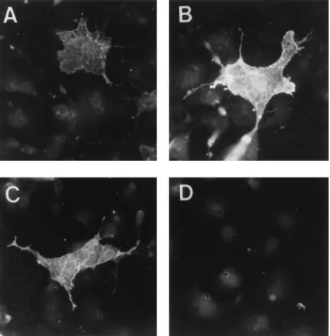
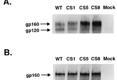
AIDS patient sera precipitated both precursor and processed forms of the wild-type and AV1/2, AV3, and AV1/2/3 glyco- proteins from transfected COS-1 cells lysates..

The positions (bases) of full-length donor template-directed DNA synthesis products (F) and products produced by strand transfer events (T) are indicated. The region where the donor