Copyright © 1994, American Society forMicrobiology
Human
Immunodeficiency Virus
Type 1
Integrase:
Effect
on
Viral
Replication
of
Mutations
at
Highly Conserved Residues
PAULAM. CANNON, WILMA WILSON,t ELAINE BYLES, SUSAN M. KINGSMAN, ANDALAN J. KINGSMAN*
Retrovirus Molecular Biology Group, Departmentof Biochemistry, University ofOxford,
Oxford
OX] 3QU, UnitedKingdom Received 7March 1994/Accepted21April 1994Sequence comparisons ofthe integrase (IN) proteins from different retroviruses have identified several highly conserved residues. Wehaveintroduced mutationsat16 of these sites into theintegrase geneof human immunodeficiency virus type 1 and analyzed the phenotypes of the resulting viruses. The viruses were all normal for p24 content and reverse transcriptase activity.In addition,all of the mutants could infect T-cell lines and undergo reversetranscription, as assessed byPCRanalysis. Most of the mutantviruses also had normalWesternblot(immunoblot) profiles, althoughthree of themutationsresulted in reducedsignalsfor IN relative to the wild typeontheimmunoblots andmutation of residue W235completelyabolishedrecognition of theprotein by pooled serafrom humanimmunodeficiencyvirustype1-positive patients.Mutations that have previously beenshown toabolishactivity in in vitrostudies producednoninfectious viruses. The substitution of W235 was notable in producing a noninfectious virus, despite previous reports of this residue being nonessential for INactivityinvitro(A. D. Leavitt, L. Shiue, andH. E.Varmus, J.Biol. Chem.268:2113-2119, 1993).Inaddition, wehave identifiedfourhighly conservedresidues that can be mutated withoutanyaffect on viral replication inT-cell lines.
Integration is an essential and characteristic step of the retrovirus life cycle. Following infection ofa susceptible cell, the viralRNAgenome iscopied intoadouble-strandedDNA molecule, containing terminally repeated longterminal repeat (LTR) sequences, which is then inserted into the host cell DNA. Integration requires the removal of two nucleotides fromthe 3' termini of theLTRs(3' processing), followed bya staggered cleavage of the host cell DNA. This produces 5' overhangs which are joined to the recessed viral termini (strand transfer). Host enzymes repair and ligate the gapped molecule so created, giving rise to short duplications of the host sequences flanking the integrated provirus (for reviews, seereferences 17 and44).
Biochemical studies have demonstrated that only one com-ponent, thevirally encoded integrase(IN) protein, is required for both the 3' processing and strand transfer reactions(4, 8, 24,28).PurifiedINcatalyzesanucleophilic attack onaspecific
phosphodiesterbondatthe terminus of the viralLTRand then positions the resulting3' hydroxyl groupsto attack the target DNA.Thesetwocleavage eventsarecoordinated inaone-step reaction, without the involvement of a covalent protein-DNA intermediate (15). Genetic evidence also points to the essential role of IN in this process, as viruses carrying mutations in HIV-1 IN are incapable of integration and correspondingly noninfectious (28, 37).
Sequence comparisons ofINproteins from different retro-viruses have identified several residues and motifs that are conserved across species (14, 19, 25, 27). The role of these residues in IN function has been investigated in vitro, using deletion and substitution mutants of IN protein from human
*Corresponding author. Mailing address: Retrovirus Molecular
Biology Group, Department of Biochemistry, University of Oxford, South Parks Road,Oxford OX1 30U,United Kingdom. Phone: 865 275248. Fax: 865275259.
tPresent address: Institute of General Microbiology, Universityof Berne,CH-3012 Berne, Switzerland.
immunodeficiencyvirustype1 (HIV-1) (11, 14, 30, 41), HIV-2 (39), murine leukemia virus (MLV) (21), and Rous sarcoma virus (RSV) (27). Such studies have identified distinct func-tional domains within IN, and mutations within individual domainscan be complemented in trans by wild-type residues within a mixed multimer (5, 13, 40).
The active site of the moleculeseems tobecontained within the central core and is characterizedby three invariant acidic residues with the pattern
D51-5,D35E.
Thisregionhas homol-ogywith similar recombinase proteins from retrotransposons andprokaryotic transposons (25, 27). The amino terminushas a conserved HHCC motif, reminiscent ofa zinc finger (19), and the specific interaction between IN and the LTR se-quenceshas been suggestedtobe a function of thisregion (5, 21, 41), although no such binding has been demonstrated directly under in vitro conditions (25, 43, 45). IN has a nonspecific affinity forDNAwhich has beenmappedtothe C terminus of the protein (43, 45), and this region may be involved in the interaction ofINwith the targetDNA.There is also evidence that IN functions as an oligomer (20, 22). A possible multimerizationregion, spanning the central catalytic domain anda regionimmediately 3' to this which includes a potential leucine zipper motif (31), has been described for HIV-1 (13).Although IN protein by itselfis sufficient to integrate an LTRoligonucleotide intoatargetDNAmolecule invitro, itis likely that other viral and cellular components playa role in this processinvivo. Viral DNArescuedfrom infected cells is contained withinahigh-molecular-weight complextermedthe preintegration complex, whichalsocontains components of the viralcore (1, 3). While studies with purifiedIN have contrib-uted much to ourunderstandingof the mechanism of integra-tion, theywill not highlightregions ofIN involved in interac-tions with other components of the preintegration complex. Furthermore, such studies may not be sensitive to mutations thatinhibitmultimerization ofIN,as mostin vitrointegration events areone-endedreactions thatmeasuretheintegrationof 4768
on November 9, 2019 by guest
http://jvi.asm.org/
MUTATIONAL ANALYSIS OF HIV-1 INTEGRASE 4769
a single LTR terminus (4). This contrasts with the concerted integration of two LTRs that occurs when extracts of infected cells are used as the source of IN (16) or that must occur in vivo (32). It is noteworthy that several highly conserved residues can be mutated without any great effect in any in vitro assays (14, 30, 39).
To investigate the role of IN in the in vivo integration process, we have created single amino acid changes at 16 highly conserved sites in HIV-1 IN. We have introduced these mutations into an infectious proviral clone and analyzed the resulting virions. We find that mutations that have previously been shown to have a deleterious effect in vitro similarly produce a protein that is unable to function in vivo. Surpris-ingly, despite their obvious conservation, several residues that were nondeleterious in in vitro assays also appear to be nonessential for integration function in our in vivo system. However, one conserved residue that appears not to be essential in vitro (30) produced noninfectious virus.
MATERIALS AND METHODS
Mutagenesis of HIV-1 integrase and proviral manipula-tions. The integrase gene from plasmid pBH10 (36) was inserted into M13, and single amino acid substitutions were introduced at highly conserved residues by site-directed mu-tagenesis. The mutated genes were inserted back into the infectious proviral clone W13 (26). W13 is a
nefr
version of HXB2 and, like pBH10, is derived fromHIVIIIB.
The se-quences of the integrase genes of pBH10 and W13 are identical (ourunpublished data). PlasmidpGEMSSAEcontains the pol sequences from W13 on a 4.3-kb SpeI-SalI fragment in apGEM5Zf backbone (Promega). The integrase gene in this plasmid has been marked by the removal of the central
EcoRI
sitefollowing end filling and religation. The mutated integrase genes were firstmoved from M13 into this construct, using the
BspMI sites ateither end of the gene; successful replacement restored theEcoRI site. These intermediates were then used to replace the equivalent SpeI-SalI fragment of plasmid pSA1, which is a W13 derivative deleted for the EcoRI fragment between nucleotides 4650 and 5745. Inserting the mutant integrase genes into this plasmid restored theEcoRI fragment andproduced afull-length proviral clone. Mutant viruses were named according to the residue that was changed (e.g.,
WI3C4oA
has thecysteine at amino acid 40 in IN changed to an alanine). In addition, the mutant proviral clone WI3AIN was derived directly from pGEMSSAE. It therefore contains a frameshift mutation at residue 142 which adds 14 nonsense codons and a termination codon, producing a protein of 156 amino acids (the wild type contains 288 amino acids). All mutations wereconfirmed by sequencing both the initial M13 clones and the final reconstituted proviral DNA.Production of virus containing integrase mutations. Virus was generated by overnight calcium phosphate transfection (18) of COS-7 or 293T (293/tsA1609neo) cells (12) (obtained from D. Baltimore, Rockefeller University). Cells were grown in Dulbecco modified Eagle medium supplemented with 10%
fetal calf serum (GIBCO BRL) and transfected at 25% confluence with 20
jig
ofplasmid DNA per 10-cm-diameter dish. Supernatants were harvested 60 h posttransfection and clarified bycentrifugation andfiltration through0.4-,um-pore-size filters. Virus stocks were treated with DNase I (Promega) for 1 h at37°C in the presence of 6 mMMgCl2 and stored in 1-mlaliquots at-70°C. Virus production was measured by p24 enzyme-linkedimmunosorbentassay (Coulter) or reverse tran-scriptase (RT) assay, using both a 32P-based RT assay (33) and a
3H-based
RT scintillation proximity assay kit that is indevelopment from Amersham International. The latter is a modification of a previously reported method (29).
Infection of cells with virus. Virus stocks were assayed for infectivity in the T-cell line C8166. A total of 5 x
105
cells were incubated in 1 ml of virus (titer of approximately 50 ng ofp24) for 1 h at37°C
and then washed and plated in 2 ml offresh medium (RPMI, 10% fetal calf serum). Infectivity was scored 3 to 4 days later by the appearance of syncytia. All of the observations reported here were made with between four and eight independently produced virus stocks for each mutant. Virus stocks were also used to infect the T-cell line H9. A total of 3x10⁶
cells were incubated with equal amounts of virus in a total volume of 1 ml for 1 h and then washed and plated in 5 ml of medium. Samples of the culture supernatant were taken every 3 to 4 days and assayed for RT activity to monitor virus production. These infections were performed with at least two independently produced virus stocks for each mutant tested.Analysis of viral proteins. COS-7 or 293T cells were trans-fected with proviral DNA as described above. Sixty hours later, virus from the filtered culture supernatant was concentrated by ultracentrifugation at 100,000 rpm for 1 h in a TL100.4 rotor (Beckman Instruments Ltd.), resuspended in 100
,ul
of loading buffer, and stored at-70°C.
Aliquots were heated to90°C
for 5min
before analysis by sodium dodecyl sulfate (SDS)-polyacrylamide gel electrophoresis. HIV-1 proteins were de-tected byWestern
blotting (immunoblotting), using pooled sera from HIV-1-infected donors at a dilution of 1:500. A rabbit antiserum directed against an amino-terminal IN pep-tide (residues 3 to 18) was made by S. Ranjbar and obtained from the MRC AIDS Directed Program Reagent Project (ADP419). In addition, a rabbit antiserum made against a hybrid Ty-integrase virus-like particle (residues 141 to 288) was supplied by British Biotechnology Ltd. Both of these anti-IN sera were used at a dilution of 1:500. The secondary antibodies used were horseradish peroxidase-conjugated goat anti-human immunoglobulin and goat anti-rabbit immunoglobulin (Vec-tor), used at dilutions of 1:1,000 and 1:4,000, respectively. Specific proteins were visualized by the enhanced chemilumi-nescence detection system (Amersham).PCR analysis. C8166 cells were infected with DNase-treated virus for 1 h as described above. Total DNA was isolated from
105
cells at time zero or 48 h following infection by resuspend-ing the cells in 200,ul
of lysis buffer containing proteinase K and Triton X-100 (7). Following incubation at65°C
for 2 h and then95°C
for 15min,
PCR was performed on 10,ul
of the sample in a20-,ul
final volume. In general, the PCR mixtures contained final concentrations of 2.5mM
MgCl2,
4 mM deoxynucleoside triphosphates, lx Taq polymerase reaction buffer (Promega), and oligonucleotide primers at 10 ng of each per ml, with 0.1 U of Taq polymerase (Promega) per reaction. The PCR conditions were 35 to 40 cycles at95°C
for 1min,
61°C
for 2min,
and72°C
for 3min.
PCR primers used to detect two-LTR circles were as described previously (38). The 595-bp band produced was detected by ethidium staining, and the identities of the bands were confirmed by Southern blotting using either a32P-end-labelled
oligonucleotide probe specific for the U3 region of the LTR (5'-TGGAAGGGCTAATTCACTCCCAA-3')
or a riboprobe containing the apical bases of the TAR stem-loop structure (TAR sequence, 5'-CCA GAUCUGAGCCUGGGAGCUCUCUGG-3').RESULTS
Generation of virus containing point mutations in the integrase gene. Comparisons of the IN sequences from
differ-VOL.68, 1994
on November 9, 2019 by guest
http://jvi.asm.org/
4770 CANNON ET AL.
W61A
C40A D64A
C43P T6A V75P
~~~~.~~~~~~~jj~~~icFiner
z
vSIF
* * 50 *
_**
*1FLDGIDKAQDEHEKYHSNWR AMASDFNLPP WAKEIVASC DKCQLKGEAMHGQVDCSPGI WQLDCTHLEG KVILVAVHVA SGYIEAEVIPAETGQETAYF
IIrim!WN-1
Iii""Aliii111 1 111m Rn - -m
TI15A
D116A 1135P
| S123A I
Centralcatalytcdomain DD(35)E l
V151A
E152P K159P
-t
ILeuill
EEEEEI0X
cinezipper
A179P
**_*_*1
**E----EI*...a...a...* 200 LLKLAGRWPV KTIHTDNGSNFTSATVKAAC WWAGIKQEFGIPYNPQSQGV VESMNKELKKIIGQVRDQAEHLKTAVQMAV FIHNFKRKGG IGGYSAGERIW235A
I Non-specificDNAbinding
g _
MIKEE M
_W 1-1-11--, o75 ~1111-11-1111119288VDIIATDIQT KELQKQITKIQNFRVYYRDS RNPLWKGPAK LLWKGEGAVVIQDNSDIKVVPRRKAKIIRDYGKQMAGDDC VASRQDED
FIG. 1. HIV-1integrase. Thecomplete amino acidsequenceofpBH10INis shown.Thepositions of 18 residues thatarecompletelyconserved withRSV,Mo-MLV,HTLV-I,bovineleukemia virus,andmousemammarytumorvirusareidentifiedbyasterisks. The 16single-sitemutations
arearrowed. Putative IN domains arehighlighted.Theseincludeanamino-terminalregionwithconservedHHCC residues(19),acentralcatalytic domain with invariantD, D, and Eresidues(27),apotential leucinezipper(31),andacarboxy-terminalregion possessingnonspecificDNAbinding activityinvitro (42).
entretroviralspecies have identified severalhighly conserved residues.Analignment ofHIV-1 INwith those ofRSV,bovine leukemia virus, Moloney MLV (Mo-MLV), human T-cell leukemia virustype I (HTLV-I), andmousemammarytumor virus, for example, produces18completelyconservedpositions (Fig. 1).Substitutions of amino acidsweremadeat16 of these positions inthe HIV-1 INgenefrom pBH10andcloned into the infectiousproviralcloneWI3.Inaddition, acontrolvirus (WI3AIN) which contains a truncated protein caused by a
frameshift mutation atthe central EcoRI sitewas made. On
thebasis of similar constructions described elsewhere(38),this virus will beincapable of integration and therefore noninfec-tious.
Virus stocks were generated from each of the mutant proviral clones, using transient transfection of the plasmids intoCOS-7and293T cells. Thisone-stepmethod ofgenerating virus isnotdependentonvirus-directed integrationand there-fore willnotdiscriminateagainst nonintegrative mutants.The virus stockssogeneratedweremeasured forp24concentration
and RT activity. Approximately equal amounts of p24 were
produced byall of theconstructs(typically1 ,ug/ml),suggesting efficient transfection and expression of the various plasmid DNAs(datanotshown).Inaddition,all of the virus stocks had reasonable levels of RTactivity anda p24/RTratio thatwas
similarto that of thewild-type W13(Table 1).
Analysisofinfectivityof viruses containing integrase muta-tions.Mutantvirusesweretested for theabilitytoestablishan
infection inT-cell lines. Stocksofviruswere standardized for
p24 content, and equivalent amounts of virus were used to
infectC8166 cells. Three tofour days following infection, the C8166cellswereinspected for theappearanceof syncytia. The wild-type virus WI3 and virus WI3AIN containing the
trun-cated IN were included as positive and negative controls,
respectively. Five mutant viruses produced visible syncytia (Table 1). Mutants WI3T66A,
WI3T115A,
WI3S123A,
andWI3vl5lA
producedsyncytiaatthesamerateasW13,but withmutantWI3KIs9P, theappearance ofsyncytia was delayed by
24 h. The remaining mutations abolished the ability of the
viruses to cause any visible cytopathic effectsin C8166cells. These observationswereconsistent for several independently produced stocks of each mutantvirus. Furthermore, noviral replication could be detected for any of the
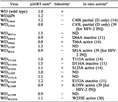
non-syncytium-TABLE 1. Properties ofHIV-1 INmutants"
Virus p24/RT ratiob Infectivityc Invitroactivityd
W13(wild type) 1.0 +
WI3AIN 1.2
-WI3c40A 1.2 - C40Spartial (D only) (14) WI3c43A 0.8 - C43Lpartial (D only) (39
[for HIV-2 IN])
WI3w61A 1.5 - ND
WI3D64A 0.9 - D64Ainactive(11)
WI3T66A 0.8 + T66Aactive(14)
WI3V75P
1.3 - NDWI3S81A 0.9 - S81A active(39 [for
HIV-2IN])
WI3T115A 1.0 + T115Aactive(14)
WI3D116A
1.5 - D116Ainactive(11)WI3s,23A
1.1 + S123Aactive(14)WI31135P
1.0 - NDWI3v151A 1.1 + ND
WI3E152P 1.2 - E152Ainactive(11)
WI3K159P 0.9 + K159V active(39[for
HIV-2IN])
WI3A179p 0.9 - ND
WI3w235A 1.1 - W235E active(30)
aSingleamino acid mutations were introduced into infectious cloneW13as
indicated. In addition, WI3AIN contains a truncated IN gene caused by a
frameshift mutation.
b p24 concentration and RTactivityweremeasuredforatleasttwo indepen-dently produced virus stocks,and thep24/RTratio wascalculated. Valueswere normalized to 1 for thewild-typevirusWI3.
Assessed bytheabilitytoinducesyncytia(+) inC8166 cells 3 to 4days followinginfection.
dActivityof HIV-1 or HIV-2 mutant INproteinin invitrointegration assays.
Mutated residues are as indicated. Sources of dataare indicatedbyreference numbers inparentheses. D,disintegration activity; ND,notdetermined.
J.VIROL.
on November 9, 2019 by guest
http://jvi.asm.org/
[image:3.612.81.532.76.278.2] [image:3.612.315.555.425.633.2]MUTATIONAL ANALYSIS OF HIV-1 INTEGRASE 4771
producing
virusesby
supernatantp24
analyses up to 21 daysfollowing
infection(data
notshown).
We also tested for thepossibility
that the nonfunctional mutants were temperaturesensitive
by carrying
out the transfections and subsequent infections with the mutant viruses at 32.5°C. However, nosyncytia
could be detected up to 8days
postinfection at thislowertemperature.
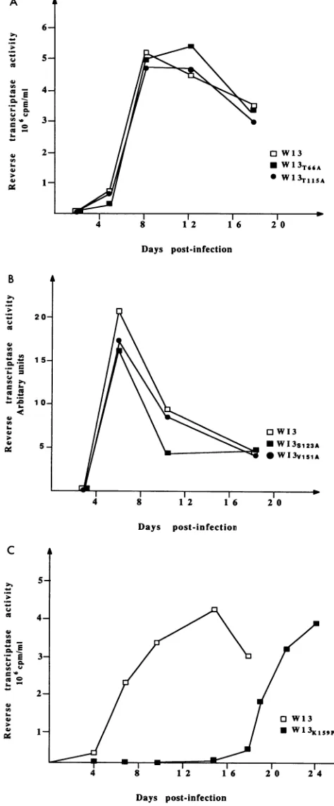
Replication
kineticsof viablemutants.Although fivemutant virusesproduced syncytia
inC8166 cells, it remained possible thattheirability
tointegrate
had been compromised by these mutations. The kinetics of their infections was thereforecompared
with that of thewild-type
virus inaninfectionof H9cells. A total of 5 x 10⁶ H9 cells were infected with 1 ml of virus stock
containing
equivalent
amounts of p24, and theinfectionwas monitored
by taking
supernatant samples every fewdays
andmeasuring
the RTactivity.
Thisanalysis demon-strated that virusesWI3T66A
and WI3Tl15A (Fig. 2A) and virusesWI3Sl23A
andWI3Vl15A
(Fig.
2B) replicated with kinetics similar to that of W13. The mutant virusWI3K159P,
however, replicated
markedly
more slowly than the wild-typeparent
(Fig. 2C).
Thegrowth
of thisvirusafter 18dayswasnot due toreversionorsuppression
of themutation,asvirus taken atthis timepoint
showed thesamedelayed growthkinetics ina new infection
(data
notshown).
The growth curves wererepeated
at least once for each mutant, using independentlyproduced
virus stocks toconfirm these observations (data notshown).
Inaddition,
theability
of each of these viruses toreplicate
at ahigher
temperature(42°C)
in C8166 cells wastested incasethe mutantIN
proteins
provedtobe less stable. Allfive of themutantscould stillproduce
syncytiaatthe highertemperature,
although
the appearance of syncytia with virusWI3K159p
wasagain delayed by
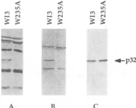
24 hrelative toW13.Analysis
ofviralproteins.
Theabsolute levels of p24 and RTactivity
in thesupernatantsof thetransfected 293T cells were similarfor all of thepanel
ofmutantviruses,
andthe ratios ofp24/RT
were alsonormal,
suggesting
that the mutations that wehad created hadnogrosseffectonvirusparticlestructure orproduction.
Toconfirmthis,
viralparticles
wereconcentratedfrom the supernatants of transfected cellsby
ultracentrifuga-tion and
analyzed
by
Westernblotting.
HIV-1 proteins were detected withpooled
humanpatient
sera,and theidentitiesof individual bands were confirmedby
using a panel of morespecific
antibodies(data
notshown).
The truncatedINmutant,WI3AIN,
asexpected
didnotproduce
aprotein
correspondingto normal
full-length
IN(Fig.
3,
lane2). (This
mutation ispredicted
to code for aprotein
of approximately half thenormal
size,
which may or maynot bestable.)
The proteinprofiles
produced
by
themajority
ofthemutantswereidenticaltothe
wild-type profile
(Fig.
3 and datanotshown).
However,four viruses had abnormal
protein profiles;
the mutationsV75P, W61A,
and I135Palways produced
viruses with arelatively
lowamountsof INprotein
compared
withthe other viralproteins (lanes
3, 4,
and7),
andnoIN proteincould be detected for virusWI3W235A
(lane 9).
Analysis
ofWI3W235A
particles
grown at32.5°C
also failed to show anyINprotein,andnoINcouldbe seen inextractsoftransfectedcells(data
not
shown).
Because theprofile
of thisvirusonWestern blotsmirrored that of the truncatedINconstruct WI3AIN,wewere
concerned that a second-site mutation could have arisen
during
our DNAmanipulations.
We therefore reconstructedthevirus from the
original
M13 clone andsequencedacrossthe entire INcoding region.
Thephenotype
of this secondWI3W235A
mutantviruswasthesameastheoriginal (datanotshown).
ResidueW235 ispartofanimmunodominantepitopeof IN. The
apparent
absence of IN in the Western blots of theA
0,
.O-.-_.
a-C
ed0,E
._E
c,0
6-
5-
4-
3-
2-Days post-infection
OW13
*WI3sl23A
*WI3vi5IA
Days post-infection
[image:4.612.318.555.93.665.2]Days post-infection
FIG. 2. Replicationkinetics of viablemutants.Equivalentamounts
ofwild-type(WI3) andmutantviruseswereusedto infect H9 cells. Virus productionwasmonitoredfor18days by analyzing RTactivity in the culture
su?ernatants
bya3H-basedscintillationproximityassay(A andC)or a 3P-basedassay(B).VOL.68, 1994
on November 9, 2019 by guest
http://jvi.asm.org/
LI' ,, e1
w , f (IX r-~2-l.el~
. 1 < Z. en i:
_ _ _ _ -~ - -_ _ -p66 __
_ _ w - l - p6o
ft Ur mm ,, gi
It ~ -gp4l
- - - ~~~~~~~~~-p32
-~~~~~~~~-01p
_
- - -ar - -p1l
1 2 3 1 .5 0 , i)
FIG. 3. Immunoblot analysis of viral proteins. Virionswere con-centratedfrom the supernatantof transfected 293Tcells by ultracen-trifugation and separated on an SDS-12.5% polyacrylamide gel. HIV-1proteins were detectedwith pooled humanpatientserum.The positions ofthemajorviralproteins areshown.
WI3W235A
viralparticles wasunexpected. It waspossible that mutation of W235 had produced a protein that was very unstable or could nolonger be incorporated into the virion. As INispresumed to be targetedtothevirionaspartofthelarger gag-pol precursor, it was unlikely that the latter explanation wascorrect.Furthermore, there was no apparent effectonthe relative amounts of the other viral proteins, RT activity was normal, and the ratio of p24 levels to RT activity was very similartothat of thewild-typevirus. Similarly,if this mutation had caused any structuralchanges inINthatpreventedcorrect processing by the viral protease at the RT/IN junction, this would be reflected in reduced levels of the processed RTproteins on the Western blots. We therefore considered the
possibilitythat themutantproteinwas nolonger recognized by
thepatient sera that we were using as the sourceof anti-IN
antibodies, even though this serum was pooled from three different individuals.Accordingly, we tested both thewild-type
and the W235A mutant viruses with two additional anti-IN sera, onederived from inoculation of rabbits with an amino-terminal peptide (residues 3 to 18) and one made against a Ty-virus-like particle containing residues 141 to 288. As
ex-pected, the wild-type INcould be detected byboth of these antisera (Fig. 4).The W235Aprotein, asbefore, failedto be detected bythepatient seraandwasalsonot detectedbythe
polyclonalserummadeagainst residues 141 to288. However,
Cn crj e e en M
_~ e_r s
,.-p32
[image:5.612.353.515.76.161.2]A B C
FIG. 4. Mutation W235Apreventsrecognition byamajor epitope
in human HIV-1-positive serum. WI3 and WI3W235A virions were
subjectedtoimmunoblot analysis usingpooled human HIV-1-positive
sera(A),arabbitantiserumdirectedagainst residues 141to288 (B),
andarabbit antiserum directed againstamino-terminal residues 3 to
18(C).Thepositionofp32IN isarrowed.
.4 595bp
FIG. 5. PCRoftwo-LTRcircles.Extractsof C8166 cells infected 3 days previouslywith virus stocks were subject to 40 cycles of PCR amplification usingprimers specificfor the two-LTR circlejunction. Amplification of suchastructureresultsin a595-bp band.
the antiserum raisedagainstresidues 3 to 18 reacted withboth the W235Aproteinand the wildtype,revealing approximately equal levels of each in the virions. The substitution of W235 had therefore prevented recognition of IN by the pooled patient sera, suggestingthat W235 is essential for an immuno-dominantepitopein IN.
Formation of two-LTR circles by nonreplicative mutants. We used PCR analysis to confirm that the noninfectious mutantswere not blocked in astep of the life cycle priorto integration. Two-LTR circles are a form ofretroviral DNA present inthe nucleus. They arethought to be produced by
host enzymes asanalternativeto correctintegration byINand are therefore formed in the absence ofa functional IN (2).
Two-LTRcirclejunctionsare notpresentintheinput plasmid
DNAused in theinitial transfection(whichcanbecarriedover into the infections and contaminate subsequent PCRs). The PCR amplification of such a structure is therefore confirma-tion of entry, uncoating, and reverse transcription by the mutant viruses. Usingtheprimers describedinMaterials and Methods,wecould amplifya 595-bpfragment corresponding to the two-LTR circle junction for all of the noninfectious mutants(Fig.5and datanotshown).Thisfindingindicatesthat the block to productive infection occurs at a step in the life
cycleafterreversetranscriptionandispresumablyat
integra-tion.
DISCUSSION
Wehavemutated16residues in theintegrasegeneof HIV-1 at positionsthatarehighlyconserved amongretroviruses and introduced them intoinfectiousproviral clones. The mutations hadnogrosseffectonviralparticlestructureorlevelsof virus production,asassessedby Westernblottingandp24 measure-ment. Furthermore, all of the mutants displayed wild-type
levels ofRTactivityin invitro assays andwerecompetentfor all stages of the HIV-1 lifecycleup to andincluding reverse transcription, asdemonstratedbythe
production
oftwo-LTR circles. The inability of several of the mutant viruses to establishaninfectioninT-celllines is thereforehighlylikelyto be theresult ofa defect inintegration.Zincfingermutants (C40Aand C43P). The amino termini of all retroviral IN proteinscontain two conserved histidines and two conserved cysteines, separated by the spacing H3 H22-32C2C (19). This structure is similar to the zinc finger
motif found insometranscriptionfactors, and theregionhas been demonstrated to bind zinc (5). Because of the known DNAbindingproperties of zincfingers, it has beenproposed
that thisstructuremaybe involved in theinteraction ofINwith DNA. In vitro binding assays have not detected any LTR-specific or nonspecific affinity for DNA in this region, and indeed the nonspecificDNAbinding ability of whole IN has
on November 9, 2019 by guest
http://jvi.asm.org/
[image:5.612.62.294.77.212.2] [image:5.612.107.245.556.666.2]MUTATIONAL ANALYSIS OF HIV-1 INTEGRASE 4773 beenmapped toits carboxyterminus (42, 45). However, other
analyses suggestan involvement of this region in LTR recog-nition (21, 41).
Substitution of any one of the conserved histidine or cysteine residues in this motif would be expected to disrupt the putative zinc finger, but mutation of IN at these residues does not
completely abolish catalytic activity in vitro. Disintegration
activityremains unaffected (14), and two groups report nearly
wild-type levels of strand transfer activity for a C43S substitu-tion of HIV-1 (11) and a C40S mutation in HIV-2 (39). However, mutation of the conserved cysteines in the context of the provirus completely abolished infectivity, suggesting a morerigidrequirement for this region in vivo. These data are in agreementwith previous studies of the effects of mutations in the analogous region of MLV IN (9, 35).
Centralcatalytic domain. Amino acidsD64, D116, andE152 arethekeyinvariantresidues in the central catalytic domain of IN. This motif is also conserved across retrotransposons and
prokaryotictransposases, and the carboxylic acid side chains of these acidicresidues have been proposed to be involved in the coordination of the divalent metal cations needed for IN
function, such as Mg2+ or Mn2+ (27). Mutation of these residues has been shown to have the most drastic effect on all invitroIN activities (11, 14, 27, 28, 30, 39) and not
unexpect-edly produces completely noninfectious viruses. Other groups have also reported noninfectious viruses for the mutations D116A and E152A(37) and also for the V151D E152Q double mutation(28).
None of the D,D35E substitutions had any effect on the
profile of the viruses on Western blots, and all three had
wild-type levels of RT activity. This finding contrasts with the
observation by Shin et al. (37) that a D116A mutation in an HXB2 provirus has a gross effect on Gag protein processing and abolishes in vitro RT activity. In our hands, the identical substitution in a similar proviral backbone (W13 is a
nef'
version ofHXB2) has no such effects. At present, we cannot accountfor these discrepancies.
Other conserved residues in this region, including V75, S81, and
1135,
abolished infectivity when they were mutated. Vi-ruses containing mutations V75P and 1135P, together withW61A, appeared to have abnormally low amounts of IN
protein
in the viral particles relative to the wild type. As all three of these mutant particles had wild-type levels of RTactivity,
normalRT/p24 ratios, and expected levels of RT p66 onWestern blots, reducedrecognition by the human antisera used for Westernblotting is the most likely explanation for this observation.Mutation of S81 to alanine also produced a noninfectious
virus, but the data from in vitro studies for this residue are
conflicting. Several different substitutions at this residue in bothHIV-1and RSVINhave been reported to severely affect 3' processingandstrand transfer (23), and other reports state thatmutations atthis site produce a protein of low solubility thatis difficulttopurify (30). Despite its lower solubility, one group hasbeen ableto purify enough of an S81A mutant of HIV-2 to test and reported wild-type activity in all of their assays
(39).
This finding suggests that substitutions at this residue mayaffectprotein folding rather than catalytic activitydirectlyand that such mutations arebettertolerated in some
expression
configurations than others.Leucinezipper(V151A,K159P,and A179P). Theseresidues lie withinaputativehelical region of the protein containing a
possible
leucinezipper(31). Residues V151, L158, V165, and L172 could contribute a hydrophobic face to a helix which could functionas adimerization domain for IN. The introduc-tion ofhelix-breaking residues (proline) at A179 and in thecenter of the region at the highly conserved K159 would be
expected to disrupt such a structure, and the resulting viruses were either completely
(WI3,179P)
or partially(WI3K159P)
defective in their replicative ability.
WI3K159p
replicated at a reduced level compared with thewild type, suggesting that this mutation had compromised integration. The ability of this mutant to replicate at all was especially surprising since K159 is one of the most highly
conserved of all the residues (14). The alignment of the potential members of the leucine zipper also abuts residues E152, K159, and R166 (31), with the first two residues being invariant and R166 also highly conserved. E152 is a memberof the proposed active site of IN, and the side chains of these adjacent amino acids may be essential for the correct environ-ment for this residue. However, the tolerance of K159 to substitution has also been demonstrated by the wild-type replication kinetics seen in SupTlcells for the somewhat less disruptive replacement by a glutamine (37).
The location ofV151 suggests that as well as being impor-tant for leucine zipper formation, it may also influence the adjacent catalytic domain residue, E152. Accordingly, the mutation that we chose to make at this site was fairly conser-vative and resulted in no effect on viral replication. It is possible that a different mutation would have had a greater effect.
Nonspecific DNA binding(W235A). The carboxy terminus of IN is the area of least sequence homology among different retroviruses. In HIV-1, it has been shown to account for the nonspecific DNA affinity of the protein which may be involved in target DNA interactions (42, 45). Mutation of the highly conserved tryptophan in this region to a glutamate has previ-ously been shown to have no effect in any in vitro integration assay (30), and the authors did not report any difficulty purifying and assaying the mutant protein. However, our W235A mutation totally abolished the ability of the provirus to replicate, so it is clearly an important residue for in vivo function of IN. We speculate that this region may therefore be involved in the correct positioning of the processed retroviral LTRs to interact with the target host cell DNA, either by virtue of an inherent affinity for DNA or by interactions with protein molecules associated with the chromatin.
W235 is probably also a key component of a major immu-nodominant region of IN. The observation that reactivity with both the pooled human sera and the rabbit serum is lost in the W235A mutant supports this notion and suggests that W235 is essential for the integrity of some local structure that is preserved in Western blotting. It is interesting that a large insertion is present just upstream of the equivalent tryptophan residue in the Mo-MLV IN relative to other retroviruses and, in addition, that Mo-MLV IN can tolerate an insertion of a further 36 amino acids into this adjacent region and still retain function (34). This finding is consistent with a role for this residue in key structural interactions within the core of the protein, possibly adjacent to a looped-out region of somewhat variable length.
Viable mutants (T66A, T115A, S123A, and V151A). It was surprising that four highly conserved residues in the central region (T66 and T115, together with S123 andV151)could be mutated without affecting the replicative ability of the viruses in T-cell lines. To guard against the possibility of revertant wild-type viruses in these cultures, we repeated these infections in H9 and C8166 cells several times with independently produced virus stocks. In every experiment, these mutants showed the same phenotype as the wild-type virus. Previously, mutation T66A has been shown to be defective in 3' processing in vitro (22% of wild-type activity) and to be partially defective VOL.68, 1994
on November 9, 2019 by guest
http://jvi.asm.org/
for integration (42% of wild-type activity) (14), although others have reported wild-type activity for mutations of the equivalent residue in RSV IN (23, 27). Mutation of residue T115 has also produced variable results in in vitro analyses. Several substitutions at this residue have been reported to
retain wild-type activity in all in vitro assays by some groups
(14, 27, 39), but a T1l5A substitution is reported to be selectivelydefective in strand transferactivity by another
(11).
Furthermore, thesameTliSAsubstitution thatweintroduced
hasalso been reportedtohavenoeffectonHIV-1 infectivity in
aT-cellline (37).
Varying in vitro observations have been made also for proteinsmutatedatS123.Thesame serine-to-alanine
replace-mentthatwemade resulted inaninsoluble proteinfor HIV-2
IN (39) butproducedaprotein with almostwild-type in vitro
activity for HIV-1 (14). No studies have been reported for single mutations at V151, although adouble mutant,
V1SiD/
E152Q, is defective both in vitro and in the context of the provirus (28). However, it is not possible to deduce anything about the role of V151 from this double mutant, as the
substitutionof E152 alone isknown tobe deleterious. Overall,ouranalysis oftheeffect of mutationsatconserved sitesin the integrasegeneonviral replicationfits with much of
the currentdataon IN.Thedrastic effectof mutations atkey residues in the D,D35E region was as expected, and the
absolute requirement for C40 and C43 in vivo confirms an
important rolefor the HHCCregion in INfunction. Residue W235 has the interesting phenotype of being completely nonessentialtoINactivity in vitrowhileabolishing integration in vivo. It willbe interestingto analyze atwhich stage in the integration process this block occurs. The finding that four
highlyconserved residuescanbe mutated withoutdeleterious
effect on viral replication is also intriguing. It raises the
possibilitythat these mutations have a phenotype that is not apparentin T-cell lines,andwe arecurrentlyanalyzing the role
of theseresidues inintegrase function in primary cells.
ACKNOWLEDGMENTS
Thisworkwassupportedbygrant G9210910from the MRC AIDS
DirectedProgram.
WethankMark Richardson for initial IN cloning. We also thank
WilliamJames (SirWilliam Dunn Schoolof Pathology, Universityof
Oxford)andthe members of his laboratoryfortheir help.
REFERENCES
1. Bowerman,B., P.O.Brown, J.M. Bishop,and H. E. Varmus.1989.
A nucleoprotein complex mediates the integration of retroviral
DNA.Genes Dev.3:469-478.
2. Bukrinsky,M. I.,N. Sharova, M. P. Dempsey, T. L.Stanwick,A. G. Bukrinskaya, S.Haggerty,andM.Stevenson.1992.Activenuclear
import of human immunodeficiency virus type 1 preintegration
complexes. Proc. Natl. Acad. Sci. USA 89:6580-6584.
3. Bukrinsky, M. I., N. Sharova, T. L. McDonald, and T.
Pushkar-skaya. 1993. Association ofintegrase, matrix, and reverse
tran-scriptase antigensof human immunodeficiency virus type 1with
viralnucleic acidsfollowing acuteinfection.Proc.Natl. Acad. Sci.
USA90:6125-6129.
4. Bushman, F. D., and R. Craigie. 1991. Activities of human
immunodeficiencyvirus (HIV)integration proteinin vitro:specific
cleavageand integrationofHIV DNA. Proc.Natl.Acad. Sci. USA 88:1339-1343.
5. Bushman, F. D., A. Engelman, I. Palmer,P.Wingfield, and R.
Craigie. 1993.Domains of the integrase proteinof human
immu-nodeficiencyvirustype 1 responsible for polynucleotidyltransfer
andzinc binding.Proc.Natl. Acad. Sci.USA90:3428-3432.
6. Bushman, F. D.,T. Fujiwara, and R. Craigie. 1990. Retroviral DNA integration directed by HIV integration protein in vitro.
Science249:1555-1558.
7. Collins, M., W. James, and S. Gordon. 1991. Development of techniques to analyse the formation of HIV provirus in primary human macrophages. Res. Virol. 142:105-112.
8. Craigie, R., T. Fujiwara, and F. Bushman. 1990. The INprotein of Moloney murine leukemia virus processes the viral DNA ends and accomplishes their integration in vitro. Cell 62:829-837.
9. Donehower, L. 1988. Analysis of mutant Moloneyleukemia viruses containing linker insertion mutations in the 3' region of
pol.
J. Virol. 62:3958-3964.10. Drelich, M., M. Haenggi, and J. Mous. 1993. Conserved residues Pro-109 and Asp-116 are required for interaction of the human immunodeficiency virus type 1 integrase protein with its viral DNA substrate. J. Virol. 67:5041-5044.
11. Drelich, M., R. Wilheim, and J. Mous. 1992. Identification of amino acid residues critical for endonuclease and integration activities of HIV-1 IN protein in vitro. Virology 188:459-468. 12. DuBridge, R. B., P. Tang, H. C. Hsia, P.-M. Leong, J. H. Miller,
and M. P. Carlos. 1987. Analysis of mutation in human cells by using an Epstein-Barr virus shuttle system. Mol. Cell. Biol. 7:379-387.
13. Engelman, A., F. D. Bushman, and R. Craigie. 1993. Identification of discrete functional domains of HIV-1 integrase and their organization within an active multimeric complex. EMBO
J.
12:3269-3275.14. Engelman, A., and R. Craigie. 1992. Identification of amino acid residues critical for human immunodeficiency virus type1 inte-grase function in vitro. J. Virol. 66:6361-6369.
15. Engelman, A., K. Mizuuchi, and R. Craigie. 1991. HIV-1 DNA integration: mechanism of viral DNA cleavage and DNA strand transfer. Cell 67:2111-1221.
16. Farnet, C. M., and W. A. Haseltine. 1990. Integration of human immunodeficiency virus type 1 DNA in vitro. Proc.Natl.Acad. Sci. USA87:4164-4168.
17. Goff, S. P. 1992. Genetics of retroviral integration. Annu. Rev. Genet. 26:527-544.
18. Gorman, C. M., L. F.Moffat,and B. H. Howard. 1982. Recombi-nant genomes which express chloramphenicol acetyltransferase in mammalian cells. Mol. Cell. Biol.32:1044-1051.
19. Johnson, M. S., M. A. McClure, D.-F. Feng, J. Gray, and R. F. Doolittle. 1986. Computer analysis of retroviral pol genes: assign-ment of enzymatic functions to specific sequences and homologies with nonviral enzymes. Proc. Natl. Acad. Sci. USA 83:7648-7652. 20. Jones, K. S., J. Coleman, G. W. Merkel, T. M. Laue, and A. M. Skalka. 1992. Retroviral integrase functions as a multimer and can turn over catalytically. J. Biol. Chem. 267:16037-16040.
21. Jonsson,C. B., and M. J. Roth. 1993. Role of the His-Cys finger of Moloney murine leukemia virus integrase protein in integration and disintegration. J. Virol. 67:5562-5571.
22. Kalpana, G. V., and S.Goff.1993. Genetic analysis of homomeric interactions of human immunodeficiency virus type 1 integrase using the yeast two-hybrid system. Proc. Natl. Acad. Sci. USA 90:10593-10597.
23. Katz,R.A., J. P. G. Mack, G. Merkel, J.Kulkosky,Z. Ge, J. Leis, and A. M. Skalka. 1992. Requirement for a conserved serine in both processing and joining activities of retroviral integrase. Proc. Natl.Acad. Sci. USA89:6741-6745.
24. Katz, R. A., G. Merkel, J.Kulkosky, J. Leis, and A. M. Skalka. 1990. The avian retroviral IN protein is both necessary and sufficient for integrative recombination in vitro. Cell 63:87-95. 25. Khan, E., J. P. G. Mack, R. A. Katz, J. Kulkosky, and A. M.
Skalka. 1991. Retroviral integrase domains: DNA binding and the recognition of LTR sequences. Nucleic Acids Res. 19:851-860. 26. Kim, S., R. Byrn, J. E. Groopman, and D. Baltimore. 1989.
Temporal aspects of DNA and RNA synthesis during human immunodeficiency virus infection: evidence for differential gene expression. J. Virol. 63:3708-3713.
27. Kulkosky, J., K S. Jones, R. A. Katz, J. P. G. Mack, and A. M. Skalka. 1992. Residues critical for retroviral integrative recombi-nation in a region that is highly conserved among retroviral/ retrotransposon integrases and bacterial insertion sequence trans-posases. Mol. Cell. Biol. 12:2331-2338.
28. LaFemina, R. L., C. L. Schneider, H. L. Robbins, P. L. Callahan, K. LeGrow, E. Roth, W. A. Schleif, and E. A. Emini. 1992.
on November 9, 2019 by guest
http://jvi.asm.org/
MUTATIONAL ANALYSIS OF HIV-1 INTEGRASE 4775 Requirement of active human immunodeficiency virus type 1
integrase enzyme for productive infection of human T-lymphoid cells. J. Virol. 66:7414-7419.
29. Lamaitre, M., T. Phan, M. J. Downes, K. B. Williams, and N. D. Cook. 1992. Poly r(A) reverse transcriptase (3H) SPA, a new enzyme assay system.Antiviral Res. 17(Suppl.):48.
30. Leavitt, A. D., L. Shiue, and H. E. Varmus. 1993. Site-directed mutagenesis of HIV-1 integrase demonstrates differential effects
onintegrase functioninvitro.J.Biol. Chem. 268:2113-2119. 31. Lin, T.-H., and D. P. Grandgenett. 1991. Retrovirus integrase:
identification of a potential leucine zipper motif. Protein Eng. 4:435-441.
32. Murphy, J. E., and S.P. Goff. 1992. Amutation at one end of Moloneyleukemia virus DNAblockscleavage of both ends by the viralintegrase in vivo. J. Virol. 66:5092-5095.
33. Potts,B.J. 1990."Mini"reversetranscriptase assay, p. 103-106.In A. Aldovini and B. Walker (ed.), Techniques in HIV research. Stockton Press, New York.
34. Roth, M.J. 1991. Mutational analysis of the carboxy terminus of the Moloney murine leukemia virus integration protein. J. Virol. 65:2141-2145.
35. Roth, M. J., P. Schwartzberg, N. Tanese, and S. P. Goff. 1990.
Analysis of mutations in the integration function of Moloney murine leukemia virus: effects on DNA binding and cutting. J. Virol. 64:4709-4717.
36. Shaw, G. M.,B.H.Hahn, S. K.Arya,J. E.Groopman, R. C. Gallo,
and F. Wong-Staal. 1984. Molecular characterization ofhuman T-cell leukemia (lymphotropic) virus type III in the acquired immunedeficiencysyndrome.Science226:1165-1171.
37. Shin,C.-G.,B.Taddeo, W.A.Haseltine,andC. M. Farnet. 1994. Genetic analysis of the human immunodeficiency virus type 1
integrase protein.J. Virol.68:1633-1642.
38. Stevenson, M., S.Haggerty,C. A.Lamonica, C. M.Meier, S.-K. Weloch,andA.J.Wasiak. 1990. Integration isnot necessaryfor expression of human immunodeficiency virus type 1 protein products.J. Virol.64:2421-2425.
39. Van Gent,D.C.,A. A.M.OudeGroeneger, and R. H.Plasterk. 1992. Mutational analysis of the integrase protein of human immunodeficiency virus type 2. Proc. Natl. Acad. Sci. USA 89: 9598-9602.
40. VanGent,D.C.,C.Vink,A.A. M.OudeGroeneger,andR. H.A. Plasterk. 1993.Complementationbetween HIVintegraseproteins mutatedin different domains. EMBO J. 12:3261-3267.
41. Vincent, K. A., V. Ellison, S. A. Chow,and P. A. Brown. 1993. Characterization of human immunodeficiency virus type I inte-grase expressed inEschenichia coli and analysisofvariants with amino-terminal mutations. J. Virol. 67:425-437.
42. Vink, C.,A. A.M. OudeGroeneger, andR.H.A. Plasterk. 1993. Identification of the catalytic and DNA-binding region of the human immunodeficiencyvirus type 1 integraseprotein. Nucleic Acids Res.21:1419-1425.
43. Vink, C.,D. C. VanGent,Y.Elgersma,andR. H. A.Plasterk. 1991. Humanimmunodeficiencyvirusintegraseprotein requiresa sub-terminal position of its viral DNA recognition sequence for efficientcleavage.J.Virol.65:4636-4644.
44. Whitcomb, J. M., and S. H. Hughes. 1992. Retroviral reverse
transcription and integration: progress and problems. Annu. Rev. Cell Biol.8:275-306.
45. Woerner,A.M.,M.Klutch, J.G.Levin,andC.J. Marcus-Sekura. 1992. Localization of DNAbinding activity of HIV-1 integraseto
theC-terminal half of the protein. AIDS Res. Hum.Retroviruses 8:297-304.
VOL.68, 1994