HYDROCHLORIDE
A dissertation submitted to
THE TAMILNADU Dr. M.G.R.MEDICAL UNIVERSITY, CHENNAI.
In partial fulfillment of the requirements for the award of degree of
MASTER OF PHARMACY IN PHARMACEUTICS
BY
REG.NO: 26091390
Under the Guidance of
Prof. S.P.SENTHIL, M.Pharm., ( Ph.D.,)
OCTOBER -2011
THE ERODE COLLEGE OF PHARMACY AND RESEARCH
INSTITUTE
DEDICATED
TO
My
Beloved Family,
Department of pharmaceutics,
Perundurai Main Road,
Veppampalayam, Erode-638112, India.
e-mail: [email protected]
CERTIFICATE
This is to certify that the investigation in this thesis entitled
“
FORMULATION AND EVALUATION OF LIPOSOMAL
DRUG
DELIVERY
SYSTEM
FOR
DOXORUBICIN
HYDROCHLORIDE
” submitted to The Tamilnadu Dr. M.G.R. MedicalUniversity Chennai. For partial fulfillment of the award of degree of Master of
pharmacy in Pharmaceutics was carried out by Reg. No: 26091390 in the
department of pharmaceutics, The Erode College of pharmacy, Erode, under
my guidance and supervision
This work is original and has not been submitted in part or full to any
other degree or diploma of this or any other university.
Place: Erode Prof. S.P.SENTHIL, M.Pharm.,(Ph.D.,)
Dr.V.Ganesan, M.Pharm., Ph.D.,
Professor and HOD of Pharmaceutics,
Perundurai Main Road,
Veppampalayam,
Erode-638112, India.
CERTIFICATE
This is to certify that the investigation in this thesis entitled
“FORMULATION AND EVALUATION OF LIPOSOMAL DRUG DELIVERY
SYSTEM FOR DOXORUBICIN HYDROCHLORIDE ” submitted to the Tamil
Nadu Dr. M.G.R Medical University, Chennai, for the partial fulfillment of the award
of Degree of Master of Pharmacy in Pharmaceutics, was carried out by
Regd. No. 26091390 in the Department of Pharmaceutics, under the guidance of
Prof. S.P. Senthil, M.Pharm., (Ph.D.,) The Erode College of Pharmacy and
Research Institute, Erode 638112.
This work is original and has not been submitted in part or full to any other
degree or diploma of this or any other university.
Place: Erode
Date: Dr.V.Ganesan, M.Pharm., Ph.D.,
This is to certify that the investigation in this thesis entitled
“
FORMULATION AND EVALUATION OF LIPOSOMAL
DRUG
DELIVERY
SYSTEM
FOR
DOXORUBICIN
HYDROCHLORIDE
” submitted in partial fulfillment of the requirements for the Degree Of MASTER OF PHARMACY in PHARMACEUTICS were carried out in the laboratories of The Erode College Of Pharmacy and Research Institute, Erode by Regd.No.26091390 under the guidance ofProf.S.P.Senthil,M.Pharm.,(Ph.D.,) The Erode College Of Pharmacy and Research Institute, Erode.
Place: Erode
The research work embodied in this dissertation work entitled
“
FORMULATION AND EVALUATION OF LIPOSOMAL
DRUG
DELIVERY
SYSTEM
FOR
DOXORUBICIN
HYDROCHLORIDE
” was carried out by me in the Department ofPharmaceutics, The Erode College of Pharmacy, Erode, under the direct supervision of Prof. S.P.Senthil, M.Pharm.,(Ph.D.,) Dept. of Pharmaceutics, The Erode College of Pharmacy, Erode – 638 112.
This dissertation submitted to The TamilNadu Dr. M.G.R. Medical University, Chennai, as a partial fulfillment for the award of degree of Master of Pharmacy in Pharmaceutics during the academic year 2009 – 2011.
The work is original and has not been submitted in part or full for the award of any other Degree or Diploma of this or any other University.
Place : Erode
Date : ( Reg. No. 26091390)
which I am eternally indebted.
I am extremely grateful to my research guide Prof.S.P.Senthil, M. Pharm., (Ph.D.,) Department of Pharmaceutics, The
Erode College of Pharmacy, for his valuable guidance, co-operation, affectionate encouragement and moral support throughout the course of my research work.
I express my profound indulge to Mr.G.Arvind, M. Pharm., Manager Formulation Development and Mr.M. Shanmukha Srinivas, M.Pharm.,
Research Associate, Celon laboratories Ltd., Hyderabad, for their valuable
guidance and providing me the required facilities and infrastructure to undergo my research work and all staff members, Celon Laboratories Ltd., for their valuable support and directions during my research work. I am also thankful to Ajay kumar Sir, Dayakar reddy Sir, Rangarao, Sanjeev Sir and all staff of m/s. Celon
laboratories Ld., Hyderabad, for providing an opportunity to carry out my project work and constant encouragement.
I now take this oppurtunity to express sincere thanks to Dr.V.GanesanM.Pharm., Ph.D., Principal, The Erode College of Pharmacy, Erode for his valuable guidance and constant encouragement.
My sincere thanks to Dr. V.S Sarvanan, M.Pharm., Ph.D., Vice Principal Mr. T. Ethiraj, M.pharm, and Mrs. T. Sudhamani,M.Pharm., Mrs.Allimalarkodi, M.Pharm., Department of Pharmaceutics, For their their
valuable support.
I also express my thanks to Dr. Paramashivam Sir, Trustee and Mr.A. Nataraajan, B.A.,H.D.C.,Secretary and correspondant, The Erode College of
STAFF and OFFICE STAFF for providing timely assistance throughout entire
work.
I would like to extend my thanks to my M.Pharm. Classmates
Ayappa, MallikaarjunaReddy, Phaniradhakrishna, Appalaraju,
Ramkumar and Ismael for their support and encouragement throughout the study. I am very thankful to my dear most friends Bala Krishna, Rajagopal reddy, Dayakar and my senior Ajith Chandra and my juniors from M.Pharm and B.Pharm.
Words fail me to express the heartfelt reverence & gratitude I feel towards my, family members, to whom I owe all I achieved in life.Their words of encouragement always helped me to keep moving on.
Last but not the least I express my sincere thanks to one and all who gave constant encouragement and help throughout my educational career.
Place : Erode Reg. No. 26091390
1
CHAPTER NO. 1 INTRODUCTION 1
2
CHAPTER NO. 2 LITERATURE REVIEW 30
3
CHAPTER NO. 3 AIM AND PLAN OF WORK 36
4
CHAPTER NO. 4 PROFILES 38
5
CHAPTER NO. 5 MATERIALS AND INSTRUMENTS 50 6
CHAPTER NO. 6 METHODOLOGY 52
7
CHAPTER NO. 7 RESULTS 63
8
CHAPTER NO. 8 DISCUSSION 96
9
CHAPTER NO. 9 SUMMARY AND DISCUSSION 98 10
3. AVE Artificial viral envelopes 4. CDR Cumulative Drug Release 5. DNA Deoxyribose nucleic acid 6. DSPC Distearoyl phosphatidyl choline 7. EE Encapsulation efficiency
8. FTIR Fourier Transform Infrared 9. GUV Gaint unilamellar vesicles 10. gm Gram
11. HPLC High performance liquid chromatography 12. IPA Iso propyl alchol
13. IR Infrared spectroscopy 14. KS Kaposis sarcoma
15. LUV Large unilamellar vesicles 16. MLV Multi laminar vesicles
17. MPS Mononuclear phagocytic system 18. MVV Multi vesicular vesicles
19. NDDS Novel Drug Delivery System 20. OLV Oligolamellar vesicles
21. PBS Phosphate buffer saline 22. PC Phosphatidyl choline 23. PDI Poly dispersive index 24. PEG Polyethylene glycol
25. RES Reticulo Endothelial system 26. RNA Ribonucleic acid
27. RPM Rotations per minute
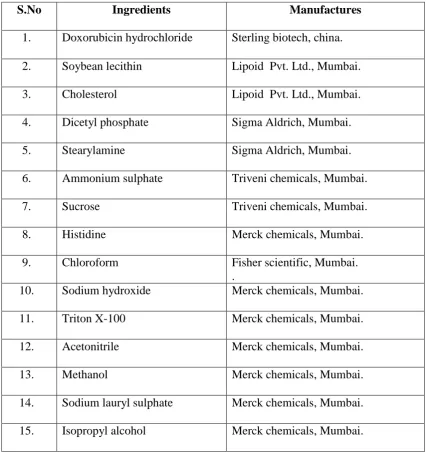
1 List of Chemicals 50
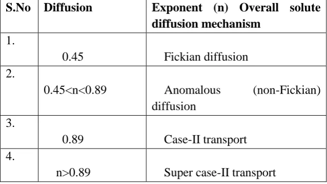
2 Diffusion Exponent And Solute Release Mechanism For Cylindrical
Shape 62
3 Standard Readings of Doxorubicin Hydrochloride in UV 63 4 Interpretations of FTIR Spectra for Pure Drug 70 5 Interpretations of FTIR Spectra for Pure Drug and Spectra-5 70 6 Interpretations of FTIR Spectra for Pure Drug and Spectra-5 71
7
The Composition and Ratios of Soy Lecithin, Cholesterol,
Stearylamine, Dicetylphosphate and Ammonium Sulphate for Different Types of Liposomes.
72
8
The Composition and Ratios of Drug, Soy Lecithin, Cholesterol, Stearylamine, Dicetylphosphate and Ammonium Sulphate for Optimized Batches.
73
9 Physicochemical Characteristics of Doxorubicin Hydrochloride
Liposomes for Optimized Batches. 73
10 Percent Free Drug of Doxorubicin Liposomal Solution for F1, F2, F3,
F4, F5 and F6 Formulations. 81
11 Assay of Doxorubicin Liposomal Solution for F1, F2, F3, F4, F5, F6
Formulations. 81
12 In Vitro Cumulative % Drug Release Profile of Doxorubicin
Hydrochloride Liposomal Formulations. 83
13 Zero Order Release Model of Doxorubicin Hydrochloride Liposomal
Optimized Formulations. 87
14 First Order Release Model of Doxorubicin Hydrochlor`IDE Liposomal
Optimized Formulations. 88
15 Higuchi Release Model of Doxorubicin Hydrochloride Liposomal
Optimized Formulations. 89
16 Korsmeyer-Peppas Model for Mechanism of Drug Release. 90 17 Curve Fitting Data of Release Rate Profile of Formulations F2, F4, F6. 90 18 Effect of Temperature on Assay of Doxorubicin HCL at 4OC. 94
19 Effect of Temperature on Assay of Doxorubicin HCL at Room
[image:13.595.77.560.115.734.2]1 Principle & Rationale of Drug Targeting 3
2 Structure of Liposome 11
3 Shape of Phospholipids Molecule. 11
4 Schematic Illustrations of Liposomes of Different Size and
Number of Lamellae. 13
5 Understanding Series of Cancer and Normal Cell Division 26 6 Accumulation of Liposomes within Solid Tumors 28 7 SEM Photography of Liposomal Solution for F2
Formulation. 80
8 SEM Photography of Liposomal Solution for F6
[image:14.595.104.533.117.404.2]NO
1 FTIR of Doxorubicin Hydrochloride 64
2 FTIR of Soy Lecithin 65
3 FTIR of Cholesterol 66
4 FTIR of soy Lecithin and Cholesterol 67
5 FTIR of Doxorubicin Hydrochloride, soy Lecithin,
Cholesterol and Dicetyl Phosphate. 68
6 FTIR of Doxorubicin Hydrochloride, Soy Lecithin,
1
Phosphate Buffer of PH 7.4. 63
2 Percent Free Drug Plot for F1, F2 F3, F4, F5, F6
Formulations 82
3 Assay Plot for F1, F2 F3, F4, F5, F6 Formulations 82
4 In Vitro Release Study for F1 Formulation. 84
5 In Vitro Release Study for F2 Formulation. 84
6 In Vitro Release Study for F3 Formulation. 85
7 In Vitro Release Study for F4 Formulation. 85
8 In Vitro Release Study for F5 Formulation. 86
9 In Vitro Release Study for F6 Formulation. 86
10 Comparison of in Vitro Release Studies for Optimized
Formulations F2, F4, F6. 91
11 Comparison of Zero Order Release Studies for Optimized
Formulations F2, F4, F6. 91
12 Comparison of First Order Release Studies for Optimized
Formulations F2, F4, F6. 92
13 Comparison of Higuchi’s Order Plot for Optimized
Formulations F2, F4, F6. 92
14 Comparison of Korsmeyer –Peppa’s Model for Optimized
Formulations F2, F4, F6. 93
15 Stability Plot for F1, F2 F3, F4, F5, F6 Formulations at 40C 95
16 Stability Plot for F1, F2 F3, F4, F5, F6 Formulations at
1
Particle Size Distribution by Wet Method of Doxorubicin HCL liposomal Solution for F2 Formulation.
74
2
Particle Size Distribution by Wet Method of Doxorubicin HCL Liposomal Solution for F4 Formulation.
75
3
Particle Size Distribution by Wet Method of Doxorubicin HCL Liposomal Solution for F6 Formulation.
76
4 Zeta Potential for Doxorubicin HCL Liposomal
Solution for F2 Formulation. 77
5 Zeta Potential for Doxorubicin HCL Liposomal
Solution for F4 Formulation. 78
6 Zeta Potential for Doxorubicin HCL Liposomal
Dept., of Pharmaceutics, The Erode College of Pharmacy & Research Institute, Erode. Page 1
1.
INTRODUCTION
The goal of any drug delivery system is to provide a therapeutic amount of
drug to the proper site in the body, to achieve promptly and then maintain the desired
drug concentration.
Conventional drug delivery system achieves as well as maintains the drug
concentration with in the therapeutically effective range needed for treatment only
when taken several times a day. This results in a significant fluctuation in drug level
(Chien YM., 1992).
The concept of designing specified delivery system to achieve selective drug
targeting has been originated from the perception of Paul Ehrlich, who proposed drug
delivery to be as a “magic bullet”.
Controlled & Novel delivery envisages optimized drug in the sense that the
therapeutic efficacy of a drug is optimized, which also implies nil or minimum side
effects. It is expected that the 21st century would witness great changes in the area of
drug delivery. The products may be more potent as well as safer. Target specific
dosage delivery is likely to overcome much of the criticism of conventional dosage
forms. The cumulative outcome could be summarized as optimized drug delivery that
encompasses greater potency & greater effectiveness, lesser side effects and toxicity,
better stability, low cost hence greater accessibility, ease of administration and best
Dept., of Pharmaceutics, The Erode College of Pharmacy & Research Institute, Erode. Page 2
1.1 RATIONALE OF DRUG TARGETING (Jain N K., 2001)
The site-specific targeted drug delivery negotiates an exclusive delivery to
specific pre identified compartments with maximum activity of drugs and
concomitantly reduced access of drug to irrelevant non-target cells. The controlled
rate & mode of drug delivery to pharmacological receptor and specific binding with
target cells as well as bioenvironmental protection of the drug in route to the site of
action are specific features of targeting. Invariably, every event stated contributes to
higher drug concentration at the site of action and resultant lowers concentration at
non-target tissue where toxicity might crop up. The high drug concentration at the
target site is relative cellular result of the uptake of the drug vehicle, liberation and
efflux of free drug from the target site.
Targeting is signified if the target compartment is distinguished from the other
compartments, where toxicity may occur and also if the active drug could be placed
predominantly in the proximity of target site. The restricted distribution of the parent
drug to the non-target site(s) with effective accessibility to the target site(s) could
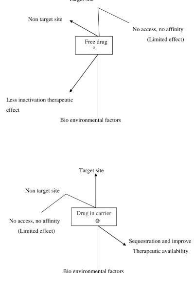
Dept., of Pharmaceutics, The Erode College of Pharmacy & Research Institute, Erode. Page 3 Figure No: 1 Principle & Rationale of drug targeting:
Target site
Non target site
No access, no affinity (Limited effect)
Less inactivation therapeutic effect
Bio environmental factors
Target site
Non target site
No access, no affinity (Limited effect)
Sequestration and improve Therapeutic availability
Bio environmental factors Free drug
°
Dept., of Pharmaceutics, The Erode College of Pharmacy & Research Institute, Erode. Page 4 The various approaches of vectoring the drug to the target site can be broadly classified as:
Passive targeting
Inverse targeting
Active targeting
Dual targeting
Double targeting
Combination targeting
PASSIVE TARGETING
System that target the systemic circulation are generally characterized as passive” delivery systems (i.e., targeting occurs because of the body’s natural response to the physicochemical characteristics of the drug or drug-carrier system. It is a sort of passive process that utilizes the natural course of biodistribution of the carrier system through which, it eventually accumulation in the organ compartment(s) of the body. The ability of some colloids to be taken up by the RES especially in liver and spleen has made them as ideal vectors for passive hepatic targeting of drugs to these compartments.
This category of targetable devices includes drug bearing bi-layer vesicular systems as well as cellular of micron or sub micron size range.
INVERSE TARGETING
Dept., of Pharmaceutics, The Erode College of Pharmacy & Research Institute, Erode. Page 5
ACTIVE TARGETING
Conceptually, active targeting exploits modification or manipulation of drug carriers to redefine its biofate. The natural distribution pattern of the drug carrier composites is enhanced using chemical, biological and physical means, so that it approaches and is identified by particular biosites. The facilitation of the drug-carrier to target cells through the use of ligands or engineered homing devices to increase receptor mediated (or I some cases receptor independent but epitopes based) localization of the drug and target specific delivery of drug(s) is referred to as active targeting.
This target approach can further be classified into three different levels of targeting
First order targeting
It refers to restricted distribution of the drug-carrier system to the capillary bed of a predetermined target site, organ or tissue. Compartmental targeting in lymphatic, peritoneal cavity, cerebral ventricles, lungs, joints, eyes, etc., represents first order targeting (it could also be categorized as level of passive targeting).
Second order targeting
The selective delivery of drugs to a specific cell type such as tumor cells and not to the normal cells is referred as second order drug targeting. The selective drug delivery to the kupffer cells in the liver exemplifiers this approach.
Third order targeting
Dept., of Pharmaceutics, The Erode College of Pharmacy & Research Institute, Erode. Page 6 Targeting components, which have been studied and exploited are pilot molecules themselves (bioconjugates) or anchored as ligands on some delivery vehicle (drug-carrier system). All the carrier systems, explored So far, in general, are colloidal in nature. They can be specifically functionalized using various biologically relevant molecular ligands including antibodies, polypeptides, oligosaccharides (carbohydrates), viral proteins and fusogenic residues. The ligands afford specific avidity to drug carrier. The engineered carrier constructs selectivity deliver the drug to the cell or group of cells generally referred to as target. The cascade of events involved in ligand negotiated specific drug delivery is termed as ligand driven receptor mediated targeting.
Physical targeting (triggered Release)
The selective drug delivery programmed and monitored at the external level
(ex vivo) with the help of physical means is referred to as physical targeting. In this
mode of targeting, some characteristics of the bioenvironmental are used either to direct the carrier to a particular location or to cause selective release of its content.
DUAL TARGETTING
This classical approach of drug targeting employs carrier molecules, which have their own intrinsic antiviral effect thus synerging the antiviral effect of the loaded active drug. Based on this approach, drug conjugates can be prepared with the fortified activity profile against the viral replication. A major advantage is that the virus replication process can be attacked at multiple points, excluding the possibilities of resistant viral strain development.
DOUBLE TARGETING
Dept., of Pharmaceutics, The Erode College of Pharmacy & Research Institute, Erode. Page 7 The temporal control of drug delivery has been developed in terms of
control drug release prior to the development of drug targeting. If spatial
targeting is combined with temporal control results in an improved therapeutic
index by the following two effects. First, if drug release or activation is occurred
locally at therapeutic sited, selectively is increased by multiplication of the spatial
selectively with the local release/activation. Second, the improvement in the
therapeutic index by a combination of a spatially selective delivery and a
preferable release pattern for a drug, such as zero order release for a longer time
period of the drugs. When these two methodologies are combined, it may be called “Double targeting”.
COMBINATION TARGETING
Petit and Gombtz., 1998 have suggested the term combination targeting
for the site specific delivery of proteins and peptides. These targeting systems are
equipped with carriers. Polymers and homing devices of molecular specificity
that could provide a direct approach to target site. Modification of proteins and
peptides with natural polymers, such as polysaccharides, or synthetic polymers,
such as poly (ethylene glycol), may alter their physical characteristics and favor
targeting the specific compartments, organs or their tissues within the
vasculature.
1.2.3 LIMITATIONS OF TARGETED DRUG DELIVERY
SYSTEMS (Cheiny M., 1992)
Several problems have been identified which require alterations in targeting strategies particularly, in vivo. These include:
Dept., of Pharmaceutics, The Erode College of Pharmacy & Research Institute, Erode. Page 8
Target tissue heterogenecity.
Problems of insufficient localizations of targeted systems into tumor cells.
Down regulation and sloughing of surface epitopes.
Diffusion and redistribution of released drug leading to no-specific accumulation.
1.2.4 CARRIERS USED IN TARGETING DRUG DELIVERY
SYSTEMS
Carrier is one of the most important entities essentially required for succ essful transportation of the loaded drug(s). They are drug vectors, which sequester, transport and retain drug en route. While eluting or delivering it within or in vicinity of target.
Colloidal carriers:
Vesicular systems: Liposomes; pharmacosomes; virosomes; immunoliposomes.
Micro particulate systems: Nanoparticles; Microparticles; Magnetic Microspheres; Nanocapsules.
Cellular carriers: Resealed erythrocytes; Serum albumin; Antibodies; Platelets; Leukocytes.
Supramolecular delivery system:
Micelles; reverse micelles; mixed micelles; polymeric micelles; liquid crystal: lipoproteins.
Polymer based systems:
Dept., of Pharmaceutics, The Erode College of Pharmacy & Research Institute, Erode. Page 9
Macromolecular carriers:
Proteins, glycoproteins, neo glycoproteins and artificial viral envelops (AVE); Glycosylated water-soluble polymers (poly-L-lysine).
Mabs; Immunological Fab fragments; antibody enzyme complex & bispesific Abs; Toxins, immunotoxin & rCD4 toxin conjugates
Lecithins (Con A) & polysaccharides.
1.3 INTRODUCTION TO LIPOSOMES
Liposomes (marc J. Ostro., 1987) have reached the clinical only recently, but
they are not a new invention Alec D. Bangham of the Agricultural Research Council’s
institute of Animal physiology in Cambridge, England, inadvertently produced the
first liposome in 1961, while evaluating the effect of phospholipids on blood clotting.
When Bangham put water in a flask containing a phospholipid film, the water
molecules to arrange themselves in to what he discovered. He found vesicles
composed of a bilayered phospholipids membrane surrounding water entrapped from
the environment.
Phospholipids form closed, fluid-filled spheres when they are mixed with
water in part because the molecules are amphipathic; they have a hydrophobic “tail”
and a hydrophilic or polar “head”. Two fatty acid chains, each composed of 10 to 24
carbon atoms, make up the hydrophobic tail of most naturally occurring phospholipids
molecules. Phosphoric acid bound to any of several water soluble molecules
composes the hydrophilic head. When a high enough concentration of phospholipids
is mixed with water, the hydrophobic tails spontaneously herd together to exclude
Dept., of Pharmaceutics, The Erode College of Pharmacy & Research Institute, Erode. Page 10 interior and the polar head groups point outward the polar groups at one surface of the membrane point toward the liposome’s interior and those at the other surface point
toward the external environment. It is this remarkable reactivity of phospholipids to
water that enables workers to load medications in to liposomes. In a liposome form,
any water soluble molecules that have been added to the water are incorporated in to
the aqueous spaces in the interior of the spheres, whereas any lipid soluble molecules
added to the solvent during vesicle formation are incorporated in to the lipid layer.
Liposomes employed for drug delivery typically range in diameter from 250
angstrom units to several micrometers and are usually suspended in a solution. They
have two standard forms: “onion-skinned” multilamellar vesicles (MLVs) made up of
several lipid bilayers separated by fluid, and unilamellar vesicles, containing of a
single bilayer surrounding an entirely fluid core. The unilamellar vesicles are typically
Dept., of Pharmaceutics, The Erode College of Pharmacy & Research Institute, Erode. Page 11
Liposome structure formed by phospholipids Figure No. 2 Structure of liposome
Shape of phospholipids molecule
Figure No. 3 Shape of phospholipids molecule.
Dept., of Pharmaceutics, The Erode College of Pharmacy & Research Institute, Erode. Page 12 The main components of liposome are
Phospholipids
o Phosphoglycerides o Sphingolipids
Cholesterol
1.5 TYPES OF LIPOSOMES (Marc J Ostro., (1987) & Amarnath
Sharma (1997)
1.5.1 Classification based on structural parameters
1. Multilamellar Large vesicles (MLV 0.1-6µm)
2. Small unilamellar vesicles (SUV 0.02-0.05 µm)
3. Large unilamellar vesicles (LUV>0.06)
4. Oligolamellar vesicles (OLV 0.1-1 µm )
5. Unilamellar vesicles (UV- wide range)
6. Gaint unilamellar vesicles (GUV –cell size vesicle s>1 µm)
7. Medium Unilamellar vesicles
Dept., of Pharmaceutics, The Erode College of Pharmacy & Research Institute, Erode. Page 13 Figure No: 4 Schematic illustrations of liposomes of different size and
number of Lamellae. SUV: Small unilamellar vesicles; LUV: Large unilamellar
vesicles; MLV: Multilamellar vesicles; MVV: Multivesicular vesicles.
1.5.2 Classification
The Liposomes are classified as vesicular systems from biological origin and non biological origin. Specialized for cellular drug targeting are grouped separately (Vyas and Dexit, 1998).
Biological origin
1) Liposomes
Dept., of Pharmaceutics, The Erode College of Pharmacy & Research Institute, Erode. Page 14 2) Polymerizable Liposomes (Fendler, 1982)
This type of Liposomes mainly contains phospholipid derivatives of polymerizable group in their fatty acyl chains. The diacetylenic lipids, methacryloyl lipids, dienoyl lipids are used as building blocks for this type.
3) Polymer capped Liposomes (Regen et al., 1984)
Here the cationic phospholipids are ion-paired with a polymerizable anion like methacrulate ion. The elute is dispersed in Liposomes followed by polymerization using irradiation or chain initiator. In this the polymeric network encases the bilayer of Liposomes without covalent linkage.
4) Polymerized phospholipid Liposomes (Weber et al., 1987)
Phospholipid polymerized with 1, 2-bis-(2-mercaptohexadecanoyl) 3-glycerol-3-phosphocholine could readily assemble to form this type of vesicles.
5) Redox Liposomes (Samvel et al., 1985)
This type of vesicles based on thiol-disulfide redox cycle.
6) Polymer grafted Liposomes (Allen et al., 1991)
These types of Liposomes are formed by natural or synthetic phospholipids with covalently linked PEG polymer.
7) Virosomes (Al-Ahdal et al., 1994)
Dept., of Pharmaceutics, The Erode College of Pharmacy & Research Institute, Erode. Page 15 8) Emulsomes (Amselem et al., 1994)
Emulsomes were constructed by phospholipids which have high concentration along with high molecular weight fatty acids.
Non-biological origin
1) Niosomes (Handijani-vila et al., 1979)
The single chain non-ionic surfactants of low HLB values were contributed mainly in construction of Liposomes.
2) Discomes (Vyas et al., 1997)
Solubilization of niosomes with non-ionic surfactant (Polyoxy ethylene acetyl ether class) forms discomes.
3) Pharmacosomes (Vizaglou et al., 1986)
The Pharmacosomes are formed by mesogenic drug which itself acts as a building block in combination with a lipid.
4) Ufasomes (Gebicks et al., 1973)
The single chain unsaturated fatty acids (Oleic, linoleic acid) are the main building blocks in this type.
5) Cryptosomes (Blume et al., 1990)
Dept., of Pharmaceutics, The Erode College of Pharmacy & Research Institute, Erode. Page 16
Specialized Liposomes
1) Glycoprotein bearing Liposomes
Incorporation of glycoprotein in bilayer membrane gives this type of vesicles. The major glycoprotein, the glycoprotein of the human erythrocyte plasma membrane has been incorporated in to Liposomes.
2) Glycolipid bearing Liposomes
Glycolipid (like monosialo-gangliaside) appended liposome are prepared and tried to target lymphocytes.
3) Glycoside bearing Liposomes
The plant glycosides, asiaticiside and corhorusin D containing rhamnose and glucose in terminal sugar respectively have been grafted on the liposomal that are tried against M.leprae and M.tuberculosis by Medda et al., (1995)
4) Protein coated Liposomes (Longaman and co-workers, 1995)
Liposomes with protein immobilized on the surface are investigated to target extra vascular spaces by using biotinylated antibody and streptavidin.
5) Peptide carrying Liposomes (Zalipsky et al., 1995)
Dept., of Pharmaceutics, The Erode College of Pharmacy & Research Institute, Erode. Page 17 6) Polysaccharide bearing Liposomes
Polysaccharide coated Liposomes used for targeting to Legionella
pneumophilia infected human monocyte and guinea pig.
7) Bacteriosomes, Proteoliposomes
Lectinization of Liposomes was used for targeting to Hela cells and streptococcus etc.,
8) Bioreactive Liposomes (Gregoriadis et al., 1983)
Enzymes were encapsulated in Liposomes and were called as Bioreactive Liposomes. Eg. Streptokinase containing Liposome.
9) Immunosomes
Immunosomes are liposomal constructs engineered by employing immunoglobulins as pilot molecules anchored on or as a structural part of vesicles to confer specificity to a wide range of target sites. These are classified in to
a) Immunoliposomes
b) Haptenated Liposomes
c) Immunotoxin anchored Liposomes
d) Immunoprotective Liposomes
e) Immunoadjuvant Liposomes
10) Transferin based Liposomes (Wagner et al., 1994)
Dept., of Pharmaceutics, The Erode College of Pharmacy & Research Institute, Erode. Page 18 11) Enzymosomes
In this type enzymes are covalently immobilized or coupled to the surface of liposome and if a prodrug is administered simultaneously, it can convert to active form at target site by immobilized enzyme.
1.6 Liposomes delivered intercellularly
Liposomes deliver their contents to the cytoplasm of cells in culture either by fusion with the outer cell membrane or by endocytosis where upon they are concentrated in acidic lysozymal sacs. (Ostro et al., 1987)
1) Inter membrane transfer
Inter membrane transfer of lipid component can take place upon close approach of the two phospholipid bilayers without the need for disruption of the liposome. Only specific phospholipids exchange (PC &PE) via intermediate of a specific cell surface exchange protein. Similarly Liposomes and lipoproteins (HDL) interactions is important liposomal stability in circulation. Addition of cholesterol retards immediate destabilization of Liposomes.
2) Contact release
Dept., of Pharmaceutics, The Erode College of Pharmacy & Research Institute, Erode. Page 19 3) Adsorption
The adsorption of Liposomes takes place either as a result of physical
attractive forces, or as a result of binding by specific receptors to ligands on the
vesicle membranes. The attraction depends on the specific cell surface protein. This
interaction was more in case of gel phase of liposome.
4) Fusion
Close approaching of Liposomes and cell membranes can lead to fusion of the two resulting in complete mixing of liposomal lipids with those of the plasma membrane of the cell and the liposomal content released in to cytoplasm. This process takes place after phagocytosis and endocytosis of Liposomes. Inside the Endosome, Liposome were affected by acidic pH. There by this will completely fuse and deliver the drug content in to cytoplasm.
1.7
Methods of liposome preparation
1.
Passive loading techniques
a.
Mechanical dispersion methods Lipid film hydration by hand shaking non-hand shaking or freeze drying.
Micro emulsification Sonication
Dept., of Pharmaceutics, The Erode College of Pharmacy & Research Institute, Erode. Page 20 Ethanol injection
Ether injection
Double emulsion vesicles
Reverse phase evaporation vesicles Stable plurilamellar vesicles
c.
Detergent removal methods
Detergent removal from mixed micelles
Dialysis
Column chromatography
Dilution
Reconstituted sandal virus enveloped vesicles2.
Active loading techniques
ADVANTAGES OF LIPOSOMES
The pharmaceutical and pharmacological justification of the use of liposomes as drug carriers is as follows:
1. Liposomal supply both a lipophilic environment and aqueous “milleu interne” in one system and are therefore suitable for the delivery of hydrophobic, amphipatic and hydrophilic drugs and agents.
2. Liposomes are chemically and physically well characterized entities. 3. The biological fate of liposomes after their administration is related to
their composition and physical properties.
4. Liosomes are biocompatible due to their biodegradability, low toxicity and lack of immunogenicity.
Dept., of Pharmaceutics, The Erode College of Pharmacy & Research Institute, Erode. Page 21 6. Liposomes help to reduce exposure of sensitive tissues to toxic drugs. 7. Liposomes can be administered through mostnroutes of administration
including ocular, pulmonary, nasal, oral, intramuscular, subcutaneous, topical and intravenous.
8. Pharmacokinetics and in-vivo distribution of liposomes can be controlled by their port of entry combined with their lipid composition and size.
DISADVANTAGES OF LIPOSOMES
1. Aggregation, fusion and drug leakage during storage.
2. Chemically instable i.e., degradable by oxidation and hydrolysis. 3. In physiological environment they are destabilized by high density
lipoproteins(HDL)
4. Purity of natural phospholipids and cost of production.
5. They undergo complete mediated phagocytosis and lipid exchange reactions.
1.8 FACTORS EFFECTING DRUG ENTRAPMENT AND RELEASE
CHARACTERISTICS
Factors effecting drug entrapment and release characteristics of Liposomes include,
1. Charge 2. Lipid content 3. partition coefficient
4. Method of preparation of Liposomes
Charge
Dept., of Pharmaceutics, The Erode College of Pharmacy & Research Institute, Erode. Page 22 entrapped volume.
Lipid content
The total amount of liposomal lipid used and thee internal volume of the Liposomes will affect the total amount of loading of non-polar and polar group in to Liposomes. Efficient capture will depend on the use of drugs at concentration which do not exceed the saturation limit of the drug in the aqueous compartment or the lipid bilayers.
Partition coefficient
The location of drug within a liposome is based on the partition coefficient of the drug between aqueous compartments and lipid bilayer and the maximum amount of drug that can be entrapped within Liposomes in dependent on its total solubility in each phase.
Method of preparation of Liposomes
The method of preparation of Liposomes can also affect drug location and overall entrapment efficacy. Several methods are now available for preparation of Liposomes. The cast film method is simple but the major drawback with MLVs prepared is the relatively low encapsulation in terms of aqueous space per mole of lipid. Dilute preparations of Liposomes with a low encapsulation efficiency is obtained, when solvent injection method is used.
1.9 APPLICATIONS OF LIPOSOME
Dept., of Pharmaceutics, The Erode College of Pharmacy & Research Institute, Erode. Page 23 the field with more than 2000 papers and reviews per year, of which most of them are related to anticancer, anti-inflammatory and anti-microbial.
1. Dinitroanilines when administered in the form of liposome shows better antimicrobial activity against leishmania than that of free drug. The improved antimicrobial activity is due to the engulfing of Liposomes by MPS cells (Mononuclear Phagocytic System) which is type of passive targeting.
2. Rifamycin in the liposomal formulation has superior effect against tuberculi when compared to that of free drug.
3. Doxorubicin can be encapsulated in liposome to reduce the serious side effects of the drug.
Hence liposome can be used as the safer and more efficient drug delivery for many problematic entities.
1.10 STABILITY OF LIPOSOMES
The stability of Liposomes is of major concern in their development for
pharmaceutical applications. A drug containing Liposomes can be unstable because of
physical or chemical stability. The stability studies could be broadly covers under two
main sections.
Stability invitro mainly covers the stability aspects prior to the administration
of the formulation and with regard to the stability of the constituted lipids.
Stability invivo, which covers the stability aspects once the formulation, is
Dept., of Pharmaceutics, The Erode College of Pharmacy & Research Institute, Erode. Page 24 I. Chemical degradation
[a] Oxidation (Hunt C et al., 1981)
The oxidative degradation of liposome can be prevented by
following the below mentioned precautions
1. Start with freshly purified lipids and freshly distilled
solvent
2. Avoid procedures which involves high temperature
3. Carryout the manufacturing process in the absence of
oxygen
4. De-oxygenate aqueous solution in an inert atmosphere
[b] Hydrolysis (Frfkjaer et al., 1984)
1. Using lipid containing ether linkage instead of ester linkage
2. Sphingomyelin prevents invivo hydrolyses
II. Physical degradation (Wong M et al., 1982)
Sedimentation, leaching of drugs aggregation or fusion. High
manufacturing temperature and many other factors can induce this.
Physical stability can be achieved by
1. Manufacturing and storing at temperature below its transition temperature
2. Adding 10% of P.A [Phosphatidic acid] or P.O to neutral liposome for
providing negative pH.
3. By cross linking membrane components covalently using gluteraldehyde
Dept., of Pharmaceutics, The Erode College of Pharmacy & Research Institute, Erode. Page 25 Approaches that can be used to increase liposome stability involve efficient
formulation and lyophilization. Formulation involves the selection of the appropriate
lipid composition and concentration of bilayer, in addition to the aqueous phase
ingredients such as buffer, antioxidants, metal-chelators and cryoprotectants. Charge
inducing lipids such as Phosphotidyl glyceride be incorporated in to the liposome
bilayer to decrease fusion, while cholesterol and sphingomyelin can be incorporated
in formulation, in order to decrease the permeability and linkage of encapsulated
drugs.
1) Buffers at neutral pH can decrease hydrolysis
2) Addition of antioxidants such as sodium ascorbate, can decrease oxidation
3) Freeze-dried liposome formulations should incorporate a lipoprotectant
like non-reducing disaccharides such as trehaloes and sucrose.
1.11 CANCER CHEMOTHERPHY
Cancer (Sydney. Basic clinical pharmacology (3rd edition)) is a group of neoplastic diseases that occur in human of all age groups and races as well as in all animals species. The incidence, geographic distribution and behavior of specific types of cancer are related to multiple factors include sex, age, race, genetic predisposition and exposure to environmental carcinogen.
Dept., of Pharmaceutics, The Erode College of Pharmacy & Research Institute, Erode. Page 26 Cancer chemotherapy has been under intensive development for the past 30 years, resulting in cures of certain types of disseminated cancers that were previously fatal. Even patients with advanced disease have improved dramatically with chemotherapy.
Mode of action of chemotherapeutic agents
Most antineoplastic agents are regarded as “cell-cycle specific”. They act specifically on processes such as DNA synthesis, transcription, or the function of mitotic spindle.
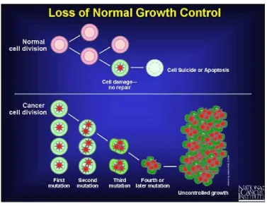
All slow growing and fast growing tumor cells display a similar pattern during the division. This may be characterized as follows:
1. There is presynthetic phase 2. The synthesis of DNA occurs
[image:44.595.128.506.107.399.2]Dept., of Pharmaceutics, The Erode College of Pharmacy & Research Institute, Erode. Page 27 4. Mitosis ensures, the G2 cell, containing a double complement of DNA, divides in to 2 daughter GI cells, may immediately re-enter to the cell cycle.
Antitumor drugs are better at killing cells during DNA synthesis and active division. When a tumor is young, most of its cells are making DNA. This is defined as large growth function in this state, tumors are destroyed by drugs because the majority of their cells are making DNA and dividing.
The major problems in cancer chemotheraphy are the toxic drugs effects on normal cells and the rapid clearance of the drug from the tumor cells. Usefull drugs without side effects do not at exist. Rapidly dividing normal cell such as hair follicles, cells lining the gastrointestinal tract and bonemarrow cells involved in the immune defence system are also destroyed by the present day chemotheraphy. Nausea, hair loss, increased susceptibility to infection and many others comprising a discouraging list.
Liposomes: An ideal “Drug Carrier” for anticancer drugs
Anticancer drugs (Sayed S. Daoud (1989)) are known to produce serious side effects to other healthy tissues. The more serious effects are myocardiopathy and pulmonary toxicity. Therefore targeting such type drugs to the cancerous cell is essential because these drugs are new for the treatment of different type of carcinomas effectively. Usually a therapeutically profitable target agent relationship is far from ideal and undesirable side effects are usually observed. The alternative is to use simple functional molecules which transport the drug to the specific site and release it to perform task. Liposomes are non-toxic, biodegradable microcapsule made up of one or multiple lipid bilayers membranes.
Dept., of Pharmaceutics, The Erode College of Pharmacy & Research Institute, Erode. Page 28 Important characteristics of drug carrier include protection of the encapsulated compound. Selective delivery of the entrapped material to specific tissues with minimal losses of drug during transit, regulation of the drug delivery rate, reduction of toxicity and removal of unused drug. All these function can co-exist in a single liposome preparation which makes it an ideal carrier of drug. Liposomes have proved to be suitable vehicles for antitumor drugs.
Figure No: 6 Accumulations of Lip osomes within Solid Tumors
Dept., of Pharmaceutics, The Erode College of Pharmacy & Research Institute. Erode Page 30
2. LITERATURE REVIEW
Amarnath Sharma et al., described that liposomes are microparticulate lipoidal vesicles which are under extensive investigation as drug carriers for improving the delivery of therapeutic agents. In this they discussed the potential applications of liposomes in drug delivery with examples of formulation approved for clinical use, and the problems associated with further exploitation of these drug delivery systems.
Antoneta et al., studied about cholesterol and other sterols are important components of biological membranes and are known to strongly influence the physical characteristics of lipid bilayers. Although this has been studied extensively in fully hydrated membranes, little is known about the effects of cholesterol on the stability of membranes in the dry state.
Chieny et al., described the concept of designing specified delivery systems to achieve selective drug targeting. In this they compared between the conventional and targeting drug delivery systems. They described about advantages and limitations of target drug delivery systems. They described that the principle and rationale of drug targeting.
Eugenia et al., reported the advantages and application of liposome with respect to anti-cancer, anti-inflammatory and anti-microbial agents. In this they described the advantages of the encapsulation macromolecules like enzymes into liposomes. In this study they incorporated the enzymes into liposomes by dried film hydration method and studied the therapeutic activity of enzymes.
Fresta et al., described the preparation of various kinds of loaded of 5FU and reported on encapsulation efficacy, storage stability and fusogenic properties. They concluded that the most suitable liposome preparation was the SPL Vs that showed both better drug loading and stability parameters than others.
Dept., of Pharmaceutics, The Erode College of Pharmacy & Research Institute. Erode Page 31
proved to be a good approach to eliminating the vehicle and improving the drug’s antitumor activity. We formulated docetaxel Liposomes containing phosphatidylcholine (soybean lecithin), cholesterol and various stabilizers by the dried thin film hydration method. Particle size analysis, drug content and entrapment efficiency in charged Liposomes were strongly affected by the different stabilizers, the stability of the lyophilized docetaxel Liposomes were evaluated after stored at 40C and the room temperature for 3 months. The Liposomes stored at 40C were found to be stable for duration of 3months. Hence it can be concluded that stabilizers like Stearylamine and Dicetylphosphate along with cholesterol were suitable carrier for the preparation of liposomal docetaxel.
Gautam Vinod et al., invented a long circulating non-pegylated liposomal doxorubicin hydrochloride composition for parenteral administration and a process for its preparation. The circulation time in Swiss albino mice is at least 25 times longer than conventional non-liposomal formulations. The non-pegylated liposomes are stable, exhibit low toxicity and have been found to be efficacious in different tumor models.
Gregoiadis et al., reported the method of liposome formulation with lecithin (egg phosphatidly choline). In this they described about method to find out the percentage of drug loading and the usage of Triton-X 100 for the percentage of drug loading in liposome. They found that Triton-X 100 can be used to disrupt the lipid bilayer in liposome. Hence thereby the bound drug liberated out of the liposome.
Dept., of Pharmaceutics, The Erode College of Pharmacy & Research Institute. Erode Page 32
cytokines and immunomodulators have been described, as have attempts to increase tumour targeting with the use of hyperthermia and antibody-coated liposomes. Leptosomes are also receiving increased attention as potentially useful vehicles for the delivery of recombinant DNA constructs in the emerging science of gene therapy, although most of this work has focused on cationic liposomes which have very different pharmacokinetic profiles to the liposomes discussed in this review article.
Jain et al., described the target drug delivery systems were likely to overcome much of the criticism of conventional dosage forms. They summarized that the optimized drug delivery that encompasses greater potency & greater effectiveness, lesser side effects and toxicity, better stability, low cost hence greater accessibility, ease of administration and best patient compliance. They also described about carriers used in targeted drug delivery systems.
Jorge et al., reported the methods of liposomal formulation for encapsulating the enzyme (L-Asparaginase). In this study they formulated liposomes with natural phospholipids (egg yolk lecithin). They also described the method to conjugated the enzyme with palmitic acid to form palmitoyl-enzyme complex, which can further prolongs the circulation time of enzyme in general circulation without affecting the enzymatic activity i.e. from 2.88 hr to 23.7 hr.
Lasic et al., described about the recent discoveries in the field of liposomes and latest application of liposomes. They described the usage of different types of liposomes and their advantages. In this they studied the efficacy of DNA encapsulation in liposomes. And in the treatment of colon carcinoma and AIDS related Kaposi sarcoma. From this study they confirmed the liposomes can be used as carrier for biological like RNA, DNA, etc.
Dept., of Pharmaceutics, The Erode College of Pharmacy & Research Institute. Erode Page 33
chosen and the presence of cholesterol. A maximum EE for 4-(N)-steroyl-gemcitabine incorporated in DSPC/DSPG 9:1. This formulation was correlated with the highest stability in vitro and in vivo. Cytotoxicity of Gemcitabine prodrugs, free or encapsulated in liposomes, was between two- and sevenfold that of free gemcitabine.
Marc Ostro et al., described the methods to reduce the dosage of drug with the help of liposomes and their potential advantages in different types of diseased states. In this they confirmed that liposomes are better dosage form than conventional dosage forms. They have also stated the drug can be targeted by active and passive targeting and the uses of both passive and targeting of liposomes.
Mirant Ahmad et al., developed a well characterized novel lyophilized liposome-based paclitaxel (LEP-ETU) formulation that is sterile, stable and easy-to-use. The mean particle size of the liposomes is about 150 nm before and after lyophilization, and the drug entrapment efficiency is greater than 90% stability data indicated that the lyophilized LEP-ETU was physically and chemically stable for at least 12 months at 2-8 and 25 8C. Moreover the formulation can diluted to about 0.25 mg/ml without drug precipitation or change in particle size. In vitro drug release study in phosphate-buffered saline (PBS, pH 7.4) showed that less than 6%of the entrapped paclitaxel was released after 120 h, indicating that the drug is highly stable in an entrapped form at physiologic temperature.
Monostoi et al., [2004] studied the stability and transdermal absorption of topical amphotericin B liposome formulation and found that the positively charged liposome might be the best formulation for AmB, dne to its higher stability than other formulations.
Dept., of Pharmaceutics, The Erode College of Pharmacy & Research Institute. Erode Page 34
Soleiman Mohammadi-Samani et al., Described Cyproterone acetate (CA) has been loaded to liposome by solvent evaporation and thin film formation technique. The effects of some formulation variables such as temperature of organic solvent evaporation, rotary evaporator speed, volume of organic solvent, volume of balloon and temperature of hydrating buffer has been evaluated. Finally percutaneous absorption of CA from simple gel and liposomal formulations was assessed. The results showed that liposomal formulation has better penetration potential than conventional CA formulation (simple gel).
Sydney et al., described the cancer chemotherapy in this they discussed incidence, geographic distribution and behavior of specific types of cancer. In that they described the mode of action, adverse effects, clinical use and dose administration of different chemotherapeutic agents.
Tyrrell et al., described the general consideration and method of preparation of liposomes and also described about liposome- protein interaction and protein entrapment liposomes. They summarized about uptake of liposomes in vivo and interaction of liposomes with cell culture and immunological aspects of liposomes.
Dept., of Pharmaceutics, The Erode College of Pharmacy & Research Institute. Erode Page 35
daunorubicin in liposomal formulation for treatment of skin cancer like cutaneous T-cell lymphoma, malignant melanoma, AIDS related Kaposi’s sarcoma. They formulated liposomes with synthetic phospholipids and stabilizing agents like stearylamine and cholesterol. They also found that the efficacy of drug is improved in liposomal formulation than that of free drug.
Xue Ming et al., studied site specific delivery of drugs and therapeutics can significantly reduce drug toxicity and increase the therapeutic effect. Transferring is one suitable ligand to conjugated to drug delivery systems to achieve site specific targeting, due to specific binding to transferring receptors, Administration of Tf-DOX to tumor-bearing mice could be used to deliver DOX effectively to the targeted site. This study indicated that the Tf-coupled PEG Liposomes could be as the targeted carriers to facilitate the delivery of the encapsulated anticancer drugs in to tumor cells by receptor-mediated way.
Yousefi et al., An improved pegylated liposomal formulation of docetaxel has
AIM AND
Dept., of Pharmaceutics, The Erode College of Pharmacy & Research Institute, Erode. Page 36
Liposomes have been used to target drug to specific organs, delay the loss of rapidly cleared, drugs, enhances therapeutic potency and offer a host of the other advantages.
Doxorubicin hydrochloride is one of the most commonly used cytotoxic anthracycline antibiotics used in cancer chemotherapy and has been shown to have activity against a wide variety of neoplasms.
Conventional compositions of doxorubicin hydrochloride are available as freeze-dried product (or) as a solution of doxorubicin hydrochloride in water. Both these products have been associated with a number of toxicities when administered intravenously. Severe myelosupression, nausea, vomiting, alopecia, mucosistis & cardio toxicity, limits the use of Doxorubicin Hcl. It also causes extravasations & necrosis at the site of injection.
To overcome these problems, an alternative approach is needed. In the present study doxorubicin Hcl liposomes are formulated using various biolipids and Stabilizers (Positive and Negative) to check effect of drug loading and particle size. Several approaches has taken in an effort to increase the circulation time of liposome by coating the liposomal surface with a hydrophilic polymer such as polyethylene glycol (PEG) to prevent adsorption of various blood plasma proteins to the liposome surface. These liposomes appeared to reduce some of the toxic effects caused by the release of their contents, but have new toxic effects appeared like skin toxicity generally known as “Hand-Foot Syndrome” and the presence of large molecules (PEG) on the liposomal surface may reduce the interaction of liposomal with cells & hinder entry of liposomes in to tumor tissue.
Dept., of Pharmaceutics, The Erode College of Pharmacy & Research Institute, Erode. Page 37
IT WAS PLANNED TO CARRY OUT THE PRESENT STUDY AS
FOLLOWS:
STAGE 1:
1. Preformulation studies
a. Standard calibration curve of Doxorubicin hydrochloride in UV b. Compatibility studies
STAGE 2:
1. Preparation of Plain Liposomes with Ammonium sulphate and stabilizers. 2. Preparation of drug loaded liposomes with Stabilizers by Dried Thin Lipid
Film Hydration Technique.
STAGE 3:
1. Physical characterization of liposomes
a. Particle size analysis b. Zeta potential
c. Scanning Electron Microscopy d. Polydispersity index
STAGE 4:
1. In vitro characterization
a. Percent free drug b. Assay
c. Study on in vitro drug release. d. Release kinetics
STAGE 5:
Dept., of Pharmaceutics, The Erode College of Pharmacy & Research Institute, Erode Page 38
4. PROFILE
4.1DRUG PROFILE:
Doxorubicin hydrochloride:
Doxorubicin is a drug used in cancer chemotherapy, it is an anthracycline topoisomerase inhibitor isolated from streptomyces peucetius var. caesius.
Doxorubicin is commonly used in the treatment of a wide range of cancers, including hematological malignancies, many types of carcinoma, and soft tissue sarcomas (Wikipedia).
Chemistry
STRUCTURE OF DOXORUBICIN HYDROCHLORIDE
Systemic (IUPAC) name
(8
S
,
10S)-10-(4-amino-5-hydroxy-6-methyl-tetrahydro-2
H
-pyran-2-yloxy)
-6, 8, 11-trihydroxy-8-(2hydroxyacetyl)
-1-methoxy-7, 8, 9,
Dept., of Pharmaceutics, The Erode College of Pharmacy & Research Institute, Erode Page 39
Molecular weight: 579.98gm.
Melting point: 204-205o C.
Physics description: Red crystalline powder.
Solubility: Sparingly soluble in cold water and Insoluble in diethyl ether.
Bioavailability: 5% (Oral).
Half-life:12–18.5 hours
Mechanism of action:
The exact mechanism of action of doxorubicin is complex and still somewhat unclear, though it is thought to interact with DNA by intercalation. Doxorubicin is known to interact with DNA by intercalation and inhibition of macromolecular biosynthesis. This inhibits the progression of the enzyme topoisomerase II, which relaxes super coils in DNA for transcription. Doxorubicin stabilizes the topoisomerase II complex after it has broken the DNA chain for replication, preventing the DNA double helix from being resealed and thereby stopping the process of replication.
The planar aromatic chromophore portion of the molecule intercalates between two base pairs of the DNA, while the six-membered daunosamine sugar sits in the minor groove and interacts with flanking base pairs immediately adjacent to the intercalation site, as evidenced by several crystal structures.
Pharmacokinetics
Dept., of Pharmaceutics, The Erode College of Pharmacy & Research Institute, Erode Page 40
patients received single doses of 10 and 20 mg/m2 with a 3-week wash-out period between doses.
Doxorubicin Hydrochloride linear pharmacokinetics is over the range of 10 to 20 mg/m2. Disposition occurred in two phases after Doxorubicin hydrochloride administration, with a relatively short first phase (5 hours) and a prolonged second phase (55 hours) that accounted for the majority of the area under the curve (AUC).
Distribution:
In contrast to the pharmacokinetics of doxorubicin, which displays a large volume of distribution, ranging from 700 to 1100 L/m2, the small steady state volume of distribution of Doxorubicin Hydrochloride shows that Doxorubicin Hydrochloride is confined mostly to the vascular fluid volume. Plasma protein binding of Doxorubicin Hydrochloride has not been determined; the plasma protein binding of doxorubicin is approximately 70%.
Metabolism:
Doxorubicinol, the major metabolites of doxorubicin, was detected at very low levels (range: of 0.8 to 26.2 ng/mL) in the plasma of patients who received 10 or 20 mg/m2 Doxorubicin Hydrochloride.
Excretion:
The plasma clearance of Doxorubicin Hydrochloride was slow, with a mean clearance value of 0.041 L/h/m2 at a dose of 20 mg/m2. This is in contrast to doxorubicin, which displays a plasma clearance value ranging from 24 to 35 L/h/m2.
Dept., of Pharmaceutics, The Erode College of Pharmacy & Research Institute, Erode Page 41
Tissue distribution in patients with Kaposi’s sarcoma:
Kaposi’s sarcoma lesions and normal skin biopsies were obtained at 48 and 98 hours pos infusion of 20 mg/m2 Doxorubicin hydrochloride in 11 patients. The concentration of Doxorubicin Hydrochloride in KS lesions was a median of 19 (range, 3-53) times higher than in normal skin at 48 hours post treatment; however, this was not corrected for likely difference in blood content between KS lesions and normal skin. The corrected ratio may lie between 1 and 22 times. Thus, higher concentrations of Doxorubicin Hydrochloride are delivered to KS lesions than to normal skin.
CLINICAL STUDIES
Ovarian cancer:
Doxorubicin Hydrochloride was studied in three open-label, single-arm, clinical studies of 176 patients with metastatic ovarian cancer. One hundred forty-five (145) of these patients were refractory to both paclitaxel- and platinum-based chemotherapy regimens. Refractory ovarian cancer is defined as disease progression while on treatment, or relapse within 6 months of completing treatment. Patients in these studies received Doxorubicin Hydrochloride at 50 mg/m2 infused over one hour every 3 or 4 weeks for 3-6 cycles or longer in the absence of dose-limiting toxicity or progression of disease.
Side effects:
The following adverse reactions are discussed in more detail in other sections of the labeling.
Cardiac Toxicity
Infusion reactions
Dept., of Pharmaceutics, The Erode College of Pharmacy & Research Institute, Erode Page 42
The most common adverse reactions observed with Doxorubicin Hydrochloride are
Asthenia
Fatigue
Fever
Nausea
Stomatitis
Vomiting
Diarrhea
Constipation
Anorexia
Hand-foot syndrome
Rash and neutropenia
Thrombocytopenia and anemia.
DOSAGE AND ADMINISTRATION
Liposomal encapsulation can substantially affect a drug’s functional properties relative to those of the unencapsulated drug. Therefore do not substitute one drug for the other.
Do not administer as a bolus injection or an undiluted solution. Rapid infusion may increase the risk of infusion-related reactions. Doxorubicin Hydrochloride must not be given by the intramuscular or subcutaneous route.
Dept., of Pharmaceutics, The Erode College of Pharmacy & Research Institute, Erode Page 43
helpful in alleviating the local reaction.
Ovarian Cancer:
Doxorubicin Hydrochloride should be administered intravenously at a dose of 50 mg/m2 at an initial rate of 1 mg/min to minimize the risk of infusion reactions.
Kaposi’s sarcoma:
Doxorubicin hydrochloride should be administered intravenously at a dose of 20 mg/m2. An initial rate of mg/min should be used to minimize the risk of infusion-related reactions.
Multiple Myeloma:
Bortezomib is administered at a dose of 1.3 mg/m2 as intravenous bolus on days 1, 4, 8 and 11, every three weeks. Doxorubicin Hydrochloride 30 mg/m2 should be administered as a 1-hr intravenous infusion on day 4 following bortezomib.
Dose Modification Guidelines:
Doxorubicin Hydrochloride exhibits nonlinear pharmacokinetics at 50 mg/m2; therefore, dose adjustments may result in a non-proportional greater change in plasma concentration and exposure to the drug.
Dept., of Pharmaceutics, The Erode College of Pharmacy & Research Institute, Erode Page 44
4.2 LIPID PROFILE:
Soy lecithin
Lecithin (Wikipedia. Org/wiki/lecithin) is a group of yellow-brownish fatty substance occurring in animal and plant tissues, and in egg yolk, composed of phosphoric acid, choline, fatty acids, glycerol, glycolipids, triglycerides, and phospholipids (e.g., Phosphatidylcholine, phosphatidylethanolamine and phosphatidylinositol). However, lecithin is sometimes used as a synonym for pure phosphatidylcholine, a phospholipids that is the major component of its phosphatide fraction. It may be isolated either from egg yolk (in Greek lekithos or from soy beans, from which