EVALUATION OF MANDIBULAR ANATOMY
RELATED TO SAGITTAL SPLIT RAMUS
OSTEOTOMY USING THREE DIMENSIONAL
COMPUTED TOMOGRAPHY SCAN IMAGES
Dissertation submitted to
THE TAMILNADU DR. MGR MEDICAL
UNIVERSITY
In partial fulfillment for the degree of
MASTER OF DENTAL SURGERY
BRANCH III
ORAL AND MAXILLOFACIAL SURGERY
APRIL 2011
1
Introduction
Sagittal split osteotomy has been performed routinely for correction of
mandibular prognathism, retrognathia, mild open bite, and asymmetry11. Obwegeser (1955)-Dal Pont (1959) sagittal osteotomy of the ramus for
correction of mandibular malformations has been widely accepted because it
can be adapted with minor variations, to the majority of malformations. Being
performed intra-orally, the procedure leaves no external scars, involves no risk
to the facial nerve, permits large displacements and, a highly important point,
modifies the obtuseness of the mandibular angle.32
One of the big objections to the sagittal split technique is the likely
damage to the inferior alveolar nerve.31 The mandibular canal is located inside the jaw and transmits the lower alveolar artery and lower alveolar nerve, a
branch of the third division of the trigeminal nerve, from the mandibular
foramen to the mental foramen (Berberi et al., 1994 And Madeira, 1995). This
plexus emits branches that supply the lower teeth and the adjacent bone tissue,
interdental papilla, periodontium, lower lip, anterior buccal mucosa and
vestibular gingival of the anterior lower teeth (Heasman, 1988 and Madeira,
1995)20.
Many authors have addressed the problem of persistent Neuro Sensory
Deficit, and the reported incidence varies from 5% to 70%. The mechanism of
2
two categories: indirect damage to the nerve by postoperative oedema or
haematoma, and direct damage to the nerve which occurs during the sagittal
osteotomy or as a result of exposure to air.31, 35
Jaaskelainen monitored the IAN during SSRO intraoperatively and
noticed that the most obvious changes in sensory nerve conduction occurred
during preparation of the medial side of the ramus for horizontal bone cuts
when the nerve was compressed and stretched at the same time by retractors31. The surgical technique of BSSO may result in direct damage to the
nerve, injury occurring from the reciprocating saw or chisels during splitting
of the bone, stretching of the nerve on the medial side by the protecting
retractors, or compressing or stretching of the nerve when the distal segment
is advanced or set back. When nerve transection occurred, it was anterior to or
in the third molar region in all instances. Confining the osteotomy to the retro
molar region provides greater protection to the neurovascular bundle, since it
is usually most lateral in this area.25, 31
Indirect damage can result from postoperative oedema or hematoma in
the mandibular canal or the wound area. 69% of all sides were “totally normal”
after 1 year, and 31% of them were “almost normal.” The patients seem to
adapt to a mild neurosensory deficit and report their sensation as “normal”
even if there is a slight difference compared with the preoperative situation31.
Jaaskelainen et al evaluated the function of the inferior alveolar nerve
3
osteotomy and found that the sensory nerve action potential remained stable in
the IANs not exposed during surgery31.
A study with 6000 panoramic x-rays evidencing the bifurcation of the
mandibular canal in 57 (0.95%) of these, had carried through. The presence
anomalies in the course of the inferior alveolar nerve increases the incidence of
nerve injury during BSSO.29,39
The mandibular sagittal ramus osteotomy must certainly be the most
"cussed" and discussed single procedure in all the history of orthognathic
surgery43, because it produces an 85 % incidence of paresthesia on immediate postoperative day, and a residual 9 % incidence 1 year after surgery.35
Long term follow up using Clinical and radiological investigation of
sagittal split technique had shown that 60% have some impairment of sensation
in the lower lip.52
BSSRO has a very important step of a horizontal bone incision in the
ascending ramus, specifically in the area located between the sigmoid notch
and mandible foramen12. Performing an osteotomy too far superiorly above the mandibular foramen may induce a fracture line in purely cortical bilaminar
zone which increases the chances of bad split. Smith et al anatomic cadaver
study of the mandibular ramus found that fusion of the buccal and lingual
cortex of the ramus occurs only in 2% below the lingula25. It is recommend that the medial horizontal cut be at or just above the tip of the lingula because a
4
incidence of unfavourable fracture25. The most obvious changes in all IAN parameters (latency, amplitude and conduction velocity) and the highest risk of
nerve injury occurred during preparation on the medial side of the ramus48.
Most importantly, surgeons are unable to operate at the osteotomy site
effectively when the exact location of the mandibular foramen and the course
of the IAN is not known. Therefore identification of mandibular foramen is
very important. Several anatomic landmarks have been proposed in the
literature to guide surgeons in locating and avoiding the IAN. The existing
measurements using dry human skulls, conventional radiographic techniques
or topographyhave severe limitations, which include shrinkage of dry skulls,
fracture of subtle structures, magnification, distortion and questionable
reproducibility of radiographic images.57
In 1954, Caldwell and Letterman first proposed to use ‘antilingula’ as
the reference for the entrance of the IAN, which was defined as ‘a very slight
rounded prominence on the lateral surface of the ramus that can be used to
identify the mandibular foramen on the mesial side’57. The antilingula has since been referred to as being located near the mandibular foramen. This guideline
has been used by many surgeons in performing medial horizontal osteotomy.
Several reports suggested that the medial horizontal osteotomy should be ‘just
above the mandibular lingula’, and should be extended as far back as possible
from the tips of the mandibular lingula. The presence of antilingula and its
5
the literature, and sometimes it is hard to recognize the true lingula due to a
poor surgical field of vision, musculotendinous attachment and morphological
variants.10, 57
Hogan and Ellis concluded that the use of this term for marking the
location of ramus osteotomies was illogical and that the antilingula was the
musculotendinous apparatus that attaches to the portion of the mandible rather
than to the entrance of the IAN. A large amount of compression and stretching
force exerted on the neurovascular bundle was found in cases where there was
minimal vertical distance between antilingula and mandibular foramen56, 57. The panoramic radiograph is an important auxiliary resource in
diagnosis and treatment plan of the dental anomalies and pathologies
involving the mandibular canal, because it allows the evaluation of its
anatomy and anatomical variations, reducing the failure risk in invasive and
non-invasive interventions in the mandibular bone. The radiographic
appearance of the mandibular canal is characterized by a radiolucent line
delimited by two radiopaque lines (WORTH, 1975), usually as a single and
bilaterally symmetrical structure, it can assume different positions inside the
body of the mandible, both superoinferiorly and mediolaterally.29, 39
The major limitations of panoramic radiographs include lower
resolution, higher distortion, potential of overlapping anatomical structures;
image is often related to the bone density and difficult to accurately identify
6
The knowledge of the mandible anatomy as well the lower alveolar
nerve course through the mandible canal is of great importance for the dental
surgeons, especially those planning to perform Orthognathic surgeries. An
accurate imaging technique might be required to give a detailed form of the
mandible including the position of the mandible foramens in relation to the
sigmoid notches41.
Three-dimensional (3-D) studies in medicine began in the early 1970s
presented by Ferencz and Graco.12 MIMICS software is an image-processing package with 3D visualization functions that interfaces with common scanner
formats. It is an interactive tool for the visualization and segmentation of CT
images.9 Measurement with the MIMICS program is a measurement on both
2D and 3D images by identifying landmarks points on a 3D
reconstructed model or on CT-scanning images. This method is quite accurate
and a comfortable method in comparison with 2D or other measurement
methods in the past.26
Poor two dimensional view, unequal magnification of the Inferior
alveolar nerve course in an OPG or Lateral Cephalogram, different anatomical
variations of the IAN course in the mandible and the high incidence of
postoperative neurosensory deficit of the IAN during BSSRO, has necessitated
for three dimensional reconstruction of CT of Mandible and analysing the IAN
1
Introduction
Sagittal split osteotomy has been performed routinely for correction of
mandibular prognathism, retrognathia, mild open bite, and asymmetry11.
Obwegeser (1955)-Dal Pont (1959) sagittal osteotomy of the ramus for
correction of mandibular malformations has been widely accepted because it
can be adapted with minor variations, to the majority of malformations. Being
performed intra-orally, the procedure leaves no external scars, involves no risk
to the facial nerve, permits large displacements and, a highly important point,
modifies the obtuseness of the mandibular angle.32
One of the big objections to the sagittal split technique is the likely
damage to the inferior alveolar nerve.31 The mandibular canal is located inside
the jaw and transmits the lower alveolar artery and lower alveolar nerve, a
branch of the third division of the trigeminal nerve, from the mandibular
foramen to the mental foramen (Berberi et al., 1994 And Madeira, 1995). This
plexus emits branches that supply the lower teeth and the adjacent bone tissue,
interdental papilla, periodontium, lower lip, anterior buccal mucosa and
vestibular gingival of the anterior lower teeth (Heasman, 1988 and Madeira,
1995)20.
Many authors have addressed the problem of persistent Neuro Sensory
Deficit, and the reported incidence varies from 5% to 70%. The mechanism of
2
two categories: indirect damage to the nerve by postoperative oedema or
haematoma, and direct damage to the nerve which occurs during the sagittal
osteotomy or as a result of exposure to air.31, 35
Jaaskelainen monitored the IAN during SSRO intraoperatively and
noticed that the most obvious changes in sensory nerve conduction occurred
during preparation of the medial side of the ramus for horizontal bone cuts
when the nerve was compressed and stretched at the same time by retractors31.
The surgical technique of BSSO may result in direct damage to the
nerve, injury occurring from the reciprocating saw or chisels during splitting
of the bone, stretching of the nerve on the medial side by the protecting
retractors, or compressing or stretching of the nerve when the distal segment
is advanced or set back. When nerve transection occurred, it was anterior to or
in the third molar region in all instances. Confining the osteotomy to the retro
molar region provides greater protection to the neurovascular bundle, since it
is usually most lateral in this area.25, 31
Indirect damage can result from postoperative oedema or hematoma in
the mandibular canal or the wound area. 69% of all sides were “totally normal”
after 1 year, and 31% of them were “almost normal.” The patients seem to
adapt to a mild neurosensory deficit and report their sensation as “normal”
even if there is a slight difference compared with the preoperative situation31.
Jaaskelainen et al evaluated the function of the inferior alveolar nerve
3
osteotomy and found that the sensory nerve action potential remained stable in
the IANs not exposed during surgery31.
A study with 6000 panoramic x-rays evidencing the bifurcation of the
mandibular canal in 57 (0.95%) of these, had carried through. The presence
anomalies in the course of the inferior alveolar nerve increases the incidence of
nerve injury during BSSO.29,39
The mandibular sagittal ramus osteotomy must certainly be the most
"cussed" and discussed single procedure in all the history of orthognathic
surgery43, because it produces an 85 % incidence of paresthesia on immediate
postoperative day, and a residual 9 % incidence 1 year after surgery.35
Long term follow up using Clinical and radiological investigation of
sagittal split technique had shown that 60% have some impairment of sensation
in the lower lip.52
BSSRO has a very important step of a horizontal bone incision in the
ascending ramus, specifically in the area located between the sigmoid notch
and mandible foramen12. Performing an osteotomy too far superiorly above the
mandibular foramen may induce a fracture line in purely cortical bilaminar
zone which increases the chances of bad split. Smith et al anatomic cadaver
study of the mandibular ramus found that fusion of the buccal and lingual
cortex of the ramus occurs only in 2% below the lingula25. It is recommend
that the medial horizontal cut be at or just above the tip of the lingula because a
4
incidence of unfavourable fracture25. The most obvious changes in all IAN
parameters (latency, amplitude and conduction velocity) and the highest risk of
nerve injury occurred during preparation on the medial side of the ramus48.
Most importantly, surgeons are unable to operate at the osteotomy site
effectively when the exact location of the mandibular foramen and the course
of the IAN is not known. Therefore identification of mandibular foramen is
very important. Several anatomic landmarks have been proposed in the
literature to guide surgeons in locating and avoiding the IAN. The existing
measurements using dry human skulls, conventional radiographic techniques
or topography have severe limitations, which include shrinkage of dry skulls,
fracture of subtle structures, magnification, distortion and questionable
reproducibility of radiographic images.57
In 1954, Caldwell and Letterman first proposed to use ‘antilingula’ as
the reference for the entrance of the IAN, which was defined as ‘a very slight
rounded prominence on the lateral surface of the ramus that can be used to
identify the mandibular foramen on the mesial side’57. The antilingula has since
been referred to as being located near the mandibular foramen. This guideline
has been used by many surgeons in performing medial horizontal osteotomy.
Several reports suggested that the medial horizontal osteotomy should be ‘just
above the mandibular lingula’, and should be extended as far back as possible
from the tips of the mandibular lingula. The presence of antilingula and its
5
the literature, and sometimes it is hard to recognize the true lingula due to a
poor surgical field of vision, musculotendinous attachment and morphological
variants.10, 57
Hogan and Ellis concluded that the use of this term for marking the
location of ramus osteotomies was illogical and that the antilingula was the
musculotendinous apparatus that attaches to the portion of the mandible rather
than to the entrance of the IAN. A large amount of compression and stretching
force exerted on the neurovascular bundle was found in cases where there was
minimal vertical distance between antilingula and mandibular foramen56, 57.
The panoramic radiograph is an important auxiliary resource in
diagnosis and treatment plan of the dental anomalies and pathologies
involving the mandibular canal, because it allows the evaluation of its
anatomy and anatomical variations, reducing the failure risk in invasive and
non-invasive interventions in the mandibular bone. The radiographic
appearance of the mandibular canal is characterized by a radiolucent line
delimited by two radiopaque lines (WORTH, 1975), usually as a single and
bilaterally symmetrical structure, it can assume different positions inside the
body of the mandible, both superoinferiorly and mediolaterally.29, 39
The major limitations of panoramic radiographs include lower
resolution, higher distortion, potential of overlapping anatomical structures;
image is often related to the bone density and difficult to accurately identify
6
The knowledge of the mandible anatomy as well the lower alveolar
nerve course through the mandible canal is of great importance for the dental
surgeons, especially those planning to perform Orthognathic surgeries. An
accurate imaging technique might be required to give a detailed form of the
mandible including the position of the mandible foramens in relation to the
sigmoid notches41.
Three-dimensional (3-D) studies in medicine began in the early 1970s
presented by Ferencz and Graco.12 MIMICS software is an image-processing
package with 3D visualization functions that interfaces with common scanner
formats. It is an interactive tool for the visualization and segmentation of CT
images.9Measurement with the MIMICS program is a measurement on both
2D and 3D images by identifying landmarks points on a 3D
reconstructed model or on CT-scanning images. This method is quite accurate
and a comfortable method in comparison with 2D or other measurement
methods in the past.26
Poor two dimensional view, unequal magnification of the Inferior
alveolar nerve course in an OPG or Lateral Cephalogram, different anatomical
variations of the IAN course in the mandible and the high incidence of
postoperative neurosensory deficit of the IAN during BSSRO, has necessitated
for three dimensional reconstruction of CT of Mandible and analysing the IAN
8
REVIEW OF LITERATURE
Dal Pont (1961)41 Changed the lower horizontal cut to a vertical
cut on the buccal cortex between the first and second molars, thereby obtaining
broader contact surfaces and requiring minimal muscle displacement in
bilateral sagittal split osteotomy.
Hunsuck (1968)41 Modified Dal Pont’s technique of BSSO, advocating
a shorter horizontal medial cut, just past the lingula, and to minimize softtissue
dissection on the medial aspect of the ramus. This modification reduced
haemorrhage, manipulation of the neurovascular bundle and postoperative
swelling.
Simpson W(1974)44 In his study, showed that the ascending ramus
was so thin that the lingual cut could only be extended as far as the
post-lingular depression. However, by use of the fine chisel and a careful technique,
the split in most of these cases extended backwards to the posterior aspect of
the ascending ramus. In none of the cases did the ascending ramus shatter.
Hans Peter M et al (1975)19 In their study, showed that the most
frequent pathological postoperative findings are Par- and Hypo- aesthesia of
the mental nerve and clicking in the TMJ. However, neither are usually
disturbing to the patient. The clicking is not associated with other disorders in
the joint area, such as pain or limited excursion of the condyle.
Reitzik et al (1976)42 They found that the greatest distance that the
9
ramus is 6 and 5 mm respectively; therefore the intersection of the horizontal
and vertical bone cuts should be placed at a point 8 mm above and 11 mm
behind the midpoint of the waist of the ascending ramus. This has proved to
be the most useful method of avoiding the inferior dental bundle in practice,
as this point is the easiest to determine at operation.
Yates C et al (1976)56 Described antilingula as a highly variable
anatomic landmark and in most instances is situated considerably anteriorly
and superiorly to the inferior dental foramen. However, a cut made between 5
and 10 mm. distal to the antilingula is within a statistically safe area, in over 72
per cent of cases, to avoid encroaching upon the inferior alveolar foramen.
Hayward et al (1977)20 A Boley gauge which allows one to read
to the nearest 0.1 mm was used in their study to measure 107 mandibles to
find the A-P position of mandibular foramen. The points of the callipers were
filed to a point to facilitate greater accuracy. The measurements were taken
from the anterior border of the ramus to the anterior portion of the mandibular
foramen and then from the anterior portion of the mandibular foramen to the
posterior border of the ramus. Their findings agree with the observations on
the location of the mandibular foramen published by miller, who stated that
the location of the mandibular foramen is just posterior to the middle of the
ramus. This study indicates that the mandibular foramen is located in the third
quadrant. Our findings indicate that the mandibular foramen, and thus the
inferior alveolar nerve’s entry into the ramus of the mandible, is located at or
10
Nortje et al (1977)39 Classified mandibular canal into 4 types:
Type 1: Bilateral single high mandibular canals - single canals either
touching or within 2 mm of the apices first and second permanent
molars.
Type 2: Bilateral single intermediate mandibular canals – single canals
not fulfilling the criteria for either high or low canals.
Type 3: Bilateral single low mandibular canals, single canals either
touching or within 2 mm of the cortical plate of the lower border of the
mandible.
Type 4: Variations including: asymmetry, duplications and absence of
mandibular canal.
Walter J. PEPEI SACK (1978)52 In his long-term postoperative
follow-up of BSSO patients, showed that 95 % of the patients are satisfied,
60% have some impairment of sensation in the lower lip. This however goes
unnoticed in 40 % of these cases. The temporo-mandibular joint does not
appear to be affected. 73 % of the patients have excellent or good occlusion,
while 18% have an unsatisfactory anterior occlusion due to some degree of
relapse. The remaining 9% have poor occlusion without sign of relapse.
Robert Bruce Macintosh (1981)43 In his study showed that an
immediate postoperative Paraesthesia incidence of almost 85 % was observed
11
paresthesias were most common in patients over 40 years of age; similarly,
healing was prolonged in patients over 40, prompting the author's
recommendation that 8 weeks intermaxillary fixation rather than 6 be
employed in these patients. The overall relapse rate was approximately 30 %.
William Simpson (1981)55 Discussed the importance in
preoperatively assessing the antero-posterior width of the mandible for BSSO
surgery. In some of his cases it is found that the A-Pwidth of the mandible is
inadequate to achieve the planned position because of lack of bony contact. He
also discussed the neurological involvement following sagittal split osteotomy
shows 20% mental nerve involvement and 2% facial nerve involvement.
Christos S. Martis (1984)11 States that, Sagittal split osteotomy has
been performed routinely for correction of mandibular prognathism,
retrognathia, mild open bite, and asymmetry. With meticulous performance of
the operation and long-term maxillomandibular fixation, complications can be
negligible, and relapse, the most problematic postoperative issue, can be
significantly reduced.
Epker (1984)15 Surgical procedures involving mandibular osteotomies,
the surgery becomes more complex with the addition of a second
neuro-vascular bundle. He emphasized the necessity of the protection of the blood
supply during those procedures. The interpretation of the panoramic
radiographic is of great importance in its location and on surgical planning.
The clinician should recognize the anatomical variations and modify the
12
Paul H. Bailey (1984)40 States that, mandibular augmentation
procedures requiring repositioning of the inferior alveolar neurovascular
bundle may lead to both subjective and objective neurosensory alterations that
may persist at long term follow up, and that the degree of subjective complaint
may correlate poorly with, and may be of much greater magnitude than, the
objectively tested level of neurosensory alteration.
Irene Karabouta et al (1985)23 In his study, 280 patients with different
types of mandibular deformities (prognathism, retrognathia, open bite,
asymmetry) had been operated on by sagittal split osteotomy of the ramus.
The patients, routinely checked preoperatively, were found to present
subjective or objective TMJ dysfunction symptoms with an incidence of 40.8
%. After surgery the incidence of such symptoms in the same patients was
11.1%. The patients with no TMJ dysfunction symptoms preoperatively,
presented such symptoms with an incidence of 3.7 % postoperatively, a
percentage very low in comparison with other statistics.
Langlais et al (1985)29 Classified bifurcated mandibular canal based on
the panoramic findings:
Type1 : Unilateral or Bilateral bifurcated mandibular canal, extending
towards retro-molar region
Type2 : Unilateral or Bilateral bifid mandibular canal limited to ramus
Unilateral or Bilateral bifid mandibular canal extending into body
Type3 : Combination of types 1 and 2
13
Henry S. Zaytoun et al (1986)21 In his study twenty-six patients who
had been treated for mandibular prognathism by either bilateral sagittal split
osteotomy or transoral vertical ramus osteotomy were evaluated by
neurosensory examination. Neuropathy was demonstrable in 28.8% of the 52
mental nerves examined. The incidence of neuropathy was significantly higher
in the bilateral sagittal split osteotomy group than in the transoral vertical
osteotomy group.
Ghali G.E et al (1989)16 In their study, patients experiencing
neurosensory alteration after orthognathic surgery are tested every other week.
This is continued for 2 months or until symptoms improve. At the 2-month
period there are three major indications for microneurosurgical intervention: 1)
persistent anesthesia 2) hyperesthesia or 3) troublesome hypoesthesia. The
sensation of static light touch and brush directional stroke are also believed to
selectively discriminate for large, myelinated, quickly adapting, A alpha
sensory nerve fibres. Pin prick selects for small, myelinated, A delta sensory
nerve fibres; on the other hand, temperature discrimination selects for small,
myelinated and unmyelinated, A delta and C sensory nerve fibres.
Brian R. Smith et al (1991)5 In their study, the following
measurements were made on 50 dried mandibles with a microcaliper capable
of measuring to the nearest 0.1 mm (Helios Microcaliper Inoxyd, Precision
Gage Co, Chicago, IL): 1) the vertical distance from the tip of the lingula to
the point at which the medial and lateral cortical plates became fused without
14
plane; 2) the vertical distance from the depth of the sigmoid notch to the point
at which the medial and lateral cortical plates became fused; 3) the thickness
of the ramus at the level of the lingula; 4) the thickness of the ramus at a level
one-half the distance between the lingula and the depth of the sigmoid notch.
They suggest that, based on considerations of fusion, there is no rationale to
extend the medial osteotomy to the posterior border (a mean distance of 3 1.5
mm from the coronoid notch) because the incidence of fusion of the cortices
increases posterior to the lingula, increasing the potential for an unfavourable
fracture. Instead, these results support the work of Dal Pant, Hunsuck, Epker,
and Jonsson, who all suggested extending the medial osteotomy only as far
posteriorly as the lingual fossa. This is more easily achieved, with less
periosteal stripping and less chance for haemorrhage. The mean length of the
medial osteotomy would, therefore, be about 18 mm.
Tammisalo T (1992)47 Conducted a study on the position of
mandibular canal in relation to the superimposed roots of 173 impacted lower
third molars was evaluated radiographically, the mandibular canal was located
buccally to the roots of impacted lower third molars in 61% cases, lingually to
the roots in 33% cases, between the roots in 3% cases and in 3% the
relationship between the roots and canal was not able to determine.
Takeuchi T et al (1994)46 In their study examined the changes in the
shape of the foramen mandibulae over a period of 6 months after BSSO
surgery during which the transient mental nerve paresthesia was recovered, and
15
distance) on 3 D film. The postoperative 3-D CT scan showed bone resorption
in front of the foramen mandibulae, and the F-S distance was shortened by an
average of 2.94 mm. These findings suggest that possible causes of the
paresthesia are due to compression of the nerve trunk resulting from posterior
movement of the mandibular ramus.
Barbara Luka (1995) 4 States that, with spiral CT the entire midfacial
skeleton can be scanned by a single 40-sec acquisition. Facial asymmetry and
deformity as well as type, shape and volume of a hard tissue implant can be
determined by 3D visualization
Kirk L. Fridrich et al (1995)27 Discussed the long term follow up after
BSSO surgery shows the chance for neurosensory recovery is good despite
intraoperative nerve manipulation. Patients seem to adapt and report normal
neurosensory function even though objective testing indicates continued
neurosensory deficit.
Hooman M. Zarrinkelk et al (1996)22 States that, Vertical maxillary
excess/retrognathia patients suffer from substantial deficiencies in their
oromotor function. Surgical correction of this particular type of dentofacial
deformity improves both the morphologic and functional deficits. Although
some changes were not statistically significant, all were toward normalization
of the presurgical values.
John A Gregg (1996)24 It is suggested that protocols for assessing
recovery from nerve injury should incorporate and quantify three different
16
1) Functional impairments (mastication, hygiene, speech, work, sleep,
social interaction)
2) patient-perceived abnormality
3) stimulus-detection deficits (discriminitive, fine and crude touch,
noxious, hot, cold, taste stimuli).
Meredith August et al (1998)35 In their study discussed the incidence
of persistent Functional Sensory Deficit more than 2 years post-BSSO
increases with increasing age in a predictable and highly significant manner.
Presurgical counselling should address this issue. Functional Sensory Deficit is
also significantly associated with “bad splits.”
Marcelo G.P. Cavalcanti (1999)33 In his study, the results showed no
statistically significant differences between the measurements made in 3D-CT
and the physical measurements. The mean difference between the image and
real measurements was less than 2 mm in all instances. It is concluded that
measurement of the skull and facial bone landmarks by 3D reconstruction is
quantitatively accurate for surgical planning and treatment evaluation of
craniofacial fractures.
Mirco Raffaini (2002)36 States that, bilateral sagittal split osteotomy
for mandibular advancement is the surgical procedure of choice for the
treatment of Class II malocclusion with mandibular deficiency. The major
advantage of sagittal mandibular osteotomy under local anesthesia and
17
conditions and without the distortions caused by gravity and muscular
relaxation commonly seen under general anesthesia.
Wan Abdul Rahman Wan Harun et al (2003)53 Showed that, there
was no significant statistical difference in the mean landmark measurements
done on 3D CAD image and direct measurement methods using the calliper
and Co-ordinate Measuring Machine. It was noted that the use of anatomical
regions and templates in MIMICS provided faster reproducibility and a
convenient method to identify craniofacial landmarks, especially those
involving angular measurements. This provides an important step in the
development of automatic landmark identification and measurement of
craniofacials.
Dennis T. Lanigan (2004)13 States that, during sagittal split procedure,
with the patient in open mouth position, the distance between posterior border
of the ascending ramus and the facial nerve is usually less than 1cm. The facial
nerve leaves the base of the skull at the Stylomastoid foramen and its main
trunk then enters the parotid gland. After sagittal osteotomies, facial nerve
injuries invariably occur distal to the Stylomastoid foramen.
Lascala CA (2004)30 States that, Cone beam CT image underestimates
the real distances between skull sites, differences are only significant for the
skull base and therefore it is reliable for linear evaluation measurements of
18
Coen Pramono D (2005)12 Panoramic radiographs analysis, integrated
with a 3D CT reconstruction proved to have an advantage to quantify the
amount of space between the mandible foramens and the sigmoid notches.
This procedure had tremendous potential for aiding in planning the surgical
procedure more accurately, and thus the risk of alveolar nerve injury was
reduced during BSSRO.
Joseph E. Ceillo Jr (2005)25 Discussed the anatomic position of the
lingula and course of IAN, presence of mandibular third molars, and the
desired direction and magnitude of distal segment movement should be
carefully reviewed before performing SSRO thereby decreasing the incidence
of unfavourable splits and associated trauma to the adjoining tissues.
Tsuji.Y. et al (2005)50 Classified the position of the mandibular
canal within the bone. (a) Separate type, bone marrow space evident; (b)
contact type, outer surface of the canal and inner surface of buccal cortical
bone in contact; and (c) fusion type, outer cortical plate of the canal not
evident.
Grant Hogan (2006)18 Reveals no evidence that the antilingula
has any relationship to the entrance of the inferior alveolar nerve. It is a bony
response to the muscles and tendons that insert in that area rather than having a
relationship to the entrance of the inferior alveolar nerve.
Kwon T.G et al (2006)28 In their study 3D CT was used for
evaluating the 3D structural correlation between the cranial structures and
19
asymmetry is a result of primary mandibular deformity or if it is influenced by
cranial base deformity and concluded that the severity of cranial asymmetry is
not the dominant factor that determines the degree of facial asymmetry.
Nizam A (2006)37 Showed that the accuracy of the replica models
produced by the stereolithography apparatus is appropriate within a margin of
error that is acceptable for clinical applications in dental and craniofacial
surgery.
Aziz Shahid R et al (2007)3 In their study showed that the position
of the lingula was posterior-inferior in relation to the position of the
antilingula. At a measurement of 5 mm posterior to the antilingula (at the level
of the antilingula), there was no risk of damaging the neurovascular bundle.
Marci.H. Levine (2007)34 In his study showed that the inferior
alveolar nerve canal was 4.9mm and 17.4mm from the buccal and superior
cortical surfaces of the mandible respectively. The bucco-lingual IAN canal
position was associated with age and race. Older patients and white patients on
average have less distance between the buccal aspect of the canal and the
buccal mandibular border.
Srinivas.M Susarla et al (2007)45 States that the additional
information provided by three dimensional imaging changed the majority of
the patients from increased risk for nerve injury to low risk for nerve injury
20
Yun-Hoa Jung et al (2007)58 Showed in his study, that the
mandibular canal was located more lingually and inferiorly in prognathic
patients than in subjects with normal occlusion.
Khemachit Sena (2008)26 States that the, measurement with the
MIMICS program is a measurement on both 2D and 3D images by identifying
landmarks points on a 3D reconstructed model or on CT-scanning images. This
method is quite accurate and a comfortable method in comparison with 2D or
other measurement methods in the past.
Yu. I.H et al(2008)57 In their study showed that, the medial
horizontal osteotomy should be done within 5mm superior to the antilingula
and extended 4-8mm posterior to the antilingula. The sagittal cut should be
extended into the medullary bone and limited within 8mm. The vertical cut
tangential to the surface of the bone at the second molar should be performed
within 5mm.
Altan Varol et al (2009)1 States that, Software programs simulating
maxillofacial surgery have become important evaluation and presentation tools.
Software providing computer aided surgery have progressed from simple
comparison programs of two dimensional (2D) preoperative/postoperative
facial profiles to sophisticated surgical simulation covering profile adjustments
and intraoperative procedures such as virtual reality osteotomies, distraction
osteogenesis and placement of dental implant.
Chizari M et al (2009)9 In their study for the reconstruction of
21
tissues) the Mimics10.1 medical imaging density segmentation software was
used. The DICOM image files generated in the CT scan are constituted by
pixels with different gray intensities. The Mimics software allows automatic
importation of the 467 slice images generated in the CT scan. A pixel size of
0.338mm was automatically calculated accounting the present image resolution
(1024x1024 pixels). The slice distance was correctly determined corresponding
to 0.4mm. The pixel size and the slice distance guarantees the coherent
dimensional reproducibility of the models generated.
Gintaras Juodzbalys (2010)17 In his study, panoramic
radiographs, showed that the vertical Mandibular Canal position can be divided
into four categories: 1) high MC (within 2 mm of the apices of the first and
second molars), 2) intermediate MC, 3) low MC, and 4) other variations –
these includes duplication or division of the canal, apparent partial or complete
absence of the canal or lack of symmetry.
Varghese S et al (2010)51 Showed that, the measurements obtained
from spiral CT images were comparable to direct skull measurements in all
three planes and were far more reliable than cephalometric measurements,
which showed significant variation from actual anatomical measurements in
most parameters. Therefore, it would be desirable for orthodontic diagnosis
and treatment planning to be based on 3D CT scans rather than on conventional
cephalograms especially when decisions depend on accurate linear
8
REVIEW OF LITERATURE
Dal Pont (1961)41 Changed the lower horizontal cut to a vertical
cut on the buccal cortex between the first and second molars, thereby obtaining
broader contact surfaces and requiring minimal muscle displacement in
bilateral sagittal split osteotomy.
Hunsuck (1968)41 Modified Dal Pont’s technique of BSSO, advocating
a shorter horizontal medial cut, just past the lingula, and to minimize softtissue
dissection on the medial aspect of the ramus. This modification reduced
haemorrhage, manipulation of the neurovascular bundle and postoperative
swelling.
Simpson W(1974)44 In his study, showed that the ascending ramus
was so thin that the lingual cut could only be extended as far as the
post-lingular depression. However, by use of the fine chisel and a careful technique,
the split in most of these cases extended backwards to the posterior aspect of
the ascending ramus. In none of the cases did the ascending ramus shatter.
Hans Peter M et al (1975)19 In their study, showed that the most
frequent pathological postoperative findings are Par- and Hypo- aesthesia of
the mental nerve and clicking in the TMJ. However, neither are usually
disturbing to the patient. The clicking is not associated with other disorders in
the joint area, such as pain or limited excursion of the condyle.
Reitzik et al (1976)42 They found that the greatest distance that the
9
ramus is 6 and 5 mm respectively; therefore the intersection of the horizontal
and vertical bone cuts should be placed at a point 8 mm above and 11 mm
behind the midpoint of the waist of the ascending ramus. This has proved to
be the most useful method of avoiding the inferior dental bundle in practice,
as this point is the easiest to determine at operation.
Yates C et al (1976)56 Described antilingula as a highly variable
anatomic landmark and in most instances is situated considerably anteriorly
and superiorly to the inferior dental foramen. However, a cut made between 5
and 10 mm. distal to the antilingula is within a statistically safe area, in over 72
per cent of cases, to avoid encroaching upon the inferior alveolar foramen.
Hayward et al (1977)20 A Boley gauge which allows one to read
to the nearest 0.1 mm was used in their study to measure 107 mandibles to
find the A-P position of mandibular foramen. The points of the callipers were
filed to a point to facilitate greater accuracy. The measurements were taken
from the anterior border of the ramus to the anterior portion of the mandibular
foramen and then from the anterior portion of the mandibular foramen to the
posterior border of the ramus. Their findings agree with the observations on
the location of the mandibular foramen published by miller, who stated that
the location of the mandibular foramen is just posterior to the middle of the
ramus. This study indicates that the mandibular foramen is located in the third
quadrant. Our findings indicate that the mandibular foramen, and thus the
inferior alveolar nerve’s entry into the ramus of the mandible, is located at or
10
Nortje et al (1977)39 Classified mandibular canal into 4 types:
Type 1: Bilateral single high mandibular canals - single canals either
touching or within 2 mm of the apices first and second permanent
molars.
Type 2: Bilateral single intermediate mandibular canals – single canals
not fulfilling the criteria for either high or low canals.
Type 3: Bilateral single low mandibular canals, single canals either
touching or within 2 mm of the cortical plate of the lower border of the
mandible.
Type 4: Variations including: asymmetry, duplications and absence of
mandibular canal.
Walter J. PEPEI SACK (1978)52 In his long-term postoperative
follow-up of BSSO patients, showed that 95 % of the patients are satisfied,
60% have some impairment of sensation in the lower lip. This however goes
unnoticed in 40 % of these cases. The temporo-mandibular joint does not
appear to be affected. 73 % of the patients have excellent or good occlusion,
while 18% have an unsatisfactory anterior occlusion due to some degree of
relapse. The remaining 9% have poor occlusion without sign of relapse.
Robert Bruce Macintosh (1981)43 In his study showed that an
immediate postoperative Paraesthesia incidence of almost 85 % was observed
11
paresthesias were most common in patients over 40 years of age; similarly,
healing was prolonged in patients over 40, prompting the author's
recommendation that 8 weeks intermaxillary fixation rather than 6 be
employed in these patients. The overall relapse rate was approximately 30 %.
William Simpson (1981)55 Discussed the importance in
preoperatively assessing the antero-posterior width of the mandible for BSSO
surgery. In some of his cases it is found that the A-P width of the mandible is
inadequate to achieve the planned position because of lack of bony contact. He
also discussed the neurological involvement following sagittal split osteotomy
shows 20% mental nerve involvement and 2% facial nerve involvement.
Christos S. Martis (1984)11 States that, Sagittal split osteotomy has
been performed routinely for correction of mandibular prognathism,
retrognathia, mild open bite, and asymmetry. With meticulous performance of
the operation and long-term maxillomandibular fixation, complications can be
negligible, and relapse, the most problematic postoperative issue, can be
significantly reduced.
Epker (1984)15 Surgical procedures involving mandibular osteotomies,
the surgery becomes more complex with the addition of a second
neuro-vascular bundle. He emphasized the necessity of the protection of the blood
supply during those procedures. The interpretation of the panoramic
radiographic is of great importance in its location and on surgical planning.
The clinician should recognize the anatomical variations and modify the
12
Paul H. Bailey (1984)40 States that, mandibular augmentation
procedures requiring repositioning of the inferior alveolar neurovascular
bundle may lead to both subjective and objective neurosensory alterations that
may persist at long term follow up, and that the degree of subjective complaint
may correlate poorly with, and may be of much greater magnitude than, the
objectively tested level of neurosensory alteration.
Irene Karabouta et al (1985)23 In his study, 280 patients with different
types of mandibular deformities (prognathism, retrognathia, open bite,
asymmetry) had been operated on by sagittal split osteotomy of the ramus.
The patients, routinely checked preoperatively, were found to present
subjective or objective TMJ dysfunction symptoms with an incidence of 40.8
%. After surgery the incidence of such symptoms in the same patients was
11.1%. The patients with no TMJ dysfunction symptoms preoperatively,
presented such symptoms with an incidence of 3.7 % postoperatively, a
percentage very low in comparison with other statistics.
Langlais et al (1985)29 Classified bifurcated mandibular canal based on
the panoramic findings:
Type1 : Unilateral or Bilateral bifurcated mandibular canal, extending
towards retro-molar region
Type2 : Unilateral or Bilateral bifid mandibular canal limited to ramus
Unilateral or Bilateral bifid mandibular canal extending into body
Type3 : Combination of types 1 and 2
13
Henry S. Zaytoun et al (1986)21 In his study twenty-six patients who
had been treated for mandibular prognathism by either bilateral sagittal split
osteotomy or transoral vertical ramus osteotomy were evaluated by
neurosensory examination. Neuropathy was demonstrable in 28.8% of the 52
mental nerves examined. The incidence of neuropathy was significantly higher
in the bilateral sagittal split osteotomy group than in the transoral vertical
osteotomy group.
Ghali G.E et al (1989)16 In their study, patients experiencing
neurosensory alteration after orthognathic surgery are tested every other week.
This is continued for 2 months or until symptoms improve. At the 2-month
period there are three major indications for microneurosurgical intervention: 1)
persistent anesthesia 2) hyperesthesia or 3) troublesome hypoesthesia. The
sensation of static light touch and brush directional stroke are also believed to
selectively discriminate for large, myelinated, quickly adapting, A alpha
sensory nerve fibres. Pin prick selects for small, myelinated, A delta sensory
nerve fibres; on the other hand, temperature discrimination selects for small,
myelinated and unmyelinated, A delta and C sensory nerve fibres.
Brian R. Smith et al (1991)5 In their study, the following
measurements were made on 50 dried mandibles with a microcaliper capable
of measuring to the nearest 0.1 mm (Helios Microcaliper Inoxyd, Precision
Gage Co, Chicago, IL): 1) the vertical distance from the tip of the lingula to
the point at which the medial and lateral cortical plates became fused without
14
plane; 2) the vertical distance from the depth of the sigmoid notch to the point
at which the medial and lateral cortical plates became fused; 3) the thickness
of the ramus at the level of the lingula; 4) the thickness of the ramus at a level
one-half the distance between the lingula and the depth of the sigmoid notch.
They suggest that, based on considerations of fusion, there is no rationale to
extend the medial osteotomy to the posterior border (a mean distance of 3 1.5
mm from the coronoid notch) because the incidence of fusion of the cortices
increases posterior to the lingula, increasing the potential for an unfavourable
fracture. Instead, these results support the work of Dal Pant, Hunsuck, Epker,
and Jonsson, who all suggested extending the medial osteotomy only as far
posteriorly as the lingual fossa. This is more easily achieved, with less
periosteal stripping and less chance for haemorrhage. The mean length of the
medial osteotomy would, therefore, be about 18 mm.
Tammisalo T (1992)47 Conducted a study on the position of
mandibular canal in relation to the superimposed roots of 173 impacted lower
third molars was evaluated radiographically, the mandibular canal was located
buccally to the roots of impacted lower third molars in 61% cases, lingually to
the roots in 33% cases, between the roots in 3% cases and in 3% the
relationship between the roots and canal was not able to determine.
Takeuchi T et al (1994)46 In their study examined the changes in the
shape of the foramen mandibulae over a period of 6 months after BSSO
surgery during which the transient mental nerve paresthesia was recovered, and
15
distance) on 3 D film. The postoperative 3-D CT scan showed bone resorption
in front of the foramen mandibulae, and the F-S distance was shortened by an
average of 2.94 mm. These findings suggest that possible causes of the
paresthesia are due to compression of the nerve trunk resulting from posterior
movement of the mandibular ramus.
Barbara Luka (1995) 4 States that, with spiral CT the entire midfacial
skeleton can be scanned by a single 40-sec acquisition. Facial asymmetry and
deformity as well as type, shape and volume of a hard tissue implant can be
determined by 3D visualization
Kirk L. Fridrich et al (1995)27Discussed the long term follow up after
BSSO surgery shows the chance for neurosensory recovery is good despite
intraoperative nerve manipulation. Patients seem to adapt and report normal
neurosensory function even though objective testing indicates continued
neurosensory deficit.
Hooman M. Zarrinkelk et al (1996)22 States that, Vertical maxillary
excess/retrognathia patients suffer from substantial deficiencies in their
oromotor function. Surgical correction of this particular type of dentofacial
deformity improves both the morphologic and functional deficits. Although
some changes were not statistically significant, all were toward normalization
of the presurgical values.
John A Gregg (1996)24 It is suggested that protocols for assessing
recovery from nerve injury should incorporate and quantify three different
16
1) Functional impairments (mastication, hygiene, speech, work, sleep,
social interaction)
2) patient-perceived abnormality
3) stimulus-detection deficits (discriminitive, fine and crude touch,
noxious, hot, cold, taste stimuli).
Meredith August et al (1998)35In their study discussed the incidence
of persistent Functional Sensory Deficit more than 2 years post-BSSO
increases with increasing age in a predictable and highly significant manner.
Presurgical counselling should address this issue. Functional Sensory Deficit is
also significantly associated with “bad splits.”
Marcelo G.P. Cavalcanti (1999)33 In his study, the results showed no
statistically significant differences between the measurements made in 3D-CT
and the physical measurements. The mean difference between the image and
real measurements was less than 2 mm in all instances. It is concluded that
measurement of the skull and facial bone landmarks by 3D reconstruction is
quantitatively accurate for surgical planning and treatment evaluation of
craniofacial fractures.
Mirco Raffaini (2002)36 States that, bilateral sagittal split osteotomy
for mandibular advancement is the surgical procedure of choice for the
treatment of Class II malocclusion with mandibular deficiency. The major
advantage of sagittal mandibular osteotomy under local anesthesia and
17
conditions and without the distortions caused by gravity and muscular
relaxation commonly seen under general anesthesia.
Wan Abdul Rahman Wan Harun et al (2003)53 Showed that, there
was no significant statistical difference in the mean landmark measurements
done on 3D CAD image and direct measurement methods using the calliper
and Co-ordinate Measuring Machine. It was noted that the use of anatomical
regions and templates in MIMICS provided faster reproducibility and a
convenient method to identify craniofacial landmarks, especially those
involving angular measurements. This provides an important step in the
development of automatic landmark identification and measurement of
craniofacials.
Dennis T. Lanigan (2004)13 States that, during sagittal split procedure,
with the patient in open mouth position, the distance between posterior border
of the ascending ramus and the facial nerve is usually less than 1cm. The facial
nerve leaves the base of the skull at the Stylomastoid foramen and its main
trunk then enters the parotid gland. After sagittal osteotomies, facial nerve
injuries invariably occur distal to the Stylomastoid foramen.
Lascala CA (2004)30 States that, Cone beam CT image underestimates
the real distances between skull sites, differences are only significant for the
skull base and therefore it is reliable for linear evaluation measurements of
18
Coen Pramono D (2005)12 Panoramic radiographs analysis, integrated
with a 3D CT reconstruction proved to have an advantage to quantify the
amount of space between the mandible foramens and the sigmoid notches.
This procedure had tremendous potential for aiding in planning the surgical
procedure more accurately, and thus the risk of alveolar nerve injury was
reduced during BSSRO.
Joseph E. Ceillo Jr (2005)25 Discussed the anatomic position of the
lingula and course of IAN, presence of mandibular third molars, and the
desired direction and magnitude of distal segment movement should be
carefully reviewed before performing SSRO thereby decreasing the incidence
of unfavourable splits and associated trauma to the adjoining tissues.
Tsuji.Y. et al (2005)50 Classified the position of the mandibular
canal within the bone. (a) Separate type, bone marrow space evident; (b)
contact type, outer surface of the canal and inner surface of buccal cortical
bone in contact; and (c) fusion type, outer cortical plate of the canal not
evident.
Grant Hogan (2006)18 Reveals no evidence that the antilingula
has any relationship to the entrance of the inferior alveolar nerve. It is a bony
response to the muscles and tendons that insert in that area rather than having a
relationship to the entrance of the inferior alveolar nerve.
Kwon T.G et al (2006)28 In their study 3D CT was used for
evaluating the 3D structural correlation between the cranial structures and
19
asymmetry is a result of primary mandibular deformity or if it is influenced by
cranial base deformity and concluded that the severity of cranial asymmetry is
not the dominant factor that determines the degree of facial asymmetry.
Nizam A (2006)37 Showed that the accuracy of the replica models
produced by the stereolithography apparatus is appropriate within a margin of
error that is acceptable for clinical applications in dental and craniofacial
surgery.
Aziz Shahid R et al (2007)3 In their study showed that the position
of the lingula was posterior-inferior in relation to the position of the
antilingula. At a measurement of 5 mm posterior to the antilingula (at the level
of the antilingula), there was no risk of damaging the neurovascular bundle.
Marci.H. Levine (2007)34 In his study showed that the inferior
alveolar nerve canal was 4.9mm and 17.4mm from the buccal and superior
cortical surfaces of the mandible respectively. The bucco-lingual IAN canal
position was associated with age and race. Older patients and white patients on
average have less distance between the buccal aspect of the canal and the
buccal mandibular border.
Srinivas.M Susarla et al (2007)45 States that the additional
information provided by three dimensional imaging changed the majority of
the patients from increased risk for nerve injury to low risk for nerve injury
20
Yun-Hoa Jung et al (2007)58 Showed in his study, that the
mandibular canal was located more lingually and inferiorly in prognathic
patients than in subjects with normal occlusion.
Khemachit Sena (2008)26 States that the, measurement with the
MIMICS program is a measurement on both 2D and 3D images by identifying
landmarks points on a 3D reconstructed model or on CT-scanning images. This
method is quite accurate and a comfortable method in comparison with 2D or
other measurement methods in the past.
Yu. I.H et al(2008)57 In their study showed that, the medial
horizontal osteotomy should be done within 5mm superior to the antilingula
and extended 4-8mm posterior to the antilingula. The sagittal cut should be
extended into the medullary bone and limited within 8mm. The vertical cut
tangential to the surface of the bone at the second molar should be performed
within 5mm.
Altan Varol et al (2009)1 States that, Software programs simulating
maxillofacial surgery have become important evaluation and presentation tools.
Software providing computer aided surgery have progressed from simple
comparison programs of two dimensional (2D) preoperative/postoperative
facial profiles to sophisticated surgical simulation covering profile adjustments
and intraoperative procedures such as virtual reality osteotomies, distraction
osteogenesis and placement of dental implant.
Chizari M et al (2009)9 In their study for the reconstruction of
21
tissues) the Mimics10.1 medical imaging density segmentation software was
used. The DICOM image files generated in the CT scan are constituted by
pixels with different gray intensities. The Mimics software allows automatic
importation of the 467 slice images generated in the CT scan. A pixel size of
0.338mm was automatically calculated accounting the present image resolution
(1024x1024 pixels). The slice distance was correctly determined corresponding
to 0.4mm. The pixel size and the slice distance guarantees the coherent
dimensional reproducibility of the models generated.
Gintaras Juodzbalys (2010)17 In his study, panoramic
radiographs, showed that the vertical Mandibular Canal position can be divided
into four categories: 1) high MC (within 2 mm of the apices of the first and
second molars), 2) intermediate MC, 3) low MC, and 4) other variations –
these includes duplication or division of the canal, apparent partial or complete
absence of the canal or lack of symmetry.
Varghese S et al (2010)51 Showed that, the measurements obtained
from spiral CT images were comparable to direct skull measurements in all
three planes and were far more reliable than cephalometric measurements,
which showed significant variation from actual anatomical measurements in
most parameters. Therefore, it would be desirable for orthodontic diagnosis
and treatment planning to be based on 3D CT scans rather than on conventional
cephalograms especially when decisions depend on accurate linear
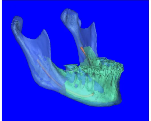
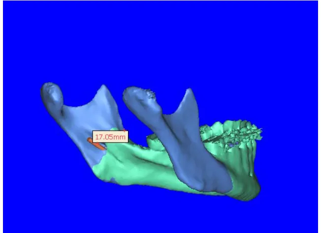
Measurement of values in 3D CT Reconstructed
[image:43.612.156.457.280.522.2]Mandible using MIMICS Software
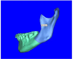
Fig 2: Sigmoid notch to Antilingula (A on right side)
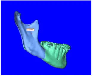
[image:44.612.152.462.392.644.2]Fig 4: Antilingula to anterior border of ramus (B on right side)
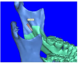
[image:45.612.151.462.393.646.2]Fig 6: Sigmoid notch to mandibular foramen (C on right side)
[image:46.612.152.462.392.642.2]Fig 8: Mandibular foramen to anterior border of ramus
(D on right side)
Fig 9: Mandibular foramen to anterior border of ramus
[image:47.612.143.469.381.618.2]Fig 10: Mandibular canal to alveolar crest level at coronal section
(E on right side)
Fig 11:
Mandibular canal to alveolar crest level at coronal
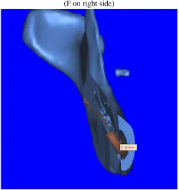
Fig 12: Mandibular canal to buccal plate at second molar
(F on right side)
Fig 13: Mandibular canal to buccal plate at second molar
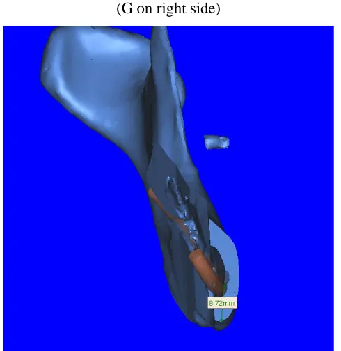
[image:49.612.177.436.366.640.2]Fig 14: Mandibular canal to lower border at second molar
(G on right side)
Fig 15: Mandibular canal to lower border at second molar
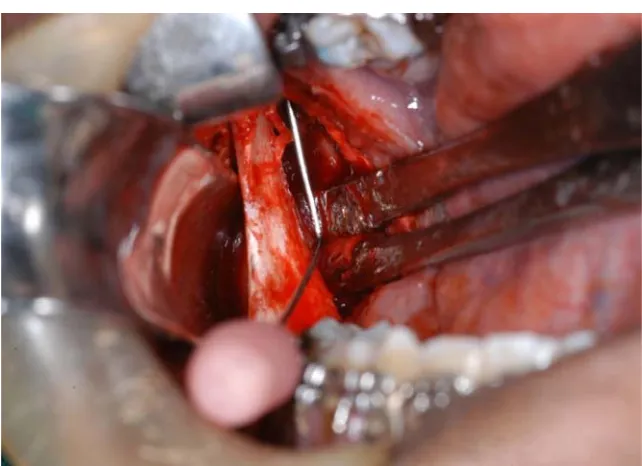
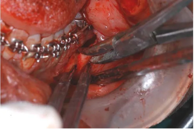
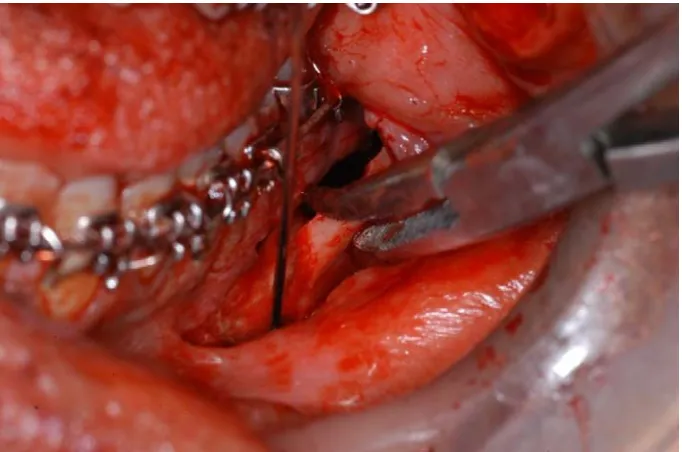
[image:50.612.185.430.366.618.2]Intra operative measurements
Fig 17: Sigmoid notch to Mandibular foramen (C)
[image:52.612.168.488.401.614.2]Fig 19: Mandibular foramen to alveolar crest level (E)
[image:53.612.167.497.358.577.2]Fig 21: Mandibular canal to lower border of mandible
Preoperative and Postoperative Lateral view photographs of three patients included in the study
Patient name: Solaimalar, Age/Sex: 21yrs/ F
Preoperative photograph
Patient name : Purusothaman , Age/sex: 24yr/ M
Preoperative photograph
Postoperative photograph
Preoperative photograph
29
Results
The mean distance from anterior border of ramus to mandibular
foramen and antilingula were 15.50mm and 15.11mm respectively. The
minimum and maximum distances were 14.08-18.01mm for the mandibular
foramen and 11.16-18.83mm for the antilingula.
The average horizontal distance between antilingula and mandibular
foramen did not differ significantly (0.65mm). But the value ranges between
-3.20 to 3.97mm.
The average distance from the sigmoid notch to the mandibular
foramen and antilingula were 17.30mm and 14.23mm respectively. The value
ranges between 11.31-21.85mm for the mandibular foramen and 9.26mm-
16.63mm for the antilingula. The average distance from antilinugla to
mandibular foramen in vertical direction was 3.01 mm. It ranges from 0.0mm
to 6.75mm.
When viewed at coronal section at the mandibular second molar region,
the average distance from the mandibular canal to alveolar crest, buccal cortex
and inferior border of mandible were 12.69mm, 6.42mm and 6.44mm
respectively. The ranges were 10.31-14.99mm for the alveolar crest, 4.09-
9.14mm for the buccal plate and 3.98-8.72mm for the inferior border of the
30
Tables 1a, 2a, 3a, 4a, 5a, 6a, 7a and 8a shows measurements between
different anatomic reference points considered for evaluating mandibular
anatomy in 3D scan images done using MIMICS software for the eight patients
included in the study.
Tables 1b, 2b, 3b, 4b, 5b, 6b, 7b and 8b shows measurements between
different anatomic reference points considered for evaluating mandibular
anatomy intraoperatively during BSSRO for the eight patients included in the
study.
Tables 1c, 2c, 3c, 4c, 5c, 6c, 7c and 8c shows measurements between
different anatomic reference points considered for evaluating mandibular
anatomy in pre-operative OPG.
Tables 10, 11 and 12 represent mean values and range of values
obtained from 3D CT scan image, OPG and intraoperative measurements
respectively.
Table 13 shows comparison of mean values obtained from 3D CT scan
image, OPG and intraoperative measurements
It shows that the value obtained from the CT scan and the values
obtained from the Intraoperative procedure have high correlation and they did
not differ from each other by more than 0.5mm. Measurements made from the
OPG is significantly different from the values obtained from CT scan and intra
31
The neurosensory deficit assessed by cotton wool test and pin prick test
is shown in table 14. Almost 100% of the population had neurosensory deficit
on the immediate post operative period and every one recovered at 2 months
post operatively.
The position of the nerve was assessed intra operatively and in all 8
patients, nerve was not encountered and it stayed in the distal fragment. None
32
Patient name: Purusothaman,
[image:61.612.121.492.182.568.2]Age/Sex : 24yrs/M
Table 1a
Anatomic reference points considered for evaluating mandibular anatomy in 3D scan
images
Right side (in mm)
Left side (in mm)
S-antilingula(A) 14.96 15.06
Antilingula –anterior border(B) 11.16 15.45
S-mandibular foramen(C) 20.16 22.38
Mandibular foramen-anterior border(D) 15.26 17.05
Antilingula –mandibular foramen (horizontal)(W)
3.97 1.60
Antilingula –mandibular foramen(vertical)(H)
5.20 7.32
Mandibular canal-alveolar crest level at coronal section (E)
10.73 9.98
Mandibular canal-buccal plate at second molar(F)
8.46 9.14
Mandibular canal-lower border at second molar(G)