*Corresponding Author Address: Dr. Anusha Avvaru. E-mail: [email protected]
Volume 05,Issue 04 Original Article
EVALUATION
OF
MANDIBULAR
INCISIVE
CANAL
ANATOMY BY CONE BEAM COMPUTED TOMOGRAPHY
Anusha Avvaru1, Jyothirmai Koneru2, R.Sudhakara Reddy3, Ramesh Thatapudi4, Anand Babu
Beeraboyina5, Rajesh Nallakunta6
1.MDS, Post Graduate,Vishnu Dental College,Bhimavaram,Andhra Pradesh 2.MDS, Reader ,Vishnu Dental College, Bhimavaram, Andhra Pradesh
3. MDS, Professor and Head of the department, Vishnu Dental College, Bhimavaram, Andhra Pradesh 4. MDS, Professor ,Vishnu Dental College, Bhimavaram, Andhra Pradesh
5. MDS, Reader ,Vishnu Dental College, Bhimavaram, Andhra Pradesh
6. MDS, Assistant professor ,Vishnu Dental College, Bhimavaram, Andhra Pradesh
ABSTRACT:
Objectives: The study was aimed to assess the prevalence of mandibular incisive nerve canal and evaluate its average location and dimension by using cone beam computed tomography in patients attending dental institute.
Methods:The cone beam computed tomography scans of 50 patients were retrospectively assessed for the presence of mandibular incisive canal bilaterally. The axial, sagittal, cross- sectional and panoramic views of the scans were analyzed and the measurements were recorded using the tools given in the ON DEMAND 3D software.
Results:The mandibular incisive canal (MIC) was visible in 98% of CBCT images with a mean length of 13 ± 5.6 mm. The mean length of the incisive canal on right and left side was 13.4±4.6mm and 14±6.4mm The distance between the origin of incisive canal and buccal plate, as well as with the lingual plates were 4.19±1.2mm and 4.1±1.0mm, and the distance between the apex of incisive canal with buccal plate, as well as with the lingual plates were 4.7±1.3mm and 4.15±1.07mm.
Conclusion: There was high prevalence of MIC with variation in length and distance up to the cortical bone. The preoperative radiographic evaluation of the MIC with CBCT must be considered for presurgical assessment to prevent complications such as post operative bleeding and paraesthesia. Key words: Cone-Beam computed tomography, anterior mandible, Mandibular incisive canal.
INTRODUCTION:
The inferior alveolar nerve and the mental foramen are the important anatomical structures that should be taken into account during preoperative planning for surgical and implant
placement in the posterior mandible.[1]
Although numerous reports describe the vital anatomical structures of the
posterior mandible, only few
contemporary radiographic and
histological studies are available about the anatomical structure of the anterior mandible. The region between the mental foramen has been considered a
safe zone for most of the surgical procedures, like insertion of endosseous
implants, genioplasty during
orthognathic surgery, bone harvesting
from the chin and mandibular
rehabilitation after trauma of the anterior region with screws and/or
plates placement.[1]
Mesial to the mental foramen, the extension of the mandibular canal in referred as the mandibular incisive canal and it contains one of the terminal branches of the inferior alveolar nerve.
Avvaru A.et al, Int J Dent Health Sci 2018; 5(4):521-530
522
Several case reports describe
neurosensory disturbances, edema,
hematoma and failure of osseo-integration of implants during or after
surgical procedures in the inter
foraminal region of the mandible and these can be attributed to the presence
of the mandibular incisive canal.[3, 4]
Cone beam computed tomography (CBCT) is considered as a useful tool for the radiographic evaluation of MIC. CBCT has been shown to improve the visibility of bony canals that cannot be clearly observed on regular panoramic or other
intraoral radiograph.[5] Chen et
al,[6]emphasized the effectiveness of
CBCT and three-dimensional
reconstruction in the identification of
important anatomical structures
relevant for the preoperative
assessment for surgical procedures in the anterior region of the mandible. The mandibular incisive canal has varied
course, with multi-morphic
representation. It may present either unilaterally or bilaterally or may be absent. Few studies stated that presence of mandibular incisive canal vary according to population. So they was a need to study in our population. The
number of cases with surgical
intervention in the inter-foraminal area
has increased considerably[7,8,9] and
failure to ascertain the exact position of the neurovascular bundle in this region may lead to complication like transient or long term paresthesia of the associated region. The present study was
conducted to assess the prevalence of mandibular incisive nerve canal and to evaluate its average location and
dimension by using Cone Beam
Computed Tomography.
MATERIALS AND METHODS:
The retrospective study consisted of 50 CBCT examinations of the mandible taken from 50 patients who were referred for various dental procedures.
Patients with any pathological
abnormalities were excluded from the
study design. This research was
approved by the institutional ethical committee. The scans were sourced fromCBCT machine SOREDEX CRANEX 3D with ON DEMAND 3D software using the following set of exposure parameters for each patient. The scans were set at 90KV and 10mA, as recommended by the manufacturer with fields of view (FOV) 61x78mm of high resolution of 300µm voxel size respectively.
The courses of the mandibular incisive
canal were assessed in images
reconstructed into multiple-plane views (axial, panoramic and cross-sectional views) and the images were analyzed and the measurements were done using the tools given in the ON DEMAND 3D software.
The image was first examined for the presence or absence of the canal the presence of the canal was noted to be unilateral or bilateral. The identified canals were analyzed. The nerve tracking tool was used to measure the length
Avvaru A.et al, Int J Dent Health Sci 2018; 5(4):521-530
523 from the origin to the end of the visible
nerve canal (Figure-1). In particular, the measurements made were:
1. Length of the incisive canal (origin to
apex)
2. Diameter of the incisive canal at its
origin
3. Distance from the origin of incisive
canal to base of the mandible
4. Distance from the apex of incisive
canal to base of the mandible
5. Distance from the origin of incisive
canal to buccal cortex of the mandible
6. Distance from the apex of incisive
canal to buccal cortex of the mandible
7. Distance from the origin of incisive
canal to lingual cortex of the mandible
8. Distance from the apex of incisive
canal to lingual cortex of the mandible
Cross sectional image of the CBCT was used to measure the distance of the nerve canal (both at the origin and the apex) to the inferior border of the mandible [Figure2].Length of the incisive canal and also the diameter (origin to apex) [figure3, 4.] The axial plane was used to measure the length distance (both at the origin and also the apex) of the nerve to the buccal and lingual cortices, respectively [Figures 5]. The data was subjected to statistical analysis
where descriptive analysis was
performed. The prevalence in
percentages was calculated for
mandibular incisive canal (Table-1). The comparison between the mean values
was performed with the unpaired t-test for (right & left side) (table-3). All statistical assessments were considered significant if p< 0.05.
RESULTS:
The study group consisted of 23 males and 27 females, age ranging from 40-80 years with a mean age of 54 years, 2% of the patients were edentulous.
PREVALENCE OF MIC
The analysis of the results showed that MIC could be seen in 95 % of the cases (50 patients, 95canals). In 92 % of the cases (46 patients) it was found bilaterally; and in 6 %( 3 patients), unilaterally. In 2%of the cases (1 patient) it was absent. As shown in table 1.
The mean Diameter of the incisive canal at its origin was 1.7 mm +/- 0.51 mm. the length of incisive canal at origin to apex was 13.7± 5mm. The distance between the origin of incisive canal and buccal plate,as well as with the lingual plates were 4.19±1.2mm and 4.1±1.0mm, and the distance between the apex of incisive canal with buccal plate, as well as with the lingual plates were 4.7±1.3mm and 4.15±1.07mm.as shown in table2.the mean length of incisive canal on right side and left side was 13.4±4.6mm and 14±6.4mm respectively . No much significant difference between the values on right and left side. Except for the distance from apex to lingual cortex.
Avvaru A.et al, Int J Dent Health Sci 2018; 5(4):521-530
524 DISCUSSION:
Studies have reported that panoramic radiographs failed to detect the incisive canal because of super imposition of various anatomical structures in 2D
image.[13,11,14,7]Hence, it is logical to
assume that, the study of the inter-foraminal area of the mandible using CBCT becomes mandatory. In the cross-sectional images of CBCT scans, MIC appears as a round hypodense area within the Mandibular trabecular bone surrounded by a hyperdense rim representing the canal walls. As reported
by Al-Ani et al [15]
The MIC was visible in all (100%) CBCT images, in a study conducted by Al-Ani et
al.[15] which was similar to that of
other authors have also found a high prevalence of MIC using CBCT, with a variable visibility of 83-97.5%. There was 93% visualization of MIC by Apostolakis
and Brown,[16] 91% by Makis et al[1] and
93.7% by Parnia et al. [17] whereas Pires
et al.[10] have found that the MIC was
present in 83% CBCT images. In spiral CT scans 93% of visualization of MIC was
shown by Jacobs et al[(7] In CBCT scans
78.75% (63 cases) showed the presence of MIC by Huang et al. [18] In this study the prevalence of MIC using CBCT, with a variable visibility of 98 % (50 cases). For all CBCT images examined, the mean length of the incisive canal for the right side was 13 ±4.6 mm and for the left side
was 14 ± 6.4mm. Pires et al.[10] verified
MIC lengths as 7.1 ±4 mm and 6.6 ± 3.7 mm for the right and left side,
respectively. Another finding was that the MIC is in close proximity to the buccal plate (4.62 ± 1.41mm) which is similar to studies conducted by Tepper G
et al.[19]
Length of the incisive canal was similar to study conducted by De Andrade et al. The mean Diameter of MIC was 1.7 mm +/-0.51 mm accordance with the study
reported by Huang et al.[18] The Distance
of the incisive canal to the lower border of the mandible was 8.95-8.88mm as
compared with 9.4– 11.15mm
accordance with the study reported by
De Andrade et al[24] Apostolakis and
Brown and Rosa et al[16] Reported that
MIC was also nearer to the buccal plate and alveolar process in its closest position. In this study the canal runs more medial course increasing its distance from the lingual cortex.
Mardinger et al.[11] reported an incisive
canal direction proceeding from the buccal plate towards the lingual plate. The distances between the origin of incisive canal and buccal plate, as well as between the origin of incisive canal and lingual plate were 4.19+/-1.2mm and 4.1+/-1.0mm, respectively.The distances between the apex of incisive canal and buccal plate, as well as between the apex of incisive canal and lingual plate were
4.7+/-1.3mm and 4.15+/-1.07mm,
respectively.
Regarding gender there was no
significant difference when comparing the proximity of the MIC to the buccal and lingual cortex. The mean values and
Avvaru A.et al, Int J Dent Health Sci 2018; 5(4):521-530
525 SD of the diameter of MIC for males and
females were 1.61 mm ± 0.41 mm and 1.46 mm ± 0.35 mm, respectively. The kappa statistics indicated an overall score of 0.74 for the inter observer agreement for identification of the MICs and a somewhat lower score (0.55) for the inter observer agreement.
This study demonstrated a high degree of occurrence of MIC’s identified on CBCT scans, these data are in union with
other observational studies.[7,20 and 16] Our
study confirmed the high reliability of CBCT in the identification of fine structures (e.g. MICs). The mean diameter of the incisive canal found in this study was 1.8mm which is similar to
the study conducted by Jacobs et al.[7]
Similar or only slight difference was found in the data reported by other
authors. For instance, Obradovic et al.[21]
found that the MIC ranged from 0.48 mm to 2.9 mm on cadaver mandibles;
Pires et al.[10] observed diameters from
0.4 mm to 4.6 mm on CBCT scans; diameters of 1.0 mm to 6.6 mm on CBCT examinations were reported by Uchida
et al.[22] In the present study we found
differences between the diameter of the canal on both sides and also between
genders. Further studies can be
conducted by taking large group of population and it can aid in forensics for the person /gender identity/variation.
CONCLUSION:
The clinical significance of this study lies in the mapping of the incisive canal and its anatomical proximity during surgical procedures in order to avoid potential injury to the incisive mandibular nerve using CBCT, which could clearly show the
three-dimensional structure and
adjacent structure of the MIC. One of the most easy and relevant location for placing an implant is the anterior mandible. Clinician should be alert while operating. As anatomical variations can be diversified, causing complications. All anatomical considerations have to be visualized, analyzed in preoperative planning itself that can help in enhance quality and prevent disasters. All surgical procedures should be carefully planned to improve patient quality of life than degrading the same.
REFERENCE:
1. Makris N, Stamatakis H, Syriopoulos
K, Tsiklakis K, van der Stelt PF. Evaluation of the visibility and the course of the mandibular incisive canal and the lingual foramen using cone-beam computed tomography. Clin. Oral Impl. Res. [21], [2010]; [766–77].
2. Ramesh AS, Rijesh K, Sharma A,
Prakash R, Kumar A, Karthik. The prevalence of mandibular incisive nerve canal and to evaluate its average location and dimension in Indian population. J Pharm Bioall Sci[ 2015];[7]: [594-6].
3. Wismeijer D, van Waas MA,
Vermeeren JI, Kalk W. Patients’ perception of sensory disturbances of the mental nerve before and after
Avvaru A.et al, Int J Dent Health Sci 2018; 5(4):521-530
526 implant surgery: A prospective study
of 110 patients. Br J Oral Maxillofac Surg[ 1997];[35]: [254‑9].
4. Hirsch JM, Brånemark PI. Fixture
stability and nerve function after transposition and lateralization of the inferior alveolar nerve and fixture installation. Br J Oral Maxillofac Surg [1995];[33]:[276‑81].
5. Juodzbalys G, Wang HL, Sabalys G
(2010) Anatomy of mandibular vital
structures. Part II: mandibular
incisive canal, mental foramen and associated neurovascular bundles in relation with dental implantology. J Oral Maxillofac Res. doi:10.5037/ jomr.[2010].[11][03-07].
6. Chen Z, Chen D, Tang L, Wang F
(2015) Relationship between the position of the mental foramen and the anterior loop of the inferior alveolar nerve as determined by cone
beam computed tomography
combined with mimics. J Comput Assist Tomogr [39]:[86–93].
7. Jacobs R, Mraiwa N, vanSteenberghe
D, Gijbels F, Quirynen M.
Appearance, location, course, and morphology of the mandibular incisive canal: an assessment on spiral CT scan. Dentomaxillofacial Radiology.[2002];[31]:[322-7].
8. Kütük N, Demirbaz AE, Gönen ZB,
Topan C, Kiliç E, Etö OA, et al. Anterior Mandibular Zone Safe for Implants. The Journal of Craniofacial Surgery. [2013];[24]: e[405-8].
9. Sokhn S, Nasseh I, Noujeim M. Using
cone beam computed tomography to determine safe regions for implant
placement. General Dentistry.[
2011];[59]:e[72-7].
10.Pires CA, Bissada NF, Becker JJ,
Kanawati A, Landers MA. Mandibular Incisive Canal: Cone Beam Computed
Tomography. Clinical Implant
Dentistry and Related Research. [2012];[14]:[67-73].
11.Mardinger O, Chaushu G, Arensburg
B, Taicher S, Kaffe I. Anatomic and radiologic course of the mandibular incisive canal. Surgical and Radiologic Anatomy. [2000];[22]:[157-61].
12.FUENTES, R.; ARIAS, A.; BUCCHI, C.;
SARAVIA, D. & DIAS, F. Prevalence and morphometric characteristics of the mandibular incisive canal through panoramic radiographs in a Chilean population. Int. J. Morphol., [2017]. [35(3]:[931-937].
13.Mraiwa N, Jacobs R, Moerman P,
Lambrichts I, vanSteenberghe D, Quirynen M. Presence and course of the incisive canal in the human mandibular interforaminal region:
two-dimensional imaging versus
anatomical observations. Surgical
and Radiologic Anatomy.
[2003];[25]:[416-23].
14.Sahman H, Sekerci AE, Sisman Y,
Payveren M. Assessment of the visibility and characteristics of the mandibular incisive canal: cone beam
computed tomography versus
panoramic radiography. The
International Journal of Oral and
Maxillofacial Implants.
[2014];[29]:[71-8].
15.Al-Ani O, Nambiar P, Ha KO, Ngeow
Avvaru A.et al, Int J Dent Health Sci 2018; 5(4):521-530
527 from the interforaminal region of the
mandible. Clin Oral Implants Res. [2013];[24] Suppl A[100]:[115-21].
16.Apostolakis D, Brown JE. The anterior
loop of the inferior alveolar nerve: prevalence, measurement of its length and a recommendation for interforaminal implant installation based on cone beam CT imaging. Clinical Oral Implants Research. [2012];[23]:[1022-30].
17.Parnia F, Moslehifard E, Hafezeqoran
A, Mahboub F, Mojaver-Kahnamoui H. Characteristics of anatomical
landmarks in the mandibular
interforaminal region: A cone-beam
computed tomography study.
Medicina Oral Patologia Oral y Cirugia Bucal. [2012];[17]:e[420-5].
18.Huang H, Liu P, Li X, Pei Z, Yang X, Bai
S, et al. Mandibular incisive canal by cone beam CT. Hua Xi Kou Qiang Yi Xue Za Zhi. [2013];[31]:[479-82].
19.Tepper G, Hofschneider UB,
Gahleitner A, Ulm C. Computed
tomographic diagnosis and
localization of bone canals in the mandibular interforaminal regions
for prevention of bleeding
complications during implant
surgery. The International Journal of
Oral & Maxillofacial Implants.[
2001];[16]:[68-72].
20.Santos T, Gomes AA, Melo DG, Melo
AR, Cavalcante JR, Araújo LG, et al.
Evaluation of reliability and
reproducibility of linear
measurements of
cone-beam-computed tomography. Indian
Journal of Dental Research.
[2012];[23]:[473-8].
21.Obradovic O., Todorovic L., Pesic V.,
Pejkovic B., Vitanovic V. Bulletin du Groupement international pour la
Recherche Scientifique en
Stomatologie et Odontologie, [1993] ,36[3-4], [109-113].
22.Uchida Y, Yamashita Y, Goto M,
Hanihara T. Measurement of anterior loop length for the mandibular canal and diameter of the mandibular incisive canal to avoid nerve damage when installing endosseous implants in the interforaminal region. Journal of Oral and Maxillofacial Surgery.[ 2007];[67]:[744-50].
23.Pereira-Maciel, Patrícia, Emerson
Tavares-de-Sousa, and
Marcelo-Augusto Oliveira-Sales. “The
Mandibular Incisive Canal and Its Anatomical Relationships: A Cone
Beam Computed Tomography
Study.” Medicina Oral, Patología Oral y Cirugía Bucal [2015]: [20.6] : e[723– e728].
24.De Andrade E, Otomo-Corgel J,
Pucher J, Ranganath KA, St George N. The intraosseous course of the mandibular incisive nerve in the
mandibular symphysis. The
International Journal of Periodontics
and Restorative Dentistry.
Avvaru A.et al, Int J Dent Health Sci 2018; 5(4):521-530
528 TABLES:
Table 1: Prevalence of mandibular incisive canal
No of CBCT scans evaluated 50(100%)
Incisive canal present bilaterally 46 (92%) Incisive canal present unilaterally 3 (6%)
Incisive canal not present 1 (2%)
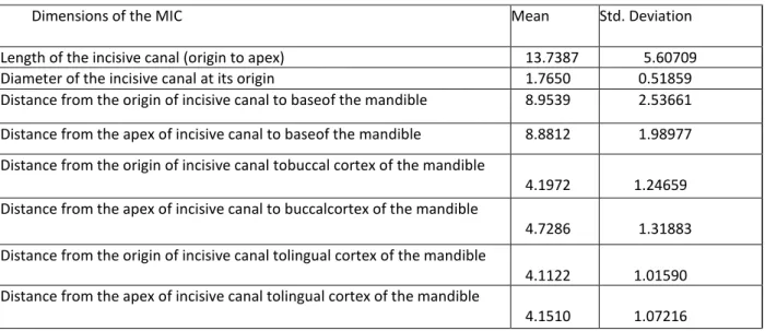
Table2: Values of various dimensions of the mandibular incisive canal (MIC) to various landmarks
Dimensions of the MIC Mean Std. Deviation
Length of the incisive canal (origin to apex) 13.7387 5.60709 Diameter of the incisive canal at its origin 1.7650 0.51859 Distance from the origin of incisive canal to baseof the mandible 8.9539 2.53661 Distance from the apex of incisive canal to baseof the mandible 8.8812 1.98977 Distance from the origin of incisive canal tobuccal cortex of the mandible
4.1972 1.24659 Distance from the apex of incisive canal to buccalcortex of the mandible
4.7286 1.31883 Distance from the origin of incisive canal tolingual cortex of the mandible
4.1122 1.01590 Distance from the apex of incisive canal tolingual cortex of the mandible
4.1510 1.07216
Table3: Comparison between groups of various dimensions of the mandibular incisive canal (MIC) to various landmarks
Dimensions of the incisive nerve GROUP N Mean Std. Deviation
p value Length of the incisive canal RIGHT 46 13.4148 4.67664
.578
LEFT 44 14.0773 6.47724
Diameter of the incisive canal at its origin RIGHT 46 1.6985 0.42293
.215
LEFT 44 1.8345 0.59980
Distance from the origin of incisive canal to base of the mandible
RIGHT 46 8.7691 2.86562
.483
LEFT 44 9.1470 2.15622
Distance from the apex of incisive canal to base ofthe mandible
RIGHT 46 8.8733 2.16581
.969
LEFT 44 8.8895 1.81261
Distance from the origin of incisive canal to buccal cortex of the mandible.
RIGHT 46 4.1443 1.30095
.683
LEFT 44 4.2525 1.19962
Distance from the apex of incisive canal to buccal cortex of the mandible
RIGHT 46 4.6243 1.16939
.447
LEFT 44 4.8375 1.46463
Distance from the origin of incisive canal to lingual cortex of the mandible
RIGHT 46 4.2035 1.01403
.387
LEFT 44 4.0168 1.02069
Distance from the apex of incisive canal to lingual cortex of the mandible
RIGHT
Avvaru A.et al, Int J Dent Health Sci 2018; 5(4):521-530
529 FIGURES:
Avvaru A.et al, Int J Dent Health Sci 2018; 5(4):521-530