0022-538X/93/052486-10$02.00/0
Copyright ©1993, American Society forMicrobiology
Mutations
in
the Putative Calcium-Binding
Domain of
Polyomavirus VP1
Affect
Capsid
Assemblyt
JOHN I. HAYNES II4 DECHING CHANG,§ANDRICHARD A. CONSIGLI* Section of VirologyandOncology, Divisionof
Biology,
AckertHall, Kansas State University,Manhattan, Kansas66506-4901 Received 25 August1992/Accepted3February 1993
Calcium ionsappeartoplayamajorrole inmaintainingthestructuralintegrityof thepolyomavirusandare
likelyinvolved in the processesofviral uncoating andassembly. Previous studies demonstrated thataVP1 fragment extending from Pro-232 to Asp-364 has calcium-binding capabilities. This fragment contains an
amino acidstretch fromAsp-266toGlu-277whichisquitesimilar insequencetothe aminoacids that make
upthecalcium-bindingEF handstructuresfoundinmanyproteins.Toassessthecontribution of this domain to polyomavirus structural integrity, the effects of mutations in this region were examined by transfecting
mutatedviral DNA into susceptible cells. Immunofluorescence studies indicated that althoughviral protein synthesisoccurrednormally,infective viralprogenywerenotproducedincells transfected withpolyomavirus
genomes encodingeithera VP1 molecule lacking amino acids Thr-262 through Gly-276 or aVP1 molecule
containingamutation ofAsp-266toAla. VP1moleculescontainingthedeletionmutationwereunable to bind
45Ca inanin vitroassay.Upon expression inEscherichiacoliandpurification by immunoaffinity chromatog-raphy,wild-type VP1wasisolatedas pentameric, capsomere-like structureswhich could be inducedtoform capsid-like structures upon addition ofCaCl2, consistent with previous studies. However, although VP1 containing the point mutation was isolated as pentamers which were indistinguishable fromwild-type VP1 pentamers,additionofCaC12didnotresult in theirassemblyintocapsid-likestructures.Immunogold labeling andelectron microscopystudies of transfected mammalian cellsprovided invivoevidence thatamutation in this region affectstheprocess ofviral assembly.
Themurinepolyomavirusiscomposed of three structural proteins,designated VP1, VP2, andVP3,whichare
assem-bledontoaDNA-protein complex (43).Asingleviruscapsid
contains 72pentameric capsomeres,each of which is
com-posedof fivemolecules of themajor capsid proteinVP1. The
presenceof the divalent cation calcium in murine
polyoma-virusvirionswasindicatedby X-ray fluorometrystudies(9).
Astructuralrole for calciumwasdemonstrated in studiesby
Bradyetal. (9, 10)which showed that chelation of calcium by ethyleneglycol-bis-N,N,N',N'-tetraacetic acid(EGTA), inconjunctionwith disulfide bonddisruption bythereducing agent dithiothreitol, results in the breakdown of the virion into its capsomere subunits and a DNA-protein complex.
Interestingly, addition ofexogenous CaCl2 to this dissoci-ated mixturepermitted reassemblyinto intact virions which partially regainedboth infectivity andhemagglutination
ac-tivity (8, 47). Calciumbinding appears to contribute to the stability ofanumber ofplant viruses aswell (1, 14-16, 22,
34, 37), possibly by helping to ensure that these viruses release their RNAs only in the host-cell cytoplasm. A characteristic swelling of these viruses which occurs upon
exposure to calcium chelators under basic conditions is
believedtorepresent aninitial stepintheiruncoating. When polyomavirus proteins were separated chromato-graphically and the isolated VP1 protein was analyzed by
electron microscopy in the absence of calcium, structures
* Correspondingauthor.
tContribution93-87-J from theKansasAgricultural Experiment Station, KansasStateUniversity, Manhattan, KS 66506.
fPresent address: Laboratoryof MolecularandDevelopmental Biology,NationalInstitutes ofHealth,Bethesda, MD 20892.
§Present address: Department of Microbiology, Chung Shan Medicaland Dental College, Taichung, Taiwan, Republic of China.
which resembled capsomeres in both size and shapewere
observed (7). Polyomavirus VP1, which was expressed in
Escherichia coli from pALVPltac (24) and purified in the
presenceofEDTA and,3-mercaptoethanol,wasalsoisolated as pentameric structures that resembled viral capsomeres
(38, 39).Removal of thechelatorandreducingagenttogether with addition of CaCl2 resulted in the assembly of these
capsomeres into structureswhich resembled viral capsids.
Besidesfurtherimplicatingarole forcalciuminmaintaining
the structural integrity of the virus, these studies also suggested that the posttranslational modifications of VP1 whichoccurineukaryoticcells(2, 3, 5, 18, 19, 26, 28, 29, 35) may not be necessary for proper capsid formation. These
studies also indicated that the minor capsid proteins were
notnecessaryfor in vitro assembly.
A 16-kDa fragment of the polyomavirus major capsid protein VP1,which isgenerated by formic acid cleavage and extends fromPro-232to Asp-364,wasshownto have calci-um-binding capabilities (27). This fragment contains a
se-quence of 12aminoacids (Asp-266 through Glu-277) thatis
quitesimilartothe12-amino-acidloopswhichmakeuppart
of the EF hand structures found in many calcium-binding proteins, includingparvalbumin, calmodulin, thrombospon-din,andtroponin C(13, 23, 42).Thepurposeof thepresent studieswas to determine the effects that alterationswithin this domain would have on polyomavirus structure and functions. The workfocused on asingle site-directedpoint
mutation within this domain, aswell as a deletion mutant
whicheliminated theputative calcium-binding domain. Both mutations resulted in the inability to form infective viral
progenyincellstransfectedwithpolyomavirusDNA
encod-ingtherespectivemutations. Inaddition,whenexpressed in E. coli,VP1moleculeslackingthe12-amino-acid motifwere
unabletobind calcium inaninvitroassay.Finally, when the
2486
on November 9, 2019 by guest
http://jvi.asm.org/
POLYOMAVIRUS VP1 CALCIUM-BINDING DOMAIN 2487
VP1 geneharboringthe point mutation was expressed in E.
coli andpurified, it was shown to be defective in the ability to assemble into capsid-like structures in the presence of calcium.
MATERIALS AND METHODS
Mutagenesis. A 1.9-kb HindIII-PvuII fragment containing 1 kb of the VP1 gene as well as about 900 bp of 3' flanking DNA was excised from the polyomavirus genome and cloned into M13mp19 to generate M13mp19-VP1. Prior to ligation, the terminal PvuII site was converted to a BamHI site to allow cloning into theHindIII-BamHI-digested vec-tor. Apoint mutation in the Asp-266 codon was created by using the Muta-gene in vitro mutagenesis kit (Bio-Rad) as described by the manufacturer and the phosphorylated mixed oligonucleotide primer 5'-CCCAACTCCAT1TTCA
(T/I)CTAG-3'
(corresponding to polyomavirus nucleotides 3265 to 3285[41]; the underlined G represents the mutation).Sequencing ofsingle-stranded phage DNA by the
dideoxy-chain termination method was used to determine whether the wild-type T nucleotide or the mutant G nucleotide
(underlined)waspresent at the variable position. The
oligo-nucleotide primer 5'-GCCCTGCACTTGGGGGAGCAT-3'
(corresponding to polyomavirus nucleotides 3061 to 3081)
(41), a- 5S-dATP (New England Nuclear) and the Sequenase kit(United StatesBiochemicals) were used in this determi-nation. A phage clone whose DNA contained the mutation was termed M13mpl9-VP1A (for alanine), and one which
contained the wild-type T nucleotide at the position of
interest was designated M13mpl9-VPlD (for aspartic acid) and servedas acontrol for the mutagenesis reactions.
Togenerate a deletion mutation in the region of the VP1 gene encoding the putative calcium-binding domain, the 504-bp HincII from wild-type polyomavirus DNA was first cloned into pUC19 to generate pCH101. This construct was subsequently treated with ApaI (which cleaves pCH101 at a unique site within the region encoding the putative calcium-binding domain corresponding to nucleotide 3264 in the
polyomavirusgenome [41]).Bal 31 slow exonuclease
(Inter-national Biotechnologies, Inc.) was used to generate the deletion, and the shortened DNA fragment was recircular-ized with T4 DNA ligase. The recircularrecircular-ized DNA was used
totransformHB101 cells, and transformants were screened
bydideoxynucleotide sequencingtodetermine the extent of
the deletions. Plasmid DNA from one colony contained a 42-bp deletion which eliminated the coding information for amino acids Thr-262 through Gly-276. An arginine codon resulted at the site of ligation, but the reading frame was preserved. This plasmid was designated pCH101A42.
Plasmid pPY501, which contains the entire 5.3-kb polyo-mavirus genome cloned into the EcoRI site of pGEM-7Zf(+)
(Promega), was used to introduce the respective mutations
into intact polyomavirus genomes. pPY501 and
M13mpl9-VP1A were digested with HincII, and the 504-bp HincIl fragment(nucleotides 2962 to 3466) fromM13mpl9-VP1was usedtoreplace thecorresponding wild-type HincII fragment inpPY501togeneratepPY5O1A. Similarly, the smallHincIl fragment (now 462 bp) frompCH101A42wasused to replace
the corresponding wild-typeHincIl fragment in pPY501 to
generate pPY501A42. The 504-bp HincII fragment derived fromM13mpl9-VPlD was used in an identical exchange to generate awild-type recombinant DNA designatedpPY5O1D.
In each case, digestion with ApaI was employed to verify
that the HincIl fragment had been cloned in the correct
orientation, and DNA was sequenced to ensure the presence ofthe mutation.
To create vectors which would allow expression of mu-tated VP1 genes in E. coli, the bacterial VP1 expression vector pALVPltac (provided by R. Garcea) (24) was di-gested with HindIll and PvuII to generate fragments of approximately 2.3 kb (vector DNA and the 5' end of the VP1 gene) and 1.9 kb (polyomavirus nucleotides 3918 to 2032). The gel-purified 1.9-kb fragment, which included the 504-bp HincII fragment, was cloned into pSP64polyA (Promega), a vector lacking HincII sites. The resulting construct was digested with HincII, and theHincII fragment from pPY501A
or pPY501A42 was used to replace the corresponding
wild-type HincIl fragment. The respective 1.9-kb HindIII-PvuII fragments were then excised and religated to the gel-purified 2.3-kb fragment (saved from the original digestion) to create
pALVPlAtac(expressing VP1 containing the point mutation)
or pALVP1A42tac (expressing VP1 containing the deletion mutation). Each construct was transformed into E. coli
RB791.
Cells andtransfections. Primary cultures of mouse kidney cells (MKCs) were prepared as described previously (12, 40) and grown oncoverslips in 35-mm dishes. In preparation for subsequent transfection studies, full-length viral DNA was released frompPY5O1D,pPY5O1A,orpPY501A42byEcoRI digestion, and thegel-purified polyomavirus DNA fragments (approximately 5.3 kb) were self-ligated at a high dilution (1 ,ug/100
RI).
Transfections were performed when cells were approximately 80% confluent with 10 ,ug of Lipofectin (Be-thesda Research Laboratories) as recommended by the manufacturer and 500 ng of circularized polyomavirus DNA per dish. The DNA-Lipofectin mixture was allowed to remain on the cells for 16 h and was then replaced with Dulbecco's modified Eagle's medium supplemented with 10% newborn calf serum. The time of addition of this medium was considered the starting time point in all trans-fection studies. At the indicated times following the change of medium, cells were fixed and the presence of large T antigen or the virus structural proteins in the cell nuclei was determined by indirect immunofluorescence (31) with a monoclonal antibody against large T antigen (provided by J. Bolen) or rabbit antipolyomavirus immunoglobulin G as the respective primary antibodies.Purification of polyomavirusVP1 from E.coli. Wild-type or mutatedpolyomavirus VP1 was expressed in E. coli RB791 frompALVPltac (24) and pALVPlAtac, respectively, and purified from bacterial cell lysates. VP1 expression was induced beginning at early logarithmic phase by the addition of
isopropyl-,3-D-thiogalactopyranoside
(IPTG) to a 1 mM final concentration. The total growth time was approxi-mately 6 h, after which time the cells were pelleted and frozen at -20°C until used. The initial steps of VP1 purifi-cation, including thepreparation and sonicationofbacterial cell lysates, were performed essentially as described by Leavitt et al. (24) with a buffer composedof 50 mM Tris (pH 7.2), 2 mM EDTA, and 250 mM NaCl. The subsequent chromatography step dictated the omission of3-mercapto-ethanol from this buffer. Following centrifugation of the sonicated lysate (10,000 xg for 20
min),
VP1 was purified from thesupernatant byimmunoaffinity chromatography. In the case of VP1 expressed frompALVP1A42tac, the bacte-rial cell pellet was sonicated in distilled water and centri-fuged underconditions identical to those used in the purifi-cation of VP1 from pALVPltac and pALVPlAtac. After centrifugation, the pellet was reserved for separation by VOL. 67,1993on November 9, 2019 by guest
http://jvi.asm.org/
sodium dodecyl sulfate-polyacrylamide gel electrophoresis
(SDS-PAGE) as described below.
To prepare the immunoaffinity column for purification of VP1expressed from pALVPltac and pALVPlAtac, purified rabbit antipolyomavirus immunoglobulin G was coupled to Affi-GelHZ hydrazide gel (Bio-Rad) as described by Chang
etal. (11). Thebacterial cell lysate was allowed to mix with
the column matrix by rocking overnight at room tempera-ture.The column wasthen treatedwith 20 volumes of wash buffer(50mMTris [pH 7.2], 2 mMEDTA, 500 mMNaCl). Bound VP1 wassubsequently collected after application of elution buffer (100 mM glycine [pH 2.5], 150 mM NaCl) to the column and was immediately neutralized by collecting 0.8-mlfractionsin 0.2 mlof 1 M Tris(pH 8.0)-150mMNaCl. The amounts ofprotein were determined by measuring the A280 of the fractions, and the peak fractions were pooled.
The purity of an aliquot of the recombinant VP1 was
evaluated by SDS-PAGE and Western blotting with
anti-polyomavirus serum.
In vitrocalcium-binding assay. Toserve asacontrolin the 45CaC12-binding studies, polyomavirusvirionswereisolated from infected primary MKCs and purified as described previously (9). VP1 expressed in E. coli RB791 from
pALVPltac, pALVPlAtac, and pALVP1A42tac was
ob-tainedasdescribed above. Polyomavirusvirions and recom-binant VP1 proteins (2.5 ,ug each) were resolved by
SDS-PAGE (12.5% acrylamide). In the case of the recombinant
VP1 deletion mutant, 10 ,ug of the E. coli lysate pellet
(generatedasdescribedabove)waselectrophoresed because
of thepurificationdifficultiesresultingfromVP1insolubility.
Theproteins were transferredto aNitroBind filter (Micron
Separations,
Inc., Westboro, Mass.), and the filter was incubated in 10 mM EGTA(pH 6.8) overnight with gentleshaking. The filterwasbrieflyrinsed three times with
Ca2+-binding buffer (0.06 M KCI, 5 mM MgCl2 6 H20, 10mM
imidazole-HCl [pH 6.8])and then incubated in
Ca2+-binding
buffer for1hwith shaking.Blotswereprobed for 15 min in 20 ml of
Ca2"-binding
buffer containing200 ,uCi of45CaC12
(ICN, Irvine, Calif.; specific activity, 14.5 mCi/mg ofCa),
washed oncewith 20 ml of 0.47 mM CaC12, allowed to air
dry, andautoradiographed.All distilledwaterusedtomake
the solutions described abovewasfurtherprocessedthrough
achelatingresin column(Sigma, St. Louis, Mo.)toremove
residualcations.
The filters that had been probed with
45CaC12
were thensubjected to Westernblottingto identifythe VP1 proteins.
The filterswerefirst incubated in 10mMEGTA overnight at room temperature with gentle shaking. They were then washed three times with water to remove the
45Ca2'
andautoradiographed to ensure that no radioactive signal
re-mained. A5% solution of nonfat dry milk in Tris-buffered saline
(TBS;
50 mMTris [pH 7.4], 150 mMNaCl)wasusedforblocking. Thefilterswerethenincubatedovernight with
rabbit antipolyomavirussera(1to300 dilution), washed five times withTBS, incubated for 2 h with
1251I-labeled
Staphy-lococcus aureus protein A (5 x 10⁵ cpm/ml), washed five
times withTBS, air dried,andautoradiographed.
ElectronmicroscopyofrecombinantVP1proteins.
Affinity-purified
VP1 derived from pALVPltac and pALVPlAtacwasdialyzed against 10mMTris (pH
7.2)-i
mMEDTA-150mMNaCl-15mM
P-mercaptoethanol-5%
glycerol for 3 daysat roomtemperatureasdescribedpreviously(39). To induce
self-assemblyofpurifiedVP1into capsid-like structures, the
preparationswere subsequently dialyzed for 3 days against
the dialysis buffer described above supplemented with 0.5
mM CaC12 but lackingEDTA and
3-mercaptoethanol.
Forelectron microscopy analysis of purified recombinant VP1 protein under alternate buffer conditions, the sample was placed on a pioloform-coated grid, and the protein was allowed to adsorb to the grid for 5 min. After a distilled-water rinse, the sample was stained with a 1% aqueous uranyl acetate solution for 5 min andexamined withaPhilips 201 electron microscope operating at 60 kV.
Fixation and embedding of transfected MKCs. Fortyhours after transfection of MKCs (as described above), the me-dium was removed and the cells were rinsed with phosphate-buffered saline (PBS; 10 mM sodium phosphate [pH7.2], 150 mMNaCl). The cells were released from the dish by a brief treatment withViokase to better preserve cellintegrity. The cells were then treated for S min with fixative (2% [wt/vol] paraformaldehyde, 0.5% [wt/vol] glutaraldehyde) made in Earle's saltssolution, transferred to an Eppendorf tube, and pelleted. The supernatants wereremoved, and fresh fixative was added to the cells, which were incubated for 30 min at roomtemperature. Free aldehydes were quenched by rinsing the cell pellet three times (30 min total) with serum-free Dulbecco's modified Eagle's medium. Following three dis-tilled-water rinses, the cells were dehydrated in 50% ethanol and stained with 2% [wt/vol] uranyl acetate in 70% ethanol (17). After a 70% ethanol rinse, the cells were infiltrated (4, 33) by treatment with a 1:1 mixture of LR White resin (Polysciences, Inc.) and 70% ethanol solution (1:1) for 60 min. This was followed by three treatments with 100% LR White over a 24-h time period. Curing was performed at55°C for 2 days in fresh LR White.
Immunogoldlabeling of thin sections and electron micros-copy. After embedding and hardening, sectioning was per-formed with a diamond knife by using a Reichart OMV-2 ultramicrotome, and silver sections were collected on piolo-form-coated nickel grids. All subsequent immunogold-label-ing procedures were performed at room temperature by floating the grids on droplets of the indicated solutions. Sections were first treated for 30 min at room temperature with a saturated solution of sodium metaperiodate to expose the antigenic sites. Following a series of washes with dis-tilled water, the sections were blocked with 3% bovine serum albumin (BSA) made in EMG buffer (0.01 M phos-phate buffer [pH 7.2], 0.5 M NaCl, 0.05% Tween 20) and were treated for 90min with rabbit antipolyomavirus serum. After being washed with EMG buffer, the grids were treated for 90minwith a 1:100 dilution of goat anti-rabbit immuno-globulin G conjugated to colloidal gold (average particle diameter, 5 nm) (Amersham). Antibody incubations were carried out in the presence of 0.5% BSA in EMG buffer. Sampleswerepostfixed with 2% glutaraldehyde in PBS for 10min and poststained with 3% uranyl acetate for 15min and lead citrate
(36)
for 10min. The sections were viewed with a Philips 201 electron microscope operating at 60 kV.RESULTS
Generationofpolyomavirus genomes encoding site-directed anddeletion mutations. Aprevious study (27), together with similarities in amino acid sequence to the calcium-binding EF hand structure, suggested that amino acids Asp-266 to Glu-277 ofpolyomavirus VP1 may have a role in calcium binding by this virus. Oligonucleotide-directed mutagenesis of Asp-266 was performed to determine the effects that substitution of this residue with a neutral amino acid (ala-nine) would have on the virus structure and function(s). In addition, a mutation which resulted in the deletion of VP1 residues Thr-262 to Gly-276 and the creation of an arginine
on November 9, 2019 by guest
http://jvi.asm.org/
POLYOMAVIRUS VP1 CALCIUM-BINDING DOMAIN 2489
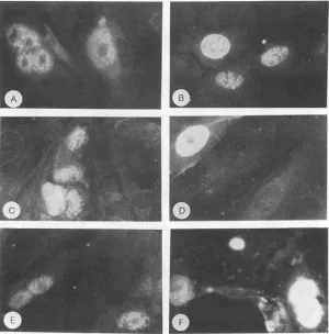
FIG. 1. Immunofluorescencestaining of MKCs transfected with polyomavirus genomes derived frompPY5O1D(Aand B),pPY5O1A(C andD),andpPY501A42(EandF).Cells werefixedand stainedby immunofluorescence for the presence of large T antigen (A, C, and E) or for the presenceof the latestructural proteins(B, D, and F).
codon at the deletionjunctionwas generated. The strategy
for introducing DNA fragments containing the mutations
into intactpolyomavirusgenomes isdescribed in Materials and Methods. Theplasmid pPY501A contains a copy of the viral genomewhich encodes the mutation of Asp-266 to Ala in VP1, while pPY501A42 contains a copy of the viral genomethatencodes the deletion mutation inVP1.
Transfection ofwild-typeand mutatedpolyomavirusgenomes
intoprimaryMKCs.Primary MKCs grown on coverslips were
transfected with polyomavirus DNA derived frompPY5O1D
(wildtype),pPY5O1A(point mutant), orpPY501A42(deletion
mutant). Immunofluorescence staining for the presence of
large Tantigenaswellasfor the late structural proteins was
performedoncells whichwerefixed 44 h after the addition of
serum-containingDulbecco's modifiedEagle'smedium. The
results, shown inFig. 1, indicated that bothlarge Tantigen
(Fig. 1A, C, and E)and the late viral proteins (Fig. 1B, D,
andF)weresynthesizedinsuccessfullytransfected cells and
properly transportedto thenucleus,the site of viral
assem-bly. This was true in cells transfected with polyomavirus
DNAencodingthe mutation ofAsp-266 to Alain VP1 (Fig.
1C andD) and viral DNAencodingaVP1moleculelacking amino acidsThr-262toGly-276(Fig. 1E andF)aswellasin cells transfected withwild-typepolyomavirus DNA(Fig. 1A andB).
A time course study was performed to examine the progression of infection in cells transfected with these DNAs. Cellsonduplicate coverslips were fixed and stained for the presence of both large Tantigen and the late viral
proteins atvarious times after transfection. Figure2 shows theresults obtained from cells transfected with DNA derived frompPY501D andpPY5O1A.Approximately2% of the cells transfected with either DNAsamplestainedpositivefor the presenceof viral proteins 2 days after transfection. Similar results were observed whether staining was for large T antigen(Fig. 2A)orfor thelate structuralproteins(Fig. 2B).
The percentage of cellstransfected withwild-typeviral DNA
(pPY501D) which stained positive increased dramatically
between 2 and 3 days after transfection and continued to increase throughout the course of the experiment. This increasewaspresumably duetoinfectionbyprogenyvirions whichwereproduced in cells that took up the viral DNA. No such increase was seen in cells transfected with DNA derived from pPY501A, nor was the increase detected in cells transfected with DNA derived frompPY501A42 (data
not shown). It is likely that the small percentage of these
cells which stainedpositive throughouttheexperimentwas
simply the result of successful transfection events.
Abun-dantcytopathic effectswerealso evident incells transfected with the wild-type genomes, while no cytopathic effects wereobserved in cells transfected with DNA derived from pPY501A or
pPY501A42
even 10 days after transfection. These results demonstrated that while expression and nu-cleartransport of themutantproteinsappearnormal, infec-tive progeny virionsare notproduced.Finally,unlikelysatesfrom cells transfected withviral DNA from pPY501D,
ly-sates from cells transfected with viral DNA derived from
pPY5O1A or
pPY501A42
were not infective(data
notVOL. 67,1993
on November 9, 2019 by guest
http://jvi.asm.org/
2490 HAYNES ET AL.
1IX
I.-u
0
c5
0
z
gow
x
us
ui z
w
0
z 'u
0
Z
CA
x
UL
D0
A
BO X .---_A+-__ _ _ _ _ _i
10 +
10 +
0 1 2 3 4 5 6 7
TIMEPOST-TRANSFECTION(DAYS)
100
0 1 2 3 4 5 6 7
TIMEPOST-TRANSFECTION(DAYS)
FIG. 2. Timecoursestudy of transfected MKCs. Primary MKCs
weretransfected with DNA derived from pPY501Dor pPY5O1A.
Duplicate plateswerefixed 1, 2, 3, 4, and 6 days aftertransfection,
and thepresence of large Tantigen (A)orpolyomavirus structural
proteins (B) was assayed by indirect immunofluorescence. The
percentage of cells exhibiting nuclear fluorescence at each time
point was determined after 1,000 cells (500 cells oneach of two coverslips)were counted. +,wild-type; A,pointmutant.
shown). Thisobservation confirms thatinfectiveviral prog-eny do not result from transfection with polyomavirus ge-nomes encoding either the point mutation or the deletion
mutation into normallysusceptiblecells. These results also revealed that largequantities of mutatedVP1 protein could not be obtained from transfected cell cultures for further analysis.
Calcium-binding analysisofrecombinant VP1 proteins. To obtain quantities ofmutated polyomavirus VP1 suitable for additional analysis, we expressed the proteins in E. coli
from versions of the VP1 expression vector, pALVPltac (24), generated as described in Materials and Methods. pALVPlAtacexpresses VP1containing theAsp-266-to-Ala
mutation, while pALVP1A42tac expresses VP1 containing
the deletion mutation. Wild-type VP1 and VP1 containing theAsp-266-to-Ala mutationwerepurified from bacterial cell
lysates by immunoaffinity chromatographyforusein
calci-um-binding analysis. Analiquot fromeachpreparationwas
also resolved bySDS-PAGE. Coomassie blue staining and Western blotting indicated that both proteins had been purified to near homogeneity (data not shown). Although Westernblotting studies ofthe E.colipelletsgeneratedafter the initial sonication andcentrifugationstepsindicated that
an 1etr ltig 2ae n 5coti 2. of purfie
[image:5.612.58.298.76.398.2]+~~~~~~~~~~~~~~~~~~~~~~~~~~~~
%_F11161WFIG. 3. Analysis of thenCa-binding abilit fied recmd viral proteins and recombinant wild-type and mutant VP1 molecules. Proteins were separated by SDS-PAGE (12.5% polyacrylamide),
transferredtoNitroBind,andanalyzed bybotha45Ca-bindingassay and Western blotting. Lanes 1 and 2 contain 2.5 ,ug of purified
polyomavitus
virionproteinsdetected by Westernblotting(lane 1) andanalyzedfortheirabilitytobindm5Ca(lane 2);lanes 3(Western blot) and 4(45iCa blot) contain 2.5 ug ofpurified recombinant wild-typeVP1; lanWe sternblot)and 6 asblot)contain 2.5ng
ofpurified recombinant VP1 with theAsp-266-to-Alamutation;lanes7(Westernblot)and 8(45Cablot)containth10 gof thepellet
generated bycentrifugationof thebacterialcell lysate fromtheE. colistrainexpressing thedeletion mutant. Thearrowtothe leftof thefigureidentifiesVP1.
VP1was
expressed
frompALVP1,&42tac,
soluble VP1 wasnotdetected in the
supernatant
component(data
notshown).
Perhaps
the deletion resulted in a foldingdifficulty
which rendered the protein insoluble under the buffer conditions used. Since we were unable to obtain the VP1 deletion mutantin asoluble form forapplication
totheimmunoaffin-ity
column, theinsoluble portionof thelysate
from E. coliexpressing
this mutantprotein
was used in thecalcium-binding
analysis
shown inFig.
3.After resolution
by
SDS-PAGE andblotting,
proteins from purifiedpolyomavirus
virions as well asrecombinantwild-type
and mutated VP1were examined for theirability
tobind45Ca(Fig.
3, lanes 2,4, 6, and8).
Theisotope
was then removed and thepresence
and location of VP1on the filterwereconfirmedby
Westernblotting
(Fig.
3,lanes1, 3, 5, and7).
Analysis
of the45Ca-binding
abilities ofproteins
derivedfrompurified virions(lane
2)
reveals amajor signal
aswellasseveral minor
signals.
Thecorresponding
Western blot(lane
1) demonstrates that theprincipal
45Ca-binding
protein
seen in lane 2 is VP1, consistent withprevious
studies(27).
These studies had also shown that the low-molecular-weight43Ca-binding
entitiesrepresent C-terminal VP1fragments resulting
frombreakdownofthe protein(27)
and that the minor
capsid proteins (detected by
thepoly-clonal antisera in lane
1)
do notbind45
Ca.Purified recombinant
wild-type
VPI and recombinant VP1containing
themutation ofAsp-266
toAla also bound45Cain this assay(Fig.
3, lanes4 and6).
Westernblotting
(Fig.
3,lanes 3 and
5)
confirmed theidentity
of theseproteins
as VP1. Additional signalsseen in lanes 3through 6 representrecognitionofproteolyticbreakdownproductsofVP1,
gen-erated duringpurification. Lane 8reveals thatthe recombi-nant VP1 deletion mutant did not bind
45Ca
in this assay, J.VIROL.a
a
4
2
on November 9, 2019 by guest
http://jvi.asm.org/
[image:5.612.318.551.79.243.2]POLYOMAVIRUS VP1 CALCIUM-BINDING DOMAIN 2491
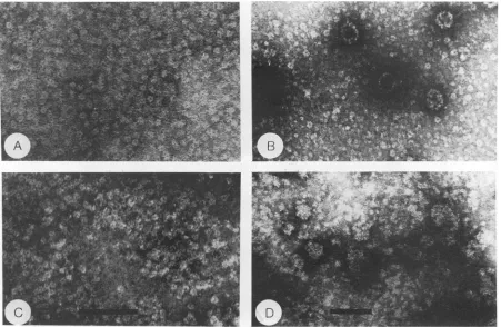
FIG. 4. Electron microscopic analysis of purified recombinantwild-type and mutatedpolyomavirus VP1 protein under different buffer conditions. (A) VP1 expressedfrompALVPltac in buffer containing EDTA andP-mercaptoethanolbutlacking calcium. (B) VP1 expressed frompALVPltacafter removal of EDTA and 3-mercaptoethanoland the addition ofCaC12; (C) VP1 expressed frompALVPlAtac under the conditions described forpanelA;(D)VP1expressedfrom pALVPlAtac under the conditions described for panel B. The bar shown in panel C is100nmandappliestopanelA aswell; the bar shown in panel D is 100 nm and applies to panel B as well.
whilelane 7identifies the location ofVP1onthefilter. These
datastronglysuggestthat aminoacidsThr-262toGly-276are
required for calcium binding by VP1 invitro. However, a
mutation ofAsp-266toAla doesnotpreventinvitrocalcium binding.
Electron microscopyanalysisofrecombinant VP1 proteins. Salunkeetal.(38, 39)demonstrated thatwhenpolyomavirus VP1wasexpressed inE. coli RB791 from pALVPltac and later purified underappropriate conditions, itwasfound in
pentameric structures which were indistinguishable from
viralcapsomeres andcouldassembleintocapsid-like
struc-tures upon additionof calcium. Figure 4 shows a seriesof
electron micrographs of purified VP1 expressed from pALVPltacandpALVPlAtac underdifferentbuffer condi-tions. Our inability to obtain purified VP1 from pALVP1A 42tac precluded its use inthese studies. Figure 4A shows
purified wild-type VP1 in buffer containing EDTA and P-mercaptoethanol. The protein formed pentameric
capso-mere-likestructuresunder theseconditions,consistent with previousreports(38, 39). Likewise, removal ofthe chelator and reducing agent, coupled with the addition of CaCl2, resulted inassemblyof thepentamersintocapsid-like struc-tures (Fig. 4B). VP1 expressed from pALVPlAtac and purifiedunder conditions favoring pentamerformationwas
indistinguishablefromwild-typeVP1 (Fig. 4C).This
similar-ity appears to indicate thatthe mutation does not cause a
grossfoldingabnormalityintheprotein. However, addition
of CaCl2 did not result in assembly into capsid-like
struc-tures. Rather, aggregates of the pentamers with variable sizesresulted, as shown in Fig. 4D. Thus, assembly of the pentamers derived from the mutated VP1 protein into cap-sid-like structures, a process shown to be calcium depen-dent,wasimpeded.
Immunogold staining of virus proteinsin transfected cells. In an attempt to study the assembly properties of the VP1
point mutant in mammalian cells, wild-type polyomavirus
DNA orDNAencoding the Asp-266-to-Ala point mutation of VP1wasfirst transfected intoprimaryMKCs.Forty-eight
hours aftertransfection, the cells were fixed and prepared forthin-section electron microscopy and immunogold label-ingasdescribed in Materials and Methods. Athin section of anormal cellwhichwassubjectedtoimmunogoldlabeling is presented in Fig. 5A, whileFig. 5B shows the insert portion of Fig. 5A at higher magnification. The complete lack of immunogold particles in these normal cells demonstrates
thatthegold particlesdonotbindtothesections
nonspecif-ically.
An immunogold-labeled thin section of a typical cell transfected withwild-type polyomavirusDNA,presentedin Fig. 5C,revealsaconcentration ofassembledvirusparticles
in an electron-dense region within the nucleus. Figure 5D shows the insertportion ofFig. 5C athighermagnification.
Most ofthe immunogoldparticlesin this region are
associ-ated with intact virions. Closer examination of Fig. 5C reveals the presence of additional gold particles scattered
throughoutthenucleus. Theseparticlesdonotappeartobe
VOL.67, 1993
on November 9, 2019 by guest
http://jvi.asm.org/
2492 HAYNES ETAL.
wS,,.^
..-XWv..t.
. ... b . . .* ^
::
... v f*.
..,f #,.
,.!,cK.^",,'*
.fw 'R ;. ,,,X,
Z..' *, 0, f:' , s;'
:. ' ;
bC ; 8 t t *26X?
I.
fs,.s*
*zss&
@ @D
At,
%:Z'At
FIG5c
-Ma;eg
4~~~~~4
derived frompPYSOlD; (D)insert region of cell shown in panel C under higher magnification; (E) cell transfected with DNA derived from
pPY5OlA;
(F) insert region 1 of cell shown in panel E, under higher magnification; (G) insert region 2 of cell shown in panel E, under higher magnification. The bar in panel E is 500 nm and applies to panels A and C as well; the bar in panel G is 100 nm and applies to panels B, D, and F aswell. Nu, nucleus; Cy, cytoplasm.associated with intact virions and arepresumablyboundto viral proteinswhich have notyet assembled into
recogniz-ablestructures.
Figure 5E depicts a thin section of a representative cell
transfected withpolyomavirusDNAderived frompPY501A.
Here,anelectron-denseregionisagain observed,butit does not appear to be composed of assembled virus particles.
Figure 5F (showing insert region 1 of Fig. SE at higher
magnification)andFig. 5G(showing higher magnificationof
insertregion 2)establish thatimmunogoldparticlesare seen in both the electron-dense region (Fig.
5F)
and in a non-electron-denseregion(Fig. 5G)of the nucleus. As inFig. SC,closerexamination ofFig. 5E reveals the presence ofgold
particles throughoutthe nucleus. Theyarealso observed in
the cytoplasm of cells transfected with polyomavirus
ge-nomesderived from eitherpPYSO1D orpPY501A(datanot
shown).Unlike the situationseeninFig.SC andD,noneof
the immunogold particles are associated with structures
resembling virions in anyregion ofthese cells (Fig. 5E, F,
and G). In addition, their presence is not the result of
nonspecific binding, since no gold particles are seen in
untransfected cells
(Fig.
5A and B). These immunogoldparticlesmusttherefore have boundtoviral proteins which
were not assembled into identifiable structures. Coupled with the immunofluorescence studies of cells transfected with DNA from pPYSO1Awhich indicated the presence of J. VIROL.
on November 9, 2019 by guest
http://jvi.asm.org/
POLYOMAVIRUS VP1 CALCIUM-BINDING DOMAIN 2493
1 2 3 4 5 6 7 8 9 10 11 12 VP1 266Asp-Glu-Asn-Gly-Val-Gly-Pro-Leu-Cys-Lys-Gly-Glu277
TnC
II 66Asp-Glu-Asp-Gly-Ser-Gly-Thr-Ile-Asp-Phe-Glu-Glu77
IV 142Asp-Lys-Asn-Asn-Asp-Gly-Arg-Ile-Asp-Phe-Asp-Glul53
CaM
[image:8.612.63.304.66.237.2]II 56Asp-Ala-Asp-Gly-Asn-Gly-Thr-Ile-Asp-Phe-Pro-Glu67 III 93Asp-Lys-Asp-Gly-Asn-Gly-Tyr-Ile-Ser-Ala-Ala-Glul04
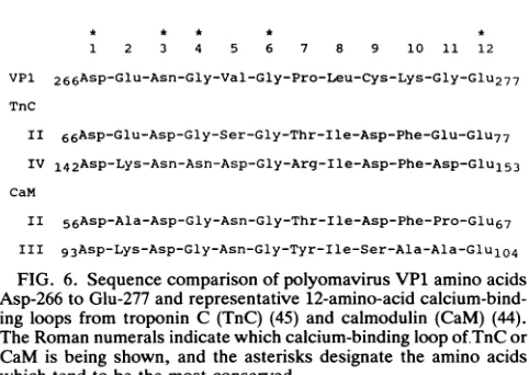
FIG. 6. Sequencecomparison of polyomavirus VP1 amino acids Asp-266 to Glu-277 andrepresentative 12-amino-acid calcium-bind-ingloops fromtroponin C (TnC) (45) and calmodulin (CaM) (44). TheRomannumeralsindicatewhichcalcium-binding loop of.TnC or CaM isbeing shown, and the asterisks designate the amino acids whichtendtobethe most conserved.
virus proteins in the nucleus (Fig. 1C and D) and with electronmicroscopy studies of recombinant VP1 expressed from pALVPlAtac(Fig. 4C and D), the results shown in Fig. 5E, F, and G stronglysuggest that this mutation results in a deficiency in virus assembly. Specifically, the calcium-de-pendent assembly of pentamers into capsid-like structures appears tobe affected.
DISCUSSION
Studies employing purified polyomavirus virions (7-10, 47) have implicated calcium as having a role in maintaining the virion structure as well as in viral uncoating and assem-bly. In addition, calcium was shown to contribute to the assembly of recombinantpolyomavirus VP1 expressed inE. coli into capsid-like structures (38, 39). Further evidence of theimportance ofcalcium in capsid assembly was obtained
whenpolyomavirus VP1 was expressed in Spodoptera
fru-giperda cells from abaculovirus construct (32). Here, VP1 wasnotassembled intorecognizable structures in the cyto-plasm, but it was foundto form capsid-like structures in the nucleus. The authorsproposed that differences in the con-centrations of availablecalcium between the cytoplasm and the nucleus could account for this result, and they further
demonstrated thataddition of the calcium ionophore
iono-mycin to the cells resultedin cytoplasmic assembly of VP1 intocapsid-like structures.
Ludlow and Consigli (27) identified a stretch of amino acids from Pro-232to Asp-364 of polyomavirus VP1 which, itself, has calcium-bindingcapabilities. Within this polypep-tide, astretch of aminoacids from Asp-266 to Glu-277 shows ahigh degree ofsequence similarity to the calcium-binding portion of the EF handmotif found in many proteins (13,42).
Acomparison between this domain of polyomavirus VP1
and representative EF hand loops from other calcium-binding proteins is shown in Fig. 6. These calcium-binding EFhand motifs usually contributeeither six or seven oxygen ligands to the metal ion.Coordination can occur through side chainoxygens (particularlyfrom acidic residues), main chain carbonyl oxygens, or oxygens from water molecules in the area. In these EF hand structures, amino acids at positions 1, 3, 5, 7, 9, and 12 tend to be involved in calcium coordination, with the conserved glutamic acid residue at position 12 supplying both oxygen atoms from its carboxyl group to the cation (13, 42).Although sequence conservation among EF hand motifs is not maintained at every residue, amino acids at certain positions are well conserved. A
comparison of sequences of 165 EF hands from various calcium-binding proteins revealed that amino acids at posi-tions 1 (Asp [98%]), 3 (AsX
[97%]),
6 (Gly [89%]), and 12 (Glu [86%]) are the most conserved (13). In addition, a glycine residue was found at position 4 in 51% of the sequences examined. Each of the amino acids is properly placed within the domain of polyomavirus VP1 extending from Asp-266 to Glu-277. Considerable variability exists at several other positions of the motif, including positions 2, 6, 9, 10, and 11, where no single amino acid was found to be present in more than one-third of the EF hands (13).The data from our transfection experiments indicate that deletion of the putative calcium-binding domain or mutation of a residue within this domain does not affect viral gene expression or nuclear transport. However, these mutations do result in a defect in the production of infective viral progeny. Evidence that an assembly problem, and more specifically an inability to bind calcium, may be the cause of this defect comes from several sources. First, recombinant VP1 containing the deletion mutation was unable to bind
45Ca
in vitro. Second,VP1
molecules containing a single point mutation within the putative calcium-binding domain were unable to carry out the calcium-dependent in vitro self-assembly step from pentameric, capsomere-like struc-tures to capsid-like strucstruc-tures. Finally, despite the fact that VP1 protein containing the point mutation could be detected in the nuclei of transfected cells, there was no evidence that these proteins were participating in viral assembly.Even though it appears to be defective in the calcium-dependent assembly steps both in vitro and in vivo, VP1 containing the Asp-266-to-Ala mutation was able to bind
45Ca
in vitro. Calcium binding by native VP1 in intact polyomavirus virions is probably of a cooperative nature, with ligands for calcium binding being contributed by do-mains from more than oneVP1
molecule. Mutation of even a single amino acid within this domain may have altered the three-dimensional relationship among the molecules in such a way that cooperative calcium binding was no longer possible. However, unlike the situation with the deletion mutant, a single amino acid substitution may not have been dramatic enough to alter the calcium-binding properties of VP1 on nitrocellulose, where such spatial relationships are less germane.Although the high-resolution crystallographic structure of the murine polyomavirus has not yet been reported, the structure of the related simian virus 40
(SV40)
has been determined to a resolution of 3.8A
(0.38 nm) (25). This study has aided in the understanding of how theSV40
capsomere subunits interact to form a virus capsid. The C-terminal arms of the individual VP1 molecules extend away from the pentamer of origin and into a subunit of an adjacent pen-tamer. Deletion of the corresponding C-terminal region from polyomavirus VP1 resulted in an assembly defect in vitro (20). In SV40, the arms are locked into place by a clamp derived from sequences near the N terminus of a VP1 molecule in the invaded pentamer. A possible site of divalent cation binding toSV40 VP1 was identified in these studies. Because of solubility considerations, the trivalent cation gadolinium(Gd3+)
was used in place of divalent calcium ions to produce difference maps between crystals treated or not treated with EGTA. It was postulated thatGd3+
would bind to theCa2+-binding
site ofSV40, as was reported to occur in similar studies of tomato bushy stunt virus (22). The differ-ence maps produced in the SV40 studies suggested thatGd3+
forms a bridge which links the C-terminal arm from one pentamer with an internal loop provided by a VP1 VOL. 67,1993on November 9, 2019 by guest
http://jvi.asm.org/
moleculein aneighboring pentamer. The investigators noted that twoglutamate residues (157 and 160) from the loop of one VP1 molecule and an aspartate residue (345) from the C-terminal arm of the other VP1 molecule were in the correctlocationtocoordinate the Gd3 .Whether the results
obtained with trivalentgadolinium ions canbeextrapolated todivalent calcium ions in SV40 isnotknownfor certain. In addition, the lack ofahigh-resolution crystallographic struc-tureof the murinepolyomavirus makes it unclear whetheran identical mechanism of calcium binding is utilized by this virus. This isespecially importantinlightof thefact that the amino acid sequences of SV40 VP1 andpolyomavirus VP1
haveonly50% identity (43). Therefore, significantstructural
variationsarequite likely.Intermsofspatial relationship,it is interesting thatasequence ofamino acids found in SV40 VP1, which isnearlyidenticaltoAsp-266 throughGlu-277 of
polyomavirus VP1, is in close proximitytothe SV40
calci-um-binding siteproposedbyLiddington etal. (25).
On the basis of the available structural and biochemical data,wepropose thatthe amino acid stretch fromAsp-266to Glu-277 ofpolyomavirusVP1is involved in calciumbinding.
This binding would involve an intermolecular interaction which would include the EF hand-like region ofone VP1 molecule and either thecorresponding region or a separate region of another VP1 molecule. The sensitivity of intact virus particlesto treatmentwith chelatingagents, as previ-ously demonstrated in virion dissociation experiments by
Brady et al. (9, 10), suggests that calcium is bound to exposed regions of thecapsid.
The early events of polyomavirus infection have been examined byelectron microscopy (30), binding studies (6),
isolation ofvirion-containing monopinocytoticvesicles(21),
and nuclear uncoating studies (46). These studies demon-strated that uncoating ofinfecting virions begins upon nu-clear entry, as evidenced by the isolation of uncoating
intermediates in the nuclei of infected cells (46). We have
postulated that theaction ofacalcium-binding protein(s)in
this cellular compartment, together with a microenviron-ment which provides the necessary reducing conditions,
maycontributetoviraluncoating.The available information suggests that calcium is alsoinvolved incapsid assembly, a late event in the virus life cycle (8, 32, 38, 39, 48). The findings presented in the present study demonstrate that mutations in the putative calcium-binding domain affect
capsid assemblyboth in vitro and in vivo.Itremainspossible
that the mutations created in thisstudyaffectedsomeaspect
of capsid structure and/or assembly other than calcium
binding; however, it must be stressed that the deletion
mutant was unable to bind
45Ca
in an in vitro assay. Inaddition,the stepofcapsid assembly previouslyshowntobe
calcium dependent was specifically hindered in VP1 mole-cules containing this point mutation. This mutation may interfere with assembly either by eliminating a calcium coordination site or by leading to a local conformational
change which renders the viral proteins unable to bind
calcium.
In summary, although it is fairly well established that
polyomavirusVP1binds calcium as ameansofmaintaining
itsstructuralintegrity,theexact natureof thebinding isnot yetunderstood. TheX-raycrystallographic determination of
thepolyomavirus structure athighresolution will be
bene-ficial inclarifyingtheexact natureofcalcium binding by its
major capsid protein VP1.
ACKNOWLEDGMENTS
Thisinvestigationwassupported byPublic Health Service grant CA07319 from the NationalCancerInstitute, byNASANAGWno.
1197and 2328, and by theWesleyFoundation ofWichita, Kans. J.I.H. is alsoapredoctoral fellowunder National Cancer Institute traininggrantCA09418.
We thank Avelina Paulsen for assistancewith electron micros-copy andToddMartin, Daryl Riley,KevinMapes,LaDonnaGrenz, and Viola Hill for technicalassistance.
REFERENCES
1. Abdel-Meguid, S. S.,T.Yamane, K.Fukuyama, and M.
Ross-mann. 1981. The location of calcium ions in Southern bean mosaic virus.Virology114:81-85.
2. Anders,D.G.,andR.A.Consigli. 1983.Chemical cleavageof polyomavirus major structural protein VP1: identification of cleavage productsand evidence thatthereceptormoietyresides in thecarboxy-terminal region.J.Virol. 48:197-205.
3. Anders,D. G., and R. A.Consigli. 1983. Comparisonof non-phosphorylated and phosphorylated species of polyomavirus majorcapsidproteinVP1and identification of themajor phos-phorylationregion.J. Virol. 48:206-217.
4. Bechtel,D.B., J.D.Wilson,andP. R.Shewry.1991. Immuno-cytochemicallocalization of the wheat storageproteintriticin in developing endospermtissue. CerealChem.60:573-577. 5. Bolen, J. B.,D.G.Anders, J. Trempy,andR. A.Consigli.1981.
Differences in thesubpopulations of the structuralproteinsof polyomavirions andcapsids: biologicalfunctionsof themultiple VP1species.J.Virol. 37:80-91.
6. Bolen,J. B.,and R.A.Consigli.1979.Differentialadsorption of polyomavirions andcapsidstomousekidneycells andguinea pig erythrocytes.J. Virol. 32:679-683.
7. Brady, J. N.,and R. A.Consigli.1978.Chromatographic sepa-ration of the polyoma virus proteins and renaturation of the isolatedVP1majorcapsid protein.J. Virol.27:436-442. 8. Brady, J. N., J.D.Kendall, and R. A. Consigli. 1979. In vitro
reassemblyof infectiouspolyomavirions. J. Virol. 32:640-647. 9. Brady, J. N.,V. D.Winston,and R. A.Consigli. 1977. Dissoci-ation ofpolyomavirus bythe chelation of calcium ionsfound associated withpurifiedvirions. J. Virol. 23:717-724.
10. Brady, J. N.,V. D.Winston,and R. A.Consigli.1978. Charac-terization ofa DNA-protein complexand capsomere subunits derived frompolyomavirus bytreatmentwith ethyleneglycol-bis-N,N'-tetraaceticacidanddithiothreitol.J. Virol.27:193-204. 11. Chang, D., J.I.Haynes, J.N.Brady,and R. A.Consigli.1992. Theuseofadditiveandsubtractiveapproachestoexaminethe nuclear localizationsequenceofthepolyomavirus majorcapsid proteinVP1.Virology189:821-827.
12. Consigli,R.A., J. Zabielski,and R.Weil.1973.Plaqueassayfor polyoma virus on primary mouse kidney cell cultures. Appl. Microbiol. 26:627-628.
13. da Silva,A. C.R., and F. C. Reinach. 1991. Calciumbinding inducesconformationalchangesin muscleregulatory proteins. Trends Biochem. Sci. 16:53-58.
14. Durham,A.C.H.,and M. A. Haidar.1977. Cationbinding by tobacco rattle virus.Virology77:520-523.
15. Durham,A.C.H.,and D. A. Hendry.1977. Cationbinding by tobacco mosaicvirus.Virology77:510-519.
16. Durham, A. C. H., D.A. Hendry, and M. B. von Wechmar. 1977. Does calcium ionbindingcontrolplantvirusdisassembly? Virology77:524-533.
17. Erickson,P.E.,D.H.Anderson,andS. K. Fisher.1987. Use of uranyl acetate en bloc to improve tissue preservation and labeling for post-embedding immuno-electron microscopy. J. Electron Microsc. Tech. 5:303-314.
18. Fattaey,A.R.,and R. A.Consigli. 1989.Synthesis, posttrans-lational modifications, and nuclear transport ofpolyomavirus major capsid proteinVP1.J. Virol. 63:3168-3175.
19. Garcea, R. L., K. Ballmer-Hofer, and T. L. Benjamin. 1985. Virion assembly defectofpolyomavirus hr-tmutants: under-phosphorylationofmajor capsid proteinVP1before viral DNA encapsidation.J.Virol. 54:311-316.
20. Garcea, R. L., D. M. Salunke, and D. L. D. Caspar. 1987.
on November 9, 2019 by guest
http://jvi.asm.org/
POLYOMAVIRUS VP1 CALCIUM-BINDING DOMAIN 2495
Site-directed mutation affecting polyomavirus capsid self-as-semblyin vitro. Nature(London)329:86-87.
21. Griffith, G. R., and R. A.Consigli. 1984. Isolation and character-ization of monopinocytotic vesicles containing polyomavirus from the cytoplasm of infected mouse kidney cells. J. Virol. 50:77-85. 22. Hogle, J., T. Kirchhausen, and S. C. Harrison. 1983. Divalent
cation sites in tomatobushy stunt virus: difference maps at 2.9 Aresolution. J. Mol. Biol. 171:95-100.
23. Lawler, J., and R. 0. Hynes. 1986. The structure of human thrombospondin, anadhesive glycoprotein with multiple calci-um-bindingsites andhomologieswith several different proteins. J. CellBiol. 103:1635-1638.
24. Leavitt, A. D., T. M. Roberts, and R. L. Garcea. 1985. Polyoma virus major capsid protein VPl: purification after high level expressioninEscherichia coli.J. Biol. Chem.260:12803-12809. 25. Liddington, R. C., Y. Yan, J. Moulai, R. Sahli, T. L.Benjamin
andS. C. Harrison. 1991. Structure ofsimianvirus 40 at 3.8X resolution. Nature(London)354:278-284.
26. Ludlow, J. W., and R. A.Consigli.1987. Differences in biolog-ical activity and structural protein VP1 phosphorylation of polyomavirus progeny resulting from infection of primary mousekidney and primary mouse embryo cell cultures. J. Virol. 61:509-517.
27. Ludlow, J. W., and R. A.Consigli.1987. Localization of calcium
onthepolyomavirus VP1capsidprotein. J. Virol. 61:2934-2937. 28. Ludlow, J. W., and R. A. Consigli. 1987. Polyomavirus major capsid protein VPl ismodified by tyrosinesulfuration. J. Virol. 61:1708-1711.
29. Ludlow, J. W., and R. A.Consigli.1989.Hydroxyprolineinthe major capsid protein VP1 of polyomavirus. J. Virol. 63:2881-2884.
30. Mackay, R. L., and R. A. Consigli. 1976. Early events in polyoma virus infection: attachment, penetration, and nuclear entry.J. Virol. 19:620-636.
31. McMillen, J., and R. A.Consigli.1977.Immunological reactivity of antisera to sodium dodecyl sulfate-derived polypeptides of polyomavirions. J. Virol. 21:1113-1120.
32. Montross, L., S. Watkins, R. B. Moreland, H. Mamon, D. L. D. Caspar,and R. L. Garcea. 1991. Nuclearassembly of polyoma-viruscapsidsininsect cellsexpressingthemajorcapsidprotein VP1.J.Virol. 65:4991-4998.
33. Newman, G. F., and J. A. Hobot. 1987. Modern acrylics for post-embedding immunostaining methods. J. Histochem. Cy-tochem. 35:971-981.
34. Olson,A.J., G. Bricogne, and S. C. Harrison. 1983.Structureof
tomato bushy stunt virus IV: the virus particle at 2.9 A resolution. J.Mol. Biol. 171:61-93.
35. Ponder, B. A., A. K. Robins, and L. V. Crawford. 1977. Phosphorylation of polyoma and SV40 virus proteins. J. Gen. Virol. 37:75-83.
36. Reynolds, E. S. 1963. The use of lead nitrate at high pH as an electron opaque stain in electron microscopy. J. Cell Biol. 17:208-212.
37. Robinson, I. K., and S. C. Harrison. 1982. Structure of the expanded stateof tomato bushy stunt virus. Nature (London) 297:563-568.
38. Salunke, D. M., D. L. D. Caspar, and R. L. Garcea. 1986. Self-assembly ofpurified polyomavirus capsidprotein VP1. Cell 46:895-904.
39. Salunke, D. M., D. L. D. Caspar, and R. L. Garcea. 1989. Polymorphism in theassembly ofpolyomaviruscapsid protein VP1.Biophys.J. 56:887-900.
40. Smith, G. L., and R. A. Consigli. 1972. Transientinhibition of polyomavirus synthesis by Sendai virus (parainfluenza I). I. Demonstrationand natureofthe inhibitionby inactivatedvirus. J.Virol. 10:1091-1097.
41. Soeda, E., J. R. Arrand, and B. E. Griffin. 1980.Polyomavirus DNA: completenucleotide sequenceofthe gene which codes forpolyomaviruscapsid proteinVP1andoverlapstheVP2/VP3 genes.J. Virol. 33:619-630.
42. Strynadkam, N. C. J., and M. N. G. James. 1989. Crystal structures of the helix-loop-helix calcium-binding proteins. Annu. Rev.Biochem. 58:951-998.
43. Tooze, J. (ed.). 1980. DNAtumorviruses,2nded. ColdSpring HarborLaboratory, ColdSpring Harbor,N.Y.
44. Watterson, D.M.,F.S.Sharief,and T.C. Vanaman. 1980. The completeamino acidsequenceoftheCa"2-dependent modula-tor protein (calmodulin) of bovine brain. J. Biol. Chem. 255: 962-971.
45. Wilkinson, J. M. 1976. The amino acid sequenceoftroponinC from chick skeletal muscle. FEBS Lett. 70:254-256.
46. Winston, V. D., J. B. Bolen, and R. A.Consigli.1980. Isolation and characterization ofpolyomauncoating intermediates from thenuclei of infectedmousecells. J. Virol. 33:1173-1181. 47. Yuen, L. K. C., and R. A.Consigli.1982.Improved infectivityof
reassembledpolyomavirus. J. Virol. 43:337-341.
48. Yuen, L. K. C., and R. A. Consigli. 1985. Identification and protein analysisofpolyomavirus assembly intermediatesfrom infectedprimarymouseembryocells.Virology 144:127-138. VOL. 67,1993