Determination of the
Origin-Specific DNA-Binding Domain
of
Polyomavirus Large
T
Antigen
NOELLE-ANNE SUNSTROM,1NICHOLAS H. ACHESON,'* ANDJOHN A. HASSELL2
DepartmentofMicrobiology and Immunology, McGill University, Montreal, Quebec H3A
2B4,1
andInstituteforMolecularBiology and Biotechnology, McMasterUniversity, Hamilton, Ontario L8S4K1,2Canada
Received 4April 1991/Accepted 9 September 1991
TomaptheDNA-bindingdomainofpolyomavirus largeTantigen,weconstructedasetofplasmids coding
for unidirectional carboxy- or amino-terminal deletion mutations in the large T antigen. Analysis of
origin-specificDNAbinding bymutantproteins expressedinCos-1 cells revealedthattheC-terminalboundary
of theDNA-binding domain isator near Glu-398. Fusion proteins oflargeT antigen lacking the first 200
N-terminal amino acids boundspecifically topolyomavirus origin DNA; however, deletions beyond this site resulted in unstable proteins which couldnotbetested for DNA binding.Testing of point mutants and internal deletions by others suggested that the N-terminal boundary of the DNA-binding domain lies between amino acids 282 and 286. Taken together, these results locate the DNA-binding domain ofpolyomavirus large T antigentothe116-amino-acid region between residues 282 and 398.
Polyomavirus largeTantigen isrequired for the initiation
ofviral DNAreplication and theregulation of early mRNA
synthesis (5, 15). These functions of large T antigen are
thoughttorequire direct binding oftheproteintosequences
withinthenoncoding regulatory region of the viralgenome
(7, 10, 13). Polyomavirus large T antigen binds to three
high-affinity binding sites, designated A, B, and C, whichare
intermingledwith elements that control early transcription,
and to three weaker binding sites, designated 1, 2, and 3,
located within the core origin for DNA replication. Each
bindingsite is characterized by thepresenceoftwo tofour
copiesof thepentanucleotidesequence5'-G(A/G)GGC-3' (7,
8, 30).
We are particularly interested in defining the functional
domainsofpolyomavirus largeTantigenimportant forDNA
replication and transcriptional control and in comparing
these to domains found inother papovavirus large T
anti-gens. Despite a wealth of information regarding the many
domainsof the largeTantigen of simian virus 40(SV40) (12),
thoseof othermembers ofthepapovavirus familyarepoorly
characterized. This information could lend insight into the evolution ofpapovaviruses and might helpinunderstanding
the species specificity of the DNA replication activity of
large Tantigen (3).
Here we report the construction and characterization of
amino-terminal and carboxy-terminal deletion mutants of
polyomavirus large Tantigen. Todefine the specific
DNA-binding domain of this protein, we expressed truncated
large-T-antigen proteins transiently in Cos-1 cells (17) and
measured specific DNA-binding activity innuclear extracts
by an immunoprecipitationassay (24, 25).
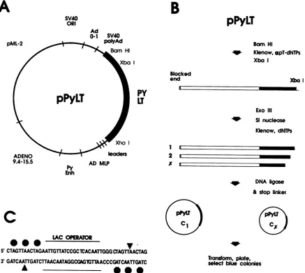
Construction of deletion mutants of large T antigen. We
usedthe expressionvectorpPyLT (24) (Fig. 1A), in whicha
cDNAfragment coding for largeTantigen (nucleotides [nt]
153 to 2962) was cloned as an XhoI-BamHI fragment.
Synthesis of large-T-antigen mRNAis driven by the
adeno-virusmajor latepromoter,andtranscriptsinitiated from this
promoterinclude the adenovirusmajor late tripartite leader
sequence. Immediately downstream ofthe large-T-antigen
* Correspondingauthor.
coding region are thepolyadenylation signals of both
poly-omavirus and SV40. The vector also contains the SV40
origin of DNA replication to allow template amplification
aftertransfection ofCos-1 cells.
A schematic representation of the steps involved in
gen-eratinglarge-T-antigen peptidestruncated atthe C terminus
is shown in Fig. 1B. Theplasmid pPyLTwaslinearized by
digestion with BamHI. The 3' recessed ends were filled in
with alpha-phosphorothioate deoxynucleosides at a final
concentration of 0.04 mM by using Klenow DNA
polymer-ase. This stepprevents subsequent digestionof the blocked endof theplasmid byexonuclease III(19). Theplasmidwas
then digested with XbaI to generate a DNA fragment
con-tainingthelarge-T-antigen coding regionuptothe XbaI site
at nt 2479 (amino acid 641 oflarge T antigen); nucleotide
numbers are assignedas for the A2 strain ofpolyomavirus
(34). This DNA (0.2 ,g/4l) was then digested with
exonu-clease III(10 U/p.l) at 37°C. Aliquotswereremovedat30-s intervals to tubes containing S1 nuclease (0.2
U/Rl),
0.4 Mpotassium acetate (pH 4.6),0.3 M NaCl, and 1.4 mM zinc
sulfate and incubated at 25°C for 30min. Afterinactivation oftheS1nuclease(70°Cfor 10min),the Klenowfragmentof
DNA polymerase and all four deoxynucleoside
triphos-phateswereaddedtoflush DNAends. A48-mer
oligonucle-otide(stop linker) containingtranslationalstopcodons in all threereadingframes(Fig. 1C), flankinga sequenceencoding
thelacrepressorbindingsite(2),wasligatedtothetruncated
cDNA. Thiswastoensureproductionofproteins containing
two or fewerforeign amino acids beyond the deletion end
pointattheir C termini. Theligation mixtureswere usedto
transform competent Escherichia coli DH5, which was
platedonminimalagarmediumcontainingX-Gal
(5-bromo-4-chloro-3-indolyl-,-D-galactopyranoside) (0.04 mg/ml) and
IPTG
(isopropyl-p-D-thiogalactopyranoside)
(2 mg/ml).De-velopmentofblue color incoloniesindicates thepresenceof
thelacrepressorbindingsite andsignalsthepresenceofthe
stop codon oligonucleotide in the recombinant plasmid. A numberof subclones from each timepointwasexamined by
digestion with restriction endonucleases to determine the
endpointof the deletions. The dideoxy method of
sequenc-ing (31)was usedtodetermine the extentof deletions.
The generationofN-terminaldeletions of large Tantigen
6998 0022-538X/91/126998-06$02.00/0
Copyright © 1991,American SocietyforMicrobiology
on November 10, 2019 by guest
http://jvi.asm.org/
pPyLT
Bom Hi
-W Klenow, apT-dNTPs Xba I
Xb I
py
LT
I _
Exo III -W SI nuclease
Kienow, dNTPs
n
2~ Xho
leaders
AD MLP
DNA Igose
& stop linker
r
0
C LAC OPERAUOR
5'CTAGTrAACTAGAATrG1TATCCGCICACAATTGGGCrAGTTAAcTAG
3'GATCAATrGATCTTAACAATAGGcGAGlGTTAACCCGATCAATrGATC
*
Transform, plate, select blue colories
FIG. 1. Construction of carboxy-terminal deletion mutants of polyomavirus large T antigen. (A) Parent plasmid pPyLT includes
large-T-antigencDNAas anXhoI-to-BamHIfragment(nt154to2962) whosetranscriptionisdirected bythe adenovirusmajorlatepromoter (AD MLP;nt5986to6038)andtripartiteleader(first,second andtwo-thirds of the thirdleader). SV40 polyAd,SV40 polyadenylation signal (nt2770to2533);Ad0-1, adenovirus left-handend(0to1mapunits, nt1to357);SV40ORI, SV40 originofreplicationand enhancerregion (nt270to0 and5243to5171);pML-2, plasmid pML-2sequences(nt650to4362); ADENO9.4-15.5,adenovirussequencesfrom 9.4to15.5
mapunits (nt3328to5788);PyEnh, polyomavirus enhancer region(nt5039to5265). (B)Schematic representationofstepsinvolved in the construction ofcarboxy-terminaldeletionmutantsoflargeTantigen. C1,
Cx:
plasmids encodingfragmentsoflargeTantigendeletedatthe Cterminus anddecreasing in size.(C) Oligonucleotide employed inthecloning proceduretofacilitate screeningofrecombinant plasmids containingthe lacrepressorbindingsite(lacoperator).Octagonsrepresentstopcodons in all threereadingframesflankingthe lacoperator. Triangles indicate thepositionofunique HpaI restriction sites.required reintroduction of an in-frame AUG translational
initiation codon upstream ofthe truncated coding region.
This was achieved by ligating a synthetic DNA fragment
(start linker) containingan ATG in the optimal context(22)
to exonuclease III-digested blunt-ended DNAs. We also
introduced in the start linker a sequence coding for an
antigenic determinant of the influenza virus hemagglutinin
(HA) protein (14), immediately downstream of the
transla-tional initiationcodon. This allowedustoimmunoprecipitate
the resulting HA-large-T-antigen-fragment fusion proteins
with an anti-HA antibody. The strategy used for the
con-struction of amino-terminal deletion mutants of large T
antigen was similar to that described for carboxy-terminal
deletion mutants. The plasmid pPyLT was digested with XhoItolinearize the DNAatthe N terminusof the
large-T-antigen coding region. Exonuclease III digestion, Si
nucle-ase treatment, and blunt ending were carried out as
de-scribedabove. The DNA wasthen cleaved with BamHI to
release fragments containing the large-T-antigen coding
re-gion with deletions at its N-terminal end. These fragments
were isolated from an agarose gel and ligatedtothe vector
pPyLT, which had been previously cleaved with XhoI and
BamHI. Ligation was carried out in the presence of a
chemically synthesized nonphosphorylated double-stranded
start linker (one strand: 5'-TCGAGATGTACCCATACGA
TGTTCCAGATTACGCTAGCTTGGGTGGTCCT-3';
com-plementary strand: 3'-_CTACATGGGTATGCTACAAGGT
CTAATGCGATCGAACCCACCAGGA-5'), which has at
one end an XhoI recognition site (underlined) followed by
the start codon fortranslation (ATG), followed by a DNA
A
SV40 ORI
pML-2
B
Ad
0-A
SV40 polyAdBlocked
end
ADENO
9A-15.5
Py
Enh
1
2 I m
n ..M
on November 10, 2019 by guest
http://jvi.asm.org/
[image:2.612.96.533.72.463.2]so cs - %O Go " "co
_ N N N - 0 co0 1 0%
A
5
z
z
0
"97 Kd 66 Kd
42 Kd
31Kd
B
1 2 3 4 5 6 7 8 9 10 11
FIG. 2. (A) Immunoprecipitation by polyclonal antiserum of
[35S]methionine-labeled large-T-antigen fragments made in Cos-1 cells transfected with C-terminal deletion mutants. Proteins were
analyzed by10% polyacrylamide gelelectrophoresis. WT, wild-type largeTantigen; C428toC298, C-terminal deletionmutantsoflarge T antigen whose termini are at the numbered positions; UT, untransfected Cos-1 cells. The numbers onthe left indicate
molec-ularsize in kilodaltons (Kd) determined from migration of marker proteins. (B) Specific DNA binding by large-T-antigen fragments described for panel A. Input DNA is Hinfl-digested recombinant polyomavirus DNA (pPHI-8). Arrow indicates the location of the 604-bp DNA fragment containing all large-T-antigen binding sites (theinput DNA digestionmixture isnot shown;seeFig. 3B).
sequenceencodinga 14-amino-acid peptideof the influenza
virus HA epitope (14), andat the other end is bluntended.
This three-part ligationwas doneata molar ratio ofvector:
fragment:linker of 1:5:50. The ligation mix was used to
transform competent E. coli DH5. Recombinant plasmids
were screened for the size of deletions by digestion with
BamHI and XhoI. Those of interest were sequenced and
plasmids whichgeneratein-frame HA-large T-antigen fusion
peptides were chosen.
Expression of large T antigenscontaining C-terminal dele-tions. Plasmid DNAs encoding large T antigen harboring
deletions atthe Cterminus wereanalyzed for their abilityto
express large-T-antigen fragments after transfection into
Cos-1 cells as described previously (3) by labeling total
cellular proteins with [35S]methionine as previously
de-scribed (24). Proteins in Cos-1 cell extracts were
immuno-precipitated with either the monoclonal antibody LT1
(On-cogene Science) or a polyclonal antiserum (obtained from
ascites fluid of brown Norwegian rats bearing tumors
in-duced by polyomavirus-transformed rat cells). Typical
re-sults are shown in Fig. 2A. In addition to large-T-antigen
fragments, a cellular protein of approximately 35 kDawas
regularly precipitated by the polyclonal antiserum. Different amounts ofboth large-T-antigen fragments and the cellular
protein were found in different extracts; these variations
may be due to differentefficiency oflabeling or extraction of
proteins. We have not attempted to quantitate levels of
large-T-antigenfragments made by each C-terminal mutant.
However, all the C-terminal mutants expressed a unique
species of large T antigen corresponding in size to that
expected from the sequenced end points of the deletions.
SV40 large T antigen, which is also present in Cos-1 cells,
was notrecognized byantibodies directedagainst
polyoma-virus large Tantigen (Fig. 2A, untransfected cellextract).
Specific DNA binding by large-T-antigen fragments
con-taining C-terminal deletions. The sequence-specific
DNA-binding activity of the carboxy-terminal truncated large T
antigens was examined by a modified McKay
immunopre-cipitation assay (24, 25). Nuclear lysates of transfected
Cos-1cells were mixed witha32P-end-labeled Hinfldigestof
the plasmid pPHI-8 (29). This Hinfl digest contains 20
fragments, one of which, 604 bp in length, contains the
region ofpolyomavirus DNA (nt 5073 to5296 and 1 to385)
to which large T antigen binds. Large T-antigen-DNA
complexes were immunoprecipitated, and bound DNAwas
released from the large-T-antigen-antibody complex and
fractionated on an agarose gel(Fig. 2B). Specificbindingby
large T antigen selectivelyprecipitates the 604-bp fragment
from themixture. Large-T-antigenfragments with C termini
up to amino acid 398 were capable ofspecific binding to
target DNA(Fig. 2B, lanes 2 to 6).However, the deletion of
14additional amino acidsfromthecarboxy terminus of large
Tantigen (C384) resulted in aprotein which was unable to
bind to the originfragment (lane 7). These results locate the
carboxy terminus of the DNA-binding domain between
amino acids 384 and 398. This domain lies upstream of a
putative metal-binding region (zinc finger) in polyomavirus
large T antigen (amino acids 452 to 472) (21). Although a
point mutation within the zinc finger was shown to abolish
DNA-binding activity (4), our results demonstrate that it
does not contribute directly to the specific DNA-binding
activity of large Tantigen.
It has been shown that SV40 and polyomavirus large T
antigen recognize and bind to similar sequence motifs in
DNA (29, 32). Therefore, it was importantto ensurethat all
DNA-binding studies were conducted in DNA excess, so
that SV40 large T antigen present in Cos-1 cell extracts
would not compete with polyomavirus large T antigen for
DNA binding. We observed that the unbound fraction of
DNAremaining in the supernatant afterprecipitation ofthe
immunecomplexescontained greater than90%of the
origin-containing fragment (not shown). Also,DNA-bindingassays
using antibodies directed against SV40 large T antigen
resulted in only a small portion of the input polyomavirus
origin-containing DNAfragment beingbound (not shown).
Expression of large T antigens containing N-terminal
dele-tions. HA-large-T-antigen fusion proteins lacking up to the
first 175 amino acids oflarge T antigen (N175) were
recog-nized by the monoclonal antibody LT1 anda ratpolyclonal
antiserum, both specific topolyomavirus largeTantigen, as
well as by theanti-HA monoclonalantibody 12CA5(Fig.3A,
lanes 4 to 9). Mutants N188 and N189 produced proteins
which wereimmunoprecipitated by the polyclonal antiserum
and anti-HA butnotby the monoclonalantibody LT1 (lanes
10to 15). These mutants therefore lack the antigenic
deter-minant recognized by the monoclonal antibody LT1 (11).
Larger deletions at the N terminus oflarge T antigen
re-sultedin theprogressive inability to detect theseproteinsby
immunoprecipitation of extracts from
[35S]methionine-la-beled Cos-1 cells. Although a small amount of N201 was
on November 10, 2019 by guest
http://jvi.asm.org/
[image:3.612.60.295.77.345.2]N156
N175
N188
N189N201
N240
N275
L P H L P H L P H L P H L P H L P H L P L P H
_~~_~~ ~ ~
_ _
--40
0
04
s4*i
mm Om
_m
_m
-__1 2 3 4 5 6 7 8 9 10 11 12 13 14 15 16 17 18 19 20 21 22 23
z 0
WT N156
L P L P H
_ _
N175
N188 N189N201
N240 N275L P H
*fW
P H P H P H L P P H
1 2 3 4 5 6 7 8 9 10 11 12 13 14 15 16 17 18 19
FIG. 3. (A) Immunoprecipitation of [35S]methionine-labeled large-T-antigen fragments made inCos-1cells transfected with N-terminal deletionmutants. Proteinswere analyzed as described inthelegendto Fig.2. WT,wild-type large Tantigen; N156toN275: N-terminal deletion mutants oflarge T antigen whose N-terminal amino acids are atthenumbered positions. Antibodies used: L, LT1 monoclonal antibody; P, polyclonal antiserum3el; H, 12CA5 monoclonal antibody directed againstinfluenza virus HAepitope. (B) SpecificDNAbinding by N-terminalmutantsoflarge T antigen. Input DNA isHinfi-digestedrecombinantpolyomavirus DNA(pPHI-8). Labelsabove eachlane designate the antibody usedtoprecipitate thelarge-T-antigen-DNA complex.
immunoprecipitated by anti-HA antibody 12CA5 and by the
polyclonalantiserum (Fig. 3A, lanes 17 and 18), no specific
protein bandwas visible afterimmunoprecipitationof N240
(this construct does not contain the HA epitope) or N275
(lanes 19to23). We also obtained other in-frame N-terminal
deletion mutants oflarge T antigen which map downstream
ofN201 (N221, N278,and N303), butnoneof thesemutant
proteins was detectable by immunoprecipitation (data not
shown).
Itremainedpossible that deletions beyond amino acid 201
masked the N-terminal HA epitope by burying it within an
inaccessible region of the protein. Therefore, weattempted
immunoprecipitation with a polyclonal antiserum directed
againstapeptide containing amino acids 273 to725 oflarge
T antigen (30a). Whereas full-length large-T-antigen and
N-terminal mutants N156, N175, N188, N189, and N201
were detected afterimmunoprecipitation using this
antise-rum, large-T-antigen mutantswithlarger deletions at the N
terminuswerenotdetected(datanotshown).Weconcluded
from these observations thatlarge-T-antigenfragments
lack-ingmorethanapproximately 200 amino acidsattheir amino
terminiare unstable in Cos-1 cells.
Specific DNA binding by large-T-antigen fragments with
N-terminal deletions. Large-T-antigen mutants N156 and
N175 were capable ofspecific origin DNA binding
compa-rabletothatoffull-length largeTantigen,independentof the
antibodyusedtoprecipitatetheprotein-DNAcomplex (Fig.
3B, lanes 2 to 9). In addition, mutants N188, N189, and
N201, which produced somewhat smaller amounts of
pro-tein, were nonetheless capable of specific origin binding
(lanes 10 to 15). Therefore, amino acids 1 to200 oflargeT
antigenare dispensable for specificDNA binding. As
men-tioned previously, proteins with N-terminal deletions
be-yond amino acid 201 could not be detected in transfected
Cos-1 cells and therefore the N-terminal boundary ofthe
specific DNA-binding domain could not be defined more
preciselyby usingthis system.
Several mutations in polyomavirus large T antigen have
been tested previously for their effects on specific
DNA-binding activity (6, 26, 36). The deletion of amino acids 130
A
WT97 Kd
66 Kd
42 Kd
31 Kd
B
I-z
Q
lo
40
41,W
.W.I
1
0
on November 10, 2019 by guest
http://jvi.asm.org/
[image:4.612.110.516.65.442.2]to 260 did not abolish specific DNA binding (26), demon-strating that the DNA-binding domain lies downstream of
amino acid 260. Inaddition, asingle-amino-acidsubstitution
at position 282 did not alter DNA-binding activity (6).
However, single-amino-acid changes at positions 293, 297,
and 300 led toloss ofDNA-binding activity,asdiddeletion
of 11 aminoacids atpositions300to310(6). Introduction of
point mutations in the G(A/G)GGC consensusbinding sites
at thereplication origin ofpolyomavirusDNAledtolossof
the capacity of these DNAs to be replicated. Mutation of
Asp-286 to Asn partially restored the capacity oflarge T
antigen to replicate DNAs with mutated origins (36). This
implies thatbinding oflarge Tantigen tothe mutatedorigin
(which was not directly measured in that study) was
im-proved by mutation atposition 286and,therefore, thatthis
amino acid is within the DNA-binding domain. Taken
to-gether, these observations suggest that the N-terminal
boundary of the DNA-binding domain lies upstream of
aminoacid 286 butprobablydownstreamof amino acid 282.
In summary, our results and those of others define the
DNA-bindingdomain of polyomaviruslargeTantigen tolie
between amino acids 282 and 398, a stretch of 116 amino
acids.
The domain responsible for SV40 large-T-antigen origin
DNA binding has been localized to a 114-residue region between amino acids 132 and 246 (1, 28, 33, 35). When
polyomavirus and SV40 large T antigens are aligned to
maximize sequence similarity, Val-132 ofSV40 is aligned
with Asp-286 of polyomavirus and Glu-246 of SV40 is
aligned with Glu-398 of polyomavirus (16, 18). These two
regions share 45% direct amino acid sequence identity.
Conservation within the DNA-binding domain would be
expected because both proteins recognize similar
pentanu-cleotide sequences in DNA targets (8, 9, 20, 30) and each
protein binds to thehigh-affinity bindingsites on the DNA of
the other virus(29, 32). Knowledgeof the amino acids that
make up the DNA-binding domains of both species of large
T antigen should aid efforts to map more precisely those
aminoacids whose side chainscontact DNAdirectly (33).
The mechanismby which large Tantigen recognizes and
binds to its target sites on DNA isnotknown. Little or no
similarity existsbetween theDNA-bindingdomain of
poly-omavirus or SV40 large T antigen and that of other
DNA-binding proteins (33). Presumably, large-T-antigen mono-mers recognize each pentanucleotide, and the strength of
bindingisinfluencedbyprotein-proteininteractions between
T-antigen molecules (8, 9, 27). ATP and other nucleotides
increase the affinity ofpolyomavirus largeT antigen for its
target DNA (23); ATP may affectbinding by altering these
protein-proteininteractions. If suchcooperativeinteractions
do takeplace,it will be of interesttodetermine theregionof
polyomavirus largeTantigeninvolvedbyanalyzingmutants such asthose described here.
WethankLynnKelly forexcellenttechnical assistanceand Carol
Privesforagiftof antiserum.
This research was supported by the NationalCancer Institute of Canada(to J.A.H.) andthe Medical Research Council ofCanada
(grant no. MT-7281 to N.H.A.). N.-A.S. was supported by a
graduatefellowshipfrom the Cancer Research Society, Inc.
(Mon-treal).
REFERENCES
1. Arthur, A. K., A. Hoss, and E. Fanning. 1988. Expression of simian virus 40 T antigen in Escherichia coli: localization of
T-antigen origin DNA-binding domain to within 129 amino
acids.J. Virol.62:1999-2006.
2. Bautista,D.S.,andF.L.Graham. 1989. Insertional
mutagene-sisusingasyntheticlac operator. Gene 82:201-208.
3. Bennett,E.R.,M. Naujokas,andJ.A. Hassell. 1989.
Require-ments for species-specific papovavirus DNA replication. J.
Virol. 63:5371-5385.
4. Bergqvist, A., M. Nilsson, K. Bondeson, and G. Magnusson.
1990. LossofDNA-binding andnew transcriptional transacti-vation function inpolyomavirus large T-antigenwith mutation
of zincfingermotif. Nucleic AcidsRes. 18:2715-2720. 5. Cogen, B. 1978.Virus-specific earlyRNAin 3T6cells infected
bya tsamutantofpolyomavirus. Virology85:220-230.
6. Cowie, A., J.deVilliers,and R. Kamen. 1986. Immortalization
of rat embryo fibroblasts by mutant polyomavirus large T
antigens deficient in DNA binding. Mol. Cell. Biol. 6:4344-4352.
7. Cowie, A., and R. Kamen. 1984. Multiple binding sites for
polyomavirus large T antigen within regulatory sequences of
polyomavirus DNA. J.Virol. 52:750-760.
8. Cowie, A., and R. Kamen. 1986. Guanine nucleotide contacts within viral DNA sequences bound by polyomavirus large T
antigen. J.Virol. 57:505-514.
9. DeLucia, A. L., B. A.Lewton, R.Tjian, and P. Tegtmeyer. 1983. Topography of simian virus 40 A protein-DNA complexes:
arrangementofpentanucleotide interaction sitesattheoriginof
replication. J. Virol. 46:143-150.
10. Dilworth, S. M., A. Cowie, R. Kamen, and B. E. Griffin. 1984. DNA binding activity ofpolyomavirus large T antigen. Proc.
Natl. Acad. Sci. USA81:1941-1945.
11. Dilworth, S. M., and B. E. Griffin. 1982.Monoclonalantibodies against polyoma virus tumor antigens. Proc. Natl. Acad. Sci.
USA 79:1059-1063.
12. Fanning, E., J. Schneider, A. Arthur, A. Hoss, I.Moarefi, and S. Modrow. 1989. Structureandfunction of SV40 largeTantigen; communication between functional domains. Curr. Top. Micro-biol. Immunol. 144:9-19.
13. Farmerie, W. G., and W. R. Folk. 1984. Regulation of polyo-mavirustranscriptionby largetumorantigen. Proc. Natl. Acad. Sci. USA 81:6919-6922.
14. Field, J., J.-I. Nikawa, D. Broek, B. MacDonald, L. Rodgers,
I. A. Wilson, R. A. Lerner, and M. Wigler. 1988. Purification of aRAS-responsive adenylylcyclase complex from
Saccharomy-ces cerevisiaeby useofanepitope addition method. Mol. Cell.
Biol.8:2159-2165.
15. Franke, B., and W. Eckhart. 1973. Polyoma gene function required forviral DNA synthesis. Virology55:127-135. 16. Friedmann, T., A. Esty, P.LaPorte, and P.Deininger. 1979.The
nucleotide sequence and genome organization of the polyoma early region: extensive nucleotide and amino acid homology withSV40. Cell 17:715-724.
17. Gluzman, Y. 1981. SV40-transformed simian cells support the replication of earlySV40mutants. Cell 23:175-182.
18. Griffin, B. E. 1982. Structureandgenomicorganization ofSV40
and polyomavirus, p. 61-123. In J. Tooze (ed.), DNA tumor viruses, 2nd ed. Cold Spring Harbor Laboratory, Cold Spring
Harbor, N.Y.
19. Henikoff, S. 1984. Unidirectional digestion with exonuclease III createstargetedbreakpointsfor DNA sequencing.Gene 28:351-355.
20. Jones, K. A., and R. Tjian. 1984. Essential contact residues withinSV40large T antigen binding sites I and II identified by
alkylation-interference. Cell36:155-162.
21. Klug, A., and D.Rhodes. 1987. "Zincfingers": a novel protein motif for nucleic acidrecognition. Trends Biochem. Sci. 12:464-469.
22. Kozak, M. 1984. Point mutations close to the AUG initiator codon affectthe efficiency oftranslation ofrat preproinsulin in vivo. Nature (London) 308:241-246.
23. Lorimer, H. E., E. H. Wang, and C. Prives. 1991. The DNA-bindingproperties of polyomavirus large T antigen are altered by ATPandother nucleotides. J. Virol. 65:687-699.
24. Massie, B., Y.Gluzman,and J. A.Hassell. 1986. Construction of a helper-free recombinant adenovirus that expresses polyoma-virus large Tantigen. Mol. Cell. Biol. 6:2872-2883.
on November 10, 2019 by guest
http://jvi.asm.org/
25. McKay, R. D. G. 1981. Binding ofa simian virus 40 large T
antigen relatedproteintoDNA. J. Mol. Biol. 145:471-488. 26. Nilsson,S.V., andG. Magnusson. 1984.Activities of
polyoma-virus large-T-antigen proteins expressed by mutantgenes. J. Virol. 51:768-775.
27. Parsons, R. E., J. E. Stenger, S. Ray, R. Welker, M. E. Anderson, and P. Tegtmeyer. 1991. Cooperative assembly of simian virus 40T-antigen hexamersonfunctional halves of the
replication origin. J. Virol. 65:2798-2806.
28. Paucha, E., D. Kalderon, R. W. Harvey, and A. E. Smith. 1986. Simianvirus 40origin DNA-binding domainonlarge T antigen.
J. Virol. 57:50-64.
29. Pomerantz, B. J., and J. A. Hassell. 1984. Polyomavirus and simian virus 40 large T antigens bind to common DNA se-quences.J. Virol. 49:925-937.
30. Pomerantz, B. J., C. R. Mueller, and J. A. Hassell. 1983. Polyomavirus large T antigen binds independently tomultiple, unique regionsonthe viralgenome.J. Virol. 47:600-610.
30a.Prives,C.Personal communication.
31. Sanger,F.,S.Nicklen,and A. R. Coulson.1977. DNA
sequenc-ing with chain-terminatsequenc-ing inhibitors. Proc. Natl. Acad. Sci. USA 74:5463-5467.
32. Scheller, A., and C. Prives. 1985. Simian virus 40 and polyoma-virus large tumor antigens have different requirements for high-affinity sequence-specific DNA binding. J. Virol. 54:532-545.
33. Simmons, D. T., W.-K. Kyungok, andW.Young. 1990. Identi-fication of simian virus 40 T-antigen residues important for specific and nonspecific binding to DNA and for helicase activity. J. Virol.64:4858-4865.
34. Soeda, E., J. R. Arrand, N. Smolar, J. E. Walsh, and B. E. Griffin. 1980. Coding potential and regulatory signals of the polyomavirusgenome. Nature(London)283:445-453. 35. Strauss, M., P. Argani, I. J. Mohr, and Y. Gluzman. 1987.
Studies on the origin-specific DNA-binding domain of simian
virus 40 large T antigen. J. Virol. 61:3326-3330.
36. Tang, W.-J., and W. R. Folk. 1989. Asp-286-->Asn-286 in polyomavirus largeTantigen relaxes thespecificityofbinding tothepolyomavirus origin. J. Virol. 63:242-249.
![FIG. 2.Tcellsanalyzeduntransfectedlargeularproteins.describedpolyomavirus604-bp(the[35S]methionine-labeled antigen (A) Immunoprecipitation by polyclonal antiserum of large-T-antigen fragments made in Cos-1 transfected with C-terminal deletion mutants](https://thumb-us.123doks.com/thumbv2/123dok_us/1312018.84552/3.612.60.295.77.345/tcellsanalyzeduntransfectedlargeularproteins-describedpolyomavirus-methionine-immunoprecipitation-polyclonal-antiserum-fragments-transfected.webp)
![FIG. 3.antibody;deletionbydesignatedeletion (A) Immunoprecipitation of [35S]methionine-labeled large-T-antigen fragments made in Cos-1 cells transfected with N-terminal mutants](https://thumb-us.123doks.com/thumbv2/123dok_us/1312018.84552/4.612.110.516.65.442/antibody-deletionbydesignatedeletion-immunoprecipitation-methionine-fragments-transfected-terminal-mutants.webp)
