Int. J. Electrochem. Sci., 7 (2012) 10607 - 10619
International Journal of
ELECTROCHEMICAL
SCIENCE
www.electrochemsci.org
Development of a Cyclic Voltammetry Method for DNA
Electrochemical Detection on Microfluidic Gene Chip
Dongneng Jiang, Guiming Xiang, ChangLiu , Juanchun Yu, Linlin Liu, Xiaoyun Pu*
Department of Clinical Laboratory, Xinqiao Hospital, Third Military Medical University, Chongqing 400037, P. R. China.
*
E-mail: [email protected]
Received: 29 September 2012 / Accepted: 16 October 2012 / Published: 1 November 2012
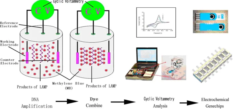
On the microfluidic gene chip, due to high difficulty in temperature changes frequently and products detecting equipment miniaturize, the conventional methods of DNA detection can’t meet the requirements. In this paper, a newly electrochemical method, cyclic voltammetry, basing on a set of special electrodes and the Loop-mediated isothermal amplification (LAMP), was introduced. The DNA amplification products of LAMP could be combined with the positive dye (Methylene blue), which leading to a reduction in the oxidation peak current (ipA) and reduction peak current (ipC) of the cyclic voltammetry. The changes of ipA/ipC were real-time measured by the special electrodes, so the copies of DNA were quantitative detected. The results show that it could be completed in 30~60 minutes with the lowest DNA to 10 CFU.mL-1, with high accuracy (96.5%), high sensitivity (96.0%), high specificity (97.0%) compare to the PCR, and good anti-interference ability against Vitamin C (up to 32mg.L-1) and Aspirin (up to 64mg.L-1) within the sample. Therefore, it was a rapid, sensitive and stable method of DNA detection, has great potential in applying on the microfluidic gene chips.
Keywords: Cyclic voltammetry, DNA detection, Electrochemical detection, Microfluidic gene chip
1. INTRODUCTION
However, due to high difficulty in temperature changes frequently and products detecting equipment miniaturize, the conventional methods of DNA detection can’t meet the requirements of the microfluidic gene chip.
The Loop-mediated isothermal amplification (LAMP) was invented by Dr. Notomi in 2000 [6], have made progress in detections of virus [7], bacteria [8], parasites [9], food safeties [10] and animal embryo sex identifications [11], characterized of rapid, high specificity and high sensitivity, without the temperature changes frequently, is suitable for the DNA detection on the microfluidic gene chip [12]. There were several determination methods of LAMP. The electrophoresis method was a common judgment [13]. The rapid electrophoresis and microchip electrophoresis promoted the level of the electrophoresis [14]. The dyes, such as Hydroxy naphthol blue [15], Calcein [16], and SYBR Green I [17] were applied in LAMP as a qualitative indicator. The turbidimetric method was another common judgment [18]. However, all of above methods required optical-electrical devices to transformate and output the results, which were expensive and bulky. It was not suitable for the construction on the gene chip. Thus, the electrochemical detection of DNA maybe becomes the better choice [19].
Researchers reported that electrochemical DNA sensor had been applied for the rapid and sensitive detection of Salmonella [20]. In the early studies of our laboratory, we have established an electrochemical method, real-time resistance measurement, for amplification and detection of DNA [21]. Compared with the real-time resistance measurement, the cyclic voltammetry method is more specific, with better anti-interference ability, more suitable for DNA detection on gene chip. The electrochemical method is fast and cheap and allows measurements under a variety of experimental conditions [22]. The electrocatalytic reduction of sevoflurane has been carried out at a platinum electrode using cyclic voltammetry [23]. The cyclic voltammetry at electrodes composed of multiple electroactive materials, where zones of one highly active material are distributed over a substrate of a second, less active material, is investigated by simulation [24]. Use of the nanogapped electrode has led to detect very small electrical signals, and therefore it was attempted for DNA sensor applications [25].
Figure 1. The scheme of the DNA electrochemical detection on the microfluidic gene chip
2. EXPERIMENTAL 2.1. Reagents
The specifical nucleotide sequences of E. coli O157:H7 were retrieved from Genbank of National Center for Biotechnology Information (NCBI, USA) as target genes. The E. coli O157:H7 were obtained from American Type Culture Collection(ATCC35150, USA). The cyclic voltammetry primers were designed by PrimerExplorer 4.0 online (Eiken,Japan), and the PCR primers were designed by PrimerPremier 5.0 (Premier, Canada). All primers were synthesized by Sangong (China) and dissolved in buffer (10 mM Tris-HCl, pH 8.0). The LAMP reagents were purchased from Eiken Chemical (Japan). The DNA extraction reagents were provided by Tiangeng (China). The PCR reagents were provided by Eurogentec Deutschland GmBH (Germany). The dyes (Malachite green, Crystal violet, Methylene blue, SYBR green I, Hydroxy naphthol blue), Vitamin C, and Aspirin were obtained from Sigma-Aldrich (USA).
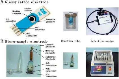
2.2. Electrode and Apparatus
[image:3.596.94.501.74.265.2]
Figure 2. The electrodes and apparatus of cyclic voltammetry method for DNA detection.
2.3. Optimization of the Cyclic Voltammetry Measurement
Five dyes (Malachite green, Crystal violet, SYBR green I, Hydroxy naphthol blue and Methylene blue) were applied synchronously with the same concentration (1.0 mg.L-1) to be compared of their cyclic voltammetry characteristics. Then the best dye was elected to applied in a series of concentration (0, 0.05, 0.1, 0.2, 0.5, 1.0, 2.0, 5.0, 10.0mg.L-1), to choose the best concentration applying in the follow-up measurements.
2.4. DNA amplification and detection
[image:4.596.86.507.73.353.2]
2.5. Sensitivity analysis of the method
Solution of E. coli was diluted into a serial of concentrations (from 1×10⁶ to 1×100 CFU.mL-1), extracted of DNA, and cyclic voltammetry detected in the same way. The ipA/ipC of cyclic voltammetry and the correlation between ipA/ipC with the copies of template DNA were analyzed in OriginPro 7.5 software.
2.6. Comparative analysis of the Cyclic Voltammetry Measurement with the PCR
200 samples (including 100 cases of E. coli and 100 cases of negative samples) were divided into two parts by each specimen. One part was identified by cyclic voltammetry, and the other part was amplificated by PCR in E-cycler 96 (Agilent, USA). The PCR assay using 25 μL multiplex-PCR mixture contained end concentration of 2.0 μmol.L-1 of the primers F and B, and 12.5 μL of the 2 × Fast qPCR MasterMix and a 2.5 μL of the template DNA. Thermal cycling conditions comprised a uracil-n-glycosylase (UNG) step at 50 °C for 2 min, a hot start DNA polymerase activation at 95 °C for 5 min, 40 cycles of denaturation at 95 °C for 3 s, an annealing at 64 °C for 30 s, and an extension at 72 °C for 10 s. The products were analyzed by electrophoresis on 2% agars gels (Biowest, Spain) at 100V for 50 minutes, and visualized in a Kodak Gel Logic 212 PRO (Eastman Kodak, USA). The results of the cyclic voltammetry and the PCR were compared of its difference, accuracy, sensitivity and specificity by Excel 2003 software.
2.7. Anti-interference ability of the Cyclic Voltammetry Measurement
This experiment were basing on the detection of redox characteristics of dyes in cyclic voltammetry. Therefore, the redox species in the samples probably have interference to the measurement. Two kinds of interference substances, Vitamin C (0, 2, 4, 8, 16, 32, 64, 128 mg.L-1,) and Aspirin (0, 2, 4, 8, 16, 32, 64, 128 mg.L-1), referring to the preliminary reports [29, 30], were added in the specimens before the DNA extraction and cyclic voltammetry measurement, to determine the anti-interference ability of the method. The same parameters as above were applied in the DNA extraction and cyclic voltammetry measurement.
3. RESULTS AND DISCUSSION
3.1. The primers design of Cyclic Voltammetry and PCR
Table 1. The primers of cyclic voltammetry and PCR. (F3: forward primer; B3: backward primer; FIP: forward inner primer; BIP: backward inner primer; F: forward primer; B: backward primer) Method Primer(5’→3’)
Cyclic Voltammetry
F3: CCAACCAAGATCCTCAGC
B3: AAAGATGTTTTTCACACTTATTG
FIP: CAAGGCCAGTTTTTTACCCTGTCGGTGCTTTTGATATTTTTCCG
BIP: CGATGAGTTTATCTGCAAGGTGAGTCTCAATTCTAACTAGGACCG
PCR F: CCAACCAAGATCCTCAGCTATAG
B: AAAGATGTTTTTCACACTTATT
Hara-Kudo et al [31] reported that the LAMP had been applied in the detection of DNA, as a rapid and sensitive method for the detection of E. coli O157 and O26. The LAMP assay as a molecular method for detecting the E. coli in enrichment culture is similar or superior to immunomagnetic separation using agar media (IMS-plating method).
Elizaquivel et al [32] demonstrate that the PCR is a suitable technique for the detection and quantification of viable pathogens and had been applied as routine tests in clinical laboratory .
Thus, the LAMP measured by the cyclic voltammetry was applied in the detection of E. coli O157:H7, and the PCR was used as the reference method in this paper.
3.2. Optimization of the Cyclic Voltammetry Measurement
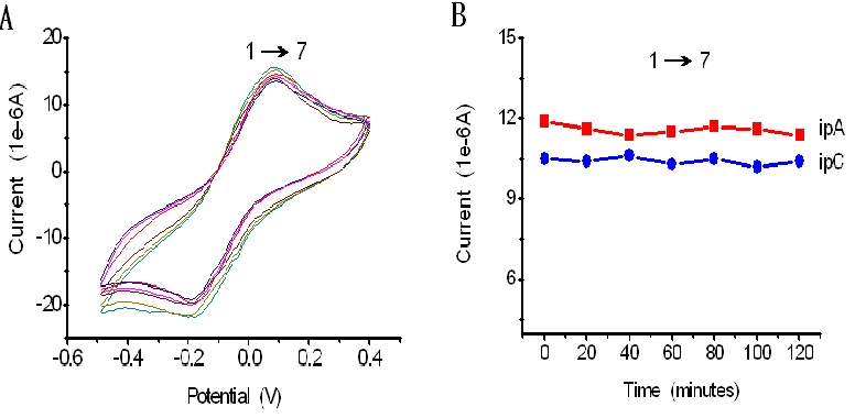
The preliminary characterization of electrode was shown in Figure 2-A, B. It was shown that the electrodes of cyclic voltammetry measurement were worked stably under 65℃ in 2 hours.
Five dyes were applied in the same concentration (1.0mg.L-1) to compares of their reaction characteristics. It shows that the Methylene blue had more efficiency than others (Figure 3-A). The results are more obviously through ipA/ipC (Figure 3-B).
Figure 3. Characterization of the five dyes in the cyclic voltammetry. (1:Methylene blue; 2:Malachite green; 3:Crystal violet; 4:SYBR green I; 5:Hydroxy naphthol blue; all in 1.0 mg.L-1)
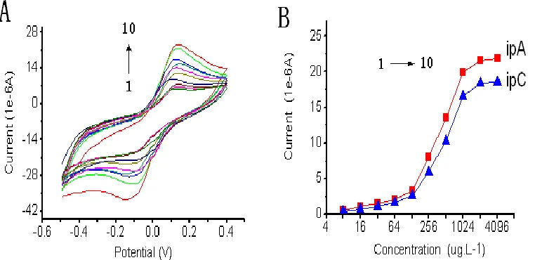
The Methylene blue was elected to applied in a series of concentrations (Figure 4-A). The results shown that it worked best in 1024~4096 μg.L-1 (Figure 4-B). Therefore, the Methylene blue was elected to apply in 2.0 mg.L-1 (middle concentration) in the follow tests.
Figure 4. The optimum concentration of Methylene blue in the cyclic voltammetry. (1:8 μg.L-1; 2:16 μg.L-1
; 3:32 μg.L-1; 4:64 μg.L-1; 5:128 μg.L-1; 6:256 μg.L-1; 7:512 μg.L-1; 8:1024 μg.L-1; 9:2048 μg.L-1
;10:4096 μg.L-1 of Methylene blue)
[image:7.596.102.494.129.313.2] [image:7.596.108.489.447.632.2]
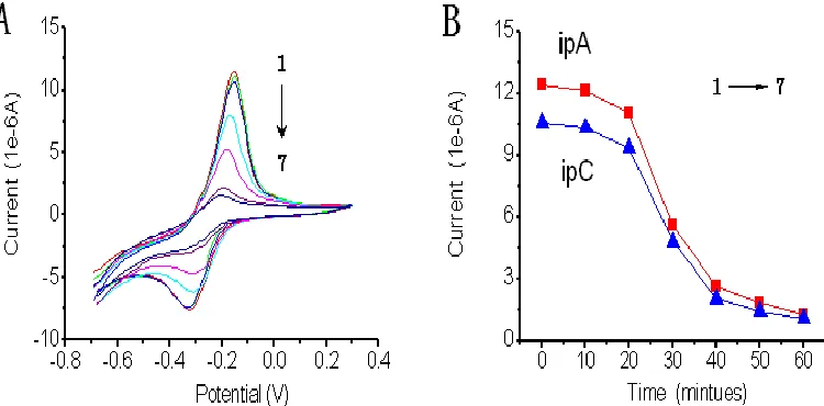
Figure 5. The DNA cyclic voltammetry measurement of E. coli O157:H7. (1→7: Cyclic voltammetry measurement every 10 minutes; E. coli O157:H7, 105 CFU.mL-1)
On the microfluidic gene chip, due to high difficulty in temperature control and products detection, the conventional methods cannot meet the requirements of the DNA detection. The electrochemical detection of DNA maybe becomes the better choice.
The cyclic voltammetry coupled with UV/VIS spectroscopic techniques were used to detection of double stranded DNA (ds-DNA) has been reported by Hajian et al [33], In the electrochemical study of Neutral red, the appearance of a pair redox peaks with DEp = 3.0 mV shows the adsorption process. The decreasing of peak currents for neutral red by addition of ds-DNA reveals a high strength binding between neutral red and DNA (Kb = 2.74 ×10⁴).
Gansen et al [34] had reported a digital loop-mediated DNA amplification (dLAMP) in a sample self-digitization (SD) chip. Digital DNA amplification has become an attractive technique to quantify absolute concentrations of DNA in a sample. He had demonstrated accurate quantification of relative and absolute DNA concentrations with sample volumes of less than 2 μL.
In this paper, the DNA was amplificated through LAMP reaction and the DNA amplification products were combined with positive dye (such as Methylene blue), which leading to a reduction of free dye and could be measured by the cyclic voltammetry methods. The reduction of ipA/ipC of the cyclic voltammetry is proportional to the amount of DNA, so the copies of template DNA was quantitative detected. We anticipate that the microfluidic gene chip basing on LAMP make it attractive as an inexpensive and easy to operate device for DNA detection, for example in point-of-care tastings.
3.3. Sensitivity of the Cyclic Voltammetry Measurement
[image:8.596.107.482.77.262.2]
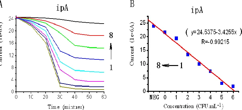
Figure 6. The sensitivity of the cyclic voltammetry measurement. (1:106 CFU.mL-1; 2:105 CFU.mL
-1
;3: 104 CFU.mL-1; 4: 103 CFU.mL-1; 5: 102 CFU.mL-1; 6:101 CFU.mL-1; 7: 100CFU.mL-1of E coli; 8:blank)
The LAMP assay is a sensitive method of DNA detection, lowest to 101~102 CFU.mL-1,higher than conventional PCR (103~104 CFU.mL-1). Shan et al [35] reported that the detection limits of the LAMP for hlyA gene were 6 colony forming units (CFU)/tube. The sensitivity in detection of L. monocytogenes by the LAMP was higher than that of PCR and none of the conventional method-positive samples was missed by the LAMP method.
Tang et al [36] had reported a sensitive reverse-transcription loop-mediated isothermal amplification (RT-LAMP) assay was developed for the rapid detection of Tembusu virus (TMUV) infection. The reaction was performed in one step in a single tube at 64 ℃ for 45 minutes, with SYBR Green I dye added prior to amplification. The detection limit of the RT-LAMP assay was approximately 10 copies.mL-1, and no cross-reaction with other avian viruses was observed. The assay was evaluated further for the diagnosis of TMUV in field samples and compared with conventional RT-PCR. The RT-LAMP with SYBR Green I dye was shown to be a sensitive, simple assay for the rapid diagnosis of TMUV infection.
In this study, the electrochemical detection of DNA, the cyclic voltammetry measurement, could be completed in 30~60 minutes, with high sensitivity (lowest to 10 CFU.mL-1), exactly consistent with the reports in these literatures. It had a good relationship between the DNA concentration (logarithm) and the ipA/ipC over a range of 1×101 to 1×10⁶ CFU.mL-1 (y=24.5375-3.4255x, R=-0.99215). Further more, the concentration of DNA in samples can be quantified. By establishing the standard curve, we could detected the DNA quantitatively.
3.4. Comparison with PCR
[image:9.596.109.494.75.250.2]
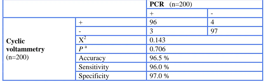
Table 2. Comparison of the cyclic voltammetry and PCR
PCR (n=200)
+ -
Cyclic voltammetry (n=200)
+ 96 4
- 3 97
X2 0.143
P a 0.706
Accuracy 96.5 %
Sensitivity 96.0 %
Specificity 97.0 %
a
There is no statistical difference between the two methods (P>0.05)
Paris et al [3] reported a LAMP method for molecular detection of Plasmodium falciparum in 115 Bangladeshi in-patients with fever. The DNA extraction for LAMP was conducted by conventional methods or simple heating of the sample; test results were either assessed visually or by gel electrophoresis. Conventional DNA extraction followed by gel electrophoresis had the highest agreement with the reference method (81.7%, kappa = 0.64), with a sensitivity (95% ) of 76.1% (68.3~83.9%), a specificity of 89.6% (84.0~95.2%) comparable to rapid diagnostic test (RDT) and microscopy. The LAMP enables molecular diagnosis in settings with limited technical resources but will need further optimization.
In our study, it also shown that the cyclic voltammetry method had a high accuracy (96.5%), sensitivity (96.0%), and specificity (97.0%), compared to standard PCR method. Compared with the PCR, the cyclic voltammetry of LAMP for the detection of DNA is a simple method that could detected DNA with high specificity, sensitivity, and rapidity, more suitable for gene chip operation. It could replace the PCR in clinically used for the detection of DNA.
3.5. Anti-interference ability of the strategy
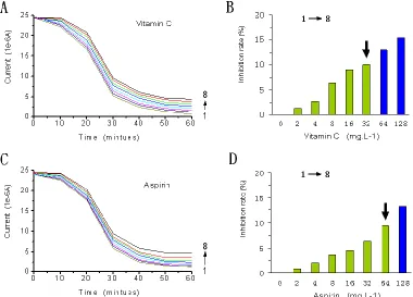
Figure 7. The anti-interference of the cyclic voltammetry against Vitamin C (A, B) and Aspirin (C, D). (1:0 mg.L-1; 2:2 mg.L-1; 3:4 mg.L-1; 4:8 mg.L-1; 5:16 mg.L-1; 6:32 mg.L-1; 7:64 mg.L-1; 8:128mg.L-1 ; Vitamin C and Aspirin); Inhibition rate (%)=1-ipA(interference)/ipA(control)
Vitamin C is one ingredient in blood of healthy human. Dherani et al [37] reported that the normal reference of Vitamin C was 17~34 μmol.L-1 (3~6 mg.L-1
). The results shown that the cyclic voltammetry measurements had good anti-interference ability against Vitamin C up to 32 mg.L-1, as the arrows shown in Figure 7-B, far more than the normal concentration of vitamin C in human blood, so the results are reliable.
Aspirin is not present in normal human blood, so its interference of DNA detection only exist while large dose of Aspirin be applied in therapy. Tehrani et al [38] reported that treatment with 75~ 320 mg/day Aspirin during 4 weeks in a crossover fashion. The reference value of Aspirin in blood was 2~10mg.L-1. The results shown that the cyclic voltammetry measurements had good anti-interference ability against Aspirin up to 64mg.L-1, as the arrows shown in Figure 7-D, far more than the normal concentration of Aspirin in human blood, the interference can be eliminated.
Therefore, the cyclic voltammetry measurement had good anti-interference ability against the redox species (such as Vitamin C and Aspirin) within the sample, could provide accurate data for the clinical detection of DNA.
4. CONCLUSIONS
[image:11.596.104.484.74.347.2]
high specificity and high sensitivity. The scientists had been paid more attentions to PCR or LAMP reacted on the microfluidic gene chip. However, it required optical-electrical devices to transformate and output the results, which were expensive and bulky. It was not suitable for the construction on the gene chip. Thus, the electrochemical detection of DNA maybe becomes the better choice. In this paper, an electrochemical method, cyclic voltammetry measurement, was proposed for the detection of DNA on the microfluidic gene chip. First, the DNA extracted from specimens was amplificated through LAMP reaction. The DNA amplification products were combined with positive dye, which leading to a reduction of free dye and could be measured by the cyclic voltammetry methods. The reduction of ipA/ipC of the cyclic voltammetry is proportional to the amount of DNA, so the copies of template DNA was quantitative detected.
The results displayed that this electrochemical detection of DNA could be completed in 30~60 minutes, with high sensitivity (lowest to 10 CFU.mL-1). The result of X2 test (P>0.05) suggested that there hadn’t significant difference between the cyclic voltammetry and the PCR. It also was shown that the cyclic voltammetry had high accuracy (96.5%), sensitivity (96.0%) and specificity (97.0%) compared to standard PCR method. It also had good anti-interference ability against Vitamin C (up to 32mg.L-1) and Aspirin (up to 64mg.L-1) within the sample. It had a good relationship between the DNA concentration (logarithm) and the ipA/ipC over a range of 1×101 to 1×10⁶ CFU.mL-1 (y=24.5375-3.4255x, R=-0.99215). Further more, the concentration of DNA in samples can be quantified. By establishing the standard curve, we could detected the DNA quantitatively. Therefore, the cyclic voltammetry measurement was a rapid, sensitive and stable method. It would provide a powerful tool for DNA detection on gene chips, pocket instruments, or other fields which needed the simple, rapid, and real-time methods of measuring.
ACKNOWLEDGEMENTS
This work was supported by the research project of Chongqing Science and Technology Commission (CSTC,2011AB5035). We appreciate the valuable comments from other members of our laboratories. References
1. K. Verstraete, J. Robyn, J. Del-Favero, P. De Rijk, M.A. Joris, L. Herman, M. Heyndrickx, L. De Zutter, K. De Reu. Food Microbiol., 29 (2012) 49
2. N. Nagatani, K. Yamanaka, H. Ushijima, R. Koketsu, T. Sasaki, K. Ikuta, M. Saito, T. Miyahara, E. Tamiya. Analyst, 137 (2012) 3422
3. D.H. Paris, S.D. Blacksell, P. Nawtaisong, K. Jenjaroen, A. Teeraratkul, W. Chierakul, V. Wuthiekanun, P. Kantipong, N.P. Day. PLoS Negl. Trop. Dis., 5 (2011) e1307
4. C.G. Cooney, D. Sipes, N. Thakore, R. Holmberg, P. Belgrader. Biomed. Microdevices, 14 (2012) 45
5. N. Pak, D.C. Saunders, C.R. Phaneuf, C.R. Forest. Biomed. Microdevices, 14 (2012) 427
6. T. Notomi, H. Okayama, H. Masubuchi, T. Yonekawa, K. Watanabe, N. Amino, T. Hase. Nucleic Acids Res., 28 (2000) E63
7. X. Wang, J.P. Zhu, Q. Zhang, Z.G. Xu, F. Zhang, Z.H. Zhao, W.Z. Zheng, L.S. Zheng. J. Virol. Methods, 179 (2012) 330
8. H.J. Han, S.J. Jung, M.J. Oh, D.H. Kim. J. Fish Dis., 34 (2011) 395
11. H. Nogami, H. Tsutsumi, T. Komuro, R. Mukoyama. Forensic. Sci. Int. Genet., 2 (2008) 349 12. V. Tjong, H. Yu, A. Hucknall, S. Rangarajan, A. Chilkoti. Anal Chem., 83 (2011) 5153
13. C.A. Le Roux, T. Kubo, A.A. Grobbelaar, P.J. van Vuren, J. Weyer, L.H. Nel, R. Swanepoel, K. Morita, J.T. Paweska. J. Clin. Microbiol., 47 (2009) 645
14. H. Iseki, A. Alhassan, N. Ohta, O.M. Thekisoe, N. Yokoyama, N. Inoue, A. Nambota, J. Yasuda, I. Igarashi. J. Microbiol. Methods, 71 (2007) 281
15. K. Nie, Y. Zhang, L. Luo, M.J. Yang, X.M. Hu, M. Wang, S.L. Zhu, F. Han, W.B. Xu, X.J. Ma. J. Virol. Methods, 175 (2011) 283
16. S.L. Wastling, K. Picozzi, A.S. Kakembo, S.C. Welburn. PLoS Negl. Trop. Dis., 4 (2010) e865 17. X.J. Zhang, Q.Y. Han, Y. Sun, S. Belak, L. Liu, H.J. Qiu. J. Virol. Methods, 171 (2011) 200 18. N. Tomita, Y. Mori, H. Kanda, T. Notomi. Nat. Protoc., 3 (2008) 877
19. M. Del Carlo, M. Di Marcello, M. Giuliani, M. Sergi, A. Pepe, D. Compagnone. Biosens. Bioelectron., 31 (2012) 270
20. Q. Li, W. Cheng, D.C. Zhang, T.X. Yu, Y.B. Yin, H.X. Ju, S.J. Ding. Int. J. Electrochem. Sci., 7 (2012) 844
21. D.N. Jiang, G.M. Xiang, J.H. Wu, C. Liu, F. Liu, X.Y. Pu. Int. J. Electrochem. Sci., 7 (2012) 5273 22. J.F. Arteaga, M. Ruiz-Montoya, A. Palma, G. Alonso-Garrido, S. Pintado, J.M. Rodriguez-
Mellado. Molecules, 17 (2012) 5126
23. J. Gonzalez, A. Molina, C.M. Soto, C. Serna. J. Electroanal. Chem., 664 (2012) 53
24. K.R. Ward, N.S. Lawrence, R.S. Hartshorne, R.G. Compton. Phys. Chem. Chem. Phys., 14 (2012) 7264
25. Y. Liu, J. Chen, N.T. Anh, C.O. Too, V. Misoska, G.G. Wallace. J. Electrochem. Soc., 155 (2008) K100
26. Y.J. Xu, X.H. Tian, X.Z. Zhang, X.H. Gong, H.H. Liu, H.J. Zhang, H. Huang, L.M. Zhang. J. Chromatogr. Sci., 50 (2012) 591
27. L. Chen, L. Zhang, T. Qiu, W. Cao. Int. J. Electrochem. Sci., 6 (2011) 5325
28. P. Norouzi, M. Pirali-Hamedani, M.R. Ganjali, F. Faridbod. Int. J. Electrochem. Sci., 5 (2010) 1434
29. S. Maggini, S. Beveridge, M. Suter. J. Int. Med. Res., 40 (2012) 28
30. S. Tehrani, A. Antovic, F. Mobarrez, K. Mageed, P.E. Lins, U. Adamson, H.N. Wallen, G. Jorneskog. Diabetes Care, 35 (2012) 404
31. Y. Hara-Kudo, N. Konishi, K. Ohtsuka, R. Hiramatsu, H. Tanaka, H. Konuma, K. Takatori. Int. J. Food Microbiol., 122 (2008) 156
32. P. Elizaquivel, G. Sanchez, R. Aznar, Food Control., 25 (2012) 704
33. R. Hajian, N. Iravani, F. Ghanbari, N. Shams, Asian J. Chem., 24 (2012) 3009
34. A. Gansen, A.M. Herrick, I.K. Dimov, L.P. Lee, D.T. Chiu, Lab Chip, 12 (2012) 2247
35. X.X. Shan, Y.Q. Zhang, Z.G. Zhang, M.R. Chen, Y.Y. Su, Y.N. Yuan, M.J. Alam, H. Yan, L. Shi, Food Sci. Biotechnol., 21 (2012) 101
36. Y. Tang, Y. Diao, C. Yu, X. Gao, L. Chen, D. Zhang, Transbound. Emerg. Dis., 59 (2012) 208 37. M. Dherani, G.V.S. Murthy, S.K. Gupta, I.S. Young, G. Maraini, M. Camparini, G.M. Price, N.
John, U. Chakravarthy, A.E. Fletcher, Invest. Ophth. Vis. Sci., 49 (2008) 3328
38. S. Tehrani, A. Antovic, F. Mobarrez, K. Mageed, P.E. Lins, U. Adamson, H.N. Wallen, G. Jorneskog, Diabetes Care, 35 (2012) 404