A structure-based method for derivation
of all-atom potentials for protein folding
The Harvard community has made this
article openly available. Please share how
this access benefits you. Your story matters
Citation
Kussell, E., J. Shimada, and E. I. Shakhnovich. 2002. “A
Structure-Based Method for Derivation of All-Atom Potentials for Protein
Folding.” Proceedings of the National Academy of Sciences99 (8):
5343–48. https://doi.org/10.1073/pnas.072665799.
Citable link
http://nrs.harvard.edu/urn-3:HUL.InstRepos:41534342
Terms of Use
This article was downloaded from Harvard University’s DASH
repository, and is made available under the terms and conditions
applicable to Other Posted Material, as set forth at
http://
A structure-based method for derivation of all-atom
potentials for protein folding
Edo Kussell*, Jun Shimada†, and Eugene I. Shakhnovich†‡
*Department of Biophysics, Harvard University, 240 Longwood Avenue, Boston, MA 02115; and†Department of Chemistry and Chemical Biology,
Harvard University, 12 Oxford Street, Cambridge, MA 02138
Edited by Alan Fersht, University of Cambridge, Cambridge, United Kingdom, and approved January 28, 2002 (received for review December 13, 2001)
A method for deriving all-atom protein folding potentials is pre-sented and tested on a three-helix bundle protein, as well as on hairpin and helical sequences. The potentials obtained are com-posed of a contact term between pairs of atoms, and a local density term for each atom, mimicking solvent exposure preferences. Using this potential in an all-atom protein folding simulation, we repeatedly folded the three-helix bundle, with the lowest energy conformations having a C␣distance rms from the native structure of less than 2 Å. Similar results were obtained for the hairpin and helices by using different potentials. We derived potentials for several different proteins and found a high correlation between the derived parameters, suggesting that a potential of this form eventually could be found that folds multiple, unrelated proteins at the atomic level of detail.
T
he problem of deriving a potential to fold proteins has been fully solved only in simple lattice models (1, 2). Most off-lattice models that are presently used to obtain complete folding trajectories use a potential, known as a Go potential, tailored to give lowest energy to the native state of a single protein (3). Because Go potentials are actually potentials de-fined on structures rather than on sequences, one cannot use them to try to fold sequences of unknown structure or even to fold two different sequences by using the same parameters. Nevertheless, Go potentials are useful for examining possible protein-folding pathways for a single protein (4–7) and for testing a particular model’s computational feasibility.In contrast, sequence-based potentials give a set of parameters that can be used to simulate any amino acid sequence. A sequence-based potential consists of an atom-typing scheme and a set of parameters giving the energetics of contacts between atom types. Given any amino acid sequence, the atom-typing scheme is used to assign a type to each atom of the sequence, and using the interaction parameters the simulation can then be performed. Recently, several groups have proposed off-lattice models using sequence-based potentials that can in principle be used to simulate any sequence (8–11). It is unclear at this time whether the simplified representation of proteins used in those models will eventually lead to correct folding of many proteins of various topologies to an acceptable level of accuracy.
In this article, we chose to work with a detailed representation that explicitly models all atoms other than hydrogen. All side-chainangles and backbone兾angles are free to rotate. At this level of detail, the basic problem of stabilizing the correct topology is further complicated by the conformational flexibility of the side chains, which can assume many different conforma-tions consistent with a given backbone conformation (12–14). The advantages of this model are that (i) packing effects caused by diverse side-chain shapes are present (14), (ii) side-chain entropy is properly accounted for, and (iii) difficulties resulting from an oversimplified protein representation are largely elim-inated, leaving only the underlying potential as a possible source of error. We have previously shown that simulations of this model using the structure-based Go potential, starting from random coils, reach the native state by a cooperative transition in a reasonable amount of computational time (6).
Finding sequence-based potentials that fold even a single protein has proven to be a difficult problem. There is no consensus on which types of potentials should be investigated, and several papers have demonstrated that the problem has no solution for certain types of potentials (15, 16). It is therefore important to focus effort on potentials of the correct form. In the present article, we derive a sequence-based potential that re-peatedly folds a single protein in the all-atom representation. The potential-derivation procedure is structure-based, that is, we find energetics tailored to fold a particular structure. The potential form, however, is sequence-based, and, unlike Go
parameters, the parameters we obtain can be used to simulate any sequence. Whether or not those same parameters would properly fold other sequences is a separate question, which we do not address here. We present data showing significant correlation between parameters derived for several different proteins, suggesting that a transferable potential of this form is likely to exist.
Methods
Density-Dependent Energy Term.We defined two atoms, A and B,
to be in contact if the distance between them was less than
(rA⫹rB), whererAandrBare their respective van der Waals
radii. We took⫽1.8 and used radii as in previous work (6). Atomic hard sphere radii were taken to be 0.75 of their van der Waals sizes (6). To each atom type A, we assigned an ideal number of contacts, nA, and a corresponding energy term
penalizing deviations from this number: if an atom of type A makesncontacts, it receives an energy ofEA⫽ ⱍn⫺nAⱍ. The
numbers nA were determined by averaging the number of
contacts,n, made by all atoms of type A in a given structure. If max(n)⫺min(n)⬎4 for a given atom type in a protein, it did not receive a density-dependent energy.
Contact Potential Derivation.For each pair of atom types A and B
in a given protein structure, we calculated NAB and N˜AB,
respectively the number of A⫺B pairs in contact and the number of A⫺B pairs not in contact. We assigned energyEABto an A⫺B
contact as follows:
EAB⫽⫺
NAB⫹共1⫺兲N˜AB NAB⫹共1⫺兲N˜AB .
[1]
Note that ifNAB⫽0 for a pair of types, thenEAB⫽1 regardless
of. The parameteris needed because the number of pairs not in contact,N˜AB, is always far larger than the number of native
contacts,NAB. If we were to take ⫽ 0.5, EAB would be an
averaging of attractive native contacts, with a value ⫺1, and repulsive non-native contacts, with a value of⫹1. Practically all
This paper was submitted directly (Track II) to the PNAS office.
Abbreviation: drms, distance rms.
‡To whom reprint requests should be addressed. E-mail: [email protected].
The publication costs of this article were defrayed in part by page charge payment. This article must therefore be hereby marked “advertisement” in accordance with 18 U.S.C. §1734 solely to indicate this fact.
values ofEABwere strongly repulsive under this⫽0.5 scheme,
so we had to give more weight (0.5⬍⬍1) to native contacts.
Combined Potential and Simulation. The contact potential and
density terms were combined as follows to give the protein folding potential,U:
U⫽␣
冘
i⬍j
EAiAj⫹共1⫺␣兲
冘
i
EAi, [2]
where Aiis the atom type of atomiof the protein. We used␣⫽
0.3, postponing systematic derivation of this parameter to future work. We turned off all side chain–backbone contacts, all local side chain–side chain contacts (up toiandi⫹2), and all local backbone–backbone contacts (up to i and i ⫹ 3). All-atom protein representation and Monte Carlo simulation were taken from previous work (6). Simulations were run at Monte Carlo temperatures between 0.4 and 0.44, at which move acceptance rates in the native state were approximately 20%. The distance rms (drms) between two structures is given by (兺(rij⫺Rij)2)1/2,
whererijis the distance between atomsiandjof one structure (and correspondinglyRijfor the other structure). We computed drms values by using only C␣atoms.
Results and Discussion
Folding Helices: Interaction with Implicit Solvent.Peptide backbones
are comprised of four heavy atom types: the carbonyl carbon (C) and oxygen (O), the amide nitrogen (N), and the␣-carbon (C␣).
Because thei⫺i⫹4 backbone hydrogen bonds are a distinct feature of ␣-helices, we thought that by introducing a strong hydrogen bonding-like interaction, we would be able to make an
␣-helical conformation the ground state for any peptide. A hydrogen bond would minimally require the NOO distance to be within 3.5 Å, and we initially thought that giving strong prefer-ence only to NOO contacts would result in a helical conforma-tion. We quickly found, however, that this was not the case: many locally crumpled, nonhelical conformations were found that had lower energy than the helical conformation. O (and N) atoms in these nonhelical conformations made contacts with multiple N
(and O) atoms. Given that the hydrogen bond is known to be strongly directional (17, 18), this naive contact potential appears to have failed because it lacked orientational specificity.
We found two ways of correcting this problem. The first method defined a potential based on four distances between backbone atoms:dO⫺N, dO⫺C, dC-amide H, and dO⫺amide H. The
potential was tuned on a database of representative protein structures, including many  and ␣兾 proteins (see Fig. 1B). Implementing only this hydrogen bonding potential, we were able to fold all peptides into helices. We were able to repeat this result by replacing some of the distances with angles (e.g., NOOOC), but opted for distances for reasons of computational efficiency. Using a potential parametrized by less than four distances led to nonhelical ground states, which is not surprising given that four independent spatial constraints are required to determine the orientation of two vectors.
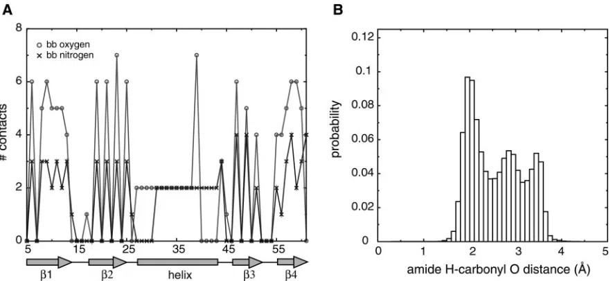
While the resulting hydrogen bond is strongly directional, and emerges naturally from a knowledge-based procedure, it is computationally expensive, slowing down simulations by at least 50%. The second method we developed is less direct, but gives equally good helix formation, at low computational cost. We recorded the number of contacts made by each backbone atom of a given protein. In Fig. 1A, we plot the number of backbone contacts made by backbone nitrogen (N) and oxygen (O) atoms at each position in protein G. We see that the helical region of the protein shows a strong signal in the number of contacts made by backbone N and O atoms: each one makes exactly two contacts when present in the helix, whereas they can make between zero and seven contacts when present in strands or loops. Other proteins yielded similar data. We therefore intro-duced an energetic term, acting on each atom, which would penalize deviations from the ideal number of contacts made by that atom (seeMethods). The ideal number of contacts depended on the atom type. We found that helices could be formed by simply setting the ideal number of contacts for O and N atoms to be two and adding an attractive contact interaction between O and N.
[image:3.630.97.541.58.261.2]Both potentials give good helices because they introduce a
Fig. 1. Statistics for hydrogen-bonding potentials. (A) Number of backbone– backbone contacts made by backbone oxygen and nitrogen atoms at each position
of protein G (Protein Data Bank code 1IGD). Local backbone contacts were turned off, so that contacts between residuesi,i⫹1,i⫹2, andi⫹3 were not included for alli.(B) Distribution of distances between backbone amide hydrogen and backbone carbonyl oxygen in proteins. The joint probabilities of observing a particular set of distances,p(dO⫺N,dO⫺C,dC⫺amide H,dO⫺amide H), were obtained from a database of representative protein structures. The database was comprised
of one representative from each homologous family in the Families of Structurally Similar Proteins database (27), giving a total of⬇2,500 structures. Distances were recorded when the CON distance was less than 5 Å. The distribution of O-amide H distances is shown here. The joint probabilities were converted to effective free energies by using the Boltzmann-like relationE⫽ ⫺ln(p). The energies obtained were then used as a potential to fold peptides.
directional hydrogen bond. The second method introduces di-rectionality indirectly by restricting the local density around each atom. This prevents too many N atoms from crowding around a given O atom, and thus strongly destabilizes the decoy confor-mations that plagued the isotropic contact potential. We find this idea attractive because it has a nice physical interpretation as well: the local density term can be thought of as an implicit way of modeling interactions with solvent molecules, which are not present in our model. The density term controls the ideal degree of solvation for a given atom.
Folding a -Hairpin: Deriving the Contact Potential. To explore
possible schemes for contact potential derivation, we started by looking at-hairpin 2 from protein G, which is known to be partially stable in solution (19–22). Because this -hairpin is relatively small and requires a balance between local and non-local interactions to fold properly, it is a good molecule for quickly testing out ideas about potentials.
To define the all-atom contact potential, we introduced an atom typing scheme in which each side-chain atom of each of the 20 amino acids is assigned a separate type. Atoms that are related by symmetry within a single amino acid are assigned the same type. Along with the four backbone atoms, we have a total of 84 atom types. The hairpin, containing a total of 130 atoms, has 44 different atom types by our scheme. Our sequence-based model of the hairpin contains⬇1兾3 the number of atom types as the Go
model would, because in the Go model each of the 130 atoms would constitute a separate type. Because the number of pa-rameters in the model scales as the square of the number of types, the sequence model contains⬇1兾9 the number of param-eters as the Gomodel. This is a very significant reduction in the number of parameters, and it is this reduction that makes the derivation of sequence-based potentials a challenging task. Because the hairpin contains five threonines, two valines, and two aspartic acid residues, there is a significant amount of reuse of the same atom types within this molecule. The problem becomes harder as the size of the protein increases, because its conformational space grows exponentially with length, while the number of types cannot exceed 84.
We used a simple guiding principle in deriving potential parameters: contacts formed in the native structure should be more favorable than contacts that are not seen. If two different
atom types are always found to contact each other, we want to assign a strong attractive interaction between those two types. On the other hand, if a pair of atom types is never seen to contact, we introduce some repulsion between their types.
To implement this idea, we assigned an energyEABto contacts
between atom types A and B, and computedEABas a function
of the number of native A–B contacts made in a given protein structure (seeMethods). If no native A–B contacts were present in the protein structure, we assignedEAB⫽1. A single parameter was introduced to control the distribution of energiesEABfor
A–B pairs that made at least one native contact. For such pairs, when⫽1,EAB⫽ ⫺1, whereas lower values ofintroduce
more repulsion and dispersion of energies.
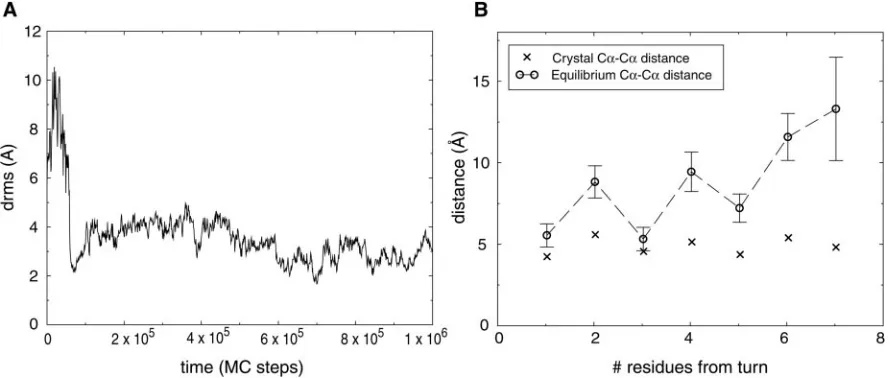
We were able to fold the hairpin repeatedly, as shown in Fig. 2A, by using values ofbetween 0.8 and 0.96. The potential-derivation scheme thus identifies many potentials that can fold a single hairpin. Lowest energy conformations had low drms values, with a typical folded conformation having a drms of⬇2 Å. We found that the hairpin was often found in a frayed-end state, in which the turn was properly formed, but the two ends were somewhat free of each other. This behavior is shown in Fig. 2B. The average distance between hydrogen-bonded residues is plotted as a function of residue number, showing that residues far from the turn tend to be less constrained than residues closer to the turn. This is precisely what is seen in experimental studies of this hairpin (19). The peptide makes brief excursions to fully native hairpin conformations, having lowest energy, and then returns to the entropically more favorable frayed-end states.
Folding a Protein: Putting the Pieces Together. We applied our
potential derivation method to the three-helix bundle protein consisting of the B domain ofStaphylococcus aureusprotein A, which has featured in both experimental (23, 24) and compu-tational (4, 7, 11, 25, 26) studies. For each of the 61 atom types present in this protein, we determined the ideal local density (see
Methods). We derived the contact potential for this protein by using a value of⫽0.98. Because simulation time was consid-erably longer for this protein than for the helices and hairpin, we could not explore a range ofvalues.
[image:4.630.94.539.60.249.2]We ran 40 folding simulations, starting from completely random coils. Nineteen of these runs folded in the allotted
Fig. 2. Folding of-hairpin 2 from protein G. (A) A folding trajectory for the hairpin using a contact potential with⫽0.93 atT⫽0.4. The trajectory was started
from a fully unfolded conformation. (B) The equilibrium C␣-C␣distances between hydrogen-bonded residues of the hairpin atT⫽0.5. Average distances (E) were computed over uncorrelated conformations from a long simulation started at the native hairpin structure taken from protein G. The error bars indicate 1.5 SDs of the distance distribution. X-marks indicate the crystal structure C␣-C␣distances.
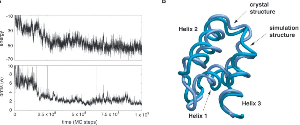
simulation time. Average drms of the folded conformations from the native crystal structure was 2.5 Å. Low energy states always corresponded to low drms conformations, with native topology and secondary structure. A sample trajectory is shown in Fig. 3. The lowest-energy structure from one trajectory, with drms of 1.9 Å, is shown superimposed on the native conformation in Fig. 3.
We found that folding proceeded by several routes. The major pathway consisted of formation of a complex of helices 2 and 3, which formed a scaffold for subsequent formation of helix 1, consistent with previous Gosimulations in a different model (7). Another pathway consisted of formation of helices 1 and 3, in native orientation, followed by slow formation of helix 2. This
pathway was necessarily somewhat slower because it is topolog-ically more difficult for helix 2 to form once helices 1 and 3 are partially stable in their native orientation. A third, rare pathway consisted of formation of a complex of helices 1 and 2, followed by formation of helix 3.
We measured the stabilities of each helix of the protein, by running equilibrium simulations for each one, using the same potential that was used to fold the entire protein. A plot of folding and unfolding transitions for helix 3 simulated at its transition temperature (Tf ⫽0.52) is given Fig. 4A. The
ther-modynamic curve is given in Fig. 4B. Because helix 2 hadTf⫽
0.58, and helix 3 hadTf⫽0.44, we found that helices 2 and 3 were
fully stable for the Monte Carlo temperature range of 0.4 to 0.44
Fig. 3. Folding of a three-helix bundle protein. (A) A folding trajectory started from a fully unfolded conformation of the three-helix bundle protein (B domain
ofStaphylococcus aureusprotein A, Protein Data Bank code 1BDD) using the all-atom sequence-based potential described in the text. The plot shows the time
course of both energy and drms from crystal structure. The trajectory reaches drms values as low as 1 Å. (B) The lowest-energy structure from a folding trajectory superimposed on the native crystal structure. The drms between the two structures is 1.9 Å.
Fig. 4. Folding of the isolated helix 3 from the three-helix bundle protein. (A) A trajectory started from the native conformation of helix 3, run near the helix’s
transition temperature (T⫽0.52). The same all-atom potential that was used to fold the entire protein is used here. Both energy and drms traces show that the helix repeatedly unfolds completely and refolds. (B) Average drms of helix 3 measured over long simulations at various temperatures. The average drms of fully unfolded conformations of the helix is⬇4 Å. Error bars indicate 1.5 SDs of the computed average.
at which simulations of the entire protein were run, whereas helix 1 was only marginally stable. Experiments have shown that, at room temperature, helix 3 is marginally stable, whereas the other two helices are unstable (24). Our Monte Carlo simulation temperature is therefore somewhat below the equivalent of room temperature, and helix stabilities need to be adjusted.
Transferability of Potentials.Having shown that a sequence-based
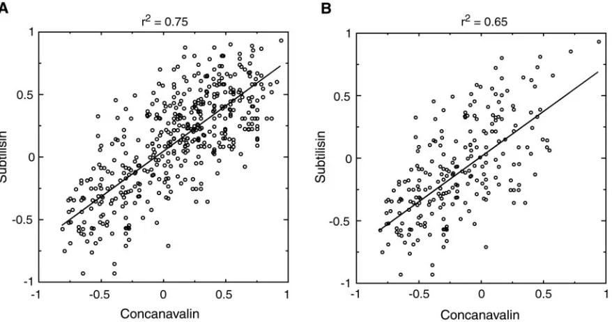
potential can fold an all-atom protein model, the question of transferability remains: does a single potential of this form exist that can fold several different proteins? The method introduced here does, after all, rely on native structure information to derive the sequence-based potential. Although future work will inves-tigate this question in full, we now give a partial answer by comparing the contact potentials derived with several different proteins. Fig. 5Ashows the correlation between parameters of two contact potentials, obtained from the proteins concanavalin and subtilisin. While these proteins have completely different folds and secondary structure content, the contact potentials derived from them have high (r2 ⫽ 0.75) correlation. Similar
results were obtained over representatives of several other folds, such as Rossman and TIM-Barrel.
The high correlation is partially explained by the following observation. Side-chain atom types that are usually found on the surface will make fewer contacts with the backbone in all proteins, whereas hydrophobic side-chain atoms will make more contacts with the backbone. This results in lower variation of our derived side chain–backbone interactions across proteins.
Nev-ertheless, when only side chain–side chain interactions are considered (Fig. 5B), we still obtain significant correlation between potentials (r2⫽ 0.65). The potential we used to fold
protein A also gave significant correlation with potentials from other proteins, but because of its small size, many contact types were not present in its structure. We therefore had to use larger proteins (Fig. 5) to have a meaningful comparison of potentials.
In this article we have shown that a sequence-based potential exists that folds a three-helix bundle protein in an all-atom representation. This finding is a marked departure from previous simulations in that we used a sequence-based potential that contains far fewer parameters than a Go potential. Because of the parameter reduction, the energy landscape for sequence-based folding becomes rougher and less biased toward the native state than in the Go model. Nevertheless, we showed that even in this rougher landscape, folding of a single protein is still possible. The next step is to develop methods that can derive a potential that stabilizes several protein structures simulta-neously. The method given here explored a very small fraction of the total parameter space of potentials and was still able to find separate potentials for folding a hairpin, a helix, and an entire protein. Additionally, we found reasonable correlation between the contact potentials derived from different structures. These observations suggest that by searching a bigger piece of potential space in a well-chosen way one could obtain a trans-ferable potential. This remains a major challenge for computa-tional protein folding.
1. Mirny, L. A. & Shakhnovich, E. I. (1996)J. Mol. Biol.264,1164–1179. 2. Hao, M. H. & Scheraga, H. A. (1996)Proc. Natl. Acad. Sci. USA93,4984–4989. 3. Go, N. & Abe, H. (1981)Biopolymers20,991–1011.
4. Zhou, Y. & Karplus, M. (1999)Nature (London)401,400–403.
5. Clementi, C., Nymeyer, H. & Onuchic, J. N. (2000)J. Mol. Biol. 298, 937–953.
6. Shimada, J., Kussell, E. & Shakhnovich, E. I. (2001)J. Mol. Biol.308,79–95.
7. Berriz, G. F. & Shakhnovich, E. I. (2001)J. Mol. Biol.310,673–685. 8. Pillardy, J., Czaplewski, C., Liwo, A., Lee, J., Ripoll, D. R., Kamierkiewicz, R.,
Oldziej, S., Wedemeyer, W. J., Gibson, K. D., Arnautova, Y. A.,et al. (2001)
Proc. Natl. Acad. Sci. USA98,2329–2333.
9. Simons, K., Strauss, C. & Baker, D. (2001)J. Mol. Biol.306,1191–1199. 10. Kihara, D., Lu, H., Kolinski, A. & Skolnick, J. (2001)Proc. Natl. Acad. Sci. USA
[image:6.630.99.538.60.291.2]98,10125–10130.
Fig. 5. Comparison of all-atom potentials derived from concanavalin (Protein Data Bank code 1NLS) and subtilisin (Protein Data Bank code 1GCI). (A) Each
point represents the contact energy for a pair of atom types that were found to make contacts in both proteins. Contact energies were derived as described
inMethods, using⫽0.995 for concanavalin and⫽0.993 for subtilisin. Note that the appropriate range ofmoves closer to 1 as protein size grows,
because the number of pairs of atoms not in contact grows much faster than the number of pairs in contact. It is clear that these values ofare not too close to 1 because we see a good dispersion of energies over both thexandyaxes. We found good correlation between potentials over a large range of values of, indicating that the trend is not sensitive to errors in. (B) Same asAexcept side chain– backbone contacts were not included in plot. The best-fit lines are shown.
11. Irback, A., Sjunnesson, F. & Wallin, S. (2000)Proc. Natl. Acad. Sci. USA97, 13614–13618.
12. Richards, F. M. & Lim, W. A. (1994)Q. Rev. Biophys.26,423–498. 13. Dunbrack, R. L., Jr. & Cohen, F. E. (1997)Protein Sci.6,1661–1681. 14. Kussell, E., Shimada, J. & Shakhnovich, E. I. (2001)J. Mol. Biol.311,183–193. 15. Vendruscolo, M. & Domany, E. (1998)J. Chem. Phys.109,11101–11108. 16. Tobi, D., Shafran, G., Linial, N. & Elber, R. (2000)Proteins40,71–85. 17. Creighton, T. E. (1993)Proteins(Freeman, New York), 2nd Ed.
18. Jeffrey, G. A. & Saenger, W. (1991)Hydrogen Bonding in Biological Structures
(Springer, Berlin).
19. Blanco, F. J., Rivas, G. & Serrano, L. (1994)Nat. Struct. Biol.1,584–590.
20. Kobayashi, N., Honda, S., Yoshii, H. & Munetaka, E. (2000)Biochemistry39, 6564–6571.
21. Zagrovic, B., Sorin, E. J. & Pande, V. (2001)J. Mol. Biol.313,151–169. 22. Klimov, D. K. & Thirumalai, D. (2000)Proc. Natl. Acad. Sci. USA97,2544–2549. 23. Bottomley, S. P., Popplewell, A. P, Scawen, M., Wan, T., Sutton, B. J. & Gore,
M. G. (1994)Protein Eng.7,1463–1470.
24. Bai, Y., Karmi, A., Dyson, H. J. & Wright, P. E. (1997) Protein Sci.6, 1449–1457.
25. Boczko, E. M. & Brooks, C. L. I.(1995) Science269,393–396.
26. Alonso, D. O. V. & Daggett, V. (2000)Proc. Natl. Acad. Sci. USA97,133–138. 27. Holm, L. & Sander, C. (1996)Nucleic Acids Res.24,206–209.