Replication
K. L. LeMay,aJ. Treadaway,aI. Angulo,bT. L. Tellinghuisena
The Scripps Research Institute, Scripps Florida, Jupiter, Florida, USAa; University of Cambridge, Department of Medicine, Addenbrooke’s Hospital, Cambridge, United
Kingdomb
The hepatitis C virus NS5A protein is essential for RNA replication and virion assembly. NS5A is phosphorylated on multiple residues during infections, but these sites remain uncharacterized. Here we identify serine 222 of genotype 2a NS5A as a phos-phorylation site that functions as a negative regulator of RNA replication. This site is a component of the hyperphosphorylated form of NS5A, which is in good agreement with previous observations that hyperphosphorylation negatively affects replication.
H
epatitis C virus (HCV) is a human pathogen of global impact, with as many as 170 million chronically infected (1). Long-term infection results in progressive liver damage, including fibro-sis, cirrhofibro-sis, and, oftentimes, hepatocellular carcinoma. HCV is a member of theFlaviviridaefamily of enveloped, positive-sense, single-stranded RNA viruses (reviewed in reference2). The non-capped, nonpolyadenylated, 9.6-kb viral genome encodes a single internal ribosome entry site (IRES)-directed open reading frame flanked by highly structured 5= and 3= nontranslated regions. Translation yields a polyprotein of approximately 3,000 amino acids that undergoes a complex series of co- and posttranslational proteolytic events catalyzed by viral and host proteases, resulting in the production of 10 mature HCV proteins. These include the structural proteins that are involved in the production of progeny virions (C, E1, E2, p7) and the nonstructural replication proteins (NS2, NS3, NS4A, NS4B, NS5A, NS5B). There is considerable overlap in functionality between these groups, with a number of nonstructural proteins serving essential functions for both RNA replication and virion assembly, including the NS5A protein (3–11).NS5A is a large, three-domain, hydrophilic phosphoprotein that exists in two distinct forms: a hypophosphorylated form of roughly 56 kDa and a hyperphosphorylated form of 58 kDa (12,
13). It is not known what residues in NS5A are phosphorylated or how various phosphorylation events contribute to the presence of the two phospho-forms (see reference14for a concise review). Evidence from mutagenesis suggests that hypophosphorylation primarily targets serine residues in domains II and III, whereas hyperphosphorylation sites cluster in and around domain I, LCSI (low complexity sequence block I), and domain II (12,15–18). The function of NS5A phosphorylation in the viral life cycle is unknown, but a general theme has emerged in which hypophos-phorylation is required for RNA replication, and hyperphosphor-ylation is a negative regulator of this process. Phosphorhyperphosphor-ylation may also play a role in the function of NS5A in virion assembly, and it could regulate the interface between replication and assem-bly events (11). A number of phosphorylation sites have been identified in NS5A using different overexpression systems, but it is unknown in some cases if these modifications are actually used in the virus life cycle or what their function(s) might be (16–22).
As no phosphorylation sites have yet been definitively mapped in NS5A in the context of the protein functioning in authentic RNA replication, we developed a method to extract NS5A from
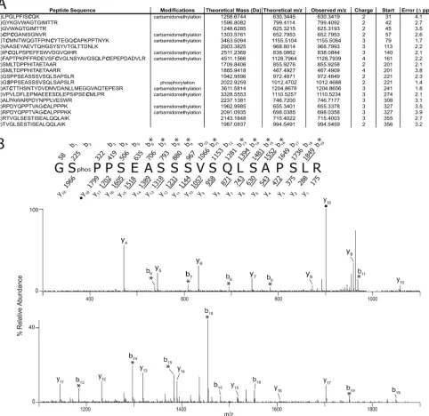
cells harboring subgenomic replicons and enrich for the protein by immunoprecipitation. Enriched fractions were then subjected to SDS-PAGE gel separation, proteolytic digestion and peptide extraction, and mass spectrometry. Briefly, 7.5⫻107Huh-7.5 cells containing the genotype 2a replicon pSGR-JFH-1 were lysed in NETN buffer containing protease and phosphatase inhibitors. The NS5A protein was extracted by immunoprecipitation using the 9E10 anti-NS5A monoclonal antibody covalently coupled to agarose beads (21). Following extensive washing in NETN, NS5A was eluted from the beads by the addition of 2⫻ SDS-PAGE nonreducing sample buffer and boiling for 5 min. Extracts were then resolved on 10% acrylamide SDS-PAGE gels, and the region of the gel corresponding to the migration point of NS5A was ex-cised and subjected to in-gel reduction, alkylation, and trypsin digestion following the standard methods of the Scripps Pro-teomic Core facility. Eluted peptides were then loaded onto a fused silica (outer diameter [OD], 360m; inner diameter [ID], 75m) capillary C18(ODS-AQ, 20m) precolumn and washed with 0.1% acetic acid. Once salts were removed, the precolumn was attached to a fused silica (OD, 360m; ID, 50m) C18 col-umn (ODS-AQ; 5m) attached to a fused silica emitter tip. Pep-tides were high-performance liquid chromatography (HPLC) gra-dient eluted and analyzed by a Thermo Electron LTQ Orbitrap ion trap mass spectrometer set to data-dependent mode to acquire tandem mass spectrometric (MS-MS) spectra on the five most abundantm/zpeaks in each MS scan. All MS-MS spectra were searched using Sequest (Thermo Electron) and Mascot (Matrix Science) software. Potential posttranslational modifications were searched (STY⫽80 [phosphorylation]) as well as static modifi-cations for carbamidomethylation (C⫽57). Search results were manually confirmed to ensure correct sequence identifications. This methodology produced 61% sequence coverage of the geno-type 2a JFH-1 isolate of NS5A and identified a single phosphory-lation posttransphosphory-lational modification at serine 222 (Fig. 1). To
Received14 August 2012 Accepted24 October 2012
Published ahead of print31 October 2012
Address correspondence to T. L. Tellinghuisen, [email protected]. This article is number 21903 from the Scripps Research Institute.
Copyright © 2013, American Society for Microbiology. All Rights Reserved.
doi:10.1128/JVI.02154-12
on November 7, 2019 by guest
http://jvi.asm.org/
provide context to the more widely studied Con1 1b NS5A pro-tein, this serine residue would reside at position 222 in that protein and position 2194 in the Con1 1b polyprotein. It should be noted that a number of serine-rich regions of NS5A were not identified in this analysis, and these may contain additional phosphorylation sites. Nonetheless, the data definitively identify serine 222 as a phosphoacceptor used during the course of authentic HCV RNA replication.
With S222 identified, we proceeded to determine what role the posttranslational modification of this residue might have in HCV biology. We generated a series of phosphomimetic mutants at this residue, using methods described previously (11), including an aspartic acid substitution to mimic phosphorylation and an ala-nine residue to generate a site incapable of phosphorylation. These were built into the genotype 2a subgenomic replicon, pSGR-JFH1, and the RNA replication kinetics of RNAs harboring these muta-FIG 1Identification of serine 222 as a phosphorylation acceptor. (A) A comprehensive list of NS5A peptides identified following LC-MS-MS analysis. Peptide sequences are listed along with their theoretical mass, theoretical and observedm/zvalues, and associated mass error (ppm). The charge state, start residue of each peptide, and identified modifications are also provided. Modifications observed are indicated, including the phosphorylation of serine 222. (B) MS-MS spectrum of phosphorylated NS5A peptide, residues 221 to 240. The [M⫹2H]⫹2ion of the phosphorylated NS5A peptide (m/z1012.47) was selected for dissociation, and the amino acid sequence of the peptide is shown above the spectrum. The masses above and below the sequence correspond to the theoretical, singly protonated b- and y-type product ions, respectively. The observed singly protonated product ions are underlined, and doubly protonated ions are denoted with filled circles. Asterisks indicate product ions that result from neutral loss of H3PO4. As indicated in the sequence, the phosphorylated residue was identified as serine residue 222.
on November 7, 2019 by guest
http://jvi.asm.org/
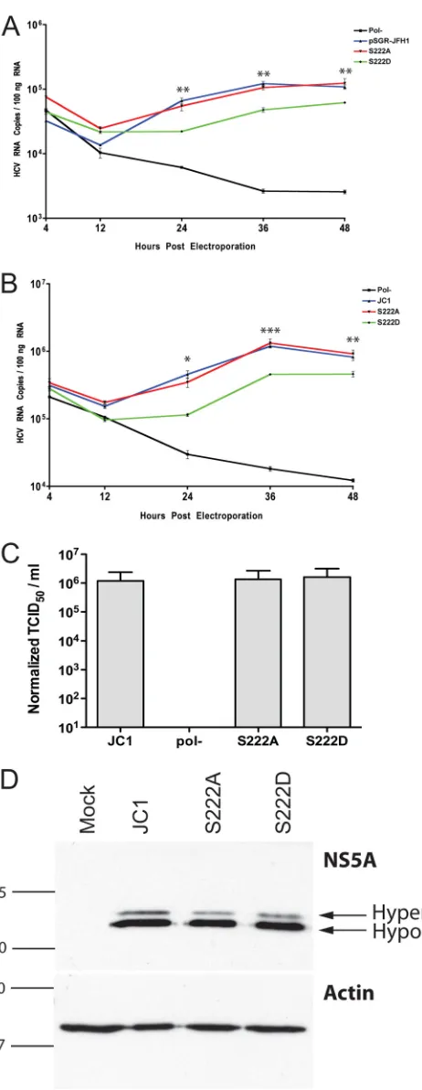
[image:2.585.50.532.71.539.2]tions were assessed relative to a wild-type pSGR-JFH1 RNA and a nonreplicative pSGR-JFH1 RNA containing a lethal lesion in the RNA-dependent RNA polymerase (Pol⫺) (23). It is important to note that the pSGR-JFH1 construct, unlike many other replicons of different HCV genotypes, replicated efficiently without adap-tive mutations, so the serine 222 mutations are analyzed in the context of a wild-type NS5A sequence.Figure 2Ashows the results of the delivery of 1g of these RNAs to Huh-7.5 cells via electro-poration and the time course analysis of total HCV RNA accumu-lation inside cells over a 48-h period via real-time reverse trans-criptase PCR analysis as described previously (11, 24). In this analysis the replication of pSGR-JFH1 with the serine 222-to-ala-nine (S222A) mutation is indistinguishable from the wild-type pSGR-JFH1 construct, suggesting that under these conditions, the inability to phosphorylate serine 222 is phenotypically null. Anal-ysis of the serine 222-to-aspartic acid mutation is more interest-ing, as pSGR-JFH1 genomes containing this mutation (S222D) are impaired in RNA replication approximately 2- to 3-fold com-pared to the parental pSGR-JFH1 construct at all data points after 24 h postelectroporation. Although this change is relatively small, it is statistically significant and reproducible. The data presented are normalized to total RNA, but it should be noted that further normalization of the data to the 4-h time point values did not alter the observed differences or their statistical validity. It is important to note that the S222D mutant containing replicons do replicate RNA, just at a lower efficiency, as can be seen by comparison to nonreplicating (Pol⫺) pSGR-JFH1 RNA. To investigate the im-pact of serine 222 modification on replication of an infectious viral genome, we built the same mutations into the JC1 HCV infectious clone and performed a similar electroporation-based RNA accu-mulation assay to that performed with the replicon (Fig. 2B). Sim-ilarly to what was observed in the replicon, the serine 222A muta-tion was identical in RNA replicamuta-tion to the JC1 parental viral genome, and the serine 222D was again attenuated in RNA repli-cation. The data presented are normalized to total RNA, but it should be noted that further normalization of the data to the 4-h time point values did not alter the observed differences or their statistical validity. As NS5A is known to be an important factor in
FIG 2Replication analysis of phosphomimetic mutations of serine 222. (A) Replication of serine 222 mutants in the context of the pSGR-JFH1 sub-genomic replicon. HCV RNA copies per 100 nanograms of total RNA as de-termined via real-time reverse transcriptase PCR analysis of cells electropo-rated with wild-type pSGR-JFH1 RNA (pSGR-JFH1), a pSGR-JFH1 replicon harboring the serine 222-to-alanine mutation (S222A), a psGR-JFH1 replicon harboring the serine 222-to-aspartic acid mutation (S222D), and a
pSGR-JFH1 replicon harboring a lethal lesion in the RNA-dependent RNA polymer-ase (Pol⫺). RNAs were harvested at indicated times postelectroporation from triplicate samples for calculation of statistical significance using a Studentttest for each indicated data point. (B) Samples as described for panel A but with mutations built into the full-length infectious JC1 virus clone (JC1). (C) Titer release from cells in panel B 48 h after electroporation. Values are in normal-ized tissue culture infectious doses, 50% value, per milliliter as determined by limiting dilution assay. Values shown are the averages of three independent electroporations of viral RNA. Samples are designated as in the previous panel. Data for this experiment were normalized using the RNA replication data from panel B. (D) NS5A expressed from VTF7-3 transfection/infection experiments separated on a 12% SDS-PAGE gel to monitor polyprotein processing and stability. The single band represents the NS5A protein. The samples shown are from wild-type JC1 virus encoding a genomic infectious clone plasmid (JC1), the JC1 infectious clone plasmid containing the serine 222-to-alanine muta-tion (S222A), and the JC1 infecmuta-tion clone plasmid containing the serine 222-to-aspartic acid mutation (S222D). NS5A was detected by Western blotting using the 9E10 anti-NS5A monoclonal antibody. An actin Western blot load-ing control is shown as the lower panel in this composite figure. Arrows indi-cate the migration distances of the hypophosphorylated (hypo) and hyper-phosphorylated (hyper) forms of NS5A. In both panels, the numbers and lines to the left of the image indicate the sizes (in kilodaltons) and mobilities of molecular weight markers.
on November 7, 2019 by guest
http://jvi.asm.org/
[image:3.585.49.280.61.657.2]infectious virus production and as this may be linked to phos-phorylation, we harvested supernatants of cells 48 h after the de-livery of our mutant RNAs and determined the titers of the amount of infectious virus in these samples by the limiting dilu-tion immunohistochemical assay described previously (21). De-spite the clear difference in RNA replication efficiency of the var-ious serine phosphomimetic mutants, we observed no statistically significant difference in infectivity, although a slight (about 1.8-fold) downward trend was seen in the S222D mutant-bearing vi-rus. Normalization of titer data to RNA replication level elimi-nated this difference, indicating that the S222D mutant does not impact virus production. As our mutations alter the sequence of the NS5A protein, we felt it was prudent to monitor the stability and processing of NS5A over the course of these experiments. To avoid differences in levels of NS5A due to different levels of RNA replication for the various mutations and other complications, we opted to express NS5A in the context of the full-length viral JC1 genome using the VTF7-3 vaccinia T7 system (25).Figure 2D
shows the results of this analysis 24 h after transfection of 1g of each plasmid via transfection as described previously (24) and infection of Huh-7.5 cells with VTF7-3 at a multiplicity of 1, prior to cell lysate preparation and SDS-PAGE and Western blot anal-ysis with the 9E10 NS5A-specific antibody and an actin control as described previously, and the results indicate that there is no gross defect in NS5A stability or polyprotein processing. Analysis of the phospho-form distribution of NS5A suggests that S222A bearing NS5A produces a 44.9% reduction in hyperphosphorylation com-pared to the wild-type construct while exhibiting only a 4.9% re-duction in hypophosphorylated NS5A relative to the control. S222D hyperphosphorylation, in contrast, is reduced only 9.8% relative to the wild type, with the hypophosphorylated form re-duced 19.3% compared to the control. This suggests that S222 might control multiple phosphorylation sites when modified. Collectively, these results indicate that serine 222 of genotype 2A NS5A is phosphorylated in the context of HCV replication, and this modification is a negative regulator of replication, albeit a minor one.
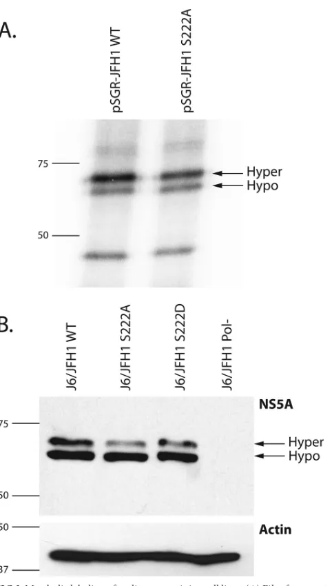
As our method of isolation of NS5A does not discriminate between hyper- and hypophosphorylated forms, we wanted to determine if serine 222 was a discrete component of one form over the other or a component of both forms. We therefore metaboli-cally labeled cells harboring the pSGR-JFH1 replicon and a repli-con harboring S222A using radioactive orthophosphate as de-scribed previously (26). Cell lysates were then prepared, NS5A was immunoprecipitated, and precipitates were analyzed by scintilla-tion counting. Samples containing Huh-7.5 cell lysates devoid of HCV and samples precipitated with isotype-matched control an-tibodies contained background levels of radioactivity after wash-ing and were not analyzed further. Samples containwash-ing NS5A were then separated by SDS-PAGE as described in the aforementioned reference, with the substitution of the 9E10 NS5A antibody for precipitations. Gels were then exposed to X-ray film to determine incorporation of radioactive phosphate into NS5A. Figure 3A
presents these data, with the electrophoretic migration of hyper-and hypophosphorylated NS5A indicated for both the wild-type and mutant replicon extracts. Measurement of radioactivity in the various phospho-forms indicated that although the hypophos-phorylated form of NS5A differed by only 3% between wild type and mutant, the hyperphosphorylated form differed by 19.1% be-tween the two samples, suggesting that phosphorylated serine 222
is predominantly a component of the hyperphosphorylated form of NS5A. We also investigated phospho-form distribution by Western blotting of NS5A from cells infected with J6/JFH1 viral genomes containing our serine 222 mutants (Fig. 3B). Densito-metric analysis of these blots shows that NS5A from S222A viruses has 36% less hyperphosphorylation than a wild-type construct, while reduction in hypophosphorylation was only 6%. Similarly, FIG 3Metabolic labeling of replicon containing cell lines. (A) Film from 10% SDS-PAGE separation of32P
i-labeled Huh-7.5 cell lysates from cells harboring wild-type pSGR-JFH1 (pSGR-FJH1 WT) or the pSGR-JFH1 replicon harbor-ing the serine 222-to-alanine mutation (pSGR-JFH1 S222A). Arrows indicate the migration distances of the hypophosphorylated (hypo) and hyperphos-phorylated (hyper) forms of NS5A. Numbers and lines to the left of the image indicate the sizes (in kilodaltons) and mobilities of molecular weight markers. (B) Upper panel, Western blot analysis detecting NS5A protein from cells electroporated with HCV J6/JFH1 viral genomic RNA containing S222A, S222D, wild-type, and Pol⫺RNAs. Arrows indicate the migration distances of the hypophosphorylated (hypo) and hyperphosphorylated (hyper) forms of NS5A. Lower panel, a Western blot of lysates identical to those in the upper panel but probed for the cellular actin protein as a loading control. In both panels, the numbers and lines to the left of the image indicate the sizes (in kilodaltons) and mobilities of molecular weight markers.
on November 7, 2019 by guest
http://jvi.asm.org/
[image:4.585.298.539.61.486.2]S222D virus showed a reduction of 23.4% hyperphosphorylation compared to the wild type, with a 7% reduction in hypophosphor-ylation. The reduction in hyperphosphorylation difference be-tween these two mutants is quite interesting, as neither is capable of being phosphorylated, and this could be interpreted as an indi-cation that S222D controls the use of other phosphorylation sites in NS5A. The location of this residue in relation to the domain model of NS5A, with serine 222 in the LCSI region between do-main I and II, is in good agreement with previous data suggesting that hyperphosphorylation sites map in this area of NS5A.
Here we have identified serine 222 of the genotype 2a NS5A protein as a site of phosphorylation used in cells harboring a func-tional replicon, the first site to be found in the context of RNA replication. Our findings are in good agreement with the concept that hyperphosphorylation is a negative regulator of replication, but the effects we observe are small, and it is therefore unlikely that serine 222 is a major regulator of RNA replication. Mutation of this residue to aspartic acid produces a different phospho-form distribution than when it is changed to alanine (or serine), and these data might be interpreted to indicate that S222 modification directs the phosphorylation of other sites in NS5A that are com-ponents of the hyperphosphorylated isoform. Unlike serine resi-dues in domain III of NS5A, serine 222 appears to have no impact on virus production, suggesting that different serine phosphory-lation events can impact different aspects of the HCV life cycle, even when those serine residues are both components of the same NS5A phospho-form. The data suggest that a more complex reg-ulation than explained in current models probably exists. Serine 222 is an absolutely conserved residue among divergent HCV geno-types, and indeed, this residue has been identified as a phospho-acceptor in an early NS5A overexpression phosphopeptide-map-ping experiment in genotype 1a (designated S2194 in that work) (16). Phosphomimetic mutation of this position in the 1b replicon does not produce dramatic replication or phospho-form distribu-tion phenotypes in the context of one set of adaptive mutadistribu-tions (15), but it does produce these effects in another adaptive genetic context (27). Adaptive mutations in genotype 1b replicons in-crease RNA replication and tend to reduce NS5A hyperphosphor-ylation (19), and one can directly manipulate hyperphosphoryla-tion to bypass the need for adaptive mutahyperphosphoryla-tions (15,17,28,29), but the mechanistic details of these observations are not understood. Perhaps different adaptive mutations produce different patterns of phosphorylation site usage on NS5A, and in the example of S222, this may be important in one context but not in another. The genotype 2a NS5A used in this work requires no adaptive mutations for efficient replication, eliminating complexities of genetic background from the analysis and simplifying the identi-fication of the phosphorylation of this residue as a minor negative regulator of replication. Perhaps the overwhelming questions sug-gest why this virus utilizes such a complex mechanism to fine-tune RNA replication efficiency and what precisely NS5A phosphory-lation actually regulates. It will likely be some time before we de-velop a comprehensive view of the role of NS5A phosphorylation, but the identification of S222 as an authentic phosphorylation site is an important step in this process.
ACKNOWLEDGMENTS
These experiments were funded, in part, by the Burroughs Wellcome Fund Investigators in the Pathogenesis of Infectious Diseases program (T.L.T.). Additional funds supporting this work came from the National
Institutes of Health, National Institute of Diabetes and Digestive and Kid-ney Diseases, and the National Institute of Allergy and Infectious Disease (T.L.T.).
We thank Marilena Fernandez, Sarae Copeland, and Melissa Telling-huisen for assistance in the preparation of the manuscript and the mem-bers of the Scripps Proteomics Core facility for technical assistance. We thank Bernard Moss for the vaccinia T7 system reagents and Charles Rice for the Huh-7.5 cells and 9E10 monoclonal antibody.
REFERENCES
1. Anonymous.1997. World Health Organization—Hepatitis C: global prevalence. Wkly. Epidemiol. Rec.72:341–344.
2.Lindenbach BD, Thiel HJ, Rice CM.2007. Flaviviridae: the viruses and their replication, p 1101–1152.InKnipe DM, Howley PM (ed), Fields virology, fifth ed, vol 1. Lippincott-Raven Publishers, Philadelphia, PA. 3.Appel N, Zayas M, Miller S, Krijnse-Locker J, Schaller T, Friebe P,
Kallis S, Engel U, Bartenschlager R.2008. Essential role of domain III of nonstructural protein 5A for hepatitis C virus infectious particle assembly. PLoS Pathog.4:e1000035. doi:10.1371/journal.ppat.1000035.
4.Hughes M, Gretton S, Shelton H, Brown DD, McCormick CJ, Angus AG, Patel AH, Griffin S, Harris M.2009. A conserved proline between domains II and III of hepatitis C virus NS5A influences both RNA repli-cation and virus assembly. J. Virol.83:10788 –10796.
5.Hughes M, Griffin S, Harris M.2009. Domain III of NS5A contributes to both RNA replication and assembly of hepatitis C virus particles. J. Gen. Virol.90:1329 –1334.
6.Jones CT, Murray CL, Eastman DK, Tassello J, Rice CM.2007. Hepatitis C virus p7 and NS2 proteins are essential for production of infectious virus. J. Virol.81:8374 – 8383.
7.Jones DM, Patel AH, Targett-Adams P, McLauchlan J.2009. The hep-atitis C virus NS4B protein can trans-complement viral RNA replication and modulates production of infectious virus. J. Virol.83:2163–2177. 8.Ma Y, Yates J, Liang Y, Lemon SM, Yi M.2008. NS3 helicase domains
involved in infectious intracellular hepatitis C virus particle assembly. J. Virol.82:7624 –7639.
9.Masaki T, Suzuki R, Murakami K, Aizaki H, Ishii K, Murayama A, Date T, Matsuura Y, Miyamura T, Wakita T, Suzuki T.2008. Interaction of hepatitis C virus nonstructural protein 5A with core protein is critical for the production of infectious virus particles. J. Virol.82:7964 –7976. 10. Pietschmann T, Kaul A, Koutsoudakis G, Shavinskaya A, Kallis S,
Steinmann E, Abid K, Negro F, Dreux M, Cosset FL, Bartenschlager R. 2006. Construction and characterization of infectious intragenotypic and intergenotypic hepatitis C virus chimeras. Proc. Natl. Acad. Sci. U. S. A. 103:7408 –7413.
11. Tellinghuisen TL, Foss KL, Treadaway J.2008. Regulation of hepatitis C virion production via phosphorylation of the NS5A protein. PLoS Pathog. 4:e1000032. doi:10.1371/journal.ppat.1000032.
12. Tanji Y, Kaneko T, Satoh S, Shimotohno K.1995. Phosphorylation of hepatitis C virus-encoded nonstructural protein NS5A. J. Virol.69:3980 – 3986.
13. Tellinghuisen TL, Marcotrigiano J, Gorbalenya AE, Rice CM.2004. The NS5A protein of hepatitis C virus is a zinc metalloprotein. J. Biol. Chem. 279:48576 – 48587.
14. Huang Y, Staschke K, De Francesco R, Tan SL.2007. Phosphorylation of hepatitis C virus NS5A nonstructural protein: a new paradigm for phos-phorylation-dependent viral RNA replication? Virology364:1–9. 15. Appel N, Pietschmann T, Bartenschlager R.2005. Mutational analysis of
hepatitis C virus nonstructural protein 5A: potential role of differential phosphorylation in RNA replication and identification of a genetically flexible domain. J. Virol.79:3187–3194.
16. Katze MG, Kwieciszewski B, Goodlett DR, Blakely CM, Neddermann P, Tan SL, Aebersold R.2000. Ser(2194) is a highly conserved major phos-phorylation site of the hepatitis C virus nonstructural protein NS5A. Vi-rology278:501–513.
17. Neddermann P, Quintavalle M, Di Pietro C, Clementi A, Cerretani M, Altamura S, Bartholomew L, De Francesco R.2004. Reduction of hep-atitis C virus NS5A hyperphosphorylation by selective inhibition of cellu-lar kinases activates viral RNA replication in cell culture. J. Virol.78: 13306 –13314.
18. Reed KE, Rice CM.1999. Identification of the major phosphorylation site of the hepatitis C virus H strain NS5A protein as serine 2321. J. Biol. Chem. 274:28011–28018.
on November 7, 2019 by guest
http://jvi.asm.org/
19. Blight KJ, Kolykhalov AA, Rice CM.2000. Efficient initiation of HCV RNA replication in cell culture. Science290:1972–1974.
20. Evans MJ, Rice CM, Goff SP.2004. Phosphorylation of hepatitis C virus nonstructural protein 5A modulates its protein interactions and viral RNA replication. Proc. Natl. Acad. Sci. U. S. A.101:13038 –13043.
21. Lindenbach BD, Evans MJ, Syder AJ, Wolk B, Tellinghuisen TL, Liu CC, Maruyama T, Hynes RO, Burton DR, McKeating JA, Rice CM. 2005. Complete replication of hepatitis C virus in cell culture. Science 309:623– 626.
22. Nordle Gilliver A, Griffin S, Harris M.2010. Identification of a novel phosphorylation site in hepatitis C virus NS5A. J. Gen. Virol.91:2428 –2432. 23. Wakita T, Pietschmann T, Kato T, Date T, Miyamoto M, Zhao Z, Murthy K, Habermann A, Krausslich HG, Mizokami M, Bartenschlager R, Liang TJ.2005. Production of infectious hepatitis C virus in tissue culture from a cloned viral genome. Nat. Med.11:791–796.
24. Tellinghuisen TL, Foss KL, Treadaway JC, Rice CM.2008. Identification of residues required for RNA replication in domains II and III of the hepatitis C virus NS5A protein. J. Virol.82:1073–1083.
25. Fuerst TR, Niles EG, Studier FW, Moss B.1986. Eukaryotic transient-expression system based on recombinant vaccinia virus that synthesizes bacteriophage T7 RNA polymerase. Proc. Natl. Acad. Sci. U. S. A.83: 8122– 8126.
26. Neddermann P.2009. NS5A phosphorylation and hyperphosphoryla-tion. Methods Mol. Biol.510:95–110.
27. Blight KJ, McKeating JA, Rice CM.2002. Highly permissive cell lines for hepatitis C virus genomic and subgenomic RNA replication. J. Virol.76: 13001–13014.
28. Quintavalle M, Sambucini S, Di Pietro C, De Francesco R, Nedder-mann P.2006. The alpha isoform of protein kinase CKI is responsible for hepatitis C virus NS5A hyperphosphorylation. J. Virol.80:11305– 11312.
29. Quintavalle M, Sambucini S, Summa V, Orsatti L, Talamo F, De Francesco R, Neddermann P.2007. Hepatitis C virus NS5A is a direct substrate of casein kinase I-alpha, a cellular kinase identified by inhibitor affinity chromatography using specific NS5A hyperphosphorylation in-hibitors. J. Biol. Chem.282:5536 –5544.