0022-538X/05/$08.00⫹0 doi:10.1128/JVI.79.13.8101–8112.2005
Copyright © 2005, American Society for Microbiology. All Rights Reserved.
Role of the Hypervariable Hinge Region of Phosphoprotein P of
Vesicular Stomatitis Virus in Viral RNA Synthesis and
Assembly of Infectious Virus Particles
Subash C. Das and Asit K. Pattnaik*
Department of Veterinary and Biomedical Sciences and Nebraska Center for Virology, University of Nebraska-Lincoln, Lincoln, Nebraska 68588-0666
Received 25 January 2005/Accepted 2 March 2005
The phosphoprotein (P protein) of vesicular stomatitis virus (VSV) is an essential subunit of the viral RNA-dependent RNA polymerase and has multiple functions residing in its different domains. In the present study, we examined the role of the hypervariable hinge region of P protein in viral RNA synthesis and recovery of infectious VSV by using transposon-mediated insertion mutagenesis and deletion mutagenesis. We observed that insertions of 19-amino-acid linker sequences at various positions within this region affected replication and transcription functions of the P protein to various degrees. Interestingly, one insertion mutant was com-pletely defective in both transcription and replication. Using a series of deletion mutants spanning the hinge region of the protein, we observed that amino acid residues 201 through 220 are required for the activity of P protein in both replication and transcription. Neither insertion nor deletion had any effect on the interaction of P protein with N or L proteins. Infectious VSVs with a deletion in the hinge region possessed retarded growth characteristics and exhibited small-plaque morphology. Interestingly, VSV containing one P protein deletion
mutant (P⌬7, with amino acids 141 through 200 deleted), which possessed significant levels of replication and
transcription activity, could be amplified only by passage in cells expressing the wild-type P protein. We con-clude that the hypervariable hinge region of the P protein plays an important role in viral RNA synthesis. Furthermore, our results provide a previously unidentified function for the P protein: it plays a critical role in the assembly of infectious VSV.
Vesicular stomatitis virus (VSV) is the prototypic member of the familyRhabdoviridaein the orderMononegavirales. VSV is an enveloped virus with a negative-stranded RNA genome of 11,161 nucleotides. The viral envelope consists of a lipid bi-layer with the spike glycoprotein protruding out and the matrix (M) protein lying underneath the lipid bilayer. The viral ge-nome which is present in the virion core is tightly encapsidated by the nucleocapsid protein (N protein) to form the ribonu-cleoprotein complex (6). The viral ribonuribonu-cleoprotein serves as the template for transcription and replication by the viral RNA-dependent RNA polymerase, which is a complex of the large protein (L protein) and phosphoprotein (P protein). The L protein is the catalytic subunit of the polymerase complex responsible for polymerization of nucleotides, capping, meth-ylation of capped mRNAs, and polyadenmeth-ylation of mRNAs (reviewed in references 1 and 45). P protein, on the other hand, acts as an accessory subunit of the viral polymerase (1, 45). In addition to having a role in polymerase functions, it binds to the L protein and stabilizes it from proteolytic degra-dation (7, 17), it complexes with the newly synthesized N pro-tein for the efficient encapsidation of nascent RNA (16, 36, 42), and it interacts with terminal sequences of viral genome for viral RNA synthesis (29, 30).
The P proteins of the two serotypes of VSV, Indiana (VSVI)
and New Jersey (VSVNJ), are small (265 and 274 amino acids,
respectively) and acidic in nature. Although these two proteins share only 30% sequence homology (19, 21, 26, 41), mutational and functional studies have identified four distinct structural regions (see Fig. 1A). Three of these structurally homologous domains (22, 41, 44) have been the targets of recent mutational studies to more precisely define their role in viral genome transcription and replication. Domain I, which encompasses the amino-terminal half of the protein (amino acid residues 1 through 150 for VSVIor 1 through 137 for VSVNJ) is a highly
acidic region that contains residues for the constitutive phos-phorylation of P protein. Phosphos-phorylation of specific amino acid residues (serine-60, threonine-62, and serine-64) in do-main I by cellular casein kinase II is important for transcrip-tional activity of P protein (2, 3, 12, 14, 22, 39, 48, 49). How-ever, replication functions of the protein are unaffected by the phosphorylation status of this domain (39). Domain II, span-ning approximately residues 210 through 244 in VSVIand 231
through 247 in VSVNJ, is phosphorylated (10, 12), although the
identity of the kinase responsible for this modification remains elusive. This domain interacts with L protein to form an active transcription complex with N-RNA template (10). Unlike the domain II of VSVNJ, which regulates transcription in vitro
(10), the phosphorylation of residues in domain II of VSVI
does not appear to have a role in transcription in vitro or in vivo (20, 27, 35). However, the phosphorylation of residues in this domain has been shown to influence the function of the P protein in replication (27). Using an infectious clone of VSV (31), we recently demonstrated that the phosphorylation of residues in both of these domains is indispensable for VSV growth (13). Although individual mutations at various
phos-* Corresponding author. Mailing address: E126 Beadle Center, 1901 Vine Street, University of Nebraska-Lincoln, Lincoln, NE 68588. Phone: (402) 472-1067. Fax: (402) 472-8722. E-mail: apattnaik2@unl .edu.
8101
on November 8, 2019 by guest
http://jvi.asm.org/
phorylation sites in either domain of P protein did not ad-versely affect virus recovery or growth, multiple mutations that resulted in the complete loss of phosphorylation in either do-main I or II were detrimental to virus recovery (13).
The extreme carboxy-terminal region, containing 21 to 25 residues, is known as domain III. The last 11 amino acids of this domain are highly conserved and contain a number of basic residues that are important for P protein function in mediating the binding of P protein to N-RNA template (15). The fourth region of P protein, which links domain I and domain II, is a stretch of approximately 50 to 60 amino acids, spanning residues 150 through 210 (11, 22). A comparison of the amino acid sequences of the P protein of several isolates of VSVNJrepresentative of the natural genetic diversity
sug-gested the highest degree of variability within this region (4). The function(s) of this hypervariable region, often referred to as the hinge region, is largely unknown. It has been shown that the hinge region is not required for the association of P protein with N protein, by using a yeast two-hybrid system (50), or with L protein, by using in vitro interaction studies (17). The exact role of this hinge region of P protein, if any, in viral RNA syn-thesis and/or the life cycle of VSV has not been addressed.
To obtain a better understanding of the function of the hinge region of the P protein, we employed two different but complementary approaches in this study: transposon-mediated insertion mutagenesis and deletion mutagenesis. The abilities of the mutant P proteins to support viral RNA synthesis and recovery of infectious VSV were examined. Our results indi-cate that the majority of the sequences within the hinge region are dispensable for viral RNA synthesis and generation of infectious VSV. However, amino acids spanning residues 201 through 220 are required for viral RNA synthesis. Further-more, our data reveal a new function for the P protein: the hinge region of the protein is required for assembly of in-fectious VSV particles.
MATERIALS AND METHODS
Cells and viruses.Baby hamster kidney (BHK-21) cells used in this study were maintained in Eagle’s minimal essential medium containing 5% fetal bovine serum (FBS) and 100 units of penicillin, 20 units of streptomycin, and 20 units of kanamycin per ml of growth medium. VSV (Indiana serotype, San Juan strain) was propagated in BHK-21 cells. Recombinant vaccinia virus (vTF7-3) express-ing bacteriophage T7 RNA polymerase (18) was grown and titrated by plaque assay in BHK-21 cells. Production and purification of defective interfering T (DI-T) particles (32), which are of the copy-back type, have been described previously (40).
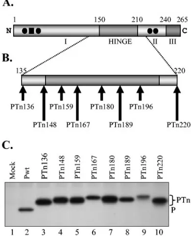
Construction of mutants by transposon (Tn)-mediated insertion mutagenesis and deletion.The P coding region from pET-P (39) was amplified by using Pfu polymerase (Stratagene) and P(⫹) and P(⫺) primers (13) digested with KpnI and PstI and was cloned into pGEM-3 vector (Promega) under the control of T7 RNA polymerase promoter. The resulting plasmid encoding the wild-type (wt) P protein was used as the template for Tn5-based in vitro mutagenesis using an EZ::TN in-frame linker insertion kit as per the instructions of the manufacturer (Epicentre). This Tn5-based insertion mutagenesis system allows random and single-hit in-frame insertion of a 48-nucleotide linker in the target DNA (23). Plasmids carrying insertions can be selected on the basis of kanamycin resistance encoded within the Tn. Following an in vitro transposase reaction, transformants were selected on kanamycin- and ampicillin-containing LB agar plates. Miniprep DNAs of individual clones were digested with NotI to release the kanamycin cassette, religated, and used for another round of transformation and selection on ampicillin-containing plates. The P coding region containing Tn-mediated insertions were identified by restriction mapping and sequence analysis. From an initial panel of insertion mutants that were identified, eight mutants spanning the hypervariable region and its flanking areas with insertions in approximately every 7 to 15 amino acid intervals were selected (Fig. 1B). The mutants were desig-nated PTn136, PTn148, PTn159, PTn167, PTn180, PTn189, PTn196, and PTn220, where the number refers to the amino acid position at the site of in-sertion in the P protein. From sequence analysis, it was observed that the Tn-induced insertions occurred in all three open reading frames (ORFs): PTn136, PTn148, PTn159, PTn180, and PTn220 contained insertions in ORF1; PTn167 and PTn189 contained insertions in ORF2; and PTn196 contained an insertion in ORF3. It must be noted that the insertion resulted in duplication of three amino acids of the target sequence at the site of insertion. Therefore, the Tn-induced in-frame insertions led to insertion of a total of 19 amino acids into the P protein.
The deletions in the hinge region were generated by overlapping PCR using DNA encoding the wild type P protein (Pwt) as the template. For each deletion, two fragments were amplified from either side of the desired region to be deleted, using primers designed to include overlapping sequences flanking the region to be deleted. The deletion mutants were then generated in a second round of PCR by amplification with P(⫹) and P(⫺) primers (13) using both the PCR-amplified fragments as templates. The final PCR product was cloned into pGEM-3 vector under T7 RNA polymerase promoter and sequenced to make sure that desired deletions were introduced into the clones and no other errors were present. The five small deletion mutants were designated P⌬1, P⌬2, P⌬3, P⌬4, and P⌬5, with amino acids 131 through 150, 151 through 170, 171 through 190, 191 through 210, and 211 through 220 deleted, respectively. In a similar manner, we also generated two other larger deletion mutants, P⌬6 and P⌬7, with amino acids 151 through 190 and 141 through 200 of the P protein deleted, respectively.
[image:2.585.64.259.72.313.2]The plasmids pN, pP, and pL, carrying the coding sequences of N, P, and L proteins of VSV under the control of T7 RNA polymerase promoter, as well as FIG. 1. Tn-mediated insertion mutants of P protein. (A) Domain
structure of P protein. The P protein with three functionally defined domains (I, II, and III) and the hinge region is shown. The phosphate acceptor sites Ser-60, Ser-64, Ser-226, and Ser-227 are represented by solid circles and Thr-62 by the solid square. The numbers on top of the rectangular boxes represent the approximate positions of the amino acid residues that define the domains. (B) Various insertion mutants used in this study. Each Tn-mediated insertion mutant is numbered according to the position at which the insertion occurred. For example, PTn136 mutant has the 19-amino-acid insertion immediately after amino acid 136 of the P protein. (C) Expression of Tn insertion mutants. BHK-21 cells infected with vTF7-3 in 35-mm six-well plates were transfected with 1g of plasmid encoding either wt or mutant P protein and radiolabeled for 2 h at 16 h posttransfection. Radiolabeled proteins were immunoprecipitated from the cytoplasmic extracts by use of rabbit anti-P antibody, analyzed by 10% SDS-PAGE, and de-tected by fluorography.
on November 8, 2019 by guest
http://jvi.asm.org/
the plasmid pVSVFL(⫹) containing an antigenomic-sense full-length VSV se-quence, have been described previously (31, 38, 40). Construction of the plasmid p10BN, encoding an antigenomic-sense VSV minigenome, has also been de-scribed earlier (28, 33). The mutant P coding regions were replaced with wt sequence in the full-length VSV genome plasmid pVSVFL(⫹) using the EcoRV sites that flank the P gene as described earlier (13).
Metabolic labeling and analysis of proteins in transfected and infected cells.
BHK-21 cells grown in 12-well plates to 90 to 95% confluence were infected with vTF7-3 at a multiplicity of infection (MOI) of 10 PFU/cell. Following adsorption for 45 min at 37°C, cells were washed with phosphate-buffered saline (PBS) and transfected with 1g of either wt or mutant P protein plasmids by using Lipo-fectamine2000 (Invitrogen) and Dulbecco’s modified Eagle’s medium (DMEM) without serum. At 4 to 6 h posttransfection, cells were washed twice with DMEM containing 5% FBS and incubated with 1 ml of the same medium for 16 to 18 h. The cells were then starved for 45 min at 37°C with 1 ml DMEM without methionine and cysteine and then labeled with 60Ci/ml of Expre35
S35 S protein-labeling mix (NEN Life Sciences, MA) in 0.4 ml of the medium described above for a period of 2 h at 37°C. Following radiolabeling, the cells were washed twice with cold PBS and lysed with 200l of radioimmunoprecipitation assay (RIPA) buffer containing phenylmethylsulfonyl fluoride (100g/ml) and leupeptin (1
g/ml) as described previously (40). After clarification at 16,000⫻gfor 5 min, the lysate was incubated with rabbit anti-P antibody (1:500) overnight at 4°C with constant rotation. A 100-microliter slurry of protein A-Sepharose (Amersham) (4 mg protein A-Sepharose in 100l RIPA buffer) was added to each sample and incubated for 2 h at 4°C with constant rotation. The immunoprecipitates were washed three times with RIPA buffer and finally resuspended in 20l RIPA buffer. The samples were then mixed with sodium dodecyl sulfate-polyacrylamide gel electrophoresis (SDS-PAGE) sample buffer, boiled for 4 min, and analyzed by SDS-PAGE. The proteins were detected by fluorography as described previ-ously (13).
For coimmunoprecipitation (co-IP) studies, BHK-21 cells infected with vTF7-3 as described above were cotransfected with 1g each of wt or mutant P protein plasmids along with pN or pL as described above. Following radiolabel-ing as described above, cells were washed twice with cold PBS and scraped into 200l co-IP buffer (0.1 M HEPES, pH 8.5, 0.05 M NH4Cl, 7 mM KCl, 4.5 mM manganese acetate, 0.25% NP-40) (8) containing phenylmethylsulfonyl fluoride (100g/ml) and leupeptin (1g/ml). The cell lysates were incubated on ice for 15 min and then clarified by centrifugation at 16,000⫻g for 5 min at 4°C. Clarified lysate was used for immunoprecipitation with either rabbit anti-P poly-clonal antibody, anti-N monopoly-clonal antibody 10G4 (5), or polypoly-clonal antibody to the NH2-terminal of L protein (47) for 18 h at 4°C with constant rotation. Subsequent analysis of proteins was performed as described above except that for all steps, the co-IP buffer was used in place of RIPA buffer.
To analyze the viral proteins in infected cells, BHK-21 cells were grown in six-well tissue culture plates to 90 to 95% confluence. Cells were infected with either wt or mutant virus at an MOI of 10 PFU/cell and incubated in 2.5 ml fresh medium containing 2% FBS for 4 h at 37°C. The cells were starved for 1 h with 0.6 ml of DMEM without methionine and cysteine and radiolabeled for 2 h as described above. The cells were washed and lysed in 300l RIPA buffer. One-tenth of the lysate was analyzed by SDS-PAGE, and the proteins were detected as described above. Western blot analysis of unlabeled proteins was performed as described previously (13).
Transfection, metabolic labeling, and analysis of RNA.BHK-21 cells were grown in six-well tissue culture plates to 90% confluence. To examine transcrip-tion activities of mutant P proteins, cells were infected with vTF7-3 at an MOI of 10 PFU/cell and subsequently transfected with 2.5g of p10BN, 1.5g of pN, 0.5
g of pL, and 1g of wt or mutant pP plasmid by using Lipofectamine2000 as described above. At 4 h posttransfection, cells were washed twice and incubated with 2 ml of fresh medium containing 5% FBS for 16 h. RNAs were radiolabeled in the presence of actinomycin D and analyzed by electrophoresis in agarose-urea gel as described previously (13). To examine the replication activities of mutant P proteins, cells infected with vTF7-3 and transfected with pN, pL, and wt or mutant P plasmid were superinfected with DI particles and RNAs were radiolabeled and analyzed as described previously (13). Quantitation of the transcription and replication products was carried out by densitometric scanning of the fluorograms with a VersaDoc system using ImageQuant software (Bio-Rad). Normalized levels of transcription were determined as described before (39) by using the following formula: (levels of transcription/levels of replication)
⫻100. Viral RNAs synthesized in infected cells were also analyzed as described previously (13).
Transfection, recovery, and identification of mutant viruses.BHK-21 cells were grown in 60-mm tissue culture dishes to 80 to 90% confluence. Cells were infected with vTF7-3 at an MOI of 10 PFU/cell. Subsequently, cells were
trans-fected with 4g of pVSVFL(⫹) encoding full-length viral genome with wt P protein or mutant P proteins, 6g of pN plasmid, 2g of pL plasmid, and 4g of pP plasmid by using Lipofectamine2000. At 4 h posttransfection, cells were washed twice and incubated with 2 ml of fresh medium containing 5% FBS for 48 h. Culture supernatant was harvested, clarified at 16,000⫻gfor 20 min, passed through a 0.45-m syringe filter to remove vaccinia virus, and used to infect fresh BHK-21 cells. If virus was recovered from transfected cells as evi-denced by development of cytopathic effects (CPE) during subsequent passage and also by indirect immunofluorescence of infected cells, the culture superna-tants from transfected cells and amplification passage (P1) were titrated by plaque assay. Stocks of mutant viruses were prepared by infecting BHK-21 cells with a plaque-purified stock virus at an MOI of 0.1 PFU/cell. To confirm the identity of mutant viruses, BHK-21 cells were infected with an MOI of 10 of each virus. At 6 h postinfection, total RNA from cells was isolated by using Trizol (Invitrogen). Reverse transcriptase PCR was performed using Moloney murine leukemia virus reverse transcriptase andTaq DNA polymerase (Invitrogen). PCR products were analyzed on agarose gels and directly sequenced to deter-mine the presence of the desired mutation.
Determination of single-step growth kinetics.Single-step growth kinetics of mutant and wt viruses in BHK-21 cells were determined essentially as described previously (13).
Indirect immunofluorescence.BHK-21 cells infected with wt virus or mutant viruses from transfected-cell supernatants were fixed with acetone-methanol (1:1) at⫺20°C and incubated with a monoclonal anti-N antibody 10G4. The cells were then incubated with the secondary antibody, anti-mouse Alexa-488 (Mo-lecular Probes) and examined by a Nikon fluorescence microscope attached with a camera (Optronics). The images were processed by using Photoshop software.
Complementation of virus recovery.BHK-21 cells grown to 80% confluence were cotransfected with two plasmids: (i) pCI-T7, a derivative of the plasmid pCI-Neo (Promega), in which the gene encoding the neomycin phosphotrans-ferase was replaced with the gene encoding the T7 RNA polymerase under the control of cytomegalovirus promoter in the plasmid, and (ii) the plasmid pP. At 24 h posttransfection, approximately 75% of cells expressed the P protein, as judged by the immunofluorescent staining of the cells. The cells were then superinfected with culture supernatant containing the VSV P⌬7 mutant (VSV-P⌬7). After 48 h postinfection, culture supernatants were collected and VSV-P⌬7 particles were titrated by plaque assay in cells expressing the P protein by using the two-plasmid system as described above.
RESULTS
Tn-induced insertion mutants of P protein.The role of the
hypervariable hinge region (Fig. 1A) of the VSV P protein in viral genome transcription and replication is not known. To examine its role, we generated a series of Tn-induced insertion mutants in this region of the P protein and determined the activities of the mutant proteins in viral RNA synthesis. From an initial panel of mutants, we selected eight mutants with insertions within the hinge region and the flanking sequences. Each mutant contained an insertion of 19 amino acids, and the insertions were approximately 7 to 15 amino acids apart (Fig. 1B). Each insertion maintained the ORF of the P protein. In addition, each mutant contained only one copy of the insert. To determine if the insertion resulted in viable and stable proteins, we examined the expression of these proteins in transfected cells. Accordingly, BHK-21 cells were transiently transfected with plasmids encoding either wt or mutant P pro-teins following vTF7-3 infection and subsequently analyzed the radiolabeled proteins by immunoprecipitation with anti-P pro-tein antibody and SDS-PAGE. As can be seen from Fig. 1C, each of the mutant P proteins was easily detected. The mutant P proteins possessed slightly reduced electrophoretic mobility compared to the wt P protein. In addition, the mutant proteins exhibited minor differences in electrophoretic mobility, a result which is most likely due to differences in the amino acid com-positions of the insertion sequences, since the mutants con-tained insertions in each of the three reading frames. It should
on November 8, 2019 by guest
http://jvi.asm.org/
be noted that the majority of the mutant proteins were de-tected at levels greater than those of the wt P proteins. The rea-son(s) for this is unclear but could be due to changes in anti-body reactivity. Pulse-chase analysis showed that the mutant proteins were as stable as the wt P protein (data not shown).
Tn-induced insertions in the hinge region affect P protein
function in viral RNA synthesis.In order to examine the effect
of insertion mutation on P protein function in viral RNA syn-thesis, we used DI particles to determine the replication activ-ities of various P mutants. The DI particle genomes are only replication competent and therefore are used to assess the replication activities of the mutant P proteins in the absence of transcription. BHK-21 cells infected with vTF7-3 were trans-fected with the plasmids encoding N, L, and wt or mutant P proteins. Cells were subsequently superinfected with DI parti-cles, and replication of DI RNA was examined by radiolabeling in the presence of actinomycin D and analyzing the total RNA by electrophoresis. Results from such an experiment (Fig. 2A) show that the mutant proteins supported replication of DI RNA to different extents. A quantitative analysis (Fig. 2B) of data from three independent experiments revealed that the level of replication supported by PTn136 and PTn180 was significantly reduced with less than 20% activity of the wt pro-tein. PTn148, PTn159, and PTn189 supported replication at levels 20 to 50% of the wt protein. PTn167 and PTn196 pro-teins were 60 to 85% as active as the wt protein. Interestingly, one insertion mutant, PTn220 was completely defective in sup-porting replication.
To examine if the mutant proteins possessed altered tran-scription activities, we used a trantran-scription- and replication-competent minigenome template encoded in the plasmid p10BN (28). RNA generated from this plasmid by T7 RNA polymer-ase is a positive-sense antigenomic RNA which must undergo replication to generate genome-sense RNA that serves as the template for transcription to generate N⌬L mRNA. To per-form the experiment, BHK-21 cells infected with vTF7-3 were transfected with the minigenome plasmid p10BN and the plas-mids encoding N, L, and wt or mutant P proteins. At 16 to 18 h posttransfection, the cells were radiolabeled in the presence of actinomycin D. Total radiolabeled RNA was isolated and an-alyzed by electrophoresis. The synthesis of N⌬L mRNA was used as a measure of transcription. Results presented in Fig. 2C show that the mutant P proteins supported different levels of transcription relative to that of the wt P protein. Since transcription of N⌬L mRNA was dependent on replication of the template, we normalized the levels of transcription relative to the levels of replication as described previously (39) and in Materials and Methods. Normalized levels of transcription would therefore measure the transcription activities of the mutant proteins relative to that of the wt P protein. A quan-titative analysis of normalized levels of transcription from three independent experiments (Fig. 2C) revealed that the majority of the insertions had no significant adverse effects on the transcription of the minigenome. Only two mutants, PTn136 and PTn180, possessed transcription activities that were 40 to 60% of that of the wt protein. Thus, the results from the studies described above suggest that insertions within the hypervariable hinge region have negative effects mostly on viral RNA replication. It should be noted that the transcription
ac-FIG. 2. Replication and transcription activities of the insertion mu-tants. (A) Replication of DI RNA supported by various insertion mutants. BHK-21 cells in 35-mm six-well plates were infected with vTF7-3 and then transfected with 1.5g of pN, 0.5g of pL, and 1.0 g of plasmid encoding wt or mutant P protein. At 16 h posttransfec-tion, cells were superinfected with purified DI-T particles and then labeled with [3H]uridine and [3H]adenosine for 6 h in the presence of
actinomycin D. The radiolabeled RNA was isolated, analyzed by elec-trophoresis in agarose-urea gel, and detected by fluorography. The minus and plus signs represent genomic and antigenomic RNAs, re-spectively, of the DI particle. (B) Quantitative determination of the levels of replication supported by various mutant P proteins. The data were obtained by densitometric scanning of fluorograms as described in Materials and Methods. The histograms represent average data from three independent experiments, with standard deviations repre-sented by error bars. The asterisk signifies undetectable levels of rep-lication. The numbers above the histograms correspond to the num-bers of the samples of mutants shown in panel A. (C) Transcription activity of insertion mutants. BHK-21 cells in 35-mm six-well plates were infected with vTF7-3 and transfected with 2.5g of p10BN, 1.5 g of pN, 0.5g of pL, and 1.0g of plasmid encoding either wt or mutant P protein. Transfected cells were labeled with [3H]uridine and
[3H]adenosine at 16 h posttransfection for 6 h in the presence of
actinomycin D. The radiolabeled RNA was isolated, analyzed by elec-trophoresis, and detected by fluorography as described above. The arrow shows the N⌬L RNA transcription product. The normalized level of transcription (Norm. Tx.) supported by the mutants was cal-culated from three independent experiments as described in Mate-rials and Methods, and the averages are shown in the boxes below the lanes.
on November 8, 2019 by guest
http://jvi.asm.org/
[image:4.585.317.521.88.435.2]tivity of PTn220 could not be directly evaluated due to the defect of PTn220 in supporting replication.
Deletion mutations in the hinge region affect P protein
func-tion in viral RNA synthesis.Since Tn-induced insertions within
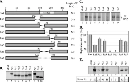
the hinge region or sequences flanking the hinge region af-fected the P protein function in viral RNA synthesis to various extents, we were interested to examine further the role of this region in P protein function by generating deletion mutants spanning this region. To this end, we generated a series of deletion mutants of P protein: P⌬1, P⌬2, P⌬3, P⌬4, and P⌬5, with amino acids 131 to 150, 151 to 170, 171 to 190, 191 to 210, and 211 to 220 deleted, respectively (Fig. 3A). All the deletion mutants expressed P proteins with sizes consistent with the extents of deletion (Fig. 3B). To analyze the effect of these deletions on replication activity of the proteins, DI RNA rep-lication in cells expressing the mutant proteins was examined. Results (Fig. 3C) show that P⌬1, P⌬2, and P⌬3 were active in supporting replication of DI RNA, whereas P⌬4 and P⌬5 were unable to support detectable levels of replication. A quantita-tive determination of the levels of replication supported by these mutants from three independent experiments (Fig. 3D) revealed that P⌬1 is about 42% as active as the wt protein. P⌬2 and P⌬3 proteins possessed 77% and 67% activity of the wt
protein. P⌬4 and P⌬5 proteins were completely inactive. These results suggest that the hypervariable region of the P protein plays a critical role in the function of the P protein in replica-tion.
We also examined the transcription activities of the deletion mutant P proteins to determine if these proteins have defects in viral RNA transcription. The experiment was performed as described in the legend to Fig. 2C, and the result from one of the experiments is shown in Fig. 3E. When the normalized level of transcription from three independent studies was cal-culated, it was observed that P⌬2 and P⌬3 were nearly as active as the wt protein, whereas P⌬1 possessed only 28% activity of the P protein. As expected, P⌬4 and P⌬5 proteins were com-pletely inactive in transcription, since in this assay, transcrip-tion was dependent on replicatranscrip-tion of the antigenomic-sense viral RNA template.
[image:5.585.68.518.71.356.2]Since some of the mutants with 20 amino acids deleted from the hinge region (P⌬2 and P⌬3) resulted in almost fully func-tional proteins, we were interested to determine if larger de-letions within this region would perturb the overall structure of the protein, rendering the mutant proteins less active or inac-tive in function. We generated two larger deletion mutants in which 40 (P⌬6) or 60 (P⌬7) amino acids from the P protein
FIG. 3. Replication and transcription activities of deletion mutants of P protein. (A) P protein deletion mutants showing the regions deleted from each mutant (dotted lines). The numbers represent the positions of the amino acids (a.a.) that are retained in the mutants. The lengths of the mutant proteins (in amino acids) are shown to the right. (B) Expression of P protein deletion mutants in transfected cells. The experiment was performed as described in the legend to Fig. 1C. (C) Replication of DI RNAs supported by the deletion mutants. The experiment was performed as described in the legend to Fig. 2A. The minus and plus signs represent genomic and antigenomic RNAs, respectively, of the DI particle. (D) Quantitative determination of the levels of replication supported by various deletion mutant P proteins. The data were obtained as described above in the legend to Fig. 2B. The histograms show the average activities from three independent experiments, with standard deviations represented by error bars. Asterisks signify undetectable levels of replication. (E) Transcription activity of insertion mutants. The experiment was performed as described in the legend to Fig. 2C. The normalized level of transcription (Norm. Tx.) supported by the mutants was calculated from three independent experiments as described in the legend to Fig. 2C, and the average values are shown in the boxes below each lane.
on November 8, 2019 by guest
http://jvi.asm.org/
were deleted (Fig. 3A). These proteins were easily detected in transfected cells (Fig. 3B). Interestingly, such large deletions in the P protein did not have major adverse effects on replication, as the levels of replication supported by these proteins were 60 to 70% of that of the wt protein (Fig. 3C and D). Normalized levels of transcription also remained nearly unaffected by the deletion of these regions from the P protein (Fig. 3E).
Insertions or deletions in the hinge region have no effect on
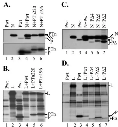
interactions with N and L proteins. Since certain insertion
(PTn220) and deletion (P⌬4 and P⌬5) mutants of P protein were nonfunctional in viral RNA synthesis, we wanted to ex-amine whether the interactions of these proteins with N and L proteins were affected. To perform these studies, plasmids encoding wt or mutant P proteins were cotransfected along with either the plasmid encoding the N or L protein into cells infected with vTF7-3. Following radiolabeling of the proteins, cell extracts were prepared and interactions of N or L proteins with the wt or mutant P proteins were examined by co-IP of P protein with antibody to either the N or L protein. Wt P protein could not be immunoprecipitated with anti-N (Fig. 4A, top panel, lane 1, and C, lane 1) or anti-L (Fig. 4B and D, lanes 1) antibodies, demonstrating that these antibodies were spe-cific only to the N or L protein, respectively. However, anti-N antibody could immunoprecipitate both N and P proteins from cell lysates of cells coexpressing both proteins (Fig. 4A and C, lanes 4), suggesting that wt P protein interacts with the N protein in cells expressing both proteins. PTn220, which is nonfunctional in viral RNA synthesis, and PTn196, which is functional, were found to interact with N protein, as these proteins could be immunoprecipitated with the N antibody (lanes 5 and 6). It should be noted that PTn220 comigrates with N protein, and therefore, in the gel shown in Fig. 4A (top panel), it could not be resolved from N protein. However, Western blot analysis of unlabeled and similarly immunopre-cipitated proteins demonstrated the presence of PTn220 (Fig. 4A, bottom panel, lane 5). In a similar study, we showed that PTn220 also interacts with the L protein as PTn220 could be coimmunoprecipitated with anti-L protein antibody (Fig. 4B, lane 5). Thus, these results show that the interactions of PTn220 with the N and L proteins remained unaffected by the insertion, even though the protein was nonfunctional in viral RNA synthesis.
In similar co-IP studies, we observed that P⌬4 and P⌬5, which are nonfunctional in viral RNA synthesis, and P⌬2, which is functional in viral RNA synthesis, interacted with the N protein (Fig. 4C) and the L protein (Fig. 4D). Taken to-gether, these studies suggest that insertions within or deletions from the hypervariable hinge region of the P protein have no effect on interactions with N or L protein.
PTn220 acts as a dominant-negative mutant of P protein
function.From the insertion and deletion mutagenesis studies
described above, we had identified mutants in the hypervari-able region that were defective in viral RNA synthesis. In addition, several mutants with insertions in other regions of the P protein were also found to be nonfunctional (S. C. Das and A. K. Pattnaik, unpublished data). Since the P protein is mod-ular with distinct domains, we were interested to determine if two inactive mutant proteins, when coexpressed, could re-constitute the activity of the P protein. Using pairwise combi-nations of various inactive mutant P proteins, we were
unsuc-cessful in reconstituting the activity of P protein in either transcription or replication (data not shown), indicating that
transcomplementation using pairs of inactive P mutants does
[image:6.585.316.520.67.287.2]not occur in this system. However, during our studies, we observed that PTn220, which is completely defective in sup-porting transcription and replication, when coexpressed with the wt P protein, inhibited the activity of the wt P protein, suggesting that PTn220 may be a dominant-negative mutant. To examine this possibility, we analyzed transcription in cells coexpressing PTn220 and Pwt proteins. The data presented in Fig. 5A show that coexpression of a fixed amount of Pwt with various amounts of PTn220 resulted in significant inhibition of transcription (lanes 6 to 8). This inhibitory activity was more evident when levels of transcription supported by a given amount of Pwt were compared with levels of transcription
FIG. 4. Interactions of mutant P proteins with N and L proteins. (A) Top panel, Tn-mediated insertion mutants interact with N protein. BHK-21 cells infected with vTF7-3 in 12-well plates were transfected with 1g of plasmids as shown on top of each lane. Transfected cells were radiolabeled, and the proteins were immunoprecipitated with anti-P (lane 3) or anti-N (lanes 1, 2, and 4 through 6) antibody. Im-munoprecipitated proteins were analyzed by 10% SDS-PAGE and de-tected by fluorography. The proteins are identified on the right. Bot-tom panel, unlabeled proteins from cells transfected with plasmids as shown were immunoprecipitated as described above and were sepa-rated by SDS-PAGE. Following transfer of the proteins onto mem-branes, the P proteins were probed with a polyclonal anti-P antibody. Wt and Tn-induced insertion mutant proteins are identified on the right. (B) Interaction of Tn-mediated mutants with L protein. The experiment was performed as described for panel A above except that the plasmid encoding the L protein was used instead of the N protein. Proteins were immunoprecipitated with anti-P (lane 3) or anti-L (lanes 1, 2, and 4 through 6) and analyzed as described above. The proteins are identified on the right. (C) Interaction of deletion mutant P pro-teins with N protein. Experimental setup was similar to that described for panel A above. Proteins were immunoprecipitated with anti-P (lane 3) or anti-N (lanes 1, 2, and 4 through 7) antibody and analyzed as described above. The proteins are identified on the right. (D) In-teraction of deletion mutant P proteins with L protein. Experimental setup was similar to that described in panel A above. Proteins were immunoprecipitated with anti-P (lane 3) or anti-L (lanes 1, 2, and 4 through 7) antibody and analyzed as described above. The proteins are identified on the right.
on November 8, 2019 by guest
http://jvi.asm.org/
supported by the same amount of both proteins together. The level of transcription with 4g of total plasmids (2g Pwt plus 2g PTn220) was about fivefold less than that with 4g of Pwt plasmid (Fig. 5A, compare lanes 3 and 7). Similarly, the amount of transcription with 6g of total plasmids (2g Pwt plus 4g PTn220) was approximately 12-fold less than that obtained with 6g of Pwt (Fig. 5A, compare lanes 4 and 8). Furthermore, when 2 g of Pwt was used with increasing amounts of PTn220, increased inhibition in a dose-dependent manner was observed (Fig. 5A, compare lane 2 with lanes 6 to 8 and the corresponding histograms in Fig. 5B). These results demonstrate that PTn220 inhibits the RNA synthesis activity of Pwt protein in vivo and therefore acts as a dominant-negative mutant of P protein function.
It should be noted that similar studies with P⌬4 and P⌬5 mutants indicated that these proteins do not function as dom-inant-negative mutants (data not shown).
Altered growth kinetics and plaque phenotype of recombi-nant VSV encoding P proteins with deletions from the hinge
region.In order to examine whether recombinant VSVs
en-coding deletion mutants of P protein can be recovered, we subcloned the mutant P protein coding regions in place of the wt P coding sequences in the plasmid encoding the full-length VSV genome (31). We chose to use the functional mutant P proteins, P⌬2, P⌬6, and P⌬7, in which 20, 40, and 60 amino acids were deleted, respectively, spanning the hypervariable hinge region of the P protein. Plasmids encoding full-length
VSV genome containing the deletion mutant P proteins were used to recover recombinant VSVs from transfected cells as described previously (13). Recombinant VSVs with deletion mutants of P protein (VSV-P⌬2, VSV-P⌬6, and VSV-P⌬7) could be recovered from transfected cells, as evidenced by examining the cells infected with the transfected-cell superna-tants for CPE and performing indirect immunofluorescence using anti-VSV antibody. Although VSV-P⌬2 and VSV-P⌬6 viruses could be amplified to high titers by passaging of the transfected-cell supernatants, VSV-P⌬7 virus could not be am-plified by passaging of the supernatants similarly. Further stud-ies on VSV-P⌬7 virus, which appeared to have a defect in growth, are reported in the next section.
Since the overall titers of these mutant viruses (VSV-P⌬2 and VSV-P⌬6) were low compared to that of wt VSV, we wanted to examine the growth kinetics of these viruses. Deter-mination of single-cycle growth curves by using plaque-purified virus stocks showed that both VSV-P⌬2 and VSV-P⌬6 exhib-ited remarkably slower growth kinetics than VSV-Pwt virus (Fig. 6A). The final highest titers of these viruses reached approximately 3 ⫻ 107 and 3 ⫻ 106 PFU/ml, respectively,
which were nearly 10- and 100-fold less than that obtained for VSV-Pwt. Interestingly, both mutant viruses possessed small-plaque phenotype. On average, the size of the small-plaques gener-ated by plaque-purified stocks of VSV-P⌬2 and VSV-P⌬6 were approximately 1.26 mm and 0.64 mm in diameter, respectively, compared to the size of 1.99 mm for VSV-Pwt virus (Fig. 6B). To determine if the plaque size and viral growth kinetics correlated with the extent of viral macromolecular synthesis in infected cells, we examined viral protein and RNA synthesis in cells infected with these viruses. Our results show that the overall synthesis of viral proteins (Fig. 6C) or viral RNA (Fig. 6D) was not significantly different from that obtained with VSV-Pwt virus-infected cells. These results indicate that in these mutant viruses, the extent of viral protein and RNA synthesis did not correlate with the viral growth kinetics or plaque size.
Defect in assembly of VSV-P⌬7 particles.During recovery of
[image:7.585.62.262.70.278.2]viruses encoding deletion mutants of the P protein, we consis-tently observed that VSV-P⌬7 virus could be detected in the supernatants of initially transfected cells as evidenced by the observation of CPE and immunofluorescent detection of viral proteins in cells subsequently infected with the transfected-cell supernatants (Fig. 7A). However, further passage of the cul-ture supernatants to amplify the virus resulted in complete loss of P⌬7 by passage 3, and the cells did not exhibit cytopathic effects nor could the viral proteins be detected by immunoflu-orescent staining. In addition, attempts to detect VSV-P⌬7 in transfected-cell supernatant by plaque assay were unsuccessful. It is possible that P⌬7 protein, which is competent to support high levels of transcription and replication of the viral genome (Fig. 3), may have some inherent defect in packaging of the viral genome into particles. The wt P protein synthesized from the support plasmid used in the initial transfection may have been responsible for generating VSV-P⌬7. As VSV-P⌬7 was passaged into naı¨ve cells, the wt P protein may have been diluted with each passage, resulting in the complete loss of VSV-P⌬7 by passage 3. We reasoned that if this was true, it may be possible to amplify VSV-P⌬7 to high titers by passaging the virus in cells expressing the wt P protein. Furthermore, it
FIG. 5. PTn220 acts as a dominant-negative mutant and inhibits wt P protein function. (A) Transcription assay demonstrating inhibition of wt P protein function by PTn220. BHK-21 cells in 35-mm six-well plates were infected with vTF7-3 and transfected with 2.5g of p10BN, 1.5g of pN, 0.5g of pL, and various amounts of plasmid (as shown on top of each lane) encoding the wt or PTn220 protein. Transfected cells were labeled with [3H]uridine and [3H]adenosine at 16 h
post-transfection for 6 h in the presence of actinomycin D. The radiolabeled RNA was isolated, analyzed by electrophoresis, and detected by fluo-rography as described above. The arrow shows the N⌬L RNA scription product. (B) The histograms show the average levels of tran-scription (Tx. Activity) from three independent experiments, with the error bars representing standard deviations. The asterisk signifies
un-detectable levels of replication.
on November 8, 2019 by guest
http://jvi.asm.org/
may also be possible to detect infectious units by plaque assay in cells expressing the wt P protein.
To examine these possibilities, BHK-21 cells were transfect-ed with two plasmids: one encoding the T7 RNA polymerase under cytomegalovirus promoter and the other encoding the wt P protein under T7 RNA polymerase promoter. P protein expression was detected in nearly 70 to 75% of the cells at 24 h posttransfection (data not shown). When VSV-P⌬7 virus from transfected-cell supernatant was used to infect the cells ex-pressing wt P protein, distinct plaques on cell monolayers could be seen by 20 to 24 h postinfection (data not shown). Generation of these plaques indicated that the wt P protein facilitated formation of VSV-P⌬7 particles, which could un-dergo multiple rounds of infection of surrounding cells to gen-erate the plaque.
To determine if VSV-P⌬7 generated from transfected cells could be amplified in cells expressing wt P protein, the trans-fected-cell supernatant was used as inoculum to infect BHK-21 cells expressing the wt P protein as described above. For a control, the same inoculum was also used to infect BHK-21
cells not expressing the wt P protein. At 48 h postinfection, culture supernatants from the infected cells were collected and the titers of VSV-P⌬7 in each were determined by using the plaque assay as described above. Results obtained from three independent experiments (Fig. 7B) indicate that VSV-P⌬7 in cells expressing wt P protein was considerably amplified and the titers on average reached 7⫻106PFU/ml, which is
10,000-to 20,000-fold higher than the titers obtained from cells not expressing the wt P protein.
In the experiments described above, it is possible that VSV-P⌬7 could have potentially recombined with sequences con-taining the wt P gene and generated a wt VSV that could grow to high titers as observed. However, by plaque assay in cells not expressing wt P protein, we were unable to obtain plaques of these viruses (data not shown), indicating that recombination to generate wt VSV did not occur. Furthermore, we also ana-lyzed viral mRNAs synthesized in cells infected with these viruses and found that VSV-P⌬7 synthesized P⌬7 mRNA with electrophoretic mobility consistent with the deletion (Fig. 7C). With the same multiplicity of infection, the levels of mRNAs
FIG. 6. Recovery and characterization of VSV encoding deletion mutants of P protein. (A) Single-cycle growth kinetics of mutant viruses. BHK-21 cells were infected plaque-purified stocks of wt (VSV-Pwt) or mutant (VSV-P⌬2 and VSV-P⌬6) viruses at an MOI of 20, and culture supernatants were collected at the indicated time points. The viruses in the supernatants were quantitated by plaque assay. The average values from three experiments are presented with error bars showing standard deviations. (B) Small-plaque morphology of mutant viruses compared to the wt virus. Plaque-purified stocks of viruses were used to perform the plaque assay, and the plaques were visualized by staining of the cell monolayer with crystal violet. The average size of each plaque was derived from 25 random plaques of each virus type and is shown at the bottom with standard deviation. (C) Analysis of proteins in cells infected with the mutant viruses. BHK-21 cells were infected with VSV-Pwt, VSV-P⌬2, and VSV-P⌬6 at an MOI of 10. At 6 h postinfection, proteins were labeled for 2 h as described in Materials and Methods. Cytoplasmic extracts were prepared, and equal amounts of cell extracts were analyzed by electrophoresis and detected by fluorography. The viral protein bands are identified on the right. (D) Analysis of VSV mRNAs in cells infected with the mutant viruses. BHK-21 cells were infected with VSV-Pwt, VSVP-P⌬2, and VSV-P⌬6 at an MOI of 10. After an incubation of 1 h at 37°C, RNAs were radiolabeled for 6 h in the presence of actinomycin D, analyzed by electrophoresis, and detected by fluorography as described in Materials and Methods. Positions of the VSV mRNAs are indicated on the right.
on November 8, 2019 by guest
http://jvi.asm.org/
synthesized in VSV-P⌬7 virus-infected cells expressing the wt P protein were greater than those synthesized in wt VSV-infected cells not expressing the plasmid-driven wt P protein (Fig. 7C, compare lanes 2 and 3). This is likely due to the expression of two functional P proteins in the system that could drive higher levels of synthesis of mRNAs, since similar
levels of mRNAs were synthesized in cells not expressing the plasmid-derived wt P protein (Fig. 7C, compare lanes 2 and 4).
DISCUSSION
In this study, by using a series of insertion and deletion mutants, we examined the role of the hypervariable hinge region and the flanking sequences of VSV P protein in viral RNA synthesis and recovery of infectious particles. Our results suggest that some of the sequences in this central region are required for the activity of the protein in viral RNA synthesis. Furthermore, our studies demonstrate that sequences present in this region play a critical role in the assembly and generation of infectious VSV, thus delineating a previously unidentified role for the P protein in the life cycle of this virus.
The hinge region of the P protein links two functionally well-characterized domains of the protein, namely, domains I and II, which have been shown to play pivotal roles in viral RNA transcription and replication, respectively (20, 27, 35, 39, 49). It is maintained throughout evolution in all vesiculovi-ruses, though with much flexibility and considerable variability. In addition, compared to the hinge region in VSVNJ, the
re-gion in Chandipura virus is longer and that in VSVIis shorter
(37). One postulated function of this hypervariable region is that it acts as a spacer to maintain the correct spatial arrange-ment of the two active domains of the protein (4). Experimen-tal demonstration using in vitro transcription studies in which the function of the P protein was shown to be affected by al-teration of the spatial organization of the two functional do-mains (I and II) through disruption of the hypervariable region (9, 11) is consistent with this interpretation. Analogous to the VSV P hinge region, putative spacer-like regions within other transcription-associated proteins with modular structures, e.g., yeast transcriptional activators, have been described (25, 34, 43).
[image:9.585.47.276.69.408.2]Although this hinge region of the P protein shows consider-able sequence plasticity, it is not necessarily without any role in the biology of the virus. Our mutational studies provide for the first time a definitive role for this domain in the function of the protein in viral RNA synthesis. Tn-mediated insertions showed that positioning of the insertions at different regions affected the transcription and replication activity of P protein to various extents. Insertions at some positions (amino acids 136 and 180) reduced the replication activities to⬍20%. The lower activities might be due to the perturbation of the functional domains which might affect their contacts with various components of the replication machinery. It is unlikely that the sequence of the insertion per se is responsible for the observed reduction in transcription and replication, as some of the insertion mutants (PTn196) retained nearly full activities in viral RNA synthesis. Although the activity of the P protein was affected by the insertions, there was no apparent correlation between the level of activity and the position of the insertion in the hinge region relative to either of the functional domains. The reason(s) for this is not clear at this time. It should be noted that although the insertion of 19 amino acids at various positions within the hinge region affected P protein function to various extents, none of the insertion mutants was fully defective. On the other hand, the insertion of larger sequences (for example, enhanced green fluorescent protein) at some of these positions led to
FIG. 7. Characterization of VSV-P⌬7 virus. (A) Immunofluores-cence analysis of proteins expressed in cells infected with cell culture supernatants collected from cells transfected with the VSV-P⌬7 plas-mid. BHK-21 cells were infected with 0.5 MOI of wt virus (VSV-Pwt) or with 1 ml of clarified supernatant from transfected cells (VSV-P⌬7). Cells were fixed at 12 h (for VSV-Pwt) or 36 h (for VSV-P⌬7) postin-fection and examined for viral proteins, using anti-N monoclonal an-tibody (10G4) as described in Materials and Methods. The fluores-cence was observed under 40⫻magnification. Immunofluorescence (IF), phase, and the merged images are shown. (B) Amplification of VSV-P⌬7 in cells expressing wt P protein. Transfected-cell supernatants containing VSV-P⌬7 were passaged in BHK-21 cells either expressing (⫹Pwt) or not expressing (⫺Pwt) the wt P protein as described in Materials and Methods. The supernatants at 48 h postinfection were collected and titers of the virus in cells expressing wt P protein determined by plaque assay. Data represent averages from three independent experiments, with error bars showing standard deviations. (C) Examination of RNA synthesized in cells infected with VSV-P⌬7 virus. BHK-21 cells either expressing (lane 3) or not expressing (lanes 1, 2, and 4) wt P protein were mock infected (lane 1) or infected with VSV-Pwt (lane 2) or VSV-P⌬7 (lanes 3 and 4) at an MOI of 10 PFU/cell. At 1 h postin-fection, RNAs were radiolabeled for 6 h in the presence of actinomycin D and analyzed as described in Materials and Methods. Positions of the VSV mRNAs are indicated on the right. P⌬7 mRNA, which mi-grates faster than the P mRNA is also shown on the right.
on November 8, 2019 by guest
http://jvi.asm.org/
completely inactive P protein, whereas functional P proteins could be obtained by the insertion of enhanced green fluores-cent protein at other positions (Das and Pattnaik, unpublished data).
We and others have previously shown that phosphorylation in domain I residues is the major determinant of transcription activity of P protein with no significant adverse effects on rep-lication (39, 49), whereas phosphorylation of domain II resi-dues is required for replication but not for transcription (20, 27, 35, 39). In the case of the insertion mutants within the hinge region, both replication and transcription functions of the P protein were affected (Fig. 2). These results suggest that although domain I and II are involved in discrete functions of the protein, the spatial arrangement of the functional domains relative to each other is important for the overall function of the P protein.
Our results from studies with the deletion mutants not only further strengthened the notion that proper spacing of the domains is important for optimal activity of the P protein but also revealed that sequences within this region are required for its function. Mutants of P protein (P⌬1, P⌬2, and P⌬3) from which 20 amino acids, spanning residues 131 to 150, 151 to 170, or 171 to 190, from the amino-terminal part of the hinge were deleted possessed significant levels of activity, whereas mutants from which 20 or 10 amino acids, spanning residues 191 to 210 or 211 to 220 (as in P⌬4 and P⌬5), from the carboxy-terminal part of the hinge region were deleted resulted in fully defective P protein (Fig. 3). From further deletion mapping studies, it became clear that the region between amino acids 140 and 201 is able to tolerate deletions without significant adverse effects on viral RNA synthesis. It is possible that the deletion of res-idues in the hinge region could bring the functional domains I and II closer to each other and potentially influence the activity of the protein by steric hindrance of the functional domains as has been seen for P⌬1, P⌬2, P⌬3, P⌬6, and P⌬7 mutants. Alternatively, since the activity of a polypeptide is determined by its properly folded structure (46), the removal of the hinge region may have an adverse effect on the three-dimensional structure of the P protein, resulting in reduced activity. We conclude that the length of the hinge region is critical for op-timal functioning of P protein. This observation is consistent with previous results obtained from various in vitro transcrip-tion studies in which disruptranscrip-tion of the linkage of the P protein functional domains negatively affected the viral polymerase activity (9, 11).
The observations that the removal of 20 (residues 191 through 210) or 10 (residues 211 through 220) amino acids from the hinge region led to completely nonfunctional P pro-teins (P⌬4 and P⌬5), whereas the removal of 60 (residues 141 through 200) amino acids resulted in a functional protein (P⌬7), suggesting that residues 201 through 220 play an im-portant role in the function of the protein. The exact role that this region plays in the function of the P protein is currently a matter of speculation.
The insertion and deletion mutants that are nonfunctional did not show any obvious loss of interactions of P protein with the other known components of the viral replication machin-ery, namely, the N and the L proteins. This observation is in agreement with previous data, obtained with a yeast two-hy-brid system, in which it was shown that the acidic N-terminal
domain I and the basic C-terminal domain III of P protein are involved in the interaction with the N protein, whereas the hinge region is not required for the interaction (50). The L pro-tein binding region on P propro-tein has also been located at the N-terminal acidic domain (17). It must be emphasized here that the insertion and deletion mutants that maintained their interactions with N and L proteins still lost their transcription activities. It is possible that this region of P protein spanning residues 201 through 220 may be involved in interactions with other unknown components of the viral replication machinery or with other cellular components required for viral RNA syn-thesis.
The observation that PTn220 acts as a dominant-negative mutant and inhibits the function of the wt P protein is quite interesting. Since PTn220 is not defective in its interaction with the N or L protein, it could therefore compete with Pwt for binding to the replication machinery resulting in the inhibition of viral RNA synthesis. Alternatively, since P protein exists in oligomeric form in infected cells (20), PTn220 may interact with the wt P protein, forming P oligomers that are inactive in polymerase functions. On the other hand, it is intriguing that P⌬4 and P⌬5 do not function as dominant-negative mutants. Both of these proteins interacted with the N and L proteins and were somewhat expected to function as dominant-negative inhibitors of P protein function. It is possible that these dele-tion mutants had lost their interacdele-tions with the cellular pro-teins involved in viral RNA synthesis, thereby not affecting the formation of functional viral polymerase complexes. Further studies using these mutants may lead to an understanding of this phenotype.
The adverse effect of the deletion in the hinge region on virus growth is remarkable. The viruses with 20 and 40 amino acids removed from the hinge region (VSV-P⌬2 and VSV-P⌬6) grew to titers that were approximately 10- and 100-fold less, respectively, than that of the wild-type VSV (Fig. 6), even though the RNA synthetic functions of the proteins were not adversely affected (Fig. 3). What is more interesting is that both mutant viruses exhibited small-plaque morphology. The sizes of the plaques generated with the mutant viruses were sig-nificantly smaller than those obtained with wt VSV. The small-plaque phenotype does not appear to depend on the extent on viral macromolecular synthesis, since approximately similar levels of viral proteins and mRNAs were synthesized in in-fected cells. These results suggest that the extents of viral pro-tein and RNA synthesis are not the sole determinants of viral growth kinetics and plaque size. Other factors, such as local-ization of viral nucleocapsids at appropriate sites for assembly and interactions of nucleocapsids with other viral proteins in-volved in viral assembly, in particular, with the M protein may have been affected in VSV-P⌬2 and VSV-P⌬6 viruses, result-ing in retarded growth and small-plaque phenotype.
Perhaps one of the most interesting aspects of the present study is the observation that VSV-P⌬7, which has 60 amino acids (residues 141 through 200) deleted from the P protein that was still functional in viral RNA synthesis, was highly debilitated in growth and could not be amplified with multiple passages. Passaging of the virus in cell culture resulted in loss of the virus, even though the virus was initially recovered from the transfected-cell supernatants. We suspected that the mu-tant P protein may have lost the ability to generate infectious
on November 8, 2019 by guest
http://jvi.asm.org/
VSV that is competent for multiple rounds of passaging. Initial recovery of VSV-P⌬7 in transfected-cell supernatant may have been due to the presence of wt P protein synthesized from the transfected P plasmid provided in the support plasmid mix. Since the P protein possesses redistributive properties (24), passaging of the virus may have resulted in dilution of the P protein, eventually leading to the complete loss of wt P protein and therefore, loss of VSV-P⌬7. This contention is supported by data from two experiments. First, VSV-P⌬7 could be am-plified to high titers by passaging the virus in cells expressing the wt P protein (Fig. 7B). Second, when the plasmid encoding P⌬7 was used in the support plasmid mix for recovery of virus, VSV-P⌬7 virus could not be detected in supernatants of trans-fected cells as examined by immunofluorescent staining of cells infected with the supernatant (data not shown). We conclude from these studies that, in addition to having a role in viral RNA transcription and replication, the P protein also plays a role in viral assembly. These two functions are separable, since the deletion of certain regions led to a protein that is defective in viral assembly while maintaining significantly high levels of RNA synthetic activity. From examination of the deletion mu-tants used in this study, it appears that the region of P protein spanning amino acids 191 through 200 may be responsible for this new function. The role of P protein in VSV assembly is not known at this time. It may mediate the interaction of the viral nucleocapsid with the M protein either directly or indirectly for the assembly of particles. Alternatively, it may be involved in the transport and proper localization of the viral nucleocapsids to the sites of viral assembly. Further detailed studies will be necessary to provide an understanding of the mechanism of action of the P protein in viral assembly.
In summary, using Tn-mediated insertion mutagenesis and deletion mutagenesis, we have shown that the hinge region of the P protein plays a role in viral RNA synthesis. Furthermore, our studies demonstrate a new function for the viral P protein: it is involved in assembly of infectious VSV particles.
ACKNOWLEDGMENTS
We thank Debasis Nayak, Angela Martinsen, and Laura Pickering for assistance in the construction of some of the mutants. We also thank You Zhou, Center for Biotechnology, University of Nebraska-Lincoln, for help in fluorescence microscopic studies.
This paper is publication no. 14918 of the Agricultural Research Division of the Institute of Agriculture and Natural Resources of the University of Nebraska-Lincoln.
This investigation was supported by Public Health Service grant AI-34956 from the National Institutes of Health and also in part by P20RR15635 from the Centers of Biomedical Research Excellence program of the National Center for Research Resources, National Institutes of Health.
REFERENCES
1.Banerjee, A. K.1987. Transcription and replication of rhabdoviruses. Micro-biol. Rev.51:66–87.
2.Barik, S., and A. K. Banerjee.1992. Phosphorylation by cellular casein kinase II is essential for transcriptional activity of vesicular stomatitis virus phos-phoprotein P. Proc. Natl. Acad. Sci. USA89:6570–6574.
3.Barik, S., and A. K. Banerjee.1992. Sequential phosphorylation of the phosphoprotein of vesicular stomatitis virus by cellular and viral protein kinases is essential for transcription activation. J. Virol.66:1109–1118. 4.Bilsel, P. A., J. E. Rowe, W. M. Fitch, and S. T. Nichol.1990. Phosphoprotein
and nucleocapsid protein evolution of vesicular stomatitis virus New Jersey. J. Virol.64:2498–2504.
5.Black, B. L., G. Brewer, and D. S. Lyles.1994. Effect of vesicular stomatitis virus matrix protein on host-directed translation in vivo. J. Virol.68:555–560.
6.Blumberg, B. M., M. Leppert, and D. Kolakofsky.1981. Interaction of VSV leader RNA and nucleocapsid protein may control VSV genome replication. Cell23:837–845.
7.Canter, D. M., and J. Perrault.1996. Stabilization of vesicular stomatitis virus L polymerase protein by P protein binding: a small deletion in the C-terminal domain of L abrogates binding. Virology219:376–386. 8.Cevik, B., D. E. Holmes, E. Vrotsos, J. A. Feller, S. Smallwood, and S. A.
Moyer.2004. The phosphoprotein (P) and L binding sites reside in the N-terminus of the L subunit of the measles virus RNA polymerase. Virology
327:297–306.
9.Chattopadhyay, D., and A. K. Banerjee.1988. NH2-terminal acidic region of the phosphoprotein of vesicular stomatitis virus can be functionally replaced by tubulin. Proc. Natl. Acad. Sci. USA85:7977–7981.
10.Chattopadhyay, D., and A. K. Banerjee.1987. Phosphorylation within a specific domain of the phosphoprotein of vesicular stomatitis virus regulates transcription in vitro. Cell49:407–414.
11.Chattopadhyay, D., and A. K. Banerjee.1987. Two separate domains within vesicular stomatitis virus phosphoprotein support transcription when added in trans. Proc. Natl. Acad. Sci. USA84:8932–8936.
12.Chen, J. L., T. Das, and A. K. Banerjee.1997. Phosphorylated states of vesicular stomatitis virus P protein in vitro and in vivo. Virology228:200–212. 13.Das, S. C., and A. K. Pattnaik.2004. Phosphorylation of vesicular stomatitis virus phosphoprotein P is indispensable for virus growth. J. Virol.78:6420– 6430.
14.Das, T., A. K. Gupta, P. W. Sims, C. A. Gelfand, J. E. Jentoft, and A. K. Banerjee.1995. Role of cellular casein kinase II in the function of the phosphoprotein (P) subunit of RNA polymerase of vesicular stomatitis virus. J. Biol. Chem.270:24100–24107.
15.Das, T., A. K. Pattnaik, A. M. Takacs, T. Li, L. N. Hwang, and A. K. Banerjee.1997. Basic amino acid residues at the carboxy-terminal eleven amino acid region of the phosphoprotein (P) are required for transcription but not for replication of vesicular stomatitis virus genome RNA. Virology
238:103–114.
16.Davis, N. L., H. Arnheiter, and G. W. Wertz.1986. Vesicular stomatitis virus N and NS proteins form multiple complexes. J. Virol.59:751–754. 17.Emerson, S. U., and M. Schubert.1987. Location of the binding domains for
the RNA polymerase L and the ribonucleocapsid template within different halves of the NS phosphoprotein of vesicular stomatitis virus. Proc. Natl. Acad. Sci. USA84:5655–5659.
18.Fuerst, T. R., E. G. Niles, F. W. Studier, and B. Moss.1986. Eukaryotic transient-expression system based on recombinant vaccinia virus that syn-thesizes bacteriophage T7 RNA polymerase. Proc. Natl. Acad. Sci. USA83:
8122–8126.
19.Gallione, C. J., J. R. Greene, L. E. Iverson, and J. K. Rose.1981. Nucleotide sequences of the mRNA’s encoding the vesicular stomatitis virus N and NS proteins. J. Virol.39:529–535.
20.Gao, Y., and J. Lenard.1995. Cooperative binding of multimeric phospho-protein (P) of vesicular stomatitis virus to polymerase (L) and template: pathways of assembly. J. Virol.69:7718–7723.
21.Gill, D. S., and A. K. Banerjee.1985. Vesicular stomatitis virus NS proteins: structural similarity without extensive sequence homology. J. Virol.55:60– 66.
22.Gill, D. S., D. Chattopadhyay, and A. K. Banerjee.1986. Identification of a domain within the phosphoprotein of vesicular stomatitis virus that is essen-tial for transcription in vitro. Proc. Natl. Acad. Sci. USA83:8873–8877. 23.Goryshin, I. Y., and W. S. Reznikoff.1998. Tn5 in vitro transposition. J. Biol.
Chem.273:7367–7374.
24.Helfman, W. B., and J. Perrault. 1989. Redistributive properties of the vesicular stomatitis virus polymerase. Virology171:319–330.
25.Hope, I. A., and K. Struhl.1986. Functional dissection of a eukaryotic transcriptional activator protein, GCN4 of yeast. Cell46:885–894. 26.Hudson, L. D., J. H. Condra, and R. A. Lazzarini.1986. Cloning and
ex-pression of a viral phosphoprotein: structure suggests vesicular stomatitis virus NS may function by mimicking an RNA template. J. Gen. Virol.67:
1571–1579.
27.Hwang, L. N., N. Englund, T. Das, A. K. Banerjee, and A. K. Pattnaik.1999. Optimal replication activity of vesicular stomatitis virus RNA polymerase requires phosphorylation of a residue(s) at carboxy-terminal domain II of its accessory subunit, phosphoprotein P. J. Virol.73:5613–5620.
28.Hwang, L. N., N. Englund, and A. K. Pattnaik.1998. Polyadenylation of vesicular stomatitis virus mRNA dictates efficient transcription termination at the intercistronic gene junctions. J. Virol.72:1805–1813.
29.Isaac, C. L., and J. D. Keene.1982. RNA polymerase-associated interactions near template promoter sequences of defective interfering particles of ve-sicular stomatitis virus. J. Virol.43:241–249.
30.Keene, J. D., B. J. Thornton, and S. U. Emerson.1981. Sequence-specific contacts between the RNA polymerase of vesicular stomatitis virus and the leader RNA gene. Proc. Natl. Acad. Sci. USA78:6191–6195.
31.Lawson, N. D., E. A. Stillman, M. A. Whitt, and J. K. Rose.1995. Recom-binant vesicular stomatitis viruses from DNA. Proc. Natl. Acad. Sci. USA92:
4477–4481.