S H O R T R E P O R T
Open Access
Non-coding RNAs change their expression profile
after Retinoid induced differentiation of the
promyelocytic cell line NB4
Annalisa Rossi
1, Oscar F D
’
Urso
2, Graziana Gatto
1, Palmiro Poltronieri
3, Manuela Ferracin
4, Paolo Remondelli
5,
Massimo Negrini
4, Maria G Caporaso
1, Stefano Bonatti
1, Massimo Mallardo
1*Abstract
Background:The importance of non-coding RNAs (ncRNAs) as fine regulators of eukaryotic gene expression has emerged by several studies focusing on microRNAs (miRNAs). miRNAs represent a newly discovered family of non coding-RNAs. They are thought to be crucial players of human hematopoiesis and related tumorigenesis and to represent a potential tool to detect the early stages of cancer. More recently, the expression regulation of numerous long ncRNAs has been linked to cell growth, differentiation and cancer although the molecular mechanism of their function is still unknown.
NB4 cells are promyelocytic cells that can be induced to differentiation upon retinoic acid (ATRA) treatment and represent a feasible model to study changes of non coding RNAs expression between cancer cells and their term-inally differentiated counterpart.
Findings:we screened, by microarray analysis, the expression of 243 miRNAs and 492 human genes transcribing for putative long ncRNAs different from miRNAs in NB4 cells before and after ATRA induced differentiation. Our data show that 8 miRNAs, and 58 long ncRNAs were deregulated by ATRA induced NB4 differentiation.
Conclusion:our data suggest that ATRA-induced differentiation lead to deregulation of a large number of the ncRNAs that can play regulatory roles in both tumorigenesis and differentiation.
Background
The past few years have revealed that the genomes of all studied eukaryotes are almost entirely transcribed, gen-erating an enormous number of long and small non-protein-coding RNAs (ncRNAs) [1-5]. In parallel, it is increasingly evident that many of these ncRNAs have regulatory functions.
Considerable work has been done on small regulatory RNAs. By miRNA microarray analysis, several labora-tories have performed the miRNA expression profile (miRNome) in cancer patients and found that miRNAs are differentially expressed in normal and tumor tissues [5-7]. These differences are often tumor-specific and, potentially, can be related to diagnosis and prognosis.
In addition to these small ncRNAs, there are thou-sands of longer transcripts whose functions are still unknown [1,8-10]. Very recently, they have been func-tionally linked to cancer and cell differentiation. Long ncRNAs, such as H19 and BIC, can exert multiple func-tions. Indeed, H19 long ncRNA promotes breast cancer cell proliferation through positive control of E2F1 [11] and can be processed into smaller RNA sequences hav-ing the feature of miRNA [12]. The longer transcript BIC originates miR-155, shown to be important in the hematopoietic function [13] as well as in the homeosta-sis and function of the immune system [14]. These examples confirm the idea that also long ncRNAs can play a role in cell transformation and differentiation.
NB4 is a promyelocytic cell line derived from the per-ipheral blood of a M3 subtype Acute Promyelocytic Leukemia (APL) patient [15]. Treatment with all-trans-retinoic acid (ATRA) is able to revert the dominant-negative effect of PML-RARa fusion protein and induce
* Correspondence: [email protected]
1Department of Biochemistry and Medical Biotechnologies, University of
Naples Federico II, Via S. Pansini 5, Napoli, Italy
cell differentiation [16]. Therefore NB4 cells represent a feasible model to study changes of ncRNAs expression between cancer cells and their terminally differentiated counterpart. While miRNAs have been implied in NB4 differentiation and tumorigenesis, there is a lack of knowledge about the expression and function of other families of ncRNAs. Fazi and co-workers showed that upregulation of miR-223 lead to repression of NF1-A translation and this is relevant in the early stages of myeloid differentiation [17]. Garzon et al. [18] showed by microarray analysis, that several miRNAs are sub-jected to changes in the expression profile during ATRA induced myeloid differentiation of promyelocytic cells.
In this work we used Ribochips to verify the expres-sion profile of 243 miRNAs and 492 long ncRNAs, dur-ing ATRA induced differentiation. We found 8 miRNAs and 58 long ncRNAs whose expression levels changed during differentiation. Our work indicates that a wide variety of ncRNAs is regulated during differentiation and suggests their involvement in cell differentiation and tumorigenesis.
Methods Chemicals
All chemicals were purchased from Sigma. All Trans Retinoic Acid (ATRA) was dissolved in ethanol at a con-centration of 1 mM stock solution and used at 0.5μM.
Cell culture
Cells were cultured in RPMI 1640 medium supplemen-ted with 10% foetal calf serum (FCS), 2 mM L-Gluta-mine in 5% CO2 atmosphere. Both NB4 and HL60 cell
lines (2 × 10⁵cells/ml) were treated with 0.5 μM ATRA. Differentiation was evaluated following the expression of the antigen CD11c and CD11b by cytofluorimetry. Cell viability was assayed by cytofluorimetry evaluating the incorporation of propidium iodine into the dead cells.
RNA extraction
At various time after ATRA treatment, 1 × 10⁷ cells were collected and washed in PBS. Total RNAs were extracted using TRIzol RNA extraction system (Invitro-gene) according to the manufacturer’s instruction. The integrity of the extracted RNA was assayed by 1% agar-ose denaturing gel electrophoresis. The ribosomal RNA 28S and 18S were considered for assessing the integrity of the total RNA.
Northern blot analysis
All RNA samples were dissolved in loading buffer (0.05% bromophenol blue, 0.05% cyanol xilene, 5% Ficoll (type 400), 80% formamide and 7 M urea), boiled for 5’ and loaded onto 15% polyacrylamide denaturing gel (15% acrylamyde-bis-acrylamide 19/1, 45 mM Tris, 45
mM boric acid, 1 mM EDTA pH 8, 7 M urea, 0.01% TEMED and 0.1% Ammonium persulphate). Samples were resolved by electrophoresis (90’ at 150 V) and transferred on nylon membrane (Hybond N+, Amer-sham) by capillary blot. The membranes, equilibrated in 1 M NaCl, were pre-hybridized in 6× SSC, 5× Denhart’s (0.1% Ficoll, 0.1% polyvinyl pirrolydone, 0.1% Bovine Serum Albumin), 100 μg/ml of sheared salmon sperm dsDNA (Ambion) at 42°C for 2 hr. 1 × 10⁶CPM/ml of [g32P-ATP] radiolabelled probe was added and the hybridization carried over night at 42°C. The mem-branes were washed twice in 2× SSC at 42°C for 30’and exposed either by autoradiography or by phosphorimage screen (Amersham). Quantification of the related signals was performed by image-scanning or Image-J software analysis.
Real Time PCR
Primers were designed (sequences are available in the additional files) for the following genes randomly selected between those found significantly expressed in
cancer cell lines: THC1225071, AK021516,
NR_002196_H19, AK022994, AK092435, AK128567,
AK027352, AK097934, AL122122, CR593144,
THC1242508. The microarray expression data was vali-dated by real-time quantitative PCR using the Platinum SYBR Green qPCR SuperMix containing uracil DNA glycosylase (UDG) (Invitrogen) according to manufac-turer instructions after cDNA synthesis with SuperScript III First-Strand Synthesis System for RT-PCR. Briefly 44
μl Master mix containing all of the reaction components except the primers was dispensed into a 96-well real-time PCR plate (Applied Biosystems) using a 8-channel pipette. The master mix contained 1.5 U PlatinumTaq DNA polymerase, 20 mM Tris-HCl (pH 8.4), 50 mM KCl, 3 mM MgCl2, 200 μM dNTP, 1μl UDG and water
content. Comparative non-coding RNA expression ana-lysis in treated vs. untreated cells was performed as fol-lows: theΔCt value was calculated by subtracting the Ct value of the non-coding RNA from the Ct value of actin. TheΔΔCt value was calculated by subtracting the ΔCt value for the untreated cell from to theΔCt value of the treated cells. Fold of enrichment values were determined according to the following formula: 2-ΔΔCT. In additional files 1 and 2 the comparative non-coding RNA expression analysis in cancer cells vs. human nor-mal tissue (control in the graphs) was performed as fol-lows: theΔCt value was calculated by subtracting the Ct value of the non-coding RNA from the Ct value of actin. TheΔΔCt value was calculated by subtracting the ΔCt value for the human normal tissue from to theΔCt value of the cancer cells Fold of enrichment values were determined according to the following formula: 2-ΔΔCT.
Details about the long ncRNAs Array Fabrication, Fluorescent Labeling, Hybridization, Image Analysis and Data Collection and Western blot are described in the additional file 3.
Results and Discussion
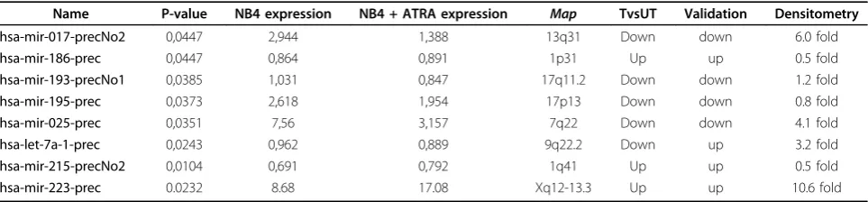
miRNAs expression profile in ATRA treated NB4 cells We compared the miRNA expression profile of termin-ally differentiated neutrophils, NB4 ATRA treated [addi-tional file 4], and untreated NB4 cells. In order to increase the significance of the microarray analysis we hybridised the RNA derived from two independent experiments. The statistical analysis (Welch t-test and SAM) identified 8 miRNAs differentially expressed (Table 1), 72 miRNAs equally expressed before and after treatment [additional file 5] and 163 miRNAs expressed within the background, hence considered as unex-pressed. In detail, the expression ofmiR-186,miR-215
and miR-223 resulted upregulated in ATRA
differen-tiated cells, while the expression of miR-17-5p, miR-25,
miR-193, miR-195, and let-7aresulted downregulated
(the miRNAs bolded were already reported as deregu-lated by ATRA in differentiated NB4 cells in refs. 17
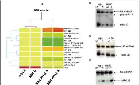
and 18). The cluster analysis, derived from two indepen-dent experiments for the 8 human miRNAs is shown in figure 1A.
miR-17-5p and miR-25 are downregulated in NB4 cells differentiated upon ATRA treatment
The microarray data indicated a strong downregulation of miR-17 and miR-25 in the differentiated phenotype (Figure 1B and 1C, NB4 lanes UT vs. T), two miRNAs not yet described as deregulated during ATRA induced differentiation of NB4 cells. Thus we focalized our ana-lysis on them. We confirmed the microarray data by Northern blot, using as DNA probes the antisense mature sequence of both deregulated miRNA. All north-ern blots were normalized against U6 snRNA. The den-sitometric analysis, carried out on three independent experiments, showed that in ATRA treated NB4 cells the expression of miR-17 decreased about 6 fold while the expression of miR-25 decreased about 4 fold. These results were confirmed also in HL60 cells, (Figure 1B and 1C, HL60 lanes UT vs. T) another promyelocytic cell line inducible to granulocyte differentiation by reti-noids. In both cell lines we observed a proportional cor-relation between the amount of the mature miR-17 and its precursor suggesting that this miRNA is mainly regu-lated at transcriptional level during myeloid differentia-tion. As positive control we checked for the expression of miR-223, upregulated during myeloid differentiation [17]. As expected, miR-223 was found expressed 10 fold higher after ATRA induced differentiation (Figure 1D) in both NB4 and HL60 cells.
[image:3.595.57.539.593.705.2]Byin silico analysis we found that both miR-25 and miR-17 could have as putative targets E2F1 and E2F4, thus, they could synergistically act in the control of pro-myelocytic proliferation and differentiation. To test our hypothesis we analyzed the expression level of E2F1 and E2F4 proteins in untreated and ATRA differentiated NB4 cells. As shown in additional file 6, both the pro-teins as well as the two miRNAs were downregulated by ATRA differentiation. These results would suggest that
Table 1 miRNAs with altered expression after ATRA induced differentation in NB4 cells by microarray analysis.
Name P-value NB4 expression NB4 + ATRA expression Map TvsUT Validation Densitometry
hsa-mir-017-precNo2 0,0447 2,944 1,388 13q31 Down down 6.0 fold
hsa-mir-186-prec 0,0447 0,864 0,891 1p31 Up up 0.5 fold
hsa-mir-193-precNo1 0,0385 1,031 0,847 17q11.2 Down down 1.2 fold
hsa-mir-195-prec 0,0373 2,618 1,954 17p13 Down down 0.8 fold
hsa-mir-025-prec 0,0351 7,56 3,157 7q22 Down down 4.1 fold
hsa-let-7a-1-prec 0,0243 0,962 0,889 9q22.2 Down up 3.2 fold
hsa-mir-215-precNo2 0,0104 0,691 0,792 1q41 Up up 0.5 fold
miR-17-5-p and miR-25 do not target E2F transcription factors, in NB4 cells from Acute Promyelocytic Leuke-mia. For this reasons, further studies on the identifica-tion of genes possessing miR-17-5p or miR-25 complementary sequences are required.
The relevance of miR-223 upregulation during the early stages of myeloid differentiation has been described [17]. We observed a ten-fold upregulation of miR-223 expression in our system, when the CD11+ positive cells are more than 90%, hence terminally dif-ferentiated [additional file 4]. Thus, miR-233 may repre-sent not only an early factor required for myeloid differentiation but it could play an additional important role in the maintenance of the differentiated phenotype.
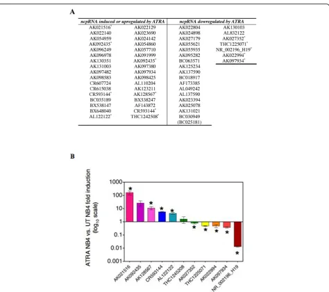
Long non-protein coding RNAs are deregulated during ATRA differentiation of NB4 cells
DNA array hybridisation experiments were performed to compare the expression levels of 492 ncRNA genes in NB4 versus NB4 + ATRA treated cells. Fifty-eight genes were found deregulated (Figure 2A), 144 genes equally
expressed in both NB4 phenotypes and 290 expressed within the background, hence considered as unexpressed. Among the 58 deregulated genes eleven randomly chosen were validated by Real Time PCR (Figure 2B). The statis-tical analysis was performed with Prism v4.0 (GraphPad software).
The variation in the expression of the detected ncRNAs in both NB4 cells phenotypes was analyzed and compared to their expression in several cancer cell lines. In additional file 1 and 2 is shown the Real Time analy-sis in cancer cell lines of twelve deregulate ncRNAs as an example. This analysis allowed us to validate the ontogeny groups assigned through differential expres-sion in proliferating versus differentiated NB4 cells.
The group of deregulated genes, represented by
AK092435, AK130351, AK131003, BC035189,
[image:4.595.55.541.90.385.2]AK023690, AK097380, AK098425 AK128567 and AL110204 was found upregulated by ATRA-induced dif-ferentiation in NB4 cells (Figure 2A). All these genes, highly expressed in cancer cells (data not shown), may therefore be considered as genes not involved in
proliferation of NB4 cells, so that their expression could result from the activity of transcription factors regulated by ATRA signalling, as NF-k-B, C/EBP-a, C/EBP-bor C/EBP-ε. A second group of transcripts found overex-pressed in cancer cell lines and downregulated in ATRA-differentiated NB4 cells is represented by
AK025078 (SLC26A2), AK027179, AK055935,
AK125234 and BC030949, a gene potentially involved in the juvenile leukaemia. These genes are potentially linked to proliferation and they could be upregulated through myc and ras signalling pathway, activated in proliferating NB4 cells [19].
[image:5.595.63.538.84.504.2]AK001558, a gene upregulated in human retinal Muller cells and protecting against oxidative stress [20], was found expressed in cancer cell lines but showed similar expres-sion in both proliferating and differentiated NB4 cells (data not shown). In addition to ncRNAs distributed randomly onto all chromosomes the Ribochip contained 70 tran-scripts from the chromosome 7, to analyse their expression changes associated to the transcripitonal state of the HOXA locus. Although most of these transcripts did not show significant variations, untreated NB4 cells showed several upregulated genes, as 7p21 (GA 3603), downstream of HOXA gene cluster, while after differentiation we found
upregulation of AK026386, in the 7p21 region, as well as GA3749 in 7p13 and AK026119 in 7p14.
Finally,H19is overexpressed in some types of cancers [12] and it was found expressed at highest levels in pro-liferating NB4 cells (Figure 2B) and in tumor cell line such as the ERBB2-overexpressing SKBR3 cell line [additional files 1 and 2] and the 883KE testis cancer cell line [additional files 1 and 2]. Thus H19 seems to be involved in cell proliferation.
All together these data show that, even if the function of long ncRNAs as tumor suppressors or as oncogenes can be only hypothesized to date, their expression is related to cell physiology and in some case is tumor specific.
In conclusion, this work shows that several ncRNAs are potentially involved in proliferation and ATRA-induced differentiation of NB4 cells. The association of functional characterization of the studied genes with their expression in cancer may provide clues on their ontology class, and possibly on their function in the aetiology of diseases. Moreover, the increasing number of ncRNAs whose expression profile changes accord-ingly to the cell phenotype (transformed or terminally differentiated) could represent, in the near future, a sui-table biomarker to characterize differentiation.
Additional file 1: Expression by Real time PCR of 15 ncRNAs in cancer cell lines. RT-PCR of ncRNA expression data using the Ribochip in different cancer cell lines, using primer pairs for selected genes AF143872, AK022994, TCAG1963768, AK094435, THC1225071, AL122122, THC1242508, AK128567, AK098425, H19, CR593144, AF143872, AK021516, AK027352, AK097934, AK097482, randomly selected, and significantly expressed in the cancer cell lines SKBR3, MCF-7, HS-578T (Breast cancer); Be[2]C, CCF-STTG1, DAOY1, (Brain cancer); LS174-T, Caco-2, WiDr, SW1116 (Colon cancer), GLC-4, H226 (Lung cancer), 833KE, NT2, Tera-1 (Testis cancer). As control we pooled human normal tissue total RNA from brain and from testis.
Click here for file
[ http://www.biomedcentral.com/content/supplementary/1756-0500-3-24-S1.PDF ]
Additional file 2: Expression by Real time PCR of 15 ncRNAs in cancer cell lines. See Additional file 1
Click here for file
[ http://www.biomedcentral.com/content/supplementary/1756-0500-3-24-S2.PDF ]
Additional file 3: Additional M&M information. Materials and Methods for Array Fabrication, Fluorescent Labeling and Hybridization, Image Analysis and Data Collection, Human genome bioinformatic analysis, Microarray analysis, ATRA-induced differentiation of promyelocytic NB4 cells. Western blot analysis and Primers sequences for Real Time validation.
Click here for file
[ http://www.biomedcentral.com/content/supplementary/1756-0500-3-24-S3.DOC ]
Additional file 4: Representative experiment of ATRA induced differentiation of NB4 cells assayed by FACS analysis. A and B) CD11
+
FACS readout in NB4 untreated (A) or ATRA treated cells (B) using PE-conjugateda-CD11c mAb; C and D) cell viability FACS readout in NB4 untreated (C) or ATRA treated cells (D) using propidium iodine. Click here for file
[ http://www.biomedcentral.com/content/supplementary/1756-0500-3-24-S4.PDF ]
Additional file 5: miRNAs equally expressed in both ATRA treated and untreated NB4 cells by microarray analysis. Hsa = human miRNAs; mmu = miRNAs
Click here for file
[ http://www.biomedcentral.com/content/supplementary/1756-0500-3-24-S5.PDF ]
Additional file 6: E2F1 and E2F4 proteins level don’t correlate with the miR-25 and miR-17 expression in NB4+ ATRA treated cells. 30
μg of total proteins were extracted at the indicated times from ATRA treated (T) or untreated (UT) NB4 cells and analyzed by western blot usinga-E2F1 anda-E2F4 polyclonal antibodies (Santa Cruz). Input proteins were equalized by detecting the endogenousg-tubulin. Click here for file
[ http://www.biomedcentral.com/content/supplementary/1756-0500-3-24-S6.PDF ]
Acknowledgements
We thank Dr. M.F. Romano for the help with the FACS analysis; Prof C.M. Croce, Dr. G. Calin and Dr. Chang-gong Liu (Ohio state university) for performing the miRNA microarray; Dr. Concetta Ambrosino for the critical reading of the manuscript.
Author details
1Department of Biochemistry and Medical Biotechnologies, University of
Naples Federico II, Via S. Pansini 5, Napoli, Italy.2Biotecgen, Ecotekne, via prov.le Monteroni, Lecce, Italy.3ISPA-CNR Ecotekne, Lecce, Italy.4Department
of Experimental and Diagnostic Medicine, Interdepartmental Center for Cancer Research, University of Ferrara, Ferrara 44100, Italy.5Department of
Pharmaceutical Sciences, University of Salerno, via Ponte Don Melillo, Fisciano SA, Italy.
Authors’contributions
A.R., G.G. and M.G.C. performed the miRNA experiments; O.F.D and P.P. performed the long ncRNAs experiments and statistical analysis; M.F. and M. N. provided the miRNA microarray statistical analysis; P.R. and S.B. contributed to manuscript preparation; M.M. conceived and supervised this study and contributed to manuscript preparation. All authors read and approved the final manuscript.
Competing interests
The authors declare that they have no competing interests.
Received: 16 July 2009
Accepted: 27 January 2010 Published: 27 January 2010
References
1. Goodrich JA, Kugel JF:Non-coding-RNA regulators of RNA polymerase II transcription.Nature Rev Mol Cell Biol2006,7:612-616.
2. Costa FF:Non-coding RNAs: new players in eukaryotic biology.Gene
2005,357:83-94.
3. Pasquinelli AE, Hunter S, Bracht J:MicroRNAs: a developing story.Curr Opin Genet Dev2005,15:200-205.
4. Ambros V:microRNAs: tiny regulators with great potential.Cell2001,
107:823-826.
5. Mallardo M, Poltronieri P, D’Urso OF:Non-protein coding RNA biomarkers and differential expression in cancers: a review.J Exp Clin Cancer Res
2008,27:19.
6. Calin GA, Croce CM:MicroRNA-cancer connection: the beginning of a new tale.Cancer Res2006,66:7390-7394.
7. Calin GA, Croce CM:MicroRNAs and chromosomal abnormalities in cancer cells.Oncogene2006,25:6202-6210.
8. Claverie JM:Fewer genes, more noncoding RNA.Science2005,
309:1529-1530.
9. Mattick JS, Makunin IV:Non-coding RNA.Hum Mol Genet2006,15:R17-29. 10. Mendes Soares LM, Valcárcel J:The expanding transcriptome: the
11. Berteaux N, Lottin S, Monté D, Pinte S, Quatannens B, Coll J, Hondermarck H, Curgy JJ, Dugimont T, Adriaenssens E:H19 mRNA-like noncoding RNA promotes breast cancer cell proliferation through positive control by E2F1.J Biol Chem2005,280:29625-29636. 12. Cai X, Cullen BR:The imprinted H19 noncoding RNA is a primary
microRNA precursor.RNA2007,13:313-316.
13. Kluiver J, Poppema S, de Jong D, Blokzijl T, Harms G, Jacobs S, Kroesen BJ, Berg van den A:BIC and miR-155 are highly expressed in Hodgkin, primary mediastinal and diffuse large B cell lymphomas.J Pathol2005,
207:243-249.
14. Rodriguez A, Vigorito E, Clare S, Warren MV, Couttet P, Soond DR, van Dongen S, Grocock RJ, Das PP, Miska EA, Vetrie D, Okkenhaug K, Enright AJ, Dougan G, Turner M, Bradley A:Requirement of bic/microRNA-155 for normal immune function.Science2007,316:608-611.
15. Lanotte M, Martin-Thouvenin V, Najman S, Balerini P, Valensi F, Berger R:
NB4, a maturation inducible cell line with t(15;17) marker isolated from a human acute promyelocytic leukemia (M3).Blood1991,77:1080-1086. 16. Melnick A, Licht JD:Deconstructing a disease: RAR alpha its fusion
partners and their roles in the pathogenesis of acute promyelocytic leukemia.Blood1999,93:3167-3215.
17. Fazi F, Rosa A, Fatica A, Gelmetti V, De Marchis ML, Nervi C, Bozzoni I:A Minicircuitry Comprised of MicroRNA-223 and Transcription Factors NFI-A and C/EBPalpha Regulates Human Granulopoiesis.Cell2005,
123:819-831.
18. Garzon R, Pichiorri F, Palumbo T, Visentini M, Aqeilan R, Cimmino A, Wang H, Sun H, Volinia S, Alder H, Calin GA, Liu CG, Andreeff M, Croce CM:
MicroRNA gene expression during retinoic acid-induced differentiation of human acute promyelocytic leukemia.Oncogene2007,26:4148-4157. 19. Sampson VB, Rong NH, Han J, Yang Q, Aris V, Soteropoulos P, Petrelli NJ, Dunn SP, Krueger LJ:MicroRNA let-7a down-regulates MYC and reverts MYC-induced growth in Burkitt lymphoma cells.Cancer Res2007,
67(20):9762-70.
20. Li C, Tang Y, Li F, Turner S, Li K, Zhou X, Centola M, Yan X, Cao W: 17b-Estradiol (bE2) Protects Human Retinal Muller Cell Against Oxidative Stress In Vitro: Evaluation of Its Effects on Gene Expression by cDNA Microarray.Glia2006,53:392-400.
doi:10.1186/1756-0500-3-24
Cite this article as:Rossiet al.:Non-coding RNAs change their expression profile after Retinoid induced differentiation of the promyelocytic cell line NB4.BMC Research Notes20103:24.
Submit your next manuscript to BioMed Central and take full advantage of:
• Convenient online submission
• Thorough peer review
• No space constraints or color figure charges
• Immediate publication on acceptance
• Inclusion in PubMed, CAS, Scopus and Google Scholar
• Research which is freely available for redistribution