Vol.44, No.1 JOURNALOFVIROLOGY, OCt. 1982,p.401-412
0022-538X/82/100401-12$02.00/0
Copyright©1982,AmericanSocietyforMicrobiology
DNA
Clone
of Avian
Fujinami
Sarcoma Virus with
Temperature-Sensitive Transforming Function in Mammalian
Cells
WEN-HWA LEE,t*CHIH-PINGLIU,tAND PETERDUESBERG DepartmentofMolecular Biology, Universityof California, Berkeley, California 94720
Received 15April 1982/Accepted 17 June 1982
Wehave molecularlycloned anintegrated proviralDNA ofFujinami sarcoma
virus (FSV) into a lambda phage vector and further subcloned it into plasmid pBR322. Thesourceofproviruswas aquailnonproducer cell clone transformed
by FSV. The FSV strain used is temperature sensitive in the maintenance of transformation of avian cells.Therecombinantplasmidwasshown to contain an
entire FSV genome by fingerprinting the hybrids formed with 32P-labeled FSV RNA. Thisanalysis also revealedapreviously undetected env-related sequence in
FSVwhichrepresents the 3' endofthegp85 envgene. Aphysicalmapofcloned FSV DNA identifying sites of several restriction enzymes is described. Upon transfection, FSV DNA cloned in pBR322 transformed mouse NIH-3T3 cells,
which proved to be temperature sensitive in maintaining transformation. Phos-phorylation butnotsynthesis of p140,theonly knowngeneproductofFSV,was
also temperature sensitiveinthese cells. The correlation between transformation
and phosphorylation ofp140 suggests that phosphorylation ofp140 isnecessary
for transformation ofmousecells, as wasshownpreviouslyfor avian cells. These results provide direct genetic evidence that the mechanisms for maintaining transformation of mammalianand aviancells involve the same FSV geneproduct, p140. Homology wasdetected by hybridizationbetween transformation-specific sequencesofFSV DNAand certainrestriction endonuclease-resistant fragments of cellular DNA of two avian species, chicken and quail. Under the same
conditionshomologywasalso detected with DNA of non-avianspecies, although apparentlyto alowerdegree thanwith avian cells.
Fujinamisarcoma virus(FSV), originally iso-latedby
Fujinami
andInamotoin 1914(12), isadefective avian sarcoma virus having an RNA genome of 4.5 kilobases (kb) (20). This RNA
contains a
contiguous
internalFSV-specific
se-quence of about 3 kb flanked by a 5'-terminalsequenceof1 kb anda 3'-terminal sequenceof
0.5 kb related to the RNA of nondefective Fujinami-associated virus (FAV), the helper vi-rus ofFSV (20). A helper-virus-unrelated,
spe-cific sequence is considered to be the hallmark ofthe onc genes of acutely oncogenic
retrovi-ruses (10) and has been shown in other highly oncogenicviruses to be closely related to cellu-larsequenceprototypes (27, 33, 35). The
specif-ic sequence of FSV has been reported to be
closely related to that of avian sarcoma virus
PRCIIand also to theSynder-Theilenand Gard-ner-Amstein strains offeline sarcoma virus by
molecular hybridization(29). Together with the 5' helpervirus gag gene-related region of FSV
tPresent address:CetusCorporation, Berkeley,CA94710.
RNA, the specific sequence encodes a
gag-related protein of140,000daltons, termed
p140
(16,20).p140 istheonlyknowngeneproductof FSV and is thought to have a transforming function (16, 19, 20). From studies ofwild-type
and temperature-sensitive mutants of FSV (15,
19), it was concluded that phosphorylation of
p140
is necessary forinitiating and maintaining transformation of infected avian cells (19, 24).Moreover,
p140
obtained by immunoprecipita-tion from infected cells is associated with akinase that phosphorylates p140 in vitro(2, 11). However, itis as yetuncertain whetherp140 is associated with a kinase or has an intrinsic kinase activity, because most of this kinase activity is separated from
p140
by sedimentation(2),
p140
synthesized in vitro or purified byimmunoaffinity chromatography does not have
detectable kinaseactivity, andp140in tempera-ture-sensitive FSV is a temperature-sensitive
substrate of phosphorylation (19; W.-H. Lee, Ph.D.thesis,Universityof
California,
Berkeley, 1981).Thispaperdescribesthepreparationand char-401
on November 10, 2019 by guest
http://jvi.asm.org/
acterization of a molecular clone of proviral FSV DNA that represents a complete comple-ment of FSV RNA and that upon transfection transforms mouse 3T3 cells. The clone was prepared from an FSV strain that is temperature sensitive in maintaining transformation of avian cells (19). As we show here, maintenance of the transformed phenotype of mouse 3T3 cells also proved to be temperature sensitive. Moreover, phosphorylation of p140 in the 3T3 cells trans-formed withcloned FSV DNA was temperature sensitive both in vivo and in vitro. It would appear that the mechanisms for maintaining
transformationinavian and mammalian cells by FSV are similar and both involve a temperature-sensitive viral gene product, p140. Previous
ex-perimentsreporting transformation of mammali-ancells with defective avian tumor viruses have not been able to distinguish between direct
transformationby aviralgene product and indi-rect transformation by positional effects of
pro-viral integration, due to the low transforming
efficiency of avian viruses (25) and proviral DNA (6, 18) on mammalian cells.
The availability of this clone should also allow a detailed investigation of the relationships be-tween the transformation-specific sequence of
FSV and those of other related viruses and of cellular prototypes in avian and mammalian cells.Our preliminary results indicate that under the same conditions ofhybridization, the DNA of quail and chicken cells was found to have counterparts related to the FSV-specific se-quence, but less homology was detected with DNAsofnon-avian origin.
MATERIALS AND METHODS
Cells and viruses. A quail cell nonproducer clone,
infected by ts FSV L-15, was grown in medium F10
supplemented with 2% tryptose phosphate broth, 2%
calf serum, 1% chicken serum, and 0.5% dimethyl
sulfoxide. NIH-3T3 cells and one NIH-3T3 cell line transformed by transfection with cloned ts FSV L-15
DNA, termedF3B,were grown in Dulbecco modified
Eagle medium containing 10% fetal calf serum.
Preparation of [32P]cDNA. [32P]cDNA from
FSV-(FAV) was prepared by using either
oligodeoxy-thymidylates12 to 18(oligo(dT) 12-18)or
oligonucleo-tides prepared byrandomdegradation of calf thymus
DNA to prime reverse transcription ofpolyadenylic
acid-selected 28-35S FSV(FAV) RNA(13). Atypical
0.1-ml reaction mixture contained 0.002 mg of
FSV(FAV) RNA,1 mMeachdATP,dGTP,andTTP,
0.5 mCi of[a-32P]dCTP (400 Ci/mmol), 0.001 mg of
oligo(dT) 12-18,20 mMTris-hydrochloride, pH 8.0,5
mM MgCl2, 10 mMdithiothreitol, 0.01 mg of
actino-mycin D, and 40 U of reverse transcriptase. The
reactionwascarried out at39°C for2hand terminated
by adding 1% sodium dodecylsulfate(SDS)and 0.1 N
NaOH,and themixturewas thenincubatedat39°Cfor
4h toallowthe RNAtemplatetobedegraded. After
being neutralized with 0.1 N acetic acid-0.1 M
Tris-hydrochloride (pH 7.4), the reaction mixture was
extractedwith phenol and chromatographed on
Sepha-dex G-50 with buffer containing 10 mM
Tris-hydro-chloride (pH 7.4), 100 mM NaCl, 1 mM EDTA, and
0.1% SDS. Usually, 5 x 10⁷ to 30 x 107 cpm of
[32P]cDNA was obtained from this reaction, with a
specific activity of2 x 105 cpm/ng of DNA.
Nick translation ofDNA. Nicktranslation of DNA
wasperformed essentially as described by Rigby et al.
(26)with one32P-labeleddeoxyribonucleotide
triphos-phate. Briefly, 0.1 ml of reaction mixture contained
0.001 mgof DNA, 0.0025 mM each dATP,dGTP, and
TTP, 100 ,uCi of[a-32P]dCTP(400 Ci/mmol), 50 mM
Tris-hydrochloride (pH 7.5), 10 mM MgCl2, 1 mM
dithiothreitol, 0.005 mg of bovineserumalbumin, 0.2
ngof DNaseI, and 5UofDNApolymerase I.After
being incubatedat 10°C for 90 min, the reactionwas
terminated by the additionof 0.01 ml of 20% SDS and
further incubation at 65°C for 10 min. The reaction
mixture was then subjected to chromatography on
Sephadex G-50. Usually, 0.001 mg of DNA with a
specificactivity of 2x 10⁴to10 x 104cpm/ng ofDNA
was obtained. The DNA was denatured with 0.1 N
NaOH at room temperature for 10 min and then
neutralized with1 Nacetic acid beforeusein
hybrid-ization experiments.
Preparation of cellular DNA. FSV L-15quail
non-producer cellDNAwas extractedasdescribed
previ-ously (3). Cells (108)werelysed and digested in10ml
of buffercontaining0.1 M EDTA(pH7.5), 1%
sarco-sinate, and 0.01 mg of proteinaseK at56°C for 2 h. The
mixture was then phenol extracted and dialyzed
against TE buffer (10 mM Tris-hydrochloride, 1 mM
EDTA, pH7.5).RNA wasthendigested by incubation
with 0.36 mg of RNaseAat40°C for 4 h. After phenol
extraction anddialysis, the DNA was quantitated by
measuring absorbancy at260nm.
Hybridizationof DNAfragmentsresolvedby agarose
gel electrophoresis. Electrophoretic DNA fragments
contained in agarosegelsweretransferredto
nitrocel-lulose filters by the method of Southern (31). The
DNAin the agarosegelwasdenaturedbysoakingthe
gel in 0.5 N NaOH-1.0M NaCl for30min and then
neutralized with1 M Tris-hydrochloride (pH 7.4)i.5
MNaCl for another30min. TheDNA wasallowedto
transfer tothe nitrocellulose filter for3 to 24h with
lOx SSC (lx SSC, 0.15 M NaCl-0.015 M sodium
citrate). Afterbeingheatedfor3 h at80°C invacuo,
the filters were incubated for 4 to 12 h at 68°C in
prehybridization buffer containing 6x SSC, 0.02%
Ficoll, 0.02%polyvinylpyrrolidone,and0.25%SDSat
pH 7.0. Hybridizations with 32P-labeled DNA
frag-ments orcDNAwerecarriedoutinthesamebuffer for 3 to24 hat68°C. Afterhybridization,the
nitrocellu-losefilterswerewashed with1x SSC-0.1% SDS for3
h at68°C, dried,andsubjected toautoradiography.
Construction and screening of a charon 27 phage library containing FSV-transformed quail cell DNA.
FSVL-15cellDNA(0.2 mg)wasdigestedcompletely
with 400 U of restriction endonucleaseBgIII at37°C for3 h andsubjectedto5to20% NaCl(inTEbuffer)
gradientcentrifugation inanSW41rotor at35,000 rpm
for3 hat20°C.Afterbeingfractionated andanalyzed
by agarosegelelectrophoresis, thefractions
contain-ing DNA fragments 6 to9 kb longwere pooled and
ethanol precipitated. The BglII-digested cell DNA
fragments (0.005 mg) were ligated with 0.02 mg of
on November 10, 2019 by guest
http://jvi.asm.org/
DNA CLONE OF TEMPERATURE-SENSITIVE FSV 403
charon 27 DNA whichhad beendigestedto
comple-tion byrestriction endonuclease BamHI. Ligationwas
performed by overnight incubation ofthe DNA in50
mM Tris-hydrochloride (pH 7.4)-10 mM MgCl2-20
mMdithiothreitol-1mM ATP-5 UofT4DNAligaseat
10°C. Thetotal DNAwasthenpackaged in vitro into
phageasdescribed previously (3).
The resulting phage, about 106 PFU, were mixed
with 8 ml ofafreshovernight culture ofEscherichia
coliK802and plated ontwocookie sheets (42by 24
cm)ataphageconcentrationof 5x 10⁵PFUpersheet.
After the phagehadgrown overnightat 37°C,
dupli-catesofnitrocellulose filters moistened in 1 MNaCl
wereplaced oneachtrayfor10min. The filterswere
thenremoved from thetrays,inverted, andprocessed
insuccessive10-min incubations by floatingthefilters
on denaturing buffer (0.5 N NaOH, 1 M NaCi) and
then onneutralizing buffer (1 MTris-hydrochloride,
pH 7.4, 1.0 M NaCI). The filters were air dried and
baked invacuoat80°Cfor 2 h. One oftwoduplicate filterswas hybridizedwith aBamHI-resistant 1.3-kb
DNA fragment of the gag gene of Schmidt-Ruppin
Rous sarcoma virus(9, 29) labeled with 32P by nick
translation, and the other was hybridized with
FSV(FAV) [32P]cDNA. Afterautoradiography,
posi-tive plaqueswerepicked, replated,andrescreenedto
purify the phage as previously described (1).
Large-scalegrowth of recombinantlambdaphagewascarried
outessentiallyasdescribedpreviously (3).
Subcloning DNA fragments into E. coli plasmid
pBR322. Typically,500ngofpBR322DNAand1,000
ng of the FSV-containing charon 27 DNA to be subcloned, respectively, were cleavedto completion
by the appropriate restriction endonucleases. These
DNA solutions were mixed, phenol extracted, and
ethanolprecipitated. The DNAwaspelletedand ligat-ed under the conditions describligat-ed above.
The procedures usedfor transformation ofE. coli
strain HB101 with the plasmid pBR322 DNA were
those described previously (22). The transformation-competentHB101 cellswereharvestedby
sedimenta-tion inaSorvall GSArotorat5,000rpmfor5 minat 2°Cduring thelog phaseofgrowth (absorbancyat600
nm,0.6)inLuria broth(10gof tryptone[Difco], 5gof yeast extract, and 5g ofNaCl per liter) andtreated twicewith 50 mMCaCl2 for 20minonice. Asampleof
HB101cellswasusedfor transfection with theligated
DNAatafinalconcentration of 25 mMCaCl2-50mM
Tris-hydrochloride (pH 7.2) at 0°C for 15 min. The
cells were heat shocked at41°C for 2 min and then chilled at0°Cfor another 30min. Luria broth (1 ml)
was added to the HB101 cells, and they were then
incubated at 37°Cfor 30min. Portionsof transfected HB101 cells were plated on agar plates containing 0.015 mgoftetracycline or0.03 mgofampicillin per
ml. Recombinant clones were purified from a
cross-selection based on the drug-resistant or -sensitive phenotype (8).
Theprocedures usedto preparethe plasmidDNA
weremainlyadoptedfromDavisetal. (8) with slight modifications. HB101 colonies which carried
recombi-nantplasmidswere picked, andeachwasgrownin 3
ml ofLuria brothovernight.Cells from 0.5 ml of each culturewerepelleted, suspendedin0.1 mlofbuffer I containing 50mMTris-hydrochloride (pH 8.5),50 mM
EDTA,15%sucrose,and 1mgoflysozymepermlfor
20 min on ice. Buffer 11(0.2 ml) containing 0.2 N
NaOH and1% SDSwasthenadded, and the cellswere
left for 5 min onice and then neutralized with 0.15 ml
of 3 M sodium acetate, pH 4.8. The mixture was
incubated on ice for 50 min and centrifuged in an
Eppendorf Microfuge for10min.PlasmidDNAin the
supernatant was ethanol precipitated, dissolved in
0.1 mM EDTA, and analyzed for FSV-related
se-quences by restriction enzyme cleavage and
hybridiza-tion.
Transfection andtransformation assays of mouse 3T3
cellswith clonedFSV DNA. Calcium phosphate-DNA precipitates used for transfection of 3T3 cells were
prepared aspreviously described (14). To about 20 ,ug
of DNA in 1 ml of 2x HBS (280 mM NaCi, 50 mM
HEPES
[N-2-hydroxyethylpiperazine-N'-2-ethanesul-fonic acid], and 1.5 mM Na2HPO4 at pH 7.05) we gradually added an equal volume of freshly prepared
0.25 mM CaC12 as air was bubbled through the
solu-tion. The mixture was blended briefly withaVortex
mixer and allowed to formprecipitates for 30 min at
room temperature. Three types of precipitates were
prepared. One type was prepared with 20 ,ug of quail
cellDNAalone; others were prepared with 0.05 to1
jig
of pFL-5 DNAwith 20 ,ugofquail cell DNA; andanother typewasprepared with 0.5 ,ug of simian virus
40DNAand 20 ,ug of quail cell DNA. Theprecipitate
(1ml) was then addedtorecipient cultures of3T3cells
ina60-mmdish in 10 ml of medium. After8 to12h,the
medium was changed. The recipient cultures were
prepared by seeding2 x 1053T3cells and incubating
the culturesovernight. One group of cellswaskeptin
liquid medium, and the medium was changed every
otherday. Fociwerescored after7 to 10daysat37°C.
The othergroupof cellswerereseeded in soft agarto
allow formation of colonies. The method for agar
colony formation was essentially that previously
de-scribed (19), except that Dulbecco modified Eagle
medium with10% fetal calfserum wasused.
RESULTS
Construction of a recombinant DNA libraryof
FSV-transformedquail cellDNA in phage lambda and isolation ofFSV DNA-containing phage. A
quail cellclone,nonproductivelytransformed by astrain of FSV(L-15)thatistemperature
sensi-tiveinmaintainingtransformation of avian cells, wasour sourceof proviral FSV DNA (19). The DNA from this cell clone was digested with restriction enzymes BamHI, EcoRI, HindIll,
and BglII. In parallel, DNA from uninfected
quailcells wasdigested withthe same enzymes.
Fragments of eachDNA were thencompared by agarose gel electrophoresis, followed by
hybrid-izationwith
[32P]cDNA
from FSV(FAV). It wasfound that a BglII-resistant DNA fragment of FSV L-15cell DNAof 8 to 9 kb which hybrid-izedwith the
32p
probedid not have a counter-partinBglII-digested uninfected quail cell DNA(Fig. 1). The other three restriction enzymes either generated too many fragments or gave
unclear results. Thus,BglIIwas chosen to digest FSV L-15 DNA for ligation with the external DNAfragments (arms) of lambda phage charon
VOL. 44,1982
on November 10, 2019 by guest
http://jvi.asm.org/
a
b
* * kb
-
12
..w
9
S
*
A
two different 32P-labeled DNA probes: a BamHI-resistant 1.3-kb DNA fragment of the
gag gene of Schmidt-Ruppin Rous sarcoma
vi-rus (RSV) thatstarts atabout 0.5 kb from the 5'
end ofthe RSV genome (9, 28) or cDNA from
FSV(FAV) prepared by transcription of FSV(FAV) RNA with reverse transcriptase in the presence of an oligo(dT) 12-18 primer (see above). Onlyoneautoradiographic spot
coincid-ed on two replicate filters of the same culture
hybridized with the two different probes. The plaque that hybridized with both probes was picked,replated, and plaque purified three times until 95% of all daughter plaqueswere positive with both hybridization probes. The recombi-nantphageselected by the above procedurewas termed CFL.
Weprepared 0.5 mg of CFLDNAforanalysis of its restriction patternby growing the phageon alarge surface of soft agar after infection at a multiplicity of about 0.01 PFU per bacterial cell. The CFLphage DNA was prepared and digested with BgIII,an 8.4- and a 4.1-kb fragment hybrid-ized with the FSV(FAV) cDNA probe, but only the 8.4-kb fragment hybridized with the gag DNAprobe as well (Fig. 2). Since the insert was
[image:4.496.75.201.50.392.2]I
FIG. 1. FSV-related sequences in the DNA of
normalquail cells inanFSV-transformed nonproducer
quail cells. DNA from uninfected quail embryo fibro-blasts (a) or from ts FSV L-15 quail nonproducer cells(b)wasdigested with BgII. After being electro-phoresedon an0.8%agarosegel, the DNA fragments were transferred to nitrocellulose filters and hybrid-ized with[32P]cDNA from FSV(FAV). Both celltypes
appeartoshareanFSV-related fragment of 13 kb. A
fragment of 8to9 kbwasdetected only in the DNA
from the FSV L-15nonproducer cell clone.
27 (4). Since BglII produces DNA fragments with thesamefour-base, single-stranded endsas
BamHI does, the DNA fragments generated by BglIIcan beconveniently inserted into the sin-gle BamHI site of charon 27 DNA by base pairing and ligation (F. Blattner, personal
com-munication). However, both the BamHI and BglII restriction sites would be lost after liga-tion.Roughly 106 phage wereobtainedby
pack-aging in vitro 0.005 mg ofBglII-cut FSV L-15 cell DNA and0.02 mgof BamHI-cut charon 27 DNA arms.
The resulting librarywasscreened for recom-binantphage containing FSV DNA by hybridiz-ing DNA from about 106 lambda plaques with
a
b
c
kb
8.4
a-@
4.1
-FIG. 2. A recombinant lambda charon 27 phage
carrying proviral FSV DNA. DNA from a
recombi-nant phage was digested withBglII and
electropho-resedon an0.8%agarosegel. After being transferred
tonitrocellulose,the DNAfragmentswerehybridized
with three different 32P-labeled DNA probes: (a) a
combination ofDNA from theoriginal lambda charon
27andFSV(FAV)cDNAprepared bytranscription of
FSV(FAV) RNA with reverse transcriptase and an
oligo(dT) primer(seethe text);(b)FSV(FAV)cDNA
asin lanea;(c) aclonedBamHI-resistant1.3-kb DNA
fragment from thegag geneof RSV (9, 28). Asingle
8.4-kb fragment was hybridized with both probes b
andc.
on November 10, 2019 by guest
http://jvi.asm.org/
[image:4.496.302.400.341.538.2]DNA CLONE OF TEMPERATURE-SENSITIVE FSV
preparedfrom FSVL-15 cell DNAdigestedwith BglII, these particular BglII sites must have
survivedtheoriginal digestion. The4.1-kb
BgIII
DNAfragment may be a deleted variant ofthe
8.4-kb fragment or the product of another
un-known sequence rearrangement. However, the DNA of the clone changed its restriction enzyme
pattern, and much of the resulting phage lost FSV-related sequences when phage-infected culturesweregrown inliquid broth. Some ofthe
resultingphage hadeithernoinsertofFSV DNA sequences or only a 1- to 1.5-kbinsert of FSV-related sequences. Therefore, we decided to
subclone the 8.4-kb fragment that hybridized
with both FSV(FAV) cDNA and gag DNA into plasmid pBR322.
Recombinant pBR322 plasmidcontaining FSV DNA (pFL-5) and its restriction endonuclease map. Toobtain stable FSV recombinant DNA,
we subcloned the BglII-resistant 8.4-kb DNA
fragment from the recombinant lambda phage
described above into the bacterial plasmid pBR322. Several recombinant plasmids were
obtained by the subcloning procedure.
One,
termed pFL-5, containedtheBglII-resistant
8.4-kb DNA fragment which hybridized with both the gag DNA and FSV(FAV) cDNA probes. pFL-5 proved tobe stable whengrown in large quantities. To establish aphysical map of FSV DNA, weanalyzedthepFL-5DNAby cleavage with restriction endonucleases BamHI, KpnI, PvuI, SmaI,andSstI. Restrictionenzymessuch
as
EcoRI, HindlIl,
BgiII,
Sall,andXbaIhad nocleavage site in the FSV DNA (Fig. 3). The
restriction enzyme map resulting from these digestions is shown in Fig. 4. Two long terminal repeat (LTR) sequences ofFSV were identified by hybridization with 32P-labeled DNA from a recombinant plasmid, p53, carrying the LTR of
RSV (23). Like the LTRs of MC29 virus(18) and Rous-associated virus-0 (17), but unlike the LTRsof RSV (7, 9, 23; D. Schwartz, R. Tizard,
and W.Gilbert, Cell, inpress), the LTRofFSV
did not contain an EcoRI restriction enzyme cleavage site. In accord with previous analyses
of FSV RNA (20), the gag gene-related se-quenceelementof FSV was located adjacentto
theleft-hand LTRof FSV(Fig. 4). The relative
locations of the gag-related and FSV-specific
elements in theclone (Fig. 4) andin viral RNA (20), and thecoincidence ofthe 2.7-kb
BamHI-resistantfragment in the clone and in the DNA
of FSV-transformed quail cells detectable in a
genomicblot (datanot shown), signaledthatthe pFL-5 clone was probably colinear with authen-tic proviral DNA. The biological activity of pFL-5, described below, confirmed this notion.
The 5' and3'junctionsbetween theFSV
provi-rus andquail cell DNA in pFL-5havenot asyet
been clearlydetermined.
pFL-5 contains the entire FSV genome. To
determine whatportion oftheFSVgenome was present inthe plasmid pFL-5, we annealed 32p_
labeled FSV(FAV) 50-70S RNA with alkali-degraded pFL-5 DNA to form RNA-DNA
hy-brids asdescribed previously(21). After RNase Ti digestion, the hybrid was isolated, and the [32P]RNA of the hybrid was fingerprinted (Fig.
A
1 3 5 7 9 11 13 15
2 4 6 8 10 12 14
B
17 19 21 23 25 27 29 16 18 20 22 24 26 28 30
-a
04
_l
46
C
31 33 35 37 39 41 43 45
32 34 36 38 40 42 44
a *n @a
-_
FIG. 3. Electrophoretic analysis of recombinant pBR322 plasmid pFL-5 carrying proviral FSV DNA and quail cell DNA after digestionwithrestrictionenzymes. Lanes:1,16,and31,BamHI; 2, 17,and32, KpnI; 3, 18, and33, SmaI; 4, 19, and34, SstI; 5, 20,and35, HindIIIIBamHI; 6,21, and36, HindIIIIKpnI; 7, 22, and37, HindIIIISalI; 8, 23,and38,HindIII/SmaI; 9, 24,and 39HindIII/SstI; 10, 25,and40,HindIIIIBgIII; 11, 26,and
41,BamHlKpnI;12, 27,and42,BamHIISalI; 13, 28,and43,BamHI/SmaI; 14, 29,and44,BamHIISstI; 15, 30,
and45,BamHI/XbaI. Electrophoresis wasdone in 0.8%agarosegelsas described in the text. Panel A: DNA fragments stained with ethidium bromide. Panels B and C: Hybridizations ofthesefragments transferred to nitrocellulose bythe method of Southern(31) withFSV(FAV) [32P]cDNAand 32P-labeled gag DNA(seethe legendtoFig. 2andthetext).
kb 23.6 9.5
6.7 4.3
2.3 2.0
0.6-VOL.44, 1982 405
on November 10, 2019 by guest
http://jvi.asm.org/
[image:5.496.57.447.436.588.2]DUESBERG
0
n
c 0 0
l l
l-&gag FSV-specific tenv Quail cell pBR 322
N
c
1. 1
kbI I
0 1 2 3 4 5 6 7 8 9 10 11 12 12.6
FIG. 4. Restrictionendonuclease mapand organization of the FSV proviral DNA insert in plasmid pFL-5. Thesize of the FSV DNA proviruswasdeduced from the sizes of the respective FSV-related fragmentsshown in Fig. 3 and is in accord with theknown size of FSV RNA (4.5 kb) (20).Asequenceelementrelatedtothegaggene
of aviantumorviruses, Agag,wasidentifiednearthe left LTR, anda sequenceelement relatedtotheenvgene,
Aenv,wasidentifiednearthe rightLTR of the FSVgenome.The locations of the junctions between the LTRs of
FSV and cell DNAaretentative. pFL-5waspreparedasfollows:aBglII digest of cellular DNA carrying FSV
proviruswasinitiallyclonedinto theBamHIsiteoflambda charon 27. ABglll-resistantfragment of 8.4 kbwas
thencut outof the charon DNAattheBgIII siteand atanadditional, previously undigestedBgII site of the cellularDNA insert(seethetext).This 8.4-kbBglII-resistant fragmentwasthen clonedintotheBamHlsite of
pBR322togeneratepFL-5. Hence,the 79 base pairs ofcharonDNA thatseparatetheBglIl site fromits original BamHl site (F. Blattner, personal communication) definetheleft-handborder of the 8.4-kbFSV proviral insert ofpFL-5. The distance between the 79-base-pairsequenceandtheleft-handLTRof FSVmustbe short but isnot
known exactly (question mark).Since the BglII-resistant insert ofthe recombinant charon phagewascloned into
theBamHI site of pBR322, theligation sites of pFL-5are nolonger sensitive toBamHIorBgII (see text).
5B). By comparing this fingerprint with that
obtained from the 4.5-kb FSV RNA(Fig. 5A), it
can be seen that all of the unique, specific
(numbers 2, 3, 6, 7, 8, 9, 10,11, 12,and13), and group-specific oligonucleotides of FSV (num-bers 1, 4, 5, cap, and probably 14)were
recov-eredfrom the DNA-RNAhybrid. Inthe
finger-print of the DNA-RNA hybrid (Fig. 5B), the autoradiographic intensity ofthegroup-specific oligonucleotides was higher than that of FSV-specific oligonucleotides. Thisreflects the
pres-ence of FSV and helper FAV-derived
group-specific oligonucleotides in hybrids formed by annealingFSV and FAV RNA withan excessof pFL-5 DNA (Fig. 5). The identity ofall of the FSVoligonucleotides in thehybridwas verified by analyzing their RNaseA-resistant fragments (20). We conclude that pFL-5 contains a DNA equivalentof the entire FSV genome.
It was surprising that FAV oligonucleotide
115, which was previously found to be part of
the env region in FAV RNA but not in FSV RNA (20), was also hybridized by cloned FSV
DNA. This result indicates that FSV probably
containsasmallregionof env-related sequences
which were not so identified previously (20).
Since FSV RNA lacks oligonucleotide 115, the
env-relatedregionof FSVappearstodiffer inits
primarystructurefrom that of FAV.
Oligonucle-otide 115 maps at sequence positions 6,198 to
6,212ofPragueRSV subgroupC cDNA, which isnearthe3'endofthegp85envgene(Schwartz et al., in press). Since the env gene encodes
a 5' product, gp85, and a 3' product, gp37, the oligonucleotide sequence we found is located approximately in the middle of the env gene
(Schwartzet al., inpress). Itwouldappearthat
FSV contains an internal fragment of the env
geneof avian retroviruses.
Biological activity of cloned FSV DNA. (i) Maintenance of mouse cell transformation
con-trolledbyatemperature-sensitivegeneproductof
FSV. Totestits biologicalactivity,in particular
the transforming function of FSV DNA, we
transfected pFL-5 DNA into mouse NIH-3T3
cells bythe calcium phosphate procedure (14).
Simian virus 40DNA servedas apositive
con-trol,andquailcellDNAwasusedasthe carrier. pFL-5-transfected 3T3 cells formed foci (Fig.
6A) which were similar to those induced by
simian virus 40 after 7 to 10 days in culture at
36°C (data not shown). The number of foci
inducedby pFL-5DNAwasroughly
proportion-altotheamountadded;e.g.,8 fociper100ng,12
4)0
I-._ -, t
I
II!
lI)
:t=E
oc
a i IU
I
b
:F=
on November 10, 2019 by guest
http://jvi.asm.org/
[image:6.496.107.386.69.251.2]DNA CLONE OF TEMPERATURE-SENSITIVE FSV foci per 200 ng, and 32 foci per 800 ng of pFL-5
DNA. The cells of a majority of these foci reverted to a normal phenotypeon propagation into mass cultures and did notcontain detectable
FSV p140 (see below). Since the number of molecules of cloned proviralDNA per cell was very high under our conditions for transfection
(about
104
molecules per cell), itispossiblethat most of the cells were initiallyinfected bymulti-ple copiesof cloned FSV DNA. Such tandemly integrated FSV DNAs are subsequently readily
lostby homologous recombination. As a conse-quence, cells initially transformed by multiple
FSV copies may revert to normal (G. M. Coo-per,personalcommunication). Itisalsopossible that the abortive transformation by FSV DNA
could have been caused by failure to integrate
stablyand subsequent loss of FSV.
3T3 cells transfected by pFL-5 DNA (100 to
800ng)also formed colonies in softagar
suspen-sion after 17 to 20 days (Fig. 6B). A stably transformed cell line, termedF3B, wasobtained
c a
rn
C 0
C
E
0
I-0
0
E
0
) ,,v 14a.
-
wlo,
*5 64 3
2 1
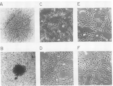
fromsuch a colony. Thetransformed 3T3 cells
had a fusiform morphology and grewinto
criss-cross patterns (Fig. 6C) similar to those of the FSV-transformed avian cells (19), but different
from 3T3 cells transformed by other retrovi-ruses.
To determine whether F3B cells were trans-formed directly by expression of the
transform-ing gene of FSV or indirectly, as for exampleby
integration at a specific cellular site, we asked whether expression of the transformed pheno-typeof the 3T3 cells was temperature sensitive, aswasthe phenotype of avian cellstransformed
by the FSV from which the pFL-5 DNA was derived. For this purpose, F3B cells were grown at36°C and then shifted to 39°C for 12 h (Fig. 6C andD). During this process, the cell morphology changed from an initially transformed to a flat
phenotypethat was indistinguishable from nor-mal 3T3cells grown at 36 or 39°C (Fig. 6E and
F). F3B cells grown at 39°C reverted to the transformed phenotype overnight after being
12~~~~~~~~~~~~ 1
IIs'ro w
11 6 7 9
2115 -
8
#2
4^
1
.~~~.. f~ ~~~~~~~~~~S * i!,
Electrophoresis
FIG. 5. RNase
Ti-resistant
oligonucleotidesof FSVRNAandFSV andFAV RNAhybridized bymolecular-ly cloned FSV DNA. (A) Electrophoretically purified 4.5-kb FSV RNA was prepared and fingerprinted as
describedpreviously(20). (B) About0.015 mgofalkali-degraded pFL-5DNA,clonedinplasmid pBR322,was
annealed with 2 x 10⁶cpm ofFSV(FAV) 50-70S [32P]RNA. Aftertheunhybridized RNA wasdigestedwith
RNaseTi,thehybridwaspurifiedandfingerprintedasdescribedpreviously (21).Theoligonucleotidesequences
recovered fromthisfingerprintwereidentifiedby theirRNaseA-resistantfragmentsand theirchromatographic
locationsandwerenumberedasdescribedpreviously (20).
VOL. 44,1982 407
on November 10, 2019 by guest
http://jvi.asm.org/
[image:7.496.109.397.321.606.2]408 LEE, LIU, AND DUESBERG
A
C
E
B
D
F
p,4 A~~
*
.. r
f,w
t
,'$,
FIG. 6. Biological activity of cloned FSV DNA.Afocus induced in NIH-3T3 cells (A) andacolony formed17
to20days aftertransfection withpFL-5DNA(B).The morphology ofanFSVDNA-transformed 3T3 cellline,
derived fromacolony like that shown in B, maintainedat36°C (C) and12 hafterashiftfrom 36to39°C(D).
Normal 3T3 cellsmaintained at36°C(E)orshiftedto39°C (F).
shiftedbackto36°C. Weconclude that
transfor-mationof3T3 cellsismaintained bya
tempera-ture-sensitivegeneproduct ofFSV.
(ii) Phosphorylation ofthe nonstructural FSV protein p140is temperaturesensitiveinF3Bcells.
The result discussed above implied thata viral
gene product rather than a positional effect of FSV DNA integration was responsible for the
transformation of mouse cells. In view of this possibility,we tested whetherp140was
synthe-sized in F3B cells by labeling F3B cells with
[35S]methionine. Thep140wasisolated from cell lysates by immunoprecipitation with anti-avian myeloblastosis virus (AMV) p27 serum. p140 withthesameserological properties and
electro-phoretic mobility as p140 of FSV L-15 quail
nonproducer cells was synthesized in the F3B
cells(Fig. 7A). FSV-transformed 3T3 cells
pro-duced about two to three times less
[35S]me-thionine-labeled p140 per cell than did FSV-transformed quail cells. No p140 was
precipitated from F3Bcells with normal serum,
nor was p140precipitated from 3T3 cells with
anti-AMV p27 serum (Fig. 7A). Two proteins, p90 and
p50,
wereoccasionally
observedin theimmunoprecipitatesof
p140
inF3B cells(Fig.7).Similar
proteins
werepreviously
observedto be associated withp140
from avian cells infected with FSV(T.Gilmore andG. S.Martin,person-al communication).
Itwaspreviously shownthat
synthesis
ofp140
in FSV L-15 isnot temperature sensitive; how-ever, phosphorylation of
p140
both invivo and in vitro is temperaturesensitive(19).
Totestthepossibility
that pFL-5 cloned DNApossessed
the same characteristics, we labeled F3B cells
with
[35S]methionine
or32P043-
for 2to3 hat36 and39°C. Cellularlysates
were immunoprecipi-tated with anti-AMV p27 serum and thenana-lyzed by
SDS-polyacrylamide
gel electrophore-sis.p140
wassynthesized
equally well in F3B and in ts FSV L-15 quail nonproducer cells ateither36or39°C(Fig. 7BandC). Itfollowsthat
synthesis of
p140
is not temperature sensitive. However,phosphorylation
ofp140
in vivo was temperature sensitive, as detected by labelingon November 10, 2019 by guest
http://jvi.asm.org/
[image:8.496.52.447.73.373.2]DNA CLONE OF TEMPERATURE-SENSITIVE FSV 409 cells with
32P043-
(Fig. 7C). It is recognized thatp140 is phosphorylated at the nonpermissive temperature (Fig. 7C) and that the difference between the extent of p140 phosphorylation at thepermissive and nonpermissive temperatures
is smaller in 3T3 mouse cells than it is in avian cells (Fig. 7C). The higher background
phos-phorylation of p140 at the nonpermissive tem-perature of 39°C in mouse cells is thought to
reflect only partial inhibition of p140
phosphor-ylation at this temperature as compared with
completeinhibition at 41.5°C in avian cells (19). It is also possible that the threshold level of phosphorylated p140 necessary to transform NIH-3T3 cells at the permissivetemperature is
lower than that required to transform avian
cells. Phosphorylation at the nonpermissive
temperature has been noted here (Fig. 7C) and
previously in avian cells (19). The
temperature-independent phosphorylation has been assigned
previouslytophosphorylation ofthegag-related portion ofp140 (19, 24).
As we showedpreviously with p140 immuno-precipitates from avian cells (19, 24), we also
observed here that in vitro labeling of immune precipitates of p140 from F3B mouse cells with
[y-32P]ATP (Fig.
7D)wastemperaturesensitive. Thecorrelation betweenmorphologicaltrans-formation and
phosphorylation
ofp140 and its1 2 3 4 5
A
W
-ap140
IN-__
p90
p50"-association with kinase suggests that fully
phos-phorylatedp140isnecessaryfor transformation of mammalian cells infected by temperature-sensitive FSV. This is in accord with similar observations on the transformation of avian cells
bytemperature-sensitive FSV (19, 24).
Cellular DNA homologous to the specific se-quence of FSV. Based on helper virus-unrelated,
transformation-specific sequences, the known
avian sarcoma viruses fall into several sub-groups (10). The specific sequences of proto-typesoftwosubgroups,RSV and avian sarcoma virus Y73, were shown to have homologous sequence counterparts inaviancells (32, 33,35),
andthe specificsequence of RSV,the src gene, also showed homology with other vertebrate
chromosomes (32). To test whether the specific sequence of FSV which is the prototype of a
third subgroup of avian sarcoma viruses, the FSV subgroup, also has a sequence counterpart
inavian andmammalian cells, wecleavedDNA
from several animalspecies with the restriction enzymeBamHI, and,afterelectrophoretic frac-tionation, the DNA fragments were hybridized with FSV-specific DNA, i.e., the PvuI-SmaI fragment ofpFL-5 DNA(Fig. 4). A single size class ofBamHI-resistant DNAfragments,
mea-suring 12 kb in chicken and8 kb in quail cells, hybridized with FSV-specific DNA (Fig. 8).
6 7 8 9 10 11 12 13 14 15 16 17
B i C D
p9OG- .
,101- pm - --i.- NW
p90 &_
FIG. 7. Synthesis, phosphorylation, and association withkinase ofp140 inF3B cells at thepermissive and
nonpermissive temperatures. (A) Serological and electrophoretic identification of[35S]methionine-labeledp140
inFSVL-15quail nonproducer cells (lane1)and F3Bmousecells(lane 2).p140 (unlabeled arrows)wasisolated
from celllysatesbyimmunoprecipitation with anti-AMV p27serumandelectrophoresedasdescribed previously
(19). Controlexperiments testing thesynthesis andspecificityof theimmunoprecipitationareshown in lanes
3-5; 3, immunoprecipitates of 35S-labeled F3B mouse cell lysates incubated with normal goat serum; 4,
immunoprecipitates ofnormal 3T3 cells incubated with anti-AMV p27 serum as used in lanes 1 and 2; 5,
immunoprecipitates of normal3T3cells incubated with normal goatserum.(B)Comparison ofp140 synthesisin
FSVL-15 quail nonproducer cells (lanes6and7) andF3Bmouse cells(lanes8 and9)atthepermissive (lanes6
and8,36°C)andnonpermissive temperatures (41.5°C for quail cells, lane 7; 39°C for F3B cells,lane9).p140was
labeled with[35S]methionineandanalyzedasdescribedforA.(C)Invivophosphorylation ofp140inFSVL-15
quail nonproducer cells (lanes10and11) andF3B mousecells(lanes12and13)atthepermissive (lanes10and
12) andnonpermissive (lanes11 and 13) temperature. p140was labeled with
32P043-
andanalyzed. (D)Invitrotestof kinaseactivity associatedwithp140isolated from FSVL-15quail nonproducer cells(lanes14and 15) and
fromF3B mousecells(lanes16 and17). p140wasimmunoprecipitated fromthecells grownatthepermissive
(lanes 14 and 16) and nonpermissive (lanes 15 and 17) temperature, and autophosphorylation activity was
assayedwith [,y-32P]ATPaspreviously described (19).
VOL.44,1982
on November 10, 2019 by guest
http://jvi.asm.org/
[image:9.496.54.452.387.507.2]ci)
0I
--ol_E aL
kb
' 23
4W
0
9.5
- 6.7
4.3
. ~ ~
2.3 2.0
FIG. 8. Electrophoretic analysis of restriction
en-zyme-resistant DNA fragments of various animal
spe-cies (genomic blots) related to FSV-specific DNA.
Mouse cell DNA (10 p.g) and chicken, quail, and
Drosophila cellDNA (5,ugeach) weredigested
com-pletely with BamHI and electrophoresed on 0.8%
agarose gels. After being blotted (31), the
nitrocellu-lose filterwas prehybridized in 50% formamide, 6x
SSC, 0.2% Ficoll, 0.2% polyvinylpyrrolidone, and
0.2% bovine serum albumin overnight and then
hy-bridized with the PuvI-SmaI [32P]DNA fragment of
cloned FSVDNA (see Fig.4) at40°C for36 h. The
filterwasthenwashed for1h with2x SSC-0.1% SDS
andfor 1 h with1xSSC-0.1%SDSat68°C. Theblot
wasautoradiographed for3days withanintensifying
screenat -70°C.
These results are consistent with the view that
possibly only one cellular locus is homologous to the FSV-specific sequence. Under the same conditions, a 20-kb fragment of Drosophila
DNAandtwofragments, 5.4 and 6 kb, ofmouse
DNA were also hybridized with less intensity.
DISCUSSION
The molecular DNA clone of FSV described here appears to represent the entire viral genome, based on its ability to hybridize a
complete complement of viral RNA and to
ex-press, intransformed mouse 3T3cells, the
pro-teinp140 which correspondstomostorallof the
coding capacity of FSV (20).
Allof theprevious experiments in transforma-tion of mammalian cells by defective avian
tu-mor viruses have been unable to distinguish
between indirect transformation (as, for exam-ple, by positional effects of integrated proviral DNA) and direct transformation via a gene prod-uctof the transforming virus. In the absence of a genetic marker, such as the
temperature-sensi-tive transforming gene of the FSV strain used
here, thisdistinction is difficult to make due to the low specific transformation activity of provi-ral DNAs from defective avian tumor viruses on
mammalian cells (6) and the failure to identify proteins with proven transforming function in such cells. Likewise, the low efficiency ofavian tumor viruses in transforming mammalian cells (25) did not allow a distinction to be made between direct transformation by a viral gene product and indirect transformation by a posi-tional effect of integrated proviral DNAs.
Ourresult, that themorphologyof mouse 3T3
cells transformed by transfection with DNA
from a temperature-sensitive strain of FSV is temperature sensitive for atransformation,
pro-vides direct evidence that a viral gene product
was responsible for maintainingtransformation of these mammalian cells. It follows that the
mechanisms of maintaining transformation in
both avian and mammalian cells by the avian FSV mustbevery similar or the same,i.e.,both
probably involvethe sameviral geneproduct(s).
TheFSVgeneproduct thatappears to be neces-saryfor maintenance of transformationin mouse 3T3cells isthephosphorylated form of p140,the
only knowngeneproductencoded by FSV(19, 20). This is consistent with previous evidence linking phosphorylated p140with transformation
of avian cells(19, 24).
The oncgenes ofacutely transforming
retro-viruseshavehomologoussequence counterparts
innormal animal cells (27, 32, 33,35). However, the degree ofhomology between agiven viral oncgene andits cellular counterpart measured
by nucleic acid
hybridization
seems to vary,greatly in some cases and in others very little, with cellular species. It was observed that the oncgeneof avian reticuloendotheliosisvirus has aclosely relatedsequence counterpart inturkey
cells but less homology with other mammalian cells (5). It was recently observed that several
viral oncgenes have homologous sequences in
Drosophila cell DNA,
although
this was ob-servedunder lessstringent
conditionsof hybrid-ization (30).Herewefoundthat DNAsfromtwoavian species and from mice and Drosophila
contain sequences homologous tothe FSV-spe-cific sequence. By
screening
agenomic
library ofchicken chromosomal DNA, wefound thatasingle 16-kbfragment of cellularDNAcontains
about 2 kb of sequences homologous to the
specific sequence of FSV (Lee and Duesberg,
J. VIROL.
on November 10, 2019 by guest
http://jvi.asm.org/
[image:10.496.93.198.69.293.2]DNA CLONE OF TEMPERATURE-SENSITIVE FSV 411 unpublished data). Thus, both lines of
experi-mentation suggest the existence of a single FSV-related locus in the chicken chromosome.
Under more stringent conditions of hybridiza-tion, the sequences homologous to the FSV-specific DNA region were only detected in avian cell DNA (unpublished data). Further work in-volving more direct sequence comparisons is necessary to determine the extent of homology between viral and cellular FSV-related se-quences, since blot hybridizations are not readi-ly quantitated.
ACKNOWLEDGMENTS
Wethank Karen Beemon, Phil Early,Mike Kriegler,Tony Shermoen, andWarrenGish for the gift of different kinds of DNAs and plasmids;WillPhares for review of the manuscript; Mike Botchan for critical discussion andsupport; and Linda Brownstein for typing the manuscript.
This research was carried out in a P2facility accordingto National Institutes of Health guidelines forrecombinantDNA research and was supportedbyPublic HealthService research grant no. CA-11426from the National Cancer Institute.
LITERATURE CITED
1. Benton, W. D., and R. W. Davis. 1977. Screening Agt recombinant clones by in situ hybridization to single plaques. Science 196:180-181.
2. Bister, K., W.-H. Lee, and P. H. Duesberg. 1980. Phos-phorylation of the nonstructural proteins encoded by three avian acute leukemiaviruses and byavianFujinami sarcoma virus. J.Virol. 36:617-621.
3. Blattner, F. R., A. E. Blechl, K. Denniston-Thompson, H. E. Faber, J. E.Richards, J. L.Slighton,P. W. Tucker, and0. Smithies. 1978.Cloninghuman fetal y globin and mouse a-type globinDNA:preparation and screening of shotgun collections. Science 202:1279-1284.
4. Blattner, F. R.,B. G.Williams, A. Blechl, K. Denniston-Thompson, H. E. Faber, L. Furlong, D.J. Grunwald, D.0.Keifer, D. D. Moore, J. W.Schumm,E. L. Sheldon, and0.Smithies. 1977. Charonphages: saferderivativesof bacteriophagelambda forDNAcloning.Science 196:161-169.
5. Chen, I.S.Y., T. W. Mak, J. J. O'Rear, and H. M. Temin. 1981. Characterization of reticuloendotheliosis virus strain T DNA and isolation of a novel variant of reticuloendotheliosis virusstrain T bymolecular cloning. J. Virol. 40:800-811.
6. Cooper, G.M., W. G. Copeland, A. D. Zelenetz, and T. Krontiris. 1980.Transformation of NIH-3T3 mouse cells by avian retrovirus DNAs. Cold Spring Harbor Symp. Quant. Biol. 44:1169-1176.
7. Czernilofsky, A. P., A. D. Levinson, H. E. Varmus, and J. M. Bishop. 1980. Nucleotide sequences of an avian sarcoma virusoncogene (src) and proposed amino acid sequencefor geneproduct. Nature (London) 287:198-203. 8. Davis, R.W., D. Botstein, and J. R. Roth (ed.). 1980. Advancedbacterial genetics: amanual for genetic engi-neering. Cold Spring Harbor Laboratory, Cold Spring Harbor,New York.
9. DeLorbe, W. J., P. A. Luciw, H. M. Goodman, H. E. Varmus, and J. M. Bishop. 1980. Molecular cloning and characterization of avian sarcoma virus circular DNA molecules.J. Virol. 36:50-61.
10. Duesberg,P. H.1980.Transforming genes of retroviruses. ColdSpringHarbor Symp. Quant. Biol.44:13-29. 11. Feldman, R. A., T. Hanafusa, and H. Hanafusa. 1980.
Characterization ofproteinkinase activityassociated with
the transforminggeneproduct of Fujinamisarcomavirus. Cell 22:757-765.
12. Fujinami, A., and K. Inamoto.1914.UeberGeschwulste beijapanischenHaushuhnern, insbesondere uber einen transplantablen Tumor.Z.Krebsforsch. 14:94-119. 13. Gilmer, T. M., and J. T. Parsons. 1979. Analysis of
cellularintegration sitesin avian sarcomavirus-infected duck embryo cells. J. Virol. 32:762-769.
14. Graham, F. L., and A. J. Van der Eb. 1973. A new techniqueforthe assayof infectivity ofhumanadenovirus 5 DNA.Virology 52:456-471.
15. Hanafusa, T., B. Mathey-Prevot, R. A. Feldman, and H. Hanafusa. 1981. Mutantsof Fujinamisarcomaviruswhich are temperaturesensitiveforcellulartransformation and protein kinaseactivity.J.Virol. 38:347-355.
16. Hanafusa, T., L.-H. Wang, R. G.Anderson, R.E.Karess, W. S. Hayward, and H.Hanafusa.1980.Characterization of the transforminggeneof Fujinamisarcomavirus. Proc. Nati. Acad. Sci.U.S.A. 77:3009-3013.
17. Hishinuma, F., P. J.DeBona, S.Astrin, and A. M. Skalka. 1981. Nucleotide sequence ofacceptorsiteandtermini of integrated avian endogenous provirus ev 1: integration creates 6 bprepeat of hostDNA.Cell 23:155-164. 18. Lautenberger, J. A., R. A. Schulz, C.F. Garon, P. H.
Tsichlis, and T. S. Papas.1981.Molecularcloning of avian myeloblastosis virus (MC29) transforming sequences. Proc. Natl.Acad. Sci. U.S.A. 78:1518-1522.
19. Lee, W.-H., K.Bister,C.Moscovici, and P. H. Duesberg. 1981. Temperature-sensitive mutantsof Fujinami sarco-ma virus: tumorigenicity and reversible phosphorylation ofthetransforming p140protein. J. Virol. 38:1064-1076. 20. Lee, W.-H., K. Bister, A. Pawson, T.Robins, C. Mosco-vici, and P. H.Duesberg. 1980. Fujinami sarcoma virus: an avian RNAtumor virus with a unique transforming gene.Proc. Natl.Acad. Sci. U.S.A. 77:2018-2022. 21. Lee, W.-H., M.Nunn, and P.H.Duesberg. 1981. src genes
oftenRous sarcoma virusstrains,includingtwo reported-lytransudced fromthe cell, arecompletely allelic; puta-tive markers oftransduction are not detected. J. Virol. 39:758-776.
22. Mandel, M., andA.Higa.1970.Calcium-dependent bacte-riophage DNAinfection. J. Mol. Biol. 53:159-162. 23. Neiman, P., K. Beemon, and J. A. Luce. 1981.
Indepen-dentrecombination between avianleukosis virus terminal sequences and host DNA in virus-induced proliferative disease. Proc.Natl. Acad. Sci. U.S.A. 78:1896-1900. 24. Pawson, T., J. Guyden, T.-H. Kung, K. Radke, T.
Gil-more, and G. S. Martin. 1980. A strain of Fujinami sarcoma virus which is temperature-sensitive in protein phosphorylation andcellulartransformation. Cell 22:767-775.
25. Quade, K. 1979. Transformation of mammalian cells by avianmyelocytomatosis virus and avianerythroblastosis virus. Virology98:461-465.
26. Rigby, P. W. J., M.Dieckmann, C. Rhodes, andP. Berg. 1977. Labeling of DNA to high specificactivity by nick translation. J. Mol. Biol. 113:237-258.
27. Scolnick, E. M., and W. P. Parks. 1974. Harvey sarcoma virus: a second murine type C sarcoma virus with rat geneticinformation. J. Virol. 13:1211-1219.
28. Shalloway, D., A. D. Zelenetz, and G. M. Cooper. 1981. Molecular cloning and characterization of the chicken genehomologous to thetransforming geneof Rous sarco-mavirus. Cell24:531-542.
29. Shibuya, M., T. Hanafusa, H. Hanafusa, and J.R. Ste-phenson. 1980. Homology exists amongthetransforming sequences of avian and feline sarcoma viruses.Proc.Natl. Acad. Sci. U.S.A.77:6536-6540.
30. Shilo, B.-Z., and R. A. Weinberg. 1981. DNA sequences homologus to vertebrate oncogenes are conserved in Drosophila melanogaster. Proc. Natl. Acad. Sci. U.S.A. 78:6789-6792.
VOL.44, 1982
on November 10, 2019 by guest
http://jvi.asm.org/
31. Southern, E. M. 1975. Detection of specific sequences amongDNAfragmentsseparated bygelelectrophoresis. J.Mol.Biol. 98:503-517.
32. Spector, D.H., H.E. Varmus, and J. M. Bishop. 1978. Nucleotidesequencesrelatedtothetransforminggeneof
avian sarcoma virus are present inDNA ofuninfected
vertebratres.Proc.Natl. Acad. Sci. U.S.A.75:4102-4106. 33. Stehelin,D.,H. E.Varmus, J. M. Bishop, and P. K. Vogt. 1976. DNA relatedtothetransforming gene(s) of avian
sarcomaviruses ispresentin normal avian DNA. Nature (London)260:170-173.
34. Vennstrom,B.,C.Moscovici, H. M.Goodman,andJ. M. Bishop.1981. Molecularcloning of the avian myelocyto-matosis virusgenomeandrecoveryof infectious virusby transfection of chicken cells. J. Virol. 39:625-631. 35. Yoshida,M.,S.Kawai,andK. Toyoshima.1980.
Uninfect-ed avian cells containstructurally unrelated progenitors of aviansarcoma genes. Nature (London) 287:653-654.