VENTRICULARSYSTOLICDYSFUNCTIO N IN
CHRONIC OBSTRUCTIVE PULMONARY DISEASE
Dissertation submitte d to
THE TAMILNADU DR. M.G.R MEDICAL UNIVERSITY
CHENNAI
In pa rtial fulfillme nt of re gulations For award of the degree of
M.D (G ENERAL MEDICINE)
BRANCH – 1
KILPAUK MEDICAL CO LLEG E
CHENNAI-10
BONAFIDE CERTIFICATE
This is to certify that dissertation named “PREVALENCE OF LEFT VENTRICULAR SYSTOLIC DYSFUNCTION IN CHRONIC OB STRUCTIVE PULMONARY DISEASE” is a bonafide work performed by Dr.J.KAMARAJ, post graduate student, Department of Medic ine, K ilpauk Medical College, Chennai-10, under my guidance and supervision in fulfillment of regulations of the Tamilnadu Dr. M.G.R Medical University for the award of M.D. Degree Branch I (General Medic ine) during the academic period from May 2011to April 2014.
Prof. Dr.N.GUNASEKARAN M.D., DTCD Prof. Dr.R.SABARATNAVEL M.D., Director Professor and Unit Chief
Institute of Non-Communicable DiseasesDepartment of General Medicine SuperintendentGovernment Royapettah Hospita l
Government Royapettah Hospital, Chennai-600014. Professor and HOD
Department of General Medicine Government Kilpauk Medical College,
Chennai-600010.
Prof. P. RAMAKRISHNAN M.D., D.L.O The Dean
Government K ilpauk Medical College
DECLARATION
I solemnly declare that this dissertation “PREVALENCE OF LEFT
VENTRICULAR SYSTOLIC DYSFUNCTION IN CHRONIC OBSTRUCTIVE PULMONARY
DISEASE”was prepared by me at Government Kilpauk Medical College and Hospital, Chennai, under the guidance and supervision ofDr.R.SABARATNAVEL M.D., Professor, Department of Internal Medicine, Government Royapettah Hospital, Chennai.
This dissertation is submitted to The Tamil Nadu Dr. M.G.R. Medical
University, Chennai in partial fulfillment of the University regulations for the
award of the degree of M.D. Branch I (General Medicine).
Place: Chennai
At the outset, I would like to thank my beloved Dean, Kilpauk Medical College Prof. Dr. P. RAMAKRISHNAN, M.D., D.L.O., for his kind permission to conduct the study in Kilpauk Medical College.
I would like to express my special thanks toProf.Dr.N.GUNASEKARAN
M.D.,DTCD , Professor and HOD, Department of General medicine, Kilpauk
Medical College and Director, Institute of Non-communicable diseases,
Superintendent, Govt. Royapettah Hospital for permitting to conduct this study.
I would like to thank wholeheartedly, Prof. Dr. R.SABARATNAVEL
M.D., my unit chief and Professor of Medicine for his encouragement and
guidance during the study.
I also express my special thanks to Prof. Dr. K.T. JEYAKUMAR M.D.,
Prof. Dr.S.MAYILVAHANAN M.D., I am extremely thankful to Assistant
Professors of Medicine, Dr.I.RohiniM.D.,Dr.N.Jayaprakash M.D., and
DR.T.Balaji M.D., for their assistance and guidance.
operation and criticism shown by my fellow post graduate colleague and friends.
I would like to extend my gratitude to my parents, my brother, my sister, my wife and my children for their unconditional support.
This receipt acknowledges that Turnitin received your paper. Below you will find the receipt information regarding your submission.
Paper ID 383643204 Paper title TRIAL4 Assignment title Medical
Author Kamaraj JAGAN
E-mail [email protected] Submission time 19-Dec-2013 05:57PM
Total words 12027
First 100 words of your submission
INTRODUCTION Chronic obstructive pulmonary disease (COPD) is a disease state “characterized by the presence of airflow obstruction due to chronic bronchitis or emphysema; the airflow obstruction is progressive and partially reversible”. COPD is now widely prevalent in both developed and
developing countries causing major health burden to the society. According to “WHO” report, COPD at present is the fourth commonest cause of death worldwide. It is proposed to be the third major cause of mortality and fifth major cause of chronic disability by the year 2030. In India every year half a million people die of COPD. Cigarette smoking is the key risk factor, other risk factors being exposure to...
CONTENTS
1. INTRODUCTION 1
2. REVIEW OF LITERATURE 3
3. AIM OF STUDY 49
4. MATERIALS AND METHODS 50
5. OBSERVATION 57
6. DISCUSSION 87
7. CONCLUSIONS 94
APPENDIX
BIBLIOGR APHY
ABBREVIATIONS
PROFORM A
MASTER CHART
ETHICAL COMMITTEE APPROVAL CERTIFICATE
PREVALENCE OF LEFT VENTRICULAR
SYSTOLIC DYSFUNCTION IN
CHRONIC OBSTRUCTIVE PULMONARY DISEASE
ABSTRACT:
Background:
COPD is now more prevalent in both developed and developing countries. It
is projected to be the third most common cause of death and fifth most common
cause of chronic disability by the year 2030. In India half a million people die of
COPD.COPD obscure the clinical signs of coexisting left ventricular dysfunction
like cough, dyspnea, paroxysmal nocturnal dyspnea and orthopnea. Symptoms of
dyspnea in COPD can be partially due to LV systolic and diastolic dysfunction.
More number of patients is either under or wrongly diagnosed, that leads to
worsening and development of complications. Early identification and treatment of LV dysfunction can improve the patient’s symptoms.
Aim of the study :
To study the prevalence of LV systolic dysfunction in COPD patients and to
assess the possible risk factors contributing to the development of LV systolic
Methods and materials:
The study design was approved by the Ethical Committee of the institution.
This study was done at Government Royapettah Hospital, Chennai between May
2013 to October 2013 .This is a cross sectional study in which 50 patients with
COPD more than 40 years of age. Severity of COPD is assessed by Spirometry.
LVEF was measured by 2D Echo. Duration of smoking and smoking index also
was calculated.
Results:
Among the 50 COPD patients, duration of smoking, smoking index and
severity of COPD correlated with LVEF. . ‘p’ value was significant. COPD is
more prevalent in males. Most patients belong to low socio economic status.
COPD severity is directly proportional to smoking index and duration of smoking.
Our study reports that 10% of the COPD patients have LV systolic dysfunction. A
higher smoking index and prolonged duration of smoking is associated with poor
LV systolic function. Increasing severity of COPD is associated with worsening of
LV systolic function
Conclusion:
LV systolic dysfunction is directly proportional to severity of COPD.
development of LVSD. Routine echocardiography is recommended for all severe
COPD patients. Smoking cessation should be a vital part of treatment plan in
COPD patients. In COPD patients with LVSD, it may be good to introduce
selective beta-1 blocker, diuretics, ACE inhibitors, angiotensin-II antagonist and
aldosterone antagonist.
Key words:
Chronic obstructive pulmonary disease, smoking, severity of COPD, Spirometry,
1
INTRODUCTION
Chronic obstructive pulmonary d isease (COPD) is a disease
state“characterized by thepresence of airflow obstruction due to chronic bronchitis
oremphysema; the airflow obstruction is progressive and partially
revers ible”.COPD is now widely prevalent in both developed and developing
countries causing major health burden to the society.
According to “WHO” report, COPD at present is the fourth commonest
cause of death worldwide. It is proposed to be the third major cause of mortality
and fifth major cause of chronic disability by the year 2030.In Ind ia every year
half a millio n people die of COPD.Cigarette smoking is the key risk factor, other
risk factors being exposure to indoor and outdoor pollution, occupationalhazards,
infection and genetic factors.
Most COPDpatients die of pulmonary and extra pulmonary
complications.Pulmonary hypertension,corpulmonale and left ventricular
dysfunction are the important cardiac predictor for mortality.
COPD obscure the clinical signs of coexisting left ventricular dysfunction
like cough,dyspnea, paroxysmal nocturnal dyspnea andorthopnea.More number of
patients is either under or wrongly d iagnosed,that leads to worsening and
2
Routinespirometeryis used to diagnoseand to assess the severity of COPD
and echocardiography is mandatory to assess the cardiac status in COPD patients.
The awareness among patients regarding COPD and its complications is
less, so more attention is needed for COPD patients pertaining to the development
3
REVIEW OF LITERATURE
HISTORICAL BACKGROUND
COPD has existed since time unknown but has been described by various terminologies in the past. Bonet described a condition named "voluminous lungs" as long back as 1679. In 1769, Giovanni Morgagniproposed lungs becoming "turgid"particularly from some airborne factor.[1]Ruysh provided the first authentic illustration and described the enlarged airspaces in emphysema in 1721.[2]
The destructive nature of the emphysematous lung was illustrated and described by Matthew Bailliein 1789.[1] In 1814 Badham denoted the word "catarrh" to the cough and mucus hypersecretion in patients with chronic bronchitis and also described it as a d isabling state.Rene Laennec, the inventor of stethoscopedescribed “emphysema" in his book as“A Treatise on the Diseases of the Chest and Mediate Auscultation” (1837).
William Briscoe in 1965 designated the term COPD.This terminology has overtaken other lesser popular termino logies and to become the established name for this disease currently. British Thoracic Society(BTS) accepted and adopted the COPD termino logy and formulated guidelines for COPD management.
4
American Thoracic Society (ATS) defined COPD is “a disease state characterized by the presence of airflow obstruction due to chronicbronchitis or emphysema; the airflow obstruction is generallyprogressive, may be accompanied by airway hyper-reactivity, and may be partially reversible.”
OTHER NAMES FOR COPD:
Chronic obstructive airway disease (COAD) Chronic obstructive lung disease (COLD) Chronic air flow limitation (CAL)
CHRONIC BRONCHITIS:
Chronic bronchitis is defined as “the presence of chronic productive cough for 3 months during each of two successive years in a patient in whom other causes of chronic cough, such as infection with mycobacterium tuberculosis, carcinoma of the lung, bronchiectasis, cystic fibrosis, and chronic congestive heart failure, have been excluded.”
The three forms of chronicbronchitis:
1. Simple bronchitis: mucus hypersecretion.
2. Chronic or recurrent mucopurulent bronchitis: intermittent or persistent mucopurulent sputum.
5
EMPHYSEMA:
Emphysema is defined as “a condition of the lung characterized by abnormal permanent enlargement of the air spaces distal to the terminal bronchio les accompanied by destruction of their walls without obvious fibrosis.” “Destruction is defined as nonuniformity in the pattern of respiratory air-space enlargement; the orderly appearance of the acinus and its components is disturbed and may be lost.”
CLASSIFICATION OF EMPHYSEMA
6
Acini are almostuniformly involved (and lobule).
CENTRIACINAREMPHYSEMA:
Dominant invo lvement of the proximal portion of the acinus (center of the
lobule)
PARASEPTAL EMPHYSEMA (DISTAL ACINAR) EMPHYSEMA:
The distal part predominantly invo lved(alveolar ducts and sacs) and the
proximal part relatively spared.
IRREGULAR EMPHYSEMA (PARACICATRICIAL EMPHYSEMA):
Acinus may be irregularly involved, usually associated with obvious
scarring.
BURDEN OF COPD
EPIDEMOLOGY
COPD currently tops the list as the major cause of morbid ity and mortality throughout the world and contributes significantly to the economic and social burden globally.(4
COPD prevalence,morbidity and mortality directly related to risk factors like smoking,indoor and outdoor pollution.
7
1995found that the prevalence of COPD in male was 4.08 percent and in females was around 2.55 percent from south India.(7) But in SEARO Region higher COPD prevalence was observed among women than men due to indoor air pollution (Pandy 1984; Behera and Jindal 1991).(9) National health interview surveyreport in United States in the year 2007 to 2009 shows the COPD prevalence was higher in older age groups, and most of the lifespan women had higher rates than men.NCMH estimates in India there were around 17 million COPD patients in 2006 but it is around 22 millio n in the next 10 year.(11)
8
COPD kills more than 3 million people every year worldwide.(5)At present it is the fourth lead ing cause of death, by the year 2030 it becomes third leading causeof death..In India half a million people die of COPD. (6)This is4 times the mortality that it causes in USA and Europe.(122)Nongkinryhet al published the WHO data of non-Communicab le d isease in Ind ia from 1998 to 2002 (JAPI,
2004). Asthma and COPD were second most cause for mortality. The morbid ity
figures around 55millio n cases for both Asthma and COPD.(12)
COPD mortality increases with age and the incidence is higher in men than women.(13,14,15,)Theincomplete recognition and diagnosis of COPD further miscalculates the prevalence, morbidity and mortality rates. Most COPD patients do not die of the disease per se andalsodue to accompanyingco-morbid conditions.(16)
9
COPD is a major health problem principally in societies where cigarette
smoking is common and nonsmokers also develops the disease.
Risk factors for the development of COPD may be broadly divided into
those related to environmental exposure and host factors.
Smoking is the major risk factor; other risk factors are alpha 1-antitrypsin
(α1-AT) deficiency, occupational exposure, childhood lower respiratoryinfections,
10
alpha1-antitrypsin deficiencysmoking exacerbatesthedevelopment ofemphysema indicating a clear genetic predisposition to COPD. (17)In India biomass smoke exposure becomesa major risk factor andburning of mosquito coils also a common indoor air pollutant.
ENVIRONMENTAL
SMOKING
In the United States therisk of developing COPD due to tobacco smoking accounts for 80% to 90%. (19) FEV1 begins to decline at 30–35 years of age in non-smokers but in non-smokers this may occur earlier.[22]Nonsmokers commonly have decline the rate FEV1 of about 30mL .Smokers on the other hand have a decline inFEV1 of about 50 ml. In males, the loss of FEV1 in excess of the normal decline with aging is 9 ml per year for each pack-year of smoking;in females, the excess
11
relationship between the numbers of cigarettes smoked andrate of decline in FEV1.[23]
Cigarette smokers had a significantly higher mortality rate compared to nonsmokers. Cessation of smoking does not substantially improvethe FEV1 but the subsequent rate of disease progression is decreased. [24,25] Indian people smoke tobaccoin various forms like beed is, hookahs and chillums. The chillum is the most harmful of the lot followed by hookahs and beedis respectively. Beed is are moredangerous than cigarettes (it contains only one fourth the amount of nicotine, but it produces four to five times more tar than cigarettes).(26)
OCCUPATIONAL EXPOSURE:
12
Several specific occupational exposures inc lud ing coal mining, gold mining,cotton textile dust, welding, construction, plastic manufacturing and utility all have been suggested as risk factors for chronic airflow obstruction. The high prevalence of smoking among workers in certain occupation has been a major confounding
factor. Occupational exposures to organic and inorganic gases and fumes also lead
to higher vulnerability to COPD. Workers exposed to cadmium can develop emphysema. [39]
OUTDOOR AIR POLLUTION:
This public health problem occurs mainly from the emission ofpollutants from motor vehiclesandindustries.(29)Particulate pollutants, ozone, nitrogen d ioxide and sulphur-d ioxide areproduce airway oxidative stress, bronchial hyper reactivity, systemic as well as pulmonary inflammation, reduction in airway mucosal cilliary activity and amplification of viral infections.(30) In community-based study,higher traffic density wasassociated withdiminished FEV1 and
FVC.(31)In low-income countries outdoor air pollutant levels tend to be higher. INDOOR AIR POLLUTION:
Commonly theindoor air pollutants are environmental tobacco smoke,particulatematter, carbon monoxide (CO), NO2, volatile organic compounds
13
contributing factors for indoor air pollution are wood, animal dung,crop residues and coal are burned in open fires and poorly functioning stoves in poorly ventilated indoors are the contributing factors for indoor air pollution. Women’s are spending more time in indoors for cooking than men, andexposed to biomass fuel combustion products, so theyare prone to develop COPD.(36,37) In India exposure to biomass smoke is a major one and burning of mosquito coils at homes are common indoor pollutants for the genesis of COPD. Burning one mosquito coil is equivalent to around 100 cigarettes because it emits as much particulate matter pollution.(35)
LOWER RESPIRATORY TRACT INFECTIONS:
Previous tuberculosis, childhood asthma and respiratory infections are the risk factors to develop COPD.Childhoodlower respiratory tract infections might produce permanent damage or impair growth and development of the lung. In
Lung Health Study COPD exacerbations in smokers were associated with an
additional loss of FEV1 of 7 ml per year for those having one exacerbation per
year.
LOW SOCIOECONOMIC STATUS:
14
HOST FACTORS:
GENETIC FACTORS:
The association between the development of early-onsetemphysema and alpha1- antitrypsin deficiency was first described by Eriksson in 1963. (40)In alpha1-AT deficiency premature development of pan lobular emphysema and early decline in lung function occurs. M allele is fornormal alpha1 antitrypsin.Mutation in the SERPINA1 gene [located on the long arm of chromosome 14 (14q31-32.3)] and forms the Z allele results severe alpha1-antitrypsin deficiency(41).In COPD variety of genes including beta1microsomal epoxide hydroxylase-1,tumor necrosis factor alpha and transforming growth factor are associated for pathogenesis.
ATOPY AND AIRWAY HYPERRESPONSIVENESS:
Dutch workers in the 1960s proposed that smokers with COPD and asthmatics shared common characters like airway hyper responsiveness, allergy and eosinophilia.(42) Atopic ind ividuals have high serum IgE and eosinophil levels. The probable mechanisms for increased AHR in smokers with COPD:
1. Shortening of the airway smooth muscle and increased thickening of the airway wall.
2. Inhaled aerosolsdeposition is in the centrally that result of airways obstruction,
15
4.Airway wall edema is due toincreased permeability of airway ep ithelium. ASTHMA:
Childhood asthma,chronic untreated and partially treated asthma can
progress to fixed airflow obstruction. Tucson epidemio logical studies had showed that asthmatics have a twelvefo ld increase risk of develops COPD than non-asthmatics.
PATHOGENESIS:
The probable pathogenesis of COPD is proteinase-antiproteinase hypothesis, immunological, imbalance of oxidant-antioxidant,apoptosis,systemic inflammation, and ineffective repair.(44)
PROTEINASE-ANTIPROTEINASE HYPOTHESIS
16
17 IMMUNOLOGICAL MECHANISM:
COPD patients have more numbers of neutrophils in sputum and lung tissue. In smoking-induced COPD immunoglobulin free light chains and IgE levels are increased.(48,49)
OXIDANT AND ANTIOXIDANT BALANCE:
Reactive oxygen species (ROS) in cigarette smoke or released by inflammatory
cells and structural cells of the lungs in response to smoke may lead to lung
injury.In smokers there isincreased theROS (hydrogen peroxide, hydroxyl rad icals, superoxide radicals, H2O2, and 8-isoprostane)and decreased plasma
antioxidantsproducts such as 4-hydroxynonenal, 3-nitrotyrosine. Oxidative attack may inactivate antiproteases. Due to decreased production of inactivated histone deacetylases oxidative damage occurs and this leads to a prolonged inflammatory response.Vasodilation and endothelial cell growth impairs in Oxidative stress. Oxidants modify the elastin and the modified elastin is more susceptible to proteolysis. Oxidant augments matrix degrading capacity which can promote emphysema.(50)
INFLAMMATION
Smoking and inhaled irritants lead to recruitment of inflammatorycells in the lungs
18
normalmechanisms of repair. In COPD neutrophils, eosinophil’s, macrophages,
and lymphocytes are associated for inflammation. Interleukin-8 (IL-8),
macrophage inflammatoryprotein-1α (MIP-1α), and monocyte chemo-attractant
protein-1(MCP-1) are up-regulated in bronchio lar ep ithelium in COPD. CD4+ and
CD8+ T cells and B cells accumulate in alveolar and airway tissue in COPD.
Among these cells CD4+ are predominantly elevated.
SYSTEMIC INFLAMMATION:
Pro-inflammatory cytokines andchemokine’s are released into the circulation as a result of persistent pulmonary inflammation.(51) These mediators stimulate bone marrow,liver and adipose tissue to release excessive amounts of leucocytes, IL-6, IL-8,C-reactive protein (CRP), fibrinogen and tumor necrosis factor-α (TNF-α) into the circulation.(52,53) Systemic inflammation may initiate or worsen the comorbid illness, such as normocytic anemia,IHD, Cardiac failure,osteoporosis, cancer lung, diabetes and depression.
APOPTOSIS:
19 INEFFECTIVE REPAIR:
The repair in emphysema not effective is due to the damaged alveoli have limited ability is to repair.
PATHOPHYSIOLOGY
In COPD Pathophysiological changes occurs in central airways, peripheral
airways, paranchyma of the lung and pulmonary vasculature. The defining physiological feature ismaximal forced expiratory flow reduction is persistent. The typical features areincreased theairway res istance, increased theresidual volume,
increased the res idual vo lume/total lung capacity ratio,decreased the inspiratory
capacity, maldistribution of ventilation, and mismatching of
ventilation-perfusion.Loss of lung elasticity, increasedairways resistanceand decreased lungcompliance are the factors that reduce forced expiratory flow.
20 MUCUS HYPER SECRETION:
In COPD patients with predominant central airway involvement the common featutre is mucus hypersecretion.It is due to enlargement submucus glands and metap las ia with increased numbers of globet cell in chronic irritation of the respiratory epithelium.
AIR FLOW LIMITATION:
The characteristic feature of COPD patient is decrease in FEV1 and FEV1/FVCratio
probably due to inflammation, fibrosis and luminal exudates in small airways.(54)
HYPERINFLATION:
21
(1) Decreases the zone of apposition between the diaphragm and the abdominal
wall, rib cage movement are hindering.
(2) Diaphragmatic muscle fiber length shortens, decreasing the force that can be
generated by the diaphragm.
(3) The radius of curvature of the diaphragm increases and decreasing
transpulmonary pressure (at constant tension).
(4) Diaphragmatic muscle fibers medially directs and impairing inflation with
diaphragmatic contraction.
PULMONARY GAS EXCHANGE ABNORMALITIES
In COPD Ventilation–perfusion (V ·a/Q ·) mismatching is the most vital cause of decreased pulmonary gas exchange. Other less importantcauses are alveo lar hypoventilation, impaired alveo lar–capillary oxygen diffusion and increased shunt. In COPD patient’sventilation distribution is unequal. As a result of emphysema reduction in b lood flow due to local destruction of alveo lar vessels, hypoxic vasoconstriction in alveolar hypoxemic areas and passive vascular obstruction as a result of increased alveolar distens ion and pressure.This results in hypoxemia and hypercapnea.
PULMONARY HYPERTENSION:
22
arteries.Progressivepulmonary hypertension leads to the development ofventricular hypertrophy and corpulmonale.
SYSTEMIC FEATURES:
COPD with smoking history have the higher comorbidities was observed in the ECLIPSE study.(62) The following comorbidities associated with COPD are cardiovascular disorders (Hypertension, CAD and Chronic cardiac failure), metabolic diseases (metabolic syndrome, obesity and diabetes), bone disease (osteopenia and osteoporosis), skeletal muscle weakness, anemia, stroke, lung cancer, cachexia, depression and cognitive decline.(63)
23
24
severe d isease.(67, 68)Anemia is common than polycythemia and it reduce the functional capacity.
CARDIO VASCULAR CHANGES:
Pulmonary hypertension and corpulmonaleare the consequence of chronic
alveo lar hypoxia; the secondary contributions are from destructionof the alveolar
capillary bed, lung hyperinflation and increased blood viscosity.The chronic alveo lar hypoxia produces a characteristic remodeling of the pulmonary arteries. The three major components remodeling are. 1. Extension of the med ial muscle into the pulmonary arterio les.[69,70]2.Proliferation of muscle that becomes progressively thickens and occludes the vascular lumen. Insevere cases Intimal fibrosis may occur. 3. Inner muscular tube develops in the smallpulmonary arteries.(71)In the late course of COPD hypoxemia and hypercapnia leads to pulmonary arterial hypertension.
The followingpulmonary vasculature changes occur in the pulmonary vascular bed in COPD patients.
1. Intimal thickening of the small pulmonary arteries is the earliest change.It is due to worsening of the airflow limitation.(72,73)
2. Medial hypertrophy of the muscular pulmonary arteries.(74)
25
In COPD patients the following factors contribute to develop of pulmonary arterial hypertens ion:
1. Pulmonary vascular bed destruction 2. Blood gas tensions abnormality 3. Pulmonary mechanics abnormality 4.Increased Cardiac output
26
Classically, pulmonary hypertens ion in emphysema has been attributed to the following factors:
1.Hypoxia leading to vasoconstriction and vascular remodeling
2. Hyperinflation compressesthe alveolar vessels and pulmonary vasculatureby physical obliteration.
3. Other possible influences inc lude elevated end-expiratory intrathoracic pressure due to dynamic or static hyperinflation or use of expiratory muscles.(77)In smokers theproduct of tobacco produces inflammatory changes and pulmonary vascular remodeling.(82)The imbalance between vasoactive constrictor and dilator substances produce pulmonary hypertension.Nitric oxideinhib its cell proliferationinthe pulmonary vasculature and prevents vascularremodeling. In COPD patients with hypoxemia there is a reduction in nitric oxide levels and development of pulmonary hypertens ion. (76)Oswald-Mammoser and colleagues observation had shown that the worsening of pulmonary hypertension is directly associated with severity of airflow obstruction.(80,81) Burrows and coworkers found that emphysematous patients had higher pulmonary vascular resistance and lower cardiac output.(79)
27
primarily affect the left side of the heart”. COPD patients have a highpulmonary vascular res istance as a result ofhypoxia.This causesincreased workload on the right ventric le leading toright ventricular hypertrophy(RVH), right ventricular dilatation and right heart failure.(77)In COPD patients the earliest sign of RV pressure overload is concentric right ventricular hypertrophy. The capacity of the RV to adapt to a high after load is overwhelmed duringacute exacerbations,overwhelmhence precipitating RV failure.(82)
In RV dilation there will be an increase in end-diastolic volume that shifts the inter-ventricular septum into the left ventric le to reduce LVend-diastolic volume and diastolic compliance.(82)In COPD large intra-thoracic pressure changes and long standing RV pressure overload can produce RV hypertrophy and dilatation that shift the inter-ventricular septum into LV cavity during systole and results in the limitation of left ventricular cavity dimensions, contractility, compliance, and rise of the left ventricular diastolic pressure(88,89,90,91)and alteration
of LV systolic function.(84,85,86,87)
In pulmonary hypertens ion blood flow through bronchial circulation
28
In Hypoxia the accumulation of excess toxic metabolic products like K+, H+ and lactic acidin the card iac tissue impair the myocardial contractility producing diminished LVEF(88).
Endothelial surface enzymes control the level of circulating compound such as bradykinin, serotonin, angiotensin and adenine nucleotides. During hypoxia there is endothelial dysfunction and altered enzyme activity may lead to atherosclerosis
and ischemia of cardiac muscles.(96)
Hyperinflation, increased work of breathing, and raised intra-thoracic pressures also have an impact on the LV functioning.
Sabit et al. had shown that the subclinical COPD have right ventricular dysfunction may be related to airways obstruction and left ventricular dysfunctionrelated to stiffness of the arteries. RV dysfunction is due to raised pulmonary arterial pressure and LV dysfunction is associated with increases afterload due to increased aortic stiffness.(93)
29
The following are also the mechanisms of heart failure. 1. Renin-angiotensin aldosterone system activation.
2. Sympathetic nervous system activation andvasopressin. 3. Generation of pro-inflammatorycytokines.
The above mentioned mechanisms are responsible for increasingvascularres istance, increasing heart rate, worsening myocardial perfusion, alteringrenal b lood flowandatrialfibrillations.Ischemia and arrhythmias are worsening the LVdysfunction and precipitating cardiac failure.(83) Skeletal muscle alterations increase the LV strain and dysfunction under the conditions of stress. Acute infections, severe anemia,hypertens ive emergencies, cardiac arrhythmias (MAT, AF), ischemia, pulmonary embolism and cardio toxicdrugs may precipitate HF in COPD patients.
SUPRAVENTRICULAR ARRHYTHMIAS :
Supraventricular tachyarrhythmiasare common in patients with COPD, as a
consequence of right atrial enlargement,increased adrenergic tone, hypoxemia and
30 EDEMA:
COPD patient’salterations in salt and water balance are due to hypoxia and hypercapnia. The activation of renin–angiotensin system bydiminution of renal blood flow andraise in AVP as a result of alteration in the blood gases concentration.
31
ANP is due to increases theatrial stretch.[98]ANPhas important beneficial effectsthat could act to prevent the formation of edema in patients with COPD, including the promotion ofnatriuresis,[99]decreasesplasma renin activity, decreases angiotensin п andaldosterone production. [100]Therenin–angiotensin system activation overwhelms the protective mechanisms and lead ing to develops edema.
PATHOLOGY:
In COPD patients the pathological changes occurs in the large and small airways and alveo li.
Large airways:(Central airway-bronchi>2mm internal diameter):
32
enlarged.In smokers goblet cells increase innumber and extend to more peripherally.Bronchi also undergo squamous metaplasia.(52)
33
Small airways(peripheral airways-bronchiole <2mm internal diameter):
Narrowing and destruction of terminal bronchioles are characteristic changes in COPD. Airway inflammation andscarring of thesmaller airways are associated with airways obstruction.
In COPD the following cellular events that occur in the small airways: 1. Mucus secreting cells replaced by Clara cells
2. Mononuclear cellinfiltration 3. Goblet cell metaplasia.(50)
Theimportant finding is Smooth muscle hypertrophy .Thecauses of airway narrowingare excessive secretion of mucus, formation of edema, cellular infiltration and fibrosis.(52)
Lung parenchyma(respiratory bronchioles andalveo li):
34 Centriacinar (syn. Centrilobular) emphysema:
This kind of emphysemais associated with tobacco smoking. The enlarged airspaces are c lustered around the terminal bronchio le. It ismore common in upper and lower lobes of the upper zones.
Panacinar (syn. pan lobular) emphysema:
35
Periac inar (syn.Paraseptal or distalacinar) emphysema:
The enlarged airspaces are along the edges of the acinar unit, where it is abuttingthe pleuraor vessel.
Scar or irregular emphysema:
It is described as enlarged airspaces around the scar margins that are notrelated to the structure of the acinus.
CLINICAL FEATURES:
Symptoms:
Cough with or without expectoration. Breathlessness
Chest pain- may be related to intercostal muscle ischemia. Haemoptysis
Weight loss, Effort into lerance,Ankle edema.
Signs:
- Nicotine staining of fingers and teeth.
- Nonspecific features of over inflation of the lungs with horizontal ribs, splaying of lower costal margin &widened xiphisternal angle.
-Paradoxical inward movement of lower ribs (Hoover’s sign). - Cardiac dullness reduced.
36 - Crepitation’s, Expiratory wheezes.
-Pursed lip breathing.
-RVH heave palpable in the subcostal angle or at lower sternal edge, loud P2.
- Third sound audible in the left fourth intercostal space, Pan-systolic murmur at the left sternal edge, Right ventricular gallop rhythm, elevated jugular venous pressure, pedal edema and tender hepatomegaly in corpulmonale.
Predominant
bronchitis
Predominant
Emphysema
General Appearance
Mesomorphic; overweight,
dusky with suffused conjunctivae, warm extremities
Thin, often emaciated, pursed lip breathing, anxious, normal or cool extremities
Age, years 40 to 55 years 50 to 75 years
Onset Cough Dyspnea
Cyanosis Marked Slight to none
Cough More evident than
dyspnea
Less evident than dyspnea
37 Blue bloaters:
Development ofhypoxemia, hypercapnia polycythemia and edema occurs
relatively early in b lue bloaters..
Pink puffer:
Pink puffers are thin,dyspneicandpreserve blood gas values until late in the course of the disease and develop pulmonary hypertens ion inadvanced disease state.
Upper respiratory Infections
Common Occasional
Breath sounds Moderately diminished Markedly diminished
Corpulmonale Common Only bouts of RTI and
also terminally Radiography Normal diaphragmatic
position; cardiomegaly; lungs normal or having increased
bronchovascular markings
Small tubular heart; low flat
diaphragm; Area of increased
radio lucency
38
INVESTIGATIONS:
SPIROMETRY:
Spirometry is asound test of airflow limitation in patients with COPD. It is the go ldstandard test for diagnosis and monitoring its progression. Normal curve has a brisk flow near total lung capacity, then a steady decline down to residual volume. Inspiratory limb is a smooth semicircle. With the development of disease in small airways the expiratory limb begins to dip at lower volumes. Spirometry measures the forced vital capacity (FVC),forced expiratory volume in one second(FEV1) and ratio of the two measurements(FEV1 /FVC).A low FEV1 and
FEV1/ FVC ratio below the normal range is the diagnostic criteria for COPD.
(104-106)
A post bronchodilator FEV1 / FVC ratio < 0.7 establishes the diagnosis of COPD. Asthma and COPD commonly coexist butCompared to the baseline FEV1 value, asthmatic patients will have 12% or greater improvement after 15 minutes use of short-acting beta2 agonist inhalation and the FEV1 also increases by> 200 ml.
CHEST X-RAY:
39
FULL BLOOD COUNT AND HAEMATOCRIT:
To identify anemia, polycythemia and leukocytosis
SPUTUM CULTURE:
For identification of theorganism.
BODY MASS INDEX (BMI):
For theassessment of prognosis.
ALPHA-1 ANTITRYPSIN:
Alpha-1 antitrypsin deficiency is an uncommon, but not rare a condition
associated with premature emphysema. Testing for α1-antitrypsin deficiency is
indicated in the likely hood of having the disorder.
A serum alpha-1 antitrypsin concentration level below 15-20% of the normal value
40
Transfer factor for carbon monoxide (TLCO) and transfer coeffic ient (KCO): To investigate symptoms thatseems disproportionate to the Spiro metric
impairment. The transfer factor test for carbon monoxide measures the ability to move carbon monoxide from inhaled gas to hemoglobin in the alveolar capillaries. A reduction in transfer factor (TLCO) and transfer coeffic ient (KCO) is the best functional indicator of the presence and severity of pulmonary emphysema.(107) In this context KCO is more specific than TLCO.(108) Asthma may reduce TLCO when severe but KCOtends to be high in asthma. Emphysema with its destruction of alveolar capillary membrane results in large air space, decreased area for gas transfer & reduction of TLCO & KCO.
PULSE OXIMETRY:
Assessthe Spo2level and need for oxygen therapy.
If corpulmonale present, or if FEV1 < 50% predicted it is indicated.
ARTERIAL BLOOD GAS MEASUREMENT:
41
CT SCAN OF THE THORAX:
CT chest is recommended only if there is a doubt inthe diagnosis of COPD.
ECG:
The characteristic ECG pattern in patients with chronic obstructive pulmonary disease is due to the insulating effect of the hyperinflated lungs,altered position of the of the heart, and caudal displacement of the diaphragm.
ECG changes in COPD
1. P waves >0.25 mV in lead II, III, or aVF – P pulmonale. 2. P wave axis to the right of 80 degrees in the frontal plane.
3. Lead I sign with an isoelectric P wave, QRS amplitude <0.15 mV, and T wave amplitude<0.05 mV
4. QRS amplitude in all limb leads <0.5 mV
5. QRS axis to the right of 90 degrees in the frontal plane
6. QRS amplitude <0.5 mV in lead V5 or V6; or R wave <0.7 mV in lead V5 or R wave <0.5 mV in lead V6
7. R/s >1 in V1 or R wave amplitude in V1 >5 mm, R/S ratio <1 in lead V5 or V6 8. S1S2S3 pattern with R/S ratio <1 in leads I, II, and III- S1S2S3 syndrome.
42
10.T wave - decrease in amplitude in all leads, may be inverted in right precordial leads
11. Right ventric le hypertrophy:
- Prominent terminal S waves in the left precordial leads. - Right axis deviation.
ECHOCARDIOGRAPHY:
Echocardiography is sensitive test to detect pulmonary hypertension, corpulmonale and left ventricular dysfunction in COPD patients. It is aneas ily available, non-invasive technique that assesses the right ventricular hypertrophy, dilatation,pulmonaryartery pressure and ejection flow dynamics. In COPD patients,the hyperinflated lungs alter the sound wave transmission.(109) Two-dimensional echocardiography gives important information on dynamics of cardiac structures and morphology (110).Continuous wave Doppler echocardiography may provide an estimate of pulmonary artery systolic pressure in the presence of tricuspid regurgitation.
Echocardiography find ings in COPD: Increase the pulmonary artery pressure. Tricuspid regurgitation.
43
Reverse movement of the inter-ventricular septum. Right atrium and ventric le enlargement
Left ventricular cavity may be normal or reduced Right ventricular dysfunction.
Systolic bowing of the inter-ventricular septum towards the left ventricle is a characteristic abnormality of right ventricular pressure overload and it is detected by two-dimensional echocardiography.[111]In patients who are d ifficult to image with 2-D echocardiography, MRI is usefulfor assessing RV structure. Pulmonary hypertens iondiagnosis is confirmed by Right-heart catheterization
BNP AND N-TERMINAL BNP:
In corpulmonale BNP and N-terminal BNP levels are elevated,secondary to RV stretch. The severity of pulmonary hypertens ion and right ventricular distension may be estimated by BNP levels and also reflects the treatment efficacy.
MANAGEMENT:
The overall approach to managing stable COPD should be characterized by an increase in treatment,depending on the severity of the disease and the clinical status of the patients.
The therapeutic goals are:
44 3.Improve daily activity and exerc ise tolerance
4. Decrease the frequency and severity of exacerbations 5. Treat the COPD exacerbations and complications. 6.Improve the health status
7. Mortality reduction
Management strategies arepharmacotherapy and non-pharmacotherapeutic approaches that improve quality of life, activity levels and symptoms of COPD. PHARMACOTHERAPY:
45
Bronchodilators are the mainstay in COPD pharmacotherapy. Bronchodilators reduce air trapping, breathlessness and improve quality of life. Bronchodilators can increase FEV1<10%.(52)Patients with mild COPD should be started onshort-acting inhaled beta2 agonist or ipratropium. If symptoms persist ipratropium( tiotropium) or long-acting beta2 agonist (LABA)) is an alternative. Concurrent use of tiotropium and ipratropium is not routinely recommended.
46
corticosteroids use associated with an increased risk of pneumonia.(52) Oral corticosteroids have numerous side effects so it is not recommended as a long-term monotherapy.(52)
Theophylline is useful in persistent symptoms despite optimal inhaled therapy. Roflumilast is a phosphodiesterase-4 (PDE-4)inhib itor, it inhibit the intracellular cyclic AMP breakdown and reduces the inflammation. It is administered 500 mg OD orally and it reduces acute exacerbations.(118)Roflumilast used concurrently with LABA has been shown to reduce the acute exacerbations.(119,120)
OXYGEN THERAPY:
The main goal of oxygen therapy is to maintain SpO2 ≥ 90% or PaO2 ≥ 60 mmHg atrest. Supplemental O2 is routinely used for patients with exertional or
nocturnal hypoxemia.Long-term oxygen therapy (LTOT) (>15 h per day) is useful in respiratory failure and with severe resting hypoxemia and has been found to have increased survival.(52)
IMMUNIZATION OF THE PNEUMOCOCCAL AND INFLUENZA
INFECTIONS:
Pneumococcal vaccination for every 5-10 years Influenza vaccination for yearly
47 SMOKING CESSATION
In long-term smokerscessation ofsmoking is more effective to prevent the progress of the disease and has been found to reduce the decline of FEV1.
PULMONARY REHABILITATION:
The components of pulmonary rehabilitation programme includesmoking cessation,nutrition counselling, exercise training and education.
SURGICAL TREATMENT:
Lung vo lume reduction surgery is ind icated in upper lobe emphysema.
Lung transplantation:
Lung transplantation is indicated in COPD patients who have FEV1 < 25% predicted or the paCO2 is > or = 55 mm Hg.
In respiratory failure either non-invas ive mechanical ventilation or mechanical ventilation ind icated.
CORPULMONALE:
The primary treatment of corpulmonale consists of continuous oxygen to
overcome hypoxemia and diuretics to control peripheral edema. Digitalis is useful
for rate control of atrial fibrillation.
PROGNOSIS:
48
status, hypoxemia, poor nutrition, the presence of corpulmonale, resting
tachycardia, low exerc ise capacity, severe dyspnea, poor health-related quality of
life, anemia, frequent exacerbations, co-morbid illnesses, and low carbon
monoxide diffusing capacity. COPD prognosis is based onthe BODE index.
BODE score greater than 7 is associated with a 30 percent 2-year mortality.
Score of 5 to 6 is associated with 15 percent 2-year mortality.
49
AIM OF THE STUDY:
50
METHODS AND MATERIALS:
Study group : COPD patients admitted in the department of
General Medic ine
Study design : Observational study
Place of Study : Govt. Royapettah Hospital
Sample size : 50 patients
Duration of study : 6 months (May 2013-October 2013)
Conflict of interest: Nil
51
Clearance was obtained from the ethical committee of Government Kilpauk Medical College.
METHODOLOGY:
In patients with COPD, CBC with ESR, RBS, FBS, PPBS, Blood urea & creatinine, lipid profile, LFT, Urine Routine, Ultra sonogram Abdomen, Chest X-ray, Pulse Oximetry for SPO2, Pulmonary function test, ECG, ECHO will be done after obtaining written informed consent. Severity COPD is assessed by Gold criteria. LVEF was measured by 2D Echo. PFT was assessed by Spirometry. LVEF was compared with severity of COPD and possible risk factor behind such development.
Inclusion criteria :
AllCOPD patients.
Exclusion criteria :
1. Systemic Hypertension 2. Diabetes Mellitus
3. H/o congenital, acquired valvular heart disease 4. IHD
52 6. Chronic AF
7. Complete RBBB or LBBB 8. H/o heart failure
9. Chronic kidney disease
10.Patients with poor echo window 11.Patients unable to perform PFT
Data collection:
The data of each patient will be collected in a specifically prepared proforma and includes demographic details proper history, clinical features, CBC with ESR, RFT, FBS, PPBS, LFT, Total Cholesterol& Triglycerides, Ultrasound Abdomen, chest X-ray, ECG, PFT and ECHO
A diagnosis of COPD should be considered in:
Persistent cough with expectoration for 3 months in two consecutive years Chronic cough - Present intermittently or present throughout the day Chronic sputum production
Dyspnea that is- Progressive (worsens over time) - Persistent (present everyday) - Worse on exercise
53
History of exposure to risk factors –smoking, air pollution
On examination: pallor, edema, elevated JVP,purse lip breathing,pulse,CVS-P2loud,RS-wheeze,crepitations.
Chest X-ray: evidence of Hypertrans luency, low flat diaphragm,widened intercostal space, and tubular heart.
SPIROMETERY:
COPD is diagnosed only if FEV1 <80%predicted and FEV1/FVC <0.7 (mild COPD FEV1 >80% predicted)
Performance of Spirometery:
Spirometry was done according to the guidelines published by the
British Thoracic Society. Patient’s sex, age and height are entered in the spirometer.Patientswereseated comfortablyandmaneuver demonstrated. They were asked to take deep full inspiration. Good seal was maintained with the mouth piece of spirometry.Patients were asked to blow the breath out, forcibly as hard and as fast as possible, until there is nothing left to expelfor at least 6 seconds and to a maximum of 15seconds.The results (FEV1 & FEV 1 /FVC) appear on the display
54
mcgof salbutamo lnebulization to rule out bronchial asthma (reversib ility more than 12% FEV1&FEV1 increases> 200 ml).
Calculation of % improvement
FEV1 (post bronchodilator) – FEV1 (base line)
--- X 100 FEV1 (base line)
FEV1 / FVC % < 70 is used to diagnose COPD For assessment of Severity of COPD by FEV1%
The COPD severity was assessed by GOLD criteria.
Electrocardiograph was taken and looked for evidence of pulmonary hypertension in chronic obstructive airways disease.
55
THE FOLLOWING PARAMETERS ARE OBTAINED FROM
ECHOCARDIOGRAM.
EJECTION FRACTION:
M-mode or two-dimensional echocardiographic measurement of LV dimensionfrom the mid-ventricular level is used to calculate LVEF as follows: LVEF = (LVEDD2 – LVESD2) / LVEDD2
EF-50% - 75%is normal.EF <50% istaken as LV systolic dysfunction. DIASTOLIC FUNCTION OF THE LV:
The trans mitral pressure gradient or the relationship between LA and LV Pressures are accurately reflected by mitral inflow Doppler velocities. Diastolic filling is usually c lassified initially on the basis of the peak mitral flow velocity of the early rap id filling wave (E), peak velocity of the late filling wave caused by atrial contraction (A), E/A ratio, and deceleration time (DT), which is the time interval for the peak E velocity to reach zero baseline. Diastolic dysfunction can be graded according to the diastolic filling pattern.
Grade 1 -mild dysfunction-(E/A ratio <1.0 and DT >240 milliseconds).
Grade 2 -moderate dysfunction- (E/A ratio of 1 to 1.5 and normal DT 160 to 240 milliseconds).
56 PULMONARY ARTERY PRESSURES:
Flow velocities recorded with Doppler echocardiography are used to determine various intra-cardiac pressures. Pulmonary artery systolic pressure equal to the calculated RV systolic pressure. RV systolic pressure can be obtained by adding the estimated RA pressure to the TR velocity.TR velocity reflects the systolic pressure difference between the right ventricle and rightatrium .Tricuspid regurgitation velocity was recording by continuous-wave Doppler echocardiography.
PA pressure levelsin PHT.
PHT GRADE PA PRESSURE
MILD 30 – 50 mmHg
MODERATE 50 – 80 mmHg
SEVERE >80 mmHg
57
There may be situations in which no tricuspid regurgitation is present or the
signal is inadequate to obtain reliab le measurements. PVAT also used toassess the
severity of PHT.
RA/RV dilation and main pulmonary artery diameter noted and correlated with the severity of PHT.
OBSERVATION
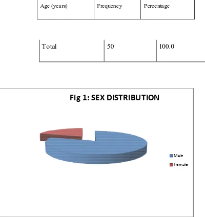
Table1: SEX DISTRIBUTION:
Sex group Frequency Percentage
Male 40 80.0
Female 10 20.0
PHT SEVERITY PVAT Normal >120 milliseconds MILD 100 – 120 milliseconds MODERATE 80 – 100 milliseconds SEVERE <80 milliseconds
58
Total 50 100.0
In our study the prevalence of COPD is more in males. Male: Female ratio is 4:1.
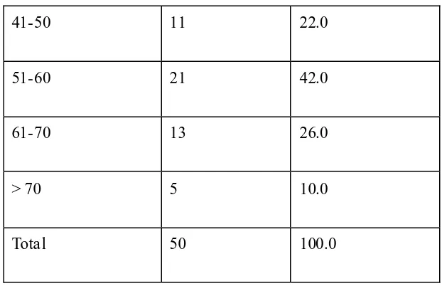
[image:69.595.71.471.142.566.2]Table 2: AGE DISTRIBUTION:
Fig 1: SEX DISTRIBUTION
Male Female
59
Lower age limit 40 was selected because many of the studies reported thatthe
41-50 11 22.0
51-60 21 42.0
61-70 13 26.0
> 70 5 10.0
Total 50 100.0
0 5 10 15 20 25
41-50 51-60 61-70 > 70
N
u
m
b
e
r
o
f
P
e
rs
o
n
s
[image:70.595.141.452.105.306.2]Age (in years)
Fig 2: AGE DISTRIBUTION
60
[image:71.595.164.430.232.452.2]overall prevalence of COPD is higher in age group more than 40 years. In ourstudy prevalence of COPD is more in the age group 51 – 60 years. i.e., 42% of patients belong to 51- 60 years.
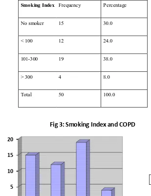
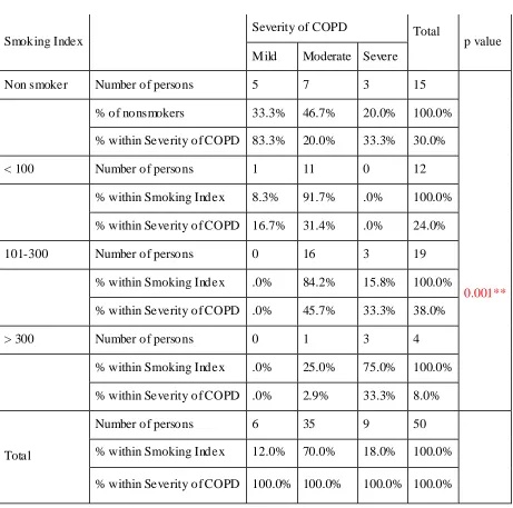
Table 3:SMOKING INDEX AND COPD
Smoking Index Frequency Percentage No smoker 15 30.0 < 100 12 24.0
101-300 19 38.0
> 300 4 8.0
Total 50 100.0
Smoking index is taken as a parameter because in India number of cigarettes,
0 5 10 15 20 No smoker
< 100 101-300 > 300
N u m b e r o f p e rs o n s Smoking Index
Fig 3: Smoking Index and COPD
[image:71.595.127.436.234.635.2]61
Beed is per pocket were quiet variable and weight of Beedis is widely d ifferent in different brands. More importantly Indian smokers often describe their smoking habit by the total number of cigarettes/Beedis per day rather than the number of pockets(121).
SMOKING INDEX GRADE
<100 Mild smoker
101 – 300 Moderate smoker
>300 Heavy smoker
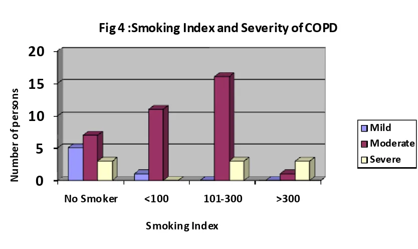
38% of our subjects were moderate smokers and 30% were nonsmokers .
Majority of the smokers have moderate to severe COPD. 91.7% mild smokers and
0
5
10
15
20
No Smoker <100 101-300 >300
[image:72.595.107.524.441.689.2]N u m b e r o f p e rs o n s Smoking Index
Fig 4 :Smoking Index and Severity of COPD
62
[image:73.595.66.527.221.678.2]84.2% of the moderate smokers had moderate COPD. 75% heavy smokers have severe COPD.“p” value is significant. Severity of COPD is directlyrelated to the smoking index .
Table 4:SMOKING INDEX AND SEVERITY OF COPD
Smoking Index
Severity of COPD Total
p value Mild Moderate Severe
Non smoker Number of persons 5 7 3 15
0.001**
% of nonsmokers 33.3% 46.7% 20.0% 100.0% % within Severity of COPD 83.3% 20.0% 33.3% 30.0%
< 100 Number of persons 1 11 0 12
% within Smoking Index 8.3% 91.7% .0% 100.0% % within Severity of COPD 16.7% 31.4% .0% 24.0%
101-300 Number of persons 0 16 3 19
% within Smoking Index .0% 84.2% 15.8% 100.0% % within Severity of COPD .0% 45.7% 33.3% 38.0%
> 300 Number of persons 0 1 3 4
% within Smoking Index .0% 25.0% 75.0% 100.0% % within Severity of COPD .0% 2.9% 33.3% 8.0%
Total
Number of persons 6 35 9 50
63
Table 5: DURATION OF SMOKING AND SEVERITY OF COPD
Duration of Smoking in years
Severity of COPD
Total p value Mild Moderate Severe
Non smoker Number of Persons 5 7 3 15
.026* % among nonsmokers 33.3% 46.7% 20.0% 100.0%
% within Severity of
COPD 83.3% 20.0% 33.3% 30.0%
< 10 Number of Persons 1 4 0 5
% within Duration of
Smoking in years 20.0% 80.0% .0% 100.0% % within Severity of
COPD 16.7% 11.4% .0% 10.0%
11-20 Number of Persons 0 12 1 13
% within Duration of
Smoking in years .0% 92.3% 7.7% 100.0% % within Severity of
COPD .0% 34.3% 11.1% 26.0%
21-30 Number of Persons 0 9 2 11
% within Duration of
Smoking in years .0% 81.8% 18.2% 100.0% % within Severity of
COPD .0% 25.7% 22.2% 22.0%
> 30 Number of Persons 0 3 3 6
% within Duration of
Smoking in years .0% 50.0% 50.0% 100.0% % within Severity of
COPD .0% 8.6% 33.3% 12.0%
Total
Number of Persons 6 35 9 50
% within Duration of
Smoking in years 12.0% 70.0% 18.0% 100.0% % within Severity of
64
In our study report those who smoked more than 10 years had moderate to severe obstruction and 50% of the smokerswho smoked more than 30 years had severe obstruction. ‘p’value is significant. prolonged duration of smoking is associated with Increased severity of COPD.
0 2 4 6 8 10 12
No Smoker <10 11--20 21-30 >30
N u m b e r o f p e rs o n s
Duration of Smoking in years
Fig 5: Duration of Smoking and Severity of COPD
Mild Moderate Severe 0 10 20 30 Low Middle N u m b e r o f P e rs o n s
Socio Economic Status
Fig 6 : Socio Economic Status and Severity of COPD
65
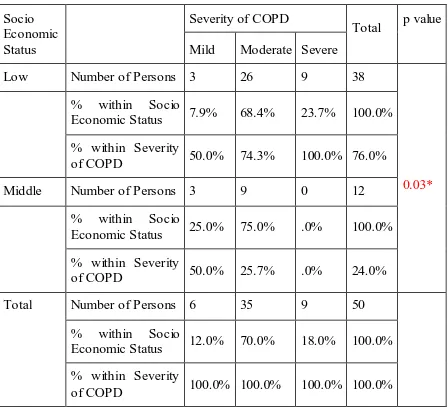
Table 6:SOCIO ECONOMIC STATUS AND SEVERITY OF COPD
Socio Economic Status
Severity of COPD
Total p value Mild Moderate Severe
Low Number of Persons 3 26 9 38
0.03* % within Socio
Economic Status 7.9% 68.4% 23.7% 100.0% % within Severity
of COPD 50.0% 74.3% 100.0% 76.0%
Middle Number of Persons 3 9 0 12
% within Socio
Economic Status 25.0% 75.0% .0% 100.0% % within Severity
of COPD 50.0% 25.7% .0% 24.0%
Total Number of Persons 6 35 9 50
% within Socio
Economic Status 12.0% 70.0% 18.0% 100.0% % within Severity
of COPD 100.0% 100.0% 100.0% 100.0%
66
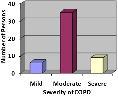
Table 7:SEVERITY OF COPD DISTRIBUTION
Severity of COPD Frequency Percent
Mild 6 12.0
Moderate 35 70.0
Severe 9 18.0
Total 50 100.0
Most of them are (70%) are moderate COPD.
0 10 20 30 40
Mild Moderate Severe
N u m b e r o f P e rs o n s
Severity of COPD
Fig 7: Severity of COPD Distribution
[image:77.595.96.303.461.630.2]67
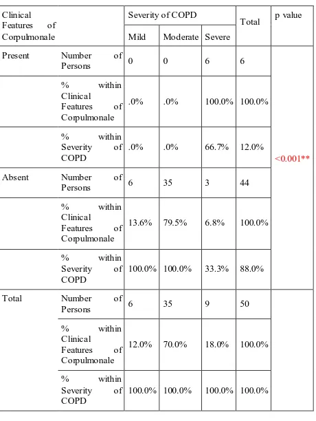
Table 8: CORPULMONALE AND SEVERITY OF COPD
Clinical
Features of Corpulmonale
Severity of COPD
Total p value Mild Moderate Severe
Present Number of
Persons 0 0 6 6
<0.001**
% within
Clinical
Features of Corpulmonale
.0% .0% 100.0% 100.0%
% within
Severity of COPD
.0% .0% 66.7% 12.0%
Absent Number of
Persons 6 35 3 44
% within
Clinical
Features of Corpulmonale
13.6% 79.5% 6.8% 100.0%
% within
Severity of COPD
100.0% 100.0% 33.3% 88.0%
Total Number of
Persons 6 35 9 50
% within
Clinical
Features of Corpulmonale
12.0% 70.0% 18.0% 100.0%
% within
Severity of COPD
68
‘p’ value is highly significant. All patients with corpulmonale had severe obstructive pattern. 66.7% of severe COPD patients developed corpulmonale.
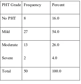
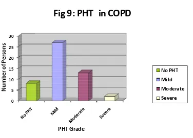
Table 9: PHT in COPD
PHT Grade Frequency Percent
No PHT 8 16.0
Mild 27 54.0
Moderate 13 26.0
Severe 2 4.0
Total 50 100.0
0 10 20 30 40 Present Absent N u m b e r o f P e rs o n s
[image:79.595.162.426.459.734.2]Clinical Features of Corpulmonale
Fig 8: Corpulmonale and Severity of COPD
Mild Moderate
69
Our study reports that 84% of them have pulmonary hypertension. This is due to chronic hypoxia in COPD that may the reason for pulmonary hypertension. Out of these 54% of them have mild, 26% moderate and 4% severe PHT.
0 5 10 15 20 25 30 N u m b e r o f P e rs o n s PHT Grade
Fig 9: PHT in COPD
No PHT Mild Moderate Severe
0
5
10
No Smoker <100 101-300 >300
[image:80.595.122.498.102.372.2]N u m b e r o f p e rs o n s Smoking Index
Fig 10 : Smoking Index and PHT
70
Table 10: SMOKING INDEX AND PHT
Smoking Index PHT Grade Total p value No
PHT Mild Moderate Severe
Non smoker Number of persons 5 9 0 1 15
<0.001**
% nonsmoker 33.3% 60.0% .0% 6.7% 100.0% % within PHT
Grade 62.5% 33.3% .0% 50.0% 30.0%
< 100 Number of persons 3 9 0 0 12
% within S moking
Index 25.0% 75.0% .0% .0% 100.0%
% within PHT
Grade 37.5% 33.3% .0% .0% 24.0%
101-300 Number of persons 0 9 9 1 19
% within S moking
Index .0% 47.4% 47.4% 5.3% 100.0%
% within PHT
Grade .0% 33.3% 69.2% 50.0% 38.0%
> 300 Number of persons 0 0 4 0 4
% within S moking
Index .0% .0% 100.0% .0% 100.0%
% within PHT
Grade .0% .0% 30.8% .0% 8.0%
Total
Number of persons 8 27 13 2 50
% within S moking
Index 16.0% 54.0% 26.0% 4.0% 100.0%
% within PHT
Grade 100.0% 100.0% 100.0% 100.0% 100.0%
71
Table 11: DURATION OF SMOKING AND PHT
Duration of Smoking in years PHT Grade Total p value No
PHT Mild Moderate Severe Non
smoker
Number of persons
5 9 0 1 15
.012* %among nonsmokers 33.3% 60.0% .0% 6.7% 100.0%
% within PHT Grade 62.5% 33.3% .0% 50.0% 30.0%
< 10 Number of persons 2 3 0 0 5
% within Duration of
Smoking in years 40.0% 60.0% .0% .0% 100.0% % within PHT Grade 25.0% 11.1% .0% .0% 10.0%
11-20 Number of persons 1 8 3 1 13
% within Duration of
Smoking in years 7.7% 61.5% 23.1% 7.7% 100.0% % within PHT Grade 12.5% 29.6% 23.1% 50.0% 26.0%
21-30 Number of persons 0 6 5 0 11
% within Duration of
Smoking in years .0% 54.5% 45.5% .0% 100.0% % within PHT Grade .0% 22.2% 38.5% .0% 22.0%
> 30 Number of persons 0 1 5 0 6
% within Duration of
Smoking in years .0% 16.7% 83.3% .0% 100.0% % within PHT Grade .0% 3.7% 38.5% .0% 12.0%
Total Number of persons 8 27 13 2 50
% within Duration of
72
‘p’ value is significant. Duration of smoking is directly proportional to the PHT severity. Moderate PHT is present in those who are smoked more than 10 years. The percentage of moderate PHT is more in those who smoked more than 20 years.
.
0 5 10
No Smoker 11--20 >30
N u m b e r o f p e rs o n s
Duration of Smoking in years
Fig 11: Duration of Smoking and PHT
No PHT Mild Moderate Severe 0 5 10 15 20 25
Mild Moderate Severe
N u m b e r o f p e rs o n s
[image:83.595.103.461.456.729.2]Severity of COPD
Fig 12: Severity of COPD and Pulmonary Hypertension
No PHT
Mild
Moderate
73
Table 12: SEVERITY OF COPD AND PHT GRADE
‘p’ value is significant. Severity of COPD is directly related to severity of PHT. Severe PHT is associated with moderate and severe COPD patients.
Severity of COPD PHT Grade Total p value No
PHT Mild Moderate Severe
Mild Number of persons 5 1 0 0 6
<.001** % within Severity
of COPD 83.3% 16.7% .0% .0% 100.0%
% within PHT
Grade 62.5% 3.7% .0% .0% 12.0%
Moderate Number of persons 3 23 8 1 35
% within Severity
of COPD 8.6% 65.7% 22.9% 2.9% 100.0%
% within PHT
Grade 37.5% 85.2% 61.5% 50.0% 70.0%
Severe Number of persons 0 3 5 1 9
% within Severity
of COPD .0% 33.3% 55.6% 11.1% 100.0%
% within PHT
Grade .0% 11.1% 38.5% 50.0% 18.0%
Total
Number of persons 8 27 13 2 50
% within Severity
of COPD 16.0% 54.0% 26.0% 4.0% 100.0%
% within PHT
74
Table 13: SEVERITY OF COPD AND PHT GRADE IN CORPULMONALE
All patients with corpulmonale have a severe obstructive pattern and 66.7% have moderate PHT. ‘p’value is significant.
0
1
2
3
4
Severe N u m b e r o f p e rs o n sSeverity of COPD
Fig 13: Severity of COPD and PHT in Corpulmonale
Mild Moderate Severe Severity of COPD PHT Grade Total p value No
PHT Mild Moderate Severe
Severe Number of persons 1 4 1 6
0.002**
% within Severity of
COPD 16.7% 66.7%