CORRELATION OF COX-2 EXPRESSION IN
COLORECTAL CARCINOMA WITH
CLINICOPATHOLOGICAL FEATURES
DISSERTATION
SUBMITTED FOR
M.D IN PATHOLOGY
THE TAMILNADU DR.M.G.R MEDICALUNIVERSITY,
DEPARTMENT OF PATHOLOGY
PSG INSTITUTE OF MEDICAL SCIENCES & RESEARCH
PEELAMEDU, COIMBATORE – 641004
TABLE OF CONTENTS
Page No Certificate
IHEC Clearance Certificate
Acknowledgement
Introduction 1
Aims and objectives 2
Review of literature 3
Materials and methods 75
Results 79
Discussion 101
Summary and conclusion 106
Bibliography
CERTIFICATE
This is to certify that the dissertation work entitled “CORRELATION OF COX-2 EXPRESSION IN COLORECTAL CARCINOMA WITH CLINICOPATHOLOGICAL FEATURES” submitted by Dr. Manjula R. is a work done by her during the period of study in the department from 31/05/2012 to 30/05/2015. This work was done under the guidance of Dr. Sandhya V, Professor, Department of Pathology, PSG IMS & R.
Dr. Prasanna N Kumar Dr. S.Ramalingam
Professor & HOD, Principal
Pathology PSG IMS&R
PSGIMS & R Coimbatore
CERTIFICATE
This is to certify that the dissertation work entitled “CORRELATION OF COX-2 EXPRESSION IN COLORECTAL CARCINOMA WITH CLINICOPATHOLOGICAL FEATURES” submitted by Dr. Manjula R to The Tamilnadu Dr M.G.R Medical University, Chennai, for the award of the degree of Doctor of Medicine in Pathology, is a bonafide record of research work carried out by her under the supervision of Dr.Sandhya V, Professor of Pathology, PSG IMS&R. The contents of this thesis, in full or in parts, have not been submitted to any other Institute or University for the award of any degree or diploma.
Dr.Manjula R Dr.Prasanna N Kumar
ACKNOWLEDGEMENT
It gives me an immense pleasure to express my sincere gratitude and
heartfelt thanks to my guide
Dr. Sandhya V
, Professor of the Pathology,
PSGIMS &R for her invaluable guidance, immense patience, timely
advice and constant support.
I am thankful to our Head of The Department
Dr. Prasanna N Kumar
,
Department of Pathology and I would like to extend my thanks to all
faculty members and whom I have had the privilege to work with
emeritus Professor
Dr. NirmalaV, Dr.Shanthakumari S
&
Dr. T M
Subba Rao
and all assistant and associate professors for their support.
I am also thankful to my colleague and friends who have helped me to
retrieve the tissue blocks & technical support
I am extremely grateful to my husband
G. Mohanraj
for his support &
to my son Dakshin Pranesh Mohanraj for his understanding and to my
parents and in laws for their prayers and blessings
I am thankful to our senior technician
Angeline Mary
and other staff
members for their technical support
Last but not least, I want to thank all the patients whose data I used for
CORRELATION OF COX- 2 EXPRESSION IN COLORECTAL
CARCINOMA WITH CLINICOPATHOLOGICAL FEATURES.
ABSTRACT
Introduction : Colorectal carcinoma is the most common neoplasm of the gastrointestinal tract. COX-2 (cyclooxygenase-2) expression is upregulated in colorectal carcinoma. Therefore its assessment would identify patients amenable to adjuvant therapy. As no studies have been done across the Indian population as compared to western , the present study was done.
Aims and objectives: To study COX-2 expression in colorectal carcinoma and correlate it with clinicopathological features like age, sex, tumor location, size, degree of differentiation, depth of invasion, lymph node status and TNM stage.
Methods: 65 consecutive cases of colorectal adenocarcinoma between January 2009 to December 2013 were retrived from records of Pathology Department at PSG IMS&R. Immunohistochemical staining for COX-2 was done and correlated with age, sex, tumor location , size, degree of differentiation, depth of invasion, lymph node status and TNM stage.
COX2 cases constituted 33.3% of stage I, 58.8% of stage II, 80% of stage III and 100% of stage IV tumors. About 56.6% of well differentiated, 66.6% of moderately differentiated and 100% of poorly differentiated carcinomas showed high COX-2 expression. The COX-2 overexpression was associated with advancing stage of tumor, more frequent lymph node and distant metastasis, decreasing degree of differentiation.
Conclusion : Determination of COX-2 expression in colorectal carcinoma gives prognostic information. COX-2 overexpression implicates advancing stage of disease. These patients can be treated with selective COX-2 inhibitors like Celecoxib, Rofecoxib, L-745,337 and SC 58125 as an adjuvant to chemo and radiotherapy.
1
INTRODUCTION
Colorectal carcinoma is the fourth common cancer in the world and
second most common cause of cancer related death1. Epidemiological studies have shown a lower incidence of colorectal adenomas and carcinomas in
subjects who have taken NSAIDS (Non Steroidal Anti-Inflammatory Drugs)
for a long time which suggest a pathogenic role for cyclooxygenase (COX -2)
in colonic tumorigenesis. COX-2, an inducible isoform of cyclooxygenase is
usually absent or present in low levels in normal colonic epithelium and is
upregulated in colorectal carcinoma. Assessment of this molecular factor would
therefore help in identifying the patients who are likely benefit from COX-2
inhibitor adjuvant therapy which attenuates the metastatic potential of
colorectal carcinoma, thereby improving the prognosis. Literature search
revealed no such study across the Indian population. Therefore the present
study aims to evaluate COX-2 expression in colorectal carcinoma and to
correlate it with clinicopathological features- age, sex, tumor location, size,
depth of invasion, histological type, degree of differentiation, lymph node
2
AIMS AND OBJECTIVES :
Primary Aim:
To study the COX-2 expression in colorectal carcinoma.
Secondary Aim :
To correlate the COX-2 expression with clinicopathological features – age, sex,
tumor location, size, histological type, degree of differentiation, depth of
3
REVIEW OF LITERATURE
Incidence and disease burden
Colorectal carcinoma is the most common neoplasm of gastrointestinal
tract. According to global cancer statistics 2002, Colorectal carcinoma is the
fourth most common cancer worldwide and second most common cause of
death1 .They are most prevalent in North America, Europe, Australia and New Zealand compared to Asia and Africa1. In India, the incidence is 30- fold lower than the former group1 . This geographical variation is due to different dietary, environmental factors. According to United States Surveillance Epidemiology
& End Results (SEER) program, colorectal carcinoma accounts for 12% of all
cancers with incidence rate of about 33.7 and 12.8 per one lakh for colon and
rectal carcinoma respectively2. Mortality rate was about 15% according to American cancer society statistics(2000-2004)2. The incidence rate has been declining in the recent years considerably in North America due to modification
of dietary habits and early screening3. Males are more frequently affected than females1. Incidence increases with age, most of the patients being above 40 years with a median age of 71 years according to SEER statistics. In patients
younger than 50 years, family history is necessary in assessing the carcinoma
risk, as 1% is associated with Familial Adenomatous Polyposis and 5% with
4
cancer in various sites of colon differs between male and female, and also
reflect environmental, genetic factors and age of presentation3 .
Location of tumor Males Females
Right colon 30% 40%
Left colon 30% 30%
Rectum 40% 30%
ETIOPATHOGENESIS :
The pathogenesis of colorectal cancer depends on environmental, diet
and genetic factors, the polyposis syndromes being the most important ones.
Environmentalfactors :
Apart from diet, decreased physical activity, occupational exposure,
alcohol, smoking, inflammatory bowel diseases are other factors having a role
in the pathogenesis of colorectal carcinoma.
Dietary factors:
... High intake of fat and animal protein provides an excess source of
calories resulting in weight gain. In addition, it also cause increase in the
5
colonic epithelium predisposing to neoplastic progression 4,5. It also increases bile acid and cholesterol synthesis, the toxic metabolites of which are
carcinogenic 4.
Good intake of fruits, vegetables with high fibre content and
micronutrients like carotenoids, flavinoids, ascorbate, isothiocyanate and
phytic acid decrease the risk of developing colorectal cancer as these increase
the defecation frequency and decrease mucosal contact time of potential
carcinogens.
The consumption of vitamin A,C,D,E, calcium, selenium reduces the risk
by their antioxidant action which prevent formation and neutralise the
carcinogens 6,7. In addition, calcium decreases the bile acids and fatty acids mediated injury of colonic epithelium by converting them into insoluble
calcium soaps, thereby decreasing epithelial cell proliferation7. Selenium, a cofactor of glutathione peroxidase reduces oxidative damage of epithelium.
The low fibre diet decreases the stool bulk and alters the intestinal flora
resulting in increased free radicals which remains in contact with colonic
mucosa due to increased transit time, causing epithelial injury 4.
Alcohol consumption results in positive energy balance and abnormal
6
Obesity, sedentary occupation, excess energy intake, decreased physical
activity in association with high fat intake is a risk factor. Increased physical
activity cause stimulation of hormonal release, increase in peristalsis and neural
reflex mechanism resulting in decreased mucosal exposure to carcinogens 10.
Individuals with prolonged exposure to metal dusts, plastics, organic
solvents, fibreglass, fumes and asbestos are at increased risk for developing
rectal cancer 1.
Intestinal factors :
Increased risk of cancer is seen in patients with diverticulosis, ulcerative
colitis, Crohn’s disease and adenomatous polyps 1. Carcinomas occurring in association with Schistosoma Japonicum infestation are multicentric and
present at an early age 1.
Hormonal factors :
Type 2 diabetes mellitus shows an increase in insulin resistance and
glucose intolerance. Increased insulin levels in Type 2 DM promotes epithelial
proliferation and inhibit apoptosis of colonic epithelial cells favouring
7
Estrogen receptors are highly expressed in colonic tumors which implicate
the protective role of estrogen.
Increased gastrin and growth hormone levels have been implicated in
colorectal tumors. In colonic cancers, serum gastrin levels may be increased ,
probably due to autocrine secretion from tumor. The gastrin has growth
promoting action. In persons with acromegaly, an increased growth hormone
status resulted in increased cell proliferation and tumor formation.1
Others :
Radiation therapy for cancers of female genital tract and prostate
increases the risk of colorectal carcinoma.
Surgical procedures like cholecystectomy, ileal conduits, gastric surgery
for peptic ulcer may contribute to carcinogenesis. Epithelial proliferation occurs
due to activation of fecal carcinogens by the diverted urine. Gastric surgery for
peptic ulcer may alter the bile acid metabolism increasing unconjugated and
8
Predisposing host factors :
Among the endogenous factors, inflammatory bowel disease, genetic
polyposis syndromes are important 2.
Inflammatory bowel disease
Inflammatory bowel disease is a long standing chronic inflammatory
process which includes Ulcerative colitis and Crohn’s disease. Numerous
studies demonstrate an association between these diseases and colorectal
carcinoma. The pathogenesis includes two hypothesis, one is a normal immune
response to abnormal environmental stimuli while other is an abnormal
immune response to normal stimuli 12.
IBD is more common around the third decade. In long standing IBD,
with increasing duration of disease, the risk of developing precursor lesions
like dysplasia or invasive carcinoma was higher 13. Patients with extensive colon involvement upto or proximal to hepatic flexure are at greater risk for
developing colorectal cancer 13. The incidence begins to increase after 8-10 years of onset of the symptoms with an incidence rate of about 5-10% and
12-20% after about 20 and 30 years of disease respectively 14. But when IBD is limited to left colon the risk of developing cancer increases after 15-20 years.
Patients with isolated ulcerative proctitis do not have greater risk for
9
surveillance for dysplasia is recommended for patients with crohn’s or
ulcerative colitis. A routine colonoscopy every 1 or 2 years for lifetime is
recommended 13.
Polyps :
Polyps of the colon are predominantly of epithelial origin, categorised as
hyperplastic, adenomatous and inflammatory polyp.
Hyperplastic polyp:
About 25-30% of large intestinal polyps are hyperplastic in nature,
grossly presenting as pale sessile nodule < 5mm in size with smooth glistening
surface1. Three subtypes have been identified namely the Microvesicular hyperplastic polyp (MVHP), Mucin poor hyperplastic polyp ( MPHP) and the
Goblet cell hyperplastic polyp (GCHP).
Hyperplastic polyps in general has low malignant potential. However
mutations have been identified in MVHP and GCHP. The microvesicular
hyperplastic polyp is associated with V600E BRAF mutation and activation
of MAPK (MAP Kinase) pathway15. The surface colonocytes, in the absence of apoptosis persist, resulting in serrated morphology. The goblet cell type
10
nuclear atypia at the crypt base, surface maturation and thicker collagen
table 2.
Adenomatous polyps:
Adenomas are common premalignant lesions of colon. They are
classified as conventional, serrated or flat types. Conventional adenomas are
subtyped as tubular, villous or tubulovillous adenomas 2. Polyps occuring in the context of hereditary colon cancer syndromes was usually multiple and
present at a younger age group 1. Among various colonic polyposis syndromes, Familial adenomatous polyposis with its attenuated form, Gardner’s syndrome,
Turcot’s syndrome, MYH associated polyposis and Lynch syndrome are most
common 2.
Familial adenomatous polyposis:
Familial adenomatous polyposis has an autosomal dominant inheritance
with APC (Adenomatous Polyposis Coli) gene mutation ( a tumor suppressor
gene). In addition KRAS and p53 mutations are also encountered. It is
characterised by presence of more than 100 tubular adenomatous polyps
involving the entire colon which by definition harbours low grade dysplasia 2. Cyclooxygenase 1 expression has been seen in stromal cells in FAP at an early
11
polyp 16. Thus in addition to COX-2, COX-1 also plays a role in the initial stages of development of polyp 16.
Gardner’s syndrome, an autosomal dominant condition with APC gene
mutation, develop in addition to adenomatous polyps, desmoid tumor and
osteomas.
Turcot’s syndrome shows coexistence of FAP or Hereditary
nonpolyposis colon cancer with central nervous system tumors. APC gene
mutation or germline defect in DNA mismatch repair genes have been
reported 2.
Attenuated FAP also known as hereditary colon cancer syndrome with
mutation similar to FAP. Morphologically the condition differs from
conventional FAP, by the presence of less than 100 polyps and the risk of
developing colorectal carcinoma at an older age group 2.
MYH associated polyposis is an autosomal recessive syndrome with
mutation in Mut Y homologue (MYH) gene which encodes an enzyme
responsible for preventing mutation after DNA damage due to oxidation.
Morphologically more than 30 adenomatous polyps are seen with a high risk
for colorectal carcinoma.
Mutation of DNA mismatch repair genes ( hMLH2, hMSH6, hMSH2,
12
Polyposis Colorectal Cancer Syndrome (HNPCC), also known as Lynch
syndrome . The polyps are of adenomatous type, usually less than 10 in number
with high grade dysplasia and increased risk for malignant transformation.
Morphology of adenomas :
Grossly, the adenomas may be pedunculated, flat or sessile. Tubular
adenomas are pedunculated, small with surface lobulation while villous
adenomas lack a stalk, present as flat or lobulated, sessile, large shaggy mass
and have a broad base. Tubulovillous adenomas show overlapping features of
tubular and villous architecture ranging between 20-79% 1.
Microscopically tubular adenomas show increase number of crypts with
dysplasia of surface epithelium, focal tubular dilation and mixed inflammatory
cells in the lamina propria. The villous adenoma consist of more than 80%
finger like fronds with a core formed by lamina propria, lined by dysplastic
epithelium. All type of adenomas are composed of mixture of absorptive cells,
intermediate cells, goblet cells, paneth cells and endocrine cells. Flat adenoma
and villous adnenomas are most commonly associated with high grade
dysplasia. Hypersecretory and clear cells are other rare types of adenomas 1.
Serrated adenomas are less frequently reported type of polyps. They may
13
stratified dysplastic cells with surface mitosis, papillary tufting and eosinophilic
cytoplasm with increased nucleo-cytoplasmic ratio and prominent nucleoli.
Hyperplastic and serrated polyps constitute 7% of premalignant lesions in
IBD 13.
Dysplasia associated lesions :
Dysplasia can be associated with both inflammatory bowel disease and polyps. In 1981, Blackstone et al, described the term DALM or Dysplasia
Associated Lesion or Mass in ulcerative colitis. The DALMs can
morphologically resembles sporadic adenoma which was termed as adenoma
like dysplasia associated lesions or masses.
The differentiating features between DALMs and sporadic adenoma is
that the former shows bottom up growth pattern with dysplastic cells at the
bottom of the crypts in the background of chronic active inflammation, while
the latter shows top-down growth pattern with luminal surface of crypts lined by
14
PATHWAYS OF COLORECTAL CARCINOGENESIS :
The molecular changes in colorectal adenocarcinoma are heterogenous
including genetic alteration and epigenetic abnormalities 4. The two distinct forms of genomic instabilities are chromosomal instability and microsatellite
instability. Till date at least three distinct pathways of colorectal carcinogenesis
have been identified 2.
1.APC / β catenin pathway
2. Microsatellite Instability Pathway
3.Serrated neoplasia pathway OR CpG Methylation pathway
1. APC / β catenin pathway :
APC gene mutation was identified commonly in individuals with familial
adenomatous polyposis (17,18). Vogelstein et al described it as a type of chromosomal instability with multistep progression from hyperproliferative
epithelium to adenoma of increasing size and dysplasia, finally resulting in
invasive carcinoma 19. The members of this pathway are APC, KRAS, p53,
β-catenin, DCC and DPC4 genes 4.
APC gene is located in chromosome 5 , undergoes germline mutation in
15
gene is a most important component of Wingless (WnT) signalling pathway. It
act as “ Gate keeper gene” with negative regulation of colonic epithelial
proliferation in colorectal neoplasia 2. Most colorectal cancer and adenoma patients have at least one allele mutation according to knudson hypothesis 4. In tumorigenesis, the second allele also mutated results in loss of tumor
suppressor effects 20.
ββββ----Catenin can be dysregulated by APC gene mutation or CTNNB1
mutation (β catenin gene) 17. Normally APC gene causes ubiquitin mediated degradation of β catenin in cytoplasm, thereby preventing it from entering the
nucleus where it induces cell proliferation by interacting with transcription of
Myc and cyclin D 21.
KRAS are protooncogenes having role in signal transduction from growth
factor receptors. Mutation cause autonomous cell proliferation. KRAS
mutations follow loss of APC in the adenoma-carcinoma pathway 19.
p53 gene, located on chromosome 17 is a tumor suppressor gene. The
most common functions are control of cell proliferation, DNA repair and
programmed cell death. Arrest of cell cycle at G1 phase, inhibition of cyclin
CDC2 or cyclin CDK complexes are also the functions of p53. Loss of wild
type p53 due to mutation or deletion therefore is a major step in carcinogenesis
16
results in amplification of unrepaired DNA and upregulated cell growth. p53
overexpression is an important predictor of survival status in patients with
lymph node positive colorectal carcinoma 1.
Loss of heterogenesity of DCC (Deleted in Colon Cancer) and
SMAD4 or DPC4 (Deleted in Pancreatic Cancer, locus 4) on chromosome 18
is commonly seen in colorectal carcinoma. These genes encode TGF-β
signalling proteins which normally inhibits cell cycle 4 and the mutation of
TGF-β causes increased cell proliferation. Loss of SMAD4 gene is seen in late
17
Sequence of events of APC /β catenin pathway 4
Normal colon with germline or somatic mutation of single allele of APC at
chromosome 5.
Hypermethylation and inactivation of second allele of the APC gene results in
increased β-catenin levels. The mucosa is put to increased risk for tumor
formation.
Subsequent KRAS and p53 mutations results in increased cell proliferation and
inhibition of apoptosis
ADENOMAS develop. COX-2 overexpression occurs (discussed in detail later
in the review of literature)
Together with loss of heterogenesity of SMAD4 &2 and increased telomerase
18
2. Microsatellite instability pathway:
Mismatch repair genes correct the genetic alteration occurring during
DNA synthesis. In conditions with DNA mismatch repair deficiency, mutation
accumulate in microsatellite repeats which is referred to as
MICROSATELLITE INSTABILITY (MSI). Many of the microsatellites are
located in noncoding regions.
When the microsatellites located in promoter or coding region of the
genes like TGF-β type II receptors, BAX proteins become unstable,
uncontrolled cell growth and proliferation occur 4. These promoter regions are
responsible for regulation of cell growth by encoding the TGF-β type II
receptor and BAX proapoptotic protein. Normally TGF-β inhibit proliferation,
so in the presence of TGF-β mutation, uncontrolled cell growth occurs with
increased survival due to BAX loss. Other mutations in BRAF and CpG
hypermethylation also develop in this defect 4.
MSI was initially discovered in HNPCC, most common cause of right
sided colonic tumor with a higher tendency to be of mucinous, poorly
19
(a) MSS( Micro Satellite Stable),
(b) low level MSI
(c) high level MSI.
low level occurs with <30% unstable microsatellite loci and
MSI-high level show >30% microsatellite instability loci 22.
Sequence of the pathway 22
In normal colon with germline or somatic mutations of mismatch repair genes
Alteration in second allele by Loss of Heterogenesity by methylation or
mutation of MLH1, MSH2,MSH6, PMS1 and PMS2.
Microsatellite instability is initiated
Accumulation of gene mutations results in uncontrolled cell proliferation and
inhibition of apoptosis by alteration in TGF-β RII, BAX, BRAF and others.
20
3. SERRATED NEOPLASIA PATHWAY OR THE CPG
METHYLATION PATHWAY
Jass et al, proposed that carcinomas arising by the serrated pathway are
heterogenous groups, the molecular profiles of which have been subtyped as
follows 15,23.
1.BRAF mutant, CIMP-H with MSI-high level.
2.BRAF mutant, CIMP-H with MSS.
3.Kras mutant, CIMP-L, MSS.
[CIMP-H or L : CpG Island Methylation Phenotype-High level or Low level,
MSI: MicroSatellite Instability, MSS: MicroSatellite Stable].
1.BRAF mutant / CIMP-H / MSI-H :
The colorectal carcinomas with this genetic profile are more
common (9-12%) in elderly women, situated in right colon, and are at
high tumor stage without lymphnode or distant metastasis at presentation.
The sessile serrated adenomas, a precursor lesion undergoes progressive
methylation and silencing of MLH1 (key promoter region) resulting in
21
Prognosis is favourable. However these tumors are resisitant to most
non-surgical treatments like chemotherapy including 5-flurouracil,
monoclonal EFGR inhibitors (cetuximab & panitumumab) 15.
2.BRAF mutant / CIMP-H / MSS:
Sessile serrated adenomas with dysplasia in the absence of loss of MLH1
gene are proposed precursor lesion of this subgroup of mutation (5-10%).
Compared to conventional colorectal carcinomas, these tumours are often
poorly differentiated and have higher incidences of tumor budding,
lymphovascular and perineural invasion with lymphnode metastasis.
The genetic abnormalities includes methylation of different promoter
region and silencing of p16, Tp53 and WnT pathway genes. These tumors have
a poor prognosis.
3. KRAS mutant / CIMP-L / MSS :
KRAS mutation with microsatellite stable subgroup is the largest group
(15-20%) among other mutants. Serrated tubulovillous adenoma are the
precursor lesions progressing via this pathway. Low level CIMP segregate these
22
Serrated morphology carcinoma :
Serrated carcinoma refers to the tumors with distinctive serrated
morphology and derived via serrated neoplastic pathway. These carcinomas
initially described by Jass and Makinon et al have defined the histological
criteria. The World Health Organisation has recognised serrated carcinomas as a
distinct subtype of colorectal carcinoma 15.
These serrated carcinomas are frequently seen in women and the common
23
SEQUENCE OF THE SERRATED PATHWAY15
NORMAL MUCOSA
BRAF / CIMP-H KRAS
Sessile serrated adenoma Traditional serrated adenoma
Loss of MLH1 Loss of p16/ MGMT
SSA with dysplasia SSA with dysplasia
Subsequent MSI results Silencing of WnT pathway genes
frameshift mutation of
TGFβ receptor II TSA with high grade dysplasia
BRAF/CIMP-H/MSI BRAF/CIMP-H/MSS KRAS/CIMP-L/MSS
Colorectal carcinoma colorectal carcinoma colorectal carcinoma
Comparing these three molecular profiles, the BRAF/ CIMP-H/ MSI has good
prognosis but resistance to chemotherapy, while the remaining two mutations
24
INFLAMMATORY BOWEL DISEASE ASSOCIATED
CARCINOGENESIS :
Inflammatory Bowel Disease (IBD) associated carcinogenesis is a
multistep process, starting from inflamed regenerative epithelium to hyperplasia
to flat dysplasia and ends with invasive adenocarcinoma. The interaction of
inflammatory cells like neutrophils and macrophages with colonic epithelium
plays a key role in IBD-induced carcinogenesis. Various factors like reactive
oxygen/nitrogen overproduction, cytokines, growth factors and arachidonic
acid metabolites, activated inflammation associated signal pathways, together
with immune dysfunction contributes to carcinogenesis in IBD.
Similar to sporadic colorectal cancer the characteristic molecular
abnormalities are mutation in oncogenes, tumor suppressor genes and DNA
repair genes with genomic instability13. Chronic inflammation results in oxidative stress which plays a characteristic important role in this
carcinogenesis. The oxygen and nitrogen free radicals bind to target protein,
DNA, RNA and result in genetic alterations, aberrant methylation and
genomic insatability.
Free radicals cause chromosomal instability secondary to DNA
translocation/ amplification/ deletion/ breakage/ telomere damage and p53
25
dysplasia25. Interaction of free radicals with the cell membranes leads to lipid peroxidation results in mutation of p53 tumor suppressor gene. Aberrant
activation of arachidonic acid pathways and COX-2 overexpression are
important in IBD associated carcinogenesis 26. In ulcerative colitis, COX-2 expression elevated along with mRNA expression in inflamed mucosa,
dysplasia and in carcinomas. COX-2 activates procarcinogens , increases free
radical production and angiogenesis 26.
IBD associated carcinogenesis and sporadic carcinogenesis shows similar
molecular changes. The difference in the timing and frequency of the alterations
distinguish both types. p53 is a key factor in IBD associated carcinogenesis
and KRAS mutation, loss of APC genes are less common and occur late in
carcinogenesis. Genes with methylation of CpG islands result in dysplasia
throughout the mucosa with ulcerative colitis.
Aneuploidy is usually seen in patients with more than 10 years disease
duration and is associated with dysplasia 15. Fluroscent In-Situ Hybridisation (FISH) analysis shows ulcerative colitis associated dysplasia or carcinoma
exhibiting monosomies and polysomies.
Microsatellite instability was frequently associated with ulcerative colitis
patient with dysplasia and carcinoma. p53 mutation in adjacent mucosa, APC
26
later stages of ulcerative colitis indicating that KRAS play a role at the later
stages of carcinogenesis.
CpG island methylation with hypermethylation of cyclin dependent
kinase inhibitors 2A (CDKN2A) or p16 and p14 are common in ulcerative
colitis associated adenocarcinoma. The p14 hypermethylation was detected in
50% cases .
In sporadic cancers the precursors are tubular adenoma. In contrast, the
dysplasia in IBD are polypoid or flat and localised or diffuse. The concept of
“Field Cancerisation” describes that the entire epithelium of upper
aerodigestive tract has increased risk for premalignant lesions in IBD due to
multiple genetic abnormalities 27. This concept was widely accepted for other organs including colon, importantly in IBD associated dysplasia. This clonally
derived mutant cells with indistinguishable histological features are seen in
inflamed segment of IBD. For example the mutant TP53 or KRAS is detected
across nondysplastic crypts as well as entire neoplasm 27.
Low grade dysplasia is associated with very short telomerases, high
level of senescences together with DEC1( Deleted in Esophageal Cancer 1)
overexpression. Above pattern is reversed in high grade dysplasia, an important
27
The level of expression of an enzyme named α-methylacyl coenzyme A
racemase (AMACR) ranging from negative in normal epithelium to high in
invasive carcinoma. Hence p53 and AMACR coexpression are demonstrated
in IBD associated carcinogenesis 15.
CLINICAL FEATURES :
Colorectal carcinomas shows wide range of clinical features from altered
bowel habits, pain and tenesmus to surgical emergency like intestinal
obstruction 17,18. The clinical symptoms differ depending on the location and stage of the tumor 1. Generally all patients have loss of weight, loss of appetite malaise, weakness and fatigue.
Right side colon cancers are often polypoidal and patients remain silent
because of the soft stool consistency in this region or have features secondary
to iron deficiency anemia, cardiac failure or angina pectoris. When the tumor
obstructs the lumen of appendix, patients may manifest with symptoms similar
to appendicitis. Most of the caecal tumours are however silent, present as an
abdominal mass with metastasis 1.
Left sided colon cancers frequently presents with altered bowel habits like
diarrhoea, alternating with constipation and incomplete rectal emptying
28
constipation and symptoms of obstruction . In case of obstruction, the proximal
colon undergoes ischemic change and rupture 1.
Bleeding is a frequent finding in rectal carcinoma 1. In advanced stages irrespective of the location , the tumours present with significant abdominal
pain, perforative peritonitis and colo-colic fistula 1.
MORPHOLOGY:
Gross appearance of colorectal carcinoma depends on the stage of
disease 2.
Small carcinomas present as red, granular, elevated circumscribed lesions
resembling adenoma 1. Aberrant crypt foci (ACF) is the earliest morphological change in the epithelial neoplasms of the colon 1.
Large carcinomas are categorised into four types:
(a)Polypoidal type growth pattern:
It is most commonly seen in caecum and ascending colon
presenting as a bulky mass 2. Also referred to as fungating or exophytic pattern they are characterised by intraluminal growth with papillary
29
usually asymptomatic until the symptoms of anemia occurs due to
chronic blood loss 1.
(b)Ulcerated or excavated type:
It is an infiltrating type with elevated edges and intramural
growth18.
(c)Annular or constricting type:
It is the circumferential involvement of colon resulting in napkin
ring constriction as seen by double contrast study 2. The colon proximal to the constriction shows attenuation of mucosal folds and dilatation 2. Both the ulcerated and annular types are commonly seen in descending
and transverse colon 18.
(d)Diffuse infiltrative or linitis plastica:
There is diffuse flattening and thickening of mucosa with more of
intramural spread in this growth pattern2. All types are relatively homogenous with areas of necrosis and infiltration upto the serosa
resulting in retraction of serosal surface 2. Tumors with high mucin contents appear gelatinous and glistening with separation of bowel
30
MICROSCOPIC APPEARANCE:
Adenocarcinoma is the most common type of colorectal cancer. The
tumor cells in colorectal carcinomas are arranged in glandular pattern and less
commonly as single infiltrating cells in the poorly differentiated tumors. The
surface of the tumors show papillary or villous pattern of arrangement 17.
The tumor cells are pleomorphic, columnar cells with increased nucleo
cytoplasmic ratio, eosinophilic cytoplasm and conspicuous nucleoli. Admixture
of goblet cells, neuroendocrine cells and paneth cells are also encountered. The
tumor confined to mucosa is grouped as intramucosal and further invasion
towards the serosa increased the tumor staging.
Inflammation and desmoplastic reaction are often seen in the surrounding
stroma. Hyperplastic changes like taller and tortuous glands are most
commonly seen at the edges of the tumor. Lymphovascular and perineural
invasion has prognostic significance 17.
Adenocarcinomas are graded on the basis of extent of glandular
differentiation into (1) Well, Moderately and Poorly differentiated or (2) Low
grade [well and moderately differentiated] and High grade [ poorly and
undifferentiated]. More than 95%, 50-95% and 5-50% of glandular
differentiation are seen in well, moderately and poorly differentiated carcinomas
31
categorised as undifferentiated type 18. These tumors have DNA mismatch repair deficiency and poor prognosis 2.
Other types of carcinomas:
Mucinous carcinoma:
Mucinous carcinoma are most frequently seen in young patients with
Hereditary Non Polyposis Colon Cancer syndrome2. The Conventional adenocarcinomas can have significant mucinous component. WHO
recommends the term mucinous carcinoma when > 50% of tumor cells
produce mucin 18.
The tumor epithelium are in acinar, clumps or single cell arrangement
and these cells are freely floating in pools of mucin 18. Microsatellite instability and previous villous adenomas are the most common associated features 1. Mucinous carcinomas have very poor prognosis due to advanced stage at
presentation, extensive lymphnode and pericolonic involvement 17.
There are two subtypes, colloid and signet ring cell carcinoma.
In colloid carcinoma, malignant disrupted glands are seen floating in
pools of mucin. Association with HNPCC is frequently seen 1.
Signet ring cell carcinoma is a rare tumor seen in young patients with
32
the presence of >50% of cells with prominent intracytoplasmic mucin 18. These cells can be distributed in mucin pools or diffusely infiltrating the bowel
wall 18. The prognosis is poor due to advanced stage and frequent metastasis 17.
Linitis plastica or Diffuse carcinoma:
Non signet ring cell carcinomas may present with diffuse growth pattern
of neoplastic cells 2. Marked desmoplasia results in thick rigid colon grossly 1. Left colon is the most common site for this subtype with poor prognosis 1.
Undifferentiated carcinoma:
The rare undifferentiated type are grossly seen as bulky soft mass with
extensive necrosis2. According to WHO, the carcinomas with no gland formation or less than 5% gland formation are described as undifferentiated
carcinoma 18.
Histologically, the neoplastic cells are seen in sheets, cords and
trabecular pattern with extensive necrosis and absence of desmoplasia . These
33
Medullary carcinoma :
Medullary variant is more frequently seen in the right colon with
associated Microsatellite Instability17. Microscopically, the malignant cells are arranged in sheets with abundant eosinophilic cytoplasm, vesicular nuclei,
prominent nucleoli and characteristic lymphocytic infilteration 18. Prognosis is favourable compared to poorly or undifferentiated carcinoma 18.
Small cell carcinoma :
Adenocarcinomas can exhibit neuroendocrine differentiation ranging
from scattered endocrine cells, mixed composition to predominantly small cell
(neuroendocrine) carcinomatous pattern. Microscopically it is characterised by
presence of sheets of small cells with hyperchromatic nuclei. Foci of glandular
differentiation can be present. Prognosis is poor with early lymphnode and
distant metastasis 17.
Adenosquamous carcinoma :
This is an unusual type of carcinoma, presents in young patients in
34
squamous carcinomatous and adenocarcinoma components are present in
separate areas or admixed 18. Prognosis is poor compared to conventional adenocarcinoma 1.
Squamous cell carcinoma :
Pure SCC is very rare in large bowel, caecum being a common
location 18. Criteria to diagnose primary squamous cell carcinoma of large bowel are:
(a) No evidence of squamous cell carcinoma in any other primary sites which
could provide a metastatic source or direct extension.
(b) No continuity with the anal squamous epithelium.
(c) No evidence of associated squamous lined fistula in the affected bowel
segment
(d) No glandular differentiation.
35
Spindle cell or Sarcomatoid carcinoma:
Extremely rare tumor of elderly persons. Grossly present as bulky tumor
with abundant hemorrhage. Microscopically, biphasic growth pattern is seen
composed of epithelial and mesenchymal components. The mesenchymal
component may be smooth muscle, osseous or cartilaginous type. The tumor
has TP53 mutation in both epithelial and mesenchymal component. Highly
aggressive tumor with poor prognosis 2.
Serrated adenocarcinoma :
Serrated adenocarcinoma, a recently recognised entity arises from
serrated or hyperplastic polyps. Pathogenesis reveals high degree of methylation
and low or high level microsatellite instability 28.
Microscopically, tumor cells are arranged in serrated, trabecular or
mucinous growth pattern with abundant eosinophilic cytoplasm, preserved
nuclear polarity, condensed chromatin without any necrosis. The characteristic
microscopic appearance has been described earlier.
Rare variants:
Apart from above mentioned types, giant or pleomorphic cell, clear cell,
basaloid, hepatoid, paneth cell rich (crypt cell carcinoma), oncocytic and
36
STAGING OF COLORECTAL CARCINOMA 17
The different staging systems for colorectal carcinoma include Duke’s,
Astler and Coller and American Joint Cancer Committee (TNM) staging. The
aim of all staging systems is to predict the prognosis and to guide treatment.
DUKE’S STAGING :
In 1932, Duke established a staging system
A – Tumor confined to mucosa and submucosa
B - Tumor extend into bowel wall
C - Tumor extend beyond bowel wall with lymph node metastasis
ASTLER COLLER STAGING :
It is a modification of Duke’s staging with the addition of stage D to
include tumors with distant metastasis :
A-Tumor confined to mucosa and submucosa
37
B1-Tumor invade into muscularis propria
B2-Tumor invade through muscularis propria into serosa
C-Tumor invades into muscularis propria with lymph node deposits
C1- Tumor invade into muscularis propria with lymph node metastasis
C2-Tumor invade through muscularis propria into serosa with lymph node
metastasis
D-Tumor with distant metastasis
AMERICAN JOINT CANCER COMMITTEE (TNM) Staging:
Primary tumor (T )
Tx – Primary tumor cannot be assessed
T0 – No evidence of primary tumor
Tis – Carcinoma in situ ( Intraepithelial or invasion of lamina propria)
T1- Tumor invades submucosa
T2 – Tumor invades into muscularis propria
T3 – Tumor invades through the muscularis propria into subserosa or into
38
T4 – Tumor directly invades other organs or structures or perforates the visceral
peritoneum or tumor adherent to other organs.
N :Regional lymph nodes:
Nx – Regional lymph nodes cannot be assessed
N0 – No regional lymph node metastasis
N1 – Metastasis in 1 to 3 regional lymph nodes
N2 – Metastasis in 4 or more regional lymph nodes.
Distant metastasis:
Mx – Presence of metastasis cannot be assessed
M0 – No distant metastasis
39
STAGE GROUPING
STAGE T N M
STAGE 0 Tis N0 M0
STAGE I T1
T2
N0
N0
M0
M0
STAGE II A T3 N0 M0
STAGE IIB T4 N0 M0
STAGE III A T1-T2 N1 M0
STAGE III B T3-T4 N1 M0
STAGE III C Any T N2 M0
STAGE IV Any T Any N M1
The TNM staging recommended by AJCC (American Joint Committee)
has replaced the Dukes and Astlers Collers staging system. The limitations of
different staging systems compared to the AJCC (TNM) staging can be assessed
40
STAGING T N M Duke’s
staging
Astler
coller’s
Staging
0 Tis N0 M0 - -
I T1
T2 N0 N0 M0 M0 A A A B1
IIA T3 N0 M0 B B2
IIB T4 N0 M0 B B3
III A T1-T2 N1 M0 C C1
III B T3-T4 N1 M0 C C2/C3
III C Any T N2 M0 C C1/C2/C3
IV Any T Any N M1 - D
In Astler collar, tumor involving mucosa alone is graded as A, but in
Duke’s staging system , stage A are tumors involving mucosa and submucosa
and in TNM, stage I are the tumors with mucosa, submucosa and / or muscularis
41
In TNM staging, the nodal involvement is subgrouped according to the
number of lymph nodes with metastasis, while in the other staging systems
only the presence or absence of lymphnode involvement is taken into
consideration.
PROGNOSTIC FACTORS IN COLORECTAL CARCINOMA: 17
1.Age and sex:
Tumors seen in very young and very old patients have poor prognosis.
In very young patients, delay in diagnosis results in advanced stages.
Association with ulcerative colitis is often present and are histologically of
signet ring and mucinous carcinoma type. Females have good prognosis than
males.
2. Tumor location:
Studies reveals that lesion in left colon have better prognosis than right
42
3. Tumor multiplicity:
Both synchronous and metachronous malignancies have similar survival
rate like solitary adenocarcinoma.
4. Local extent of tumor:
Polyp with focal microscopic carcinoma confined to mucosa and
submucosa has excellent prognosis. Tumors with extension beyond the wall
with regional lymph node metastasis have poor prognosis.
5.Tumor size:
A minor relationship was noted between tumor size and lymph node
metastasis, prognosis.
6. Tumor edge:
Adenocarcinoma with nonpolypoidal edge have worse prognosis than
43
7. Obstruction and perforation:
Both occur when there is extensive infiltration of bowel wall and it
indicates worse prognosis.
8. Tumor budding:
Tumor cells singly or >5% cells clusters at the invasive tumor front is a
strong prognostic indicator for poor outcome.
9. Microscopic type:
Among the various types of adenocarcinoma, mucinous, signet ring
and anaplastic carcinoma have worse prognosis while medullary carcinoma has
better prognosis.
10. Tumor margin and inflammatory reaction:
Tumor with pushing type margins and marked lymphoplasmacytic
infilterate at the interphase between tumor and adjacent bowel tissue have a
44
associated with good prognosis. But the presence of mast cells indicate a
comparatively poor prognosis.
11. Lymph node involvement:
The extent of lymphnode metastasis determines the survival rate of the
patients. Greater, the number of lymph nodes involved, the worser is the
prognosis. Micrometastasis of lymph node detected by IHC(CK 20) or PCR
(for CEA) have prognostic significance.
12. Tumor angiogenesis :
Tumors with significant angiogenesis have high chance of recurrence, hence decreased survival rate.
13. Tumor thickness:
Increased thickness of tumor in central depressed region correlates with
45
14. Angiolymphatic invasion:
The tumor invading veins particularly extramural vessels, is associated
with more frequent distant metastasis and decreased survival rate. Lymphatic
vessel invasion has adverse prognosis if patients have stage III disease.
15. Perineural invasion: Perineural invasion has unfavourable prognosis.
16. Pericolonic tumor deposits: They are associated with poor prognosis.
17. Surgical margin:
Presence of tumor < 2mm from the radial margin is a bad prognostic
indicator with increased local recurrence.
18. Microscopic grading:
The less differentiated tumors have poor prognosis. The grading is
46
19. Pattern of lymphnode reaction:
The regional lymphnodes may show evidence of cell mediated immune
response characterised by paracortical expansion with immunoblasts and sinus
histiocytosis. These patients has better survival rate than others without any
change.
20. Staging of tumor:
The patients in advanced stage of the tumor have poor prognosis.
21. Serum CEA levels:
Increased serum carcinoembryonic antigen about more than 5 ng/ml
have an adverse prognosis.
22. Mucin related antigens:
The colorectal carcinomas with mucin associated antigens like sialyl-Tn
and sialyl-lewis expression have aggressive outcome. MUC1, is an
independent prognostic factor associated with high chance of progression of
47
23. HLA-DR and BCl-2 expression:
Both HLA-DR and BCl-2 expression are associated with good prognosis.
24. TGF-ββββ Mutation:
TGF-β type II receptor mutation with high levels of Microsatellite
Instability have favourable prognosis.
25.Oncogenes and tumor suppressor gene expression:
Presence of MSI is associated with better patient survival. On the other
hand, KRAS mutation, overexpression p53 and lack of p27 expression are
associated with poor prognosis.
26. Allele loss of chromosome 18q:
Loss of chr18q allele results in unfavourable prognosis. But retention of
the alleles in microsatellite stable tumors indicate favourable outcome with
48
27. DNA ploidy:
In rectal carcinoma, tumors with aneuploidy have increased risk of
recurrence and unfavourable prognosis.
28. pRb and p16:
Poorer outcome was seen in patients with aberrant expression of pRb and
p16.
29. Claudin 1:
Claudin is a tight junction associated protein. Loss of claudin results in
poor prognosis with high chance of recurrence.
30. Fascin:
Immunohistochemical detection of increased fascin expression indicates
49
WHAT IS CYCLOOXYGENASE ?
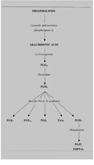
Cyclooxygenase (COX) or prostaglandin H2 synthase (PGHS) is the
enzyme that catalyses the first two steps in the biosynthesis of prostaglandins
( PGs) from the substrate arachidonic acid ( AA ). These include the oxidation
of AA to PGG2 ( hydroperoxy endoperoxide) and reduction to PGH2 ( hydroxy
endoperoxide) .
Two isoforms of this enzyme exist : COX-1 and COX-2 29. The PGH2 is then converted to PGE2, PGF2 , prostacyclin, PGD2 and thromboxane by
specific enzymes as shown in the flow chart below (Fig 1) 30 .
Cyclooxygenase-1 (COX-1) was first obtained from bovine vesicular
50
51
Tissue distribution of cox-1 and cox-2:
COX-1 is the ‘constitutive isoform’ expressed in nearly all cell types
under basal conditions. It has been found in blood vessels, interstitial cells,
smooth muscle cells, platelets, mesothelial cells, stomach and kidney 33.
COX-2 isoform is not constitutive in most tissues except in placenta,
macula densa of the kidney and brain 31. COX-2 is however inducible in many cell types- synoviocytes, endothelial cells, chondrocytes and macrophages by
cytokines like IL- 1 (Interleukin-1), Interleukin-2, Tumor Necrosis Factor-alpha
and bacterial lipopolysaccharide (LPS) 34. Cytokines such as interleukin-4, IL-10, IL-13 and corticosteroids decrease the induction of COX-2.
Functions of cox-1 and cox-2:
Stomach:
Cytoprotective prostaglandins eg: prostacyclins are synthesized by
COX-1, although small quantities of COX-2 is expressed constitutively 35. COX-1 promotes crypt stem cell survival and proliferation. It maintains the
integrity of the mucosal epithelium by enhancing mucosal blood flow by
52
Kidney:
PGs are synthesized mainly by COX-1, low levels of COX-2 has been
detected. PGs do not maintain normal renal blood flow, but are important in a
compromised kidney and in patients with congestive heart failure, cirrhosis and
renal insufficiency 29.
Platelet:
The only isoform detectable in the platelet is COX-1 which produces
thromboxane. Thus prophylaxis against thromboembolic disease can be
achieved by aspirin.
Gestation and parturition:
Both COX-1 and COX-2 are expressed in the uterine epithelium and are
important for implantation of the ovum and in angiogenesis during placenta
formation. COX-2 plays a role in inducing uterine contractions during labor. In
addition both COX-1 and COX-2, the former at a higher level, is expressed in
fetal hearts, kidneys, lungs, brain and the decidual lining of uterus 36.
CNS:
COX-1 is distributed in neurons throughout the brain, being most
53
hypothalamus and spinal cord. In addition, COX-2 is also detected in the
non-neuronal cells.
The major PGs of the CNS are PGE2 and PGD2 which are involved in
modulation of autonomic nervous system and sensory processing 37. PGE2
generates signals that activate the thermoregulatory centre of the anterior
hypothalamus thus playing a role in febrile response. The COX-2 induced in the
endothelial cells of cerebral blood vessels is responsible for PGE2 release 38 .
In the spinal cord, the nociceptive process is brought about by COX-2
causing hyperalgesia.
Cox-2 in colon:
COX-2 expression in the normal colonic mucosa is either low or
absent 39. Low levels of COX-2 are derived from macrophages, vascular endothelial cells and neuroendocrine cells in the normal mucosa 39. However it is an early response gene that is induced rapidly in response to growth factors,
cytokines, oncogenes and phorbol esters 40.
The promoter region of COX-2 consist of many transcription factor
binding sites such as nuclear factor Kb, nuclear factor of IL-6, AMP (Cyclic
Adenosine Monophosphate) and hypoxia inducible factors (HIF-1) – all of
54
STRUCTURE OF CYCLOOXYGENASE:
COX-1 and COX-2 have a similar molecular weight of 70K.Da and are
almost identical in length. The tertiary and quarternary structures are also
identical (Fig 2A). Each subunit has three structural domains: a short N-terminal
epidermal growth factor (EGF) domain, an alpha-helical membrane binding
moiety and a C-terminal catalytic domain 42.
COX-1 and COX-2 are bifunctional enzymes that carry out two
consecutive chemical reactions. Both the cyclooxygenase and the peroxidase
active sites are located in the catalytic domain, which accounts for 80% of the
protein.
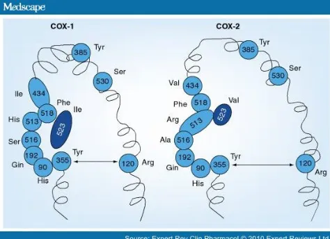
Around 63% of their 600 aminoacids are in an identical sequence. Some
substitutions are seen among the two namely- Isoleucine in COX-1 is
exchanged for valine in COX-2 at positions 434 and 523 (Fig 2B).
Inspite of the structural identity, differences are seen in substrate and
inhibitor selectivity. COX-2 acts on a wider range of fatty acids as substrate
55
Fig 2A : Shows the overlapping structure of COX-1 and COX-242
[image:67.595.179.416.407.579.2]56
COX-2 AND INFLAMMATORY BOWEL DISEASE :
Crohn’s disease characterized by chronic inflammation may occur at any
level of the digestive tract. Mutations in the NOD2 / CARD 15 gene located on
chromosome 16 is the predisposing event for the development and maintenance
of the inflammatory process.
Overexpression of COX-2 has been seen in both the epithelial cells and
inflammatory cells in response to the inflammatory cytokines 43. Romero and others have found a significant association between COX-2 expression and
epithelial alterations such as ulceration, mucin depletion, presence of paneth
cells 43. COX-2 overexpression was more frequently seen in the epithelial cells than in inflammatory cells. COX-2 immunostaining was demonstrated in
normal mucosa in the control group of Romero’s study in contrast to other
studies which have reported the absence of the expression in normal
epithelium 44.
As already reviewed, extensive ulcerative colitis of more than 8 years
duration is an important risk factor for colonic epithelial dysplasia and
adenocarcinoma13 . Agoff and others have observed COX-2 expression in both the actively inflamed, non dysplastic mucosa and in non inflamed mucosa in all
stages of neoplastic progression. COX-2 overexpression has been demonstrated
57
authors documented a more uniform diffuse cytoplasmic expression in all
grades of dysplasia compared to adenocarcinoma where the COX-2 expression
was not uniform with only 50% of cells staining in foci, although some foci had
marked, uniform COX-2 overexpression 45 .
ROLE OF COX-2 IN CARCINOGENESIS:
COX-2 induction has been associated with various premalignant and
malignant lesions of colon, lung, breast, prostate, bladder, stomach and
esophagus 41. COX2 and its major downstream product PGE2 play an important
role at multiple levels in colorectal carcinogenesis 47. COX-2 is involved in activation and formation of carcinogens, inhibition of apoptosis, providing
replicative potential, production of angiogenic factors and enhancing the
metastatic potential40.
Mechanism of overexpression of COX-2 in colorectal carcinoma :
Activating mutations in the PTGS2 gene, the gene encoding COX-2 in
humans have not been reported but other mechanisms that cause COX-2
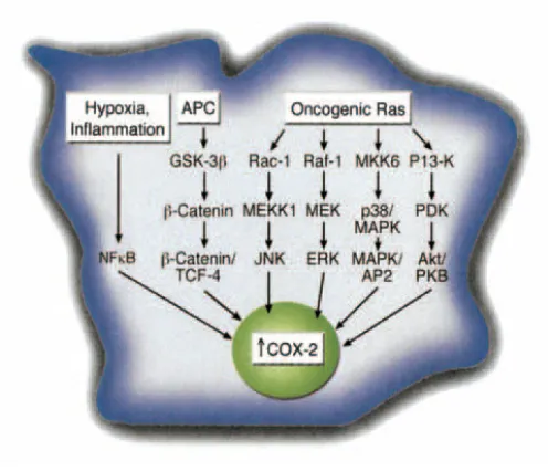
overexpression include the activation of WnT pathway, loss of APC gene and
58
Mechanism of cox-2 carcinogenesis 47 :
(i)Evasion of apoptosis
(ii) Providing self sufficiency of growth signals
(iii) Decreasing the sensitivity to antigrowth signals
(iv) Providing limitless replicative potential
(v) Sustaining angiogenesis
(vi) Tumor progression- Invasion and metastasis
[image:70.595.173.421.71.284.2](vii) Evasion of antitumor immune response.
59
(i)Evasion of apoptosis:
COX-2 increases the expression of bcl-2, antiapoptotic protein 47 . In addition it also increases the cell survival by activating the survival pathways
such as PI3K /AKT (Phosphatidyl Inositol triphosphate / AKT), cAMP / protein
kinase A signalling and PPAR (nuclear peroxisome proliferator – activator
receptor – delta ) pathways 47. The inhibition of Fas- mediated apoptosis is another mechanism observed in cholangiocarcinoma 48. Thus both the extrinsic and intrinsic pathways of apoptosis are inhibited.
The PPAR acts as a transcriptor factor, controlling the expression of
genes involved in lipid metabolism. The dietary fat provides agonist ligands for
PPARs. Thus COX-2 overexpression along with increased intake of dietry fat
together play a role in carcinogenesis 49 .
The suppression of apoptosis by COX-2 also determines the susceptibility
of tumor cells to radiotherapy and chemotherapy. COX-2 overexpression
decreases the sensitivity of tumor cells to cytotoxic therapy 47 .
(ii) Providing self-sufficiency of growth signals :
As described above COX-2 / PGE2 activate PI3K / AKT, RAS-MAPK /
ERK, EGFR signalling and cAMP / protein kinase A pathways by which tumor
60
More important is the activation of the APC/β catenin pathway 47. Mechanisms
prevail within the tumor to maintain the overexpression of COX-2.
During hypoxia, the HIF-1 (Hypoxia- Inducible Factor-1) upregulates
COX-2 expression which activates the RAS-MAPK pathway which acts in a
positive feedback loop to maintain the COX-2 / PGE250.
(iii) Insensitivity to antigrowth signals :
Overexpression of COX-2 down regulates the TGFβ type II receptor
(Transforming Growth Factor β) which normally blocks the cell progression
through G1 phase of cell cycle by suppression of c-MYC and activation of
cycle cycle inhibitors.
Another mechanism is to block the differentiation of crypt epithelial cells
in the upper third of the crypt, a physiological process occurring in the normal
crypt epithelium. The progenitor proliferating cells in the lower third of the
crypt become differentiated and are shed into the lumen. This is blocked by
COX-2 / PGE2 by the activation of APC / β catenin pathway which maintain
cells in a progenitor state. Few studies have shown that the COX-2 /PGE2 can
61
(iv) Limitless replicative potential :
The proliferative compartments in the intestinal crypts are maintained
through activation of the WnT pathway. Perturbations of the APC /β catenin
pathway have been detected in aberrant crypt foci, the earliest lesions of
colorectal tumor.
Following the loss of APC gene, there is increased expression of COX-2.
This further activates the activity of the already elevated levels of the β catenin
following the loss of APC. The β catenin translocates to the nucleus and acts as
a transcription factor along with TCF-4 (T-Cell Factor 4) complex. The
β catenin – TCF4 complex binds to specific target genes – c MYC, cyclin D,
PPARs, COX-2 and increase their levels thereby causing cell proliferation.
On the other hand, by increasing the COX-2 levels, a positive loop is formed 41 (Fig 4,5). TCF binding sites have been identified in the promoter region of
62