2015
The identification of effectors of retinal cell fate
determination through single cell transcriptomics
Jillian JoAnne Goetz Iowa State University
Follow this and additional works at:https://lib.dr.iastate.edu/etd
Part of theDevelopmental Biology Commons,Molecular Biology Commons, and the Neuroscience and Neurobiology Commons
This Dissertation is brought to you for free and open access by the Iowa State University Capstones, Theses and Dissertations at Iowa State University Digital Repository. It has been accepted for inclusion in Graduate Theses and Dissertations by an authorized administrator of Iowa State University Digital Repository. For more information, please [email protected].
Recommended Citation
Goetz, Jillian JoAnne, "The identification of effectors of retinal cell fate determination through single cell transcriptomics" (2015). Graduate Theses and Dissertations. 14585.
The identification of effectors of retinal cell fate determination
through single cell transcriptomics
by
Jillian JoAnne Goetz
A dissertation submitted to the graduate faculty
in partial fulfillment of the requirements for the degree of
DOCTOR OF PHILOSOPHY
Major: Neuroscience
Program of Study Committee: Jeffrey Trimarchi, Major Professor
Drena Dobbs M. Heather Greenlee
Don Sakaguchi Jeanne Serb
Iowa State University
Ames, Iowa
2015
DEDICATION
This dissertation is dedicated to my grandpa, the first Doc Goetz. Grandpa
Doc is the total package: not only is he resilient and tough, but also quick with a joke
or an offering of his own eye if it could help my research. The past few years have
been so hard for our entire family, Grandpa Doc especially, and I regret not being
TABLE OF CONTENTS
DEDICATION ... ii
ACKNOWLEDGEMENTS... v
CHAPTER 1. GENERAL INTRODUCTION ... 1
1. Background information ... 1
1.1 Introduction to the retina... 1
1.2 Cell types that comprise the retina ... 2
2. Retinal cell fate specification ... 9
2.1 Retinal progenitor cells are multipotent ...11
2.2 Competence in retinal cell fate determination ...13
2.3 Models of retinal cell fate determination ...15
3. “Omics” approaches to understanding retinal cell fate decisions...18
3.1 Whole retina studies ...18
3.2 Single cell studies ...20
4. Intrinsic signals and retinal development ...22
4.1 Basic helix-loop-helix factors ...23
4.2 Homeodomain-containing transcription factors ...31
4.3 Other transcription factors and their effects on retinal cell fate ...36
5. Other influences on cell fate determination ...37
5.1 Asymmetric versus symmetric cell divisions ...37
5.2 The cell cycle’s influence on cell fate ...39
6. Conclusions ...40
7. References ...42
8. Figures and legends ...54
CHAPTER 2. ONECUT1 AND ONECUT2 PLAY CRITICAL ROLES IN THE DEVELOPMENT OF THE MOUSE RETINA ... 56
1. Abstract ...57
2. Introduction ...58
3. Materials and methods ...62
4. Results ...69
5. Discussion ...79
6. Acknowledgements ...84
7. References ...84
8. Figures and legends ...89
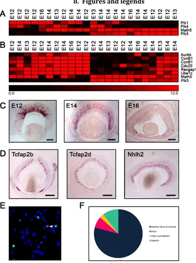
CHAPTER 3. THE EXPRESSION OF POLO-LIKE KINASE 3 AND ITS ROLE IN RETINAL DEVELOPMENT ... 102
1. Abstract ... 102
2. Introduction ... 103
4. Results ... 112
5. Discussion ... 121
6. Acknowledgements ... 123
7. References ... 123
8. Figures and legends ... 127
CHAPTER 4. SINGLE CELL TRANSCRIPTOME PROFILING OF DEVELOPING CHICK RETINAL NEURONS ... 138
1. Abstract ... 138
2. Introduction ... 139
3. Materials and methods ... 141
4. Results and discussion ... 146
5. Conclusions ... 156
6. References ... 157
7. Figures and legends ... 160
CHAPTER 5. GENERAL DISCUSSION... 166
1. Introduction ... 166
2. Retinal transcriptomes reveal subsets of transcription factors present in Math5+ cells ... 168
3. Explorations of genes expressed during retinogenesis with roles outside of direct transcription regulation... 173
4. The future of single-cell isolation in the retina ... 178
5. Conclusions ... 182
7. References ... 183
8. Figures and legends ... 187
APPENDIX. THE OTX2 AND ONECUT FACTORS PROMOTE CONE PHOTORECEPTOR AND HORIZONTAL CELL GENESIS OVER ROD PHOTORECEPTORS ... 188
1. Abstract ... 188
2. Introduction ... 189
3. Materials and methods ... 194
4. Results ... 198
5. Discussion ... 213
6. Acknowledgements ... 218
7. References ... 219
ACKNOWLEDGEMENTS
I would like to thank the members of my Program of Study Committee, Drs. Drena Dobbs, Heather Greenlee, Don Sakaguchi, and Jeanne Serb, for their valuable help throughout the course of my time at Iowa State.
Many members of the Trimarchi lab were indispensable to my research. Our lab tech, Greg Martin, was especially helpful in acclimating me to life as a new lab’s first graduate student during my first few years with a sink-or-swim approach to labwork. My fellow graduate student, Lauren Laboissonniere, has provided incomparable insights and moral support as a talented colleague and a wonderful friend. I would also like to thank Caitlin Farris for bringing me down to earth and Rebecca Chowdhury, who will be taking the tough job of Jeff’s senior graduate student when I leave. The undergraduate and high school students who have come through the lab were also integral parts of my time here, especially Brock Pope, Miranda Lee, Kallie Weinand, Diane Fru, Mary Horton, Annie Wester, Anuradha Gore, and Maddie Lynch, who have gone above and beyond in terms of their dedication to our projects.
My friends, especially Ian Lunderskov, Abbie Caldwell, and Markus Hippen, were always ready to dole out support or calm me down when things were tough.
My family has been amazingly supportive, even when I was at my most stressed and wanting to quit. I hope my mom, Nancy, knows what her unswerving dedication to her children means to us and how amazing it feels to have someone I can always call for help. My father, Steve, is my definition of a role model in terms of his kindness, professionalism, sense of humor, and sense of calm even in the midst of the rest of our family.
Lastly, my PI, Jeff Trimarchi, is the reason I could make it through graduate school. I was definitely a challenge to work with, especially as a first graduate student. However, Jeff’s talents as an educator and scientist, as well as his
CHAPTER 1. GENERAL INTRODUCTION
1. Background information
1.1 Introduction to the retina
The proper integration of sensory information as vital as vision requires a
precisely functioning instrument. In vertebrates, this process begins in the retina.
This photosensitive tissue at the back of the eye is responsible for
phototransduction – the conversion of light energy to neural signals – and the initial
processing of visual information before the signals are passed on to the brain (Burns
and Arshavsky, 2005). Specialized retinal neurons contribute to basic signal
modulation even before those features are forwarded on to the visual centers of the
brain. These nerve cells range from the light-sensing photoreceptors with their
unique outer segments to the retinal ganglion cells whose long axons must connect
with specific partners in the brain. Interestingly, each mature retinal neuron
emanates from the same retinal progenitor population and precisely integrates into
Understanding the factors directing each progenitor cell to its final fate as a
specialized neuron is important for multiple reasons. First, the factors that drive the
transition from an uncommitted progenitor cell to a fully functioning neuron may be
shared among developing central nervous system (CNS) neurons beyond the retina.
Knowledge regarding how these factors function in concert would, therefore, inform
our understanding of how different neuronal populations are generated. Second, a
greater understanding of how each retinal progenitor cell determines its fate is of
great interest not only for developmental biologists, but also for those interested in
diseases of the visual system. Many degenerative conditions affecting the retina
could potentially be treated through cellular replacement therapy. These cell-based
treatments involve either regenerating lost retinal neurons in the diseased tissue or
injecting new cells that have been produced in culture to replace those that have
been lost (Schmeer et al., 2012). Therapies such as these require a thorough
knowledge of how to generate a significant number of different types of neurons to
replace those that are deteriorating. We can only uncover all of the factors to be
used in these cutting edge replacement strategies through studies of the
mechanisms behind retinal cell fate decisions.
1.2 Cell types that comprise the retina
1.2.1 Photoreceptors
Although much remains to be elucidated regarding how the precise retinal
different cells that make up the retina. The retina possesses a laminar organization
that is well conserved throughout vertebrate evolution. It is composed of six major
types of neurons and one type of glia, all of which are located within specific layers
of the retina (Masland, 2012; Rodieck, 1998). The two types of photoreceptors, rods
and cones, array themselves at the outer portion of the eye in the outer nuclear
layer (ONL) (see Figure 1A). As their name suggests, photoreceptors function as the
primary sensory neurons in the retina, converting light photons into chemical
signals that can then be passed on to interneurons in the inner nuclear layer (INL)
for further processing. In general, photoreceptors are depolarized in the absence of
light, which leads to a constant sodium influx and release of the neurotransmitter
glutamate to activate downstream interneurons (Yau, 1994). Conversely, when
stimulated by light, photoreceptors hyperpolarize and cease to release glutamate
(Yau, 1994). Rod photoreceptors, containing the photopigment rhodopsin, are best
suited for low light conditions and distinguish between light and dark (Morrow et
al., 1998).
Cone photoreceptor cells are distinguishable from rod photoreceptors by the
presence of specialized opsins that react to ranges of brighter light at different
wavelengths (Bruhn and Cepko, 1996; Masland, 2012). Although different numbers
of opsins are present in various vertebrates, many, including humans, possess three
distinct opsins – L or red opsins, which have a peak spectral sensitivity of 560nm; M
or green opsins, with a peak sensitivity of 530nm; and S or blue opsins, with a peak
sensitivity around 430nm (Cheung et al., 2013). Possibly owing to the fact that mice
contain only the second two varieties of opsins, although multiple cone phenotypes
can arise from the expression of different combinations of opsins (Applebury et al.,
2000). The diurnal chick, while possessing the same three cone opsins noted in
primates, contains an additional violet opsin, as well as a specialized double cone
that generally expresses red opsin in each of its two connected cell bodies (Bruhn
and Cepko, 1996; Enright et al., 2015). Observations of the generation of specific
cone subtypes in vertebrate species have indicated that subtype-specific
transcription factors lead to the early generation of the more numerous
longer-wavelength opsins considerably ahead of their more rare short-longer-wavelength
counterparts during retinal development (Bruhn and Cepko, 1996; Cheung et al.,
2013; Enright et al., 2015).
1.2.2. Interneurons
Regardless of each photoreceptor’s range of excitability, once a given
photoreceptor hyperpolarizes in response to photon absorption, their glutamate
inhibition decreases, allowing for signals to pass from the ONL through to the
interneurons of the INL (Masland, 2001a; Masland and Raviola, 2000). As opposed
to sensory or output neurons, interneurons are recognized by their role as
intermediate relays for circuits between those two types. While the specific
responsibilities of interneurons vary based on their connectivity, retinal
interneurons are specifically responsible for mediating the communication between
the sensory neurons (photoreceptors) and output neurons (retinal ganglion cells) of
retina: horizontal cells arrayed in a mosaic pattern along the apical side of the INL
neighboring the ONL, bipolar interneurons stretching from the apical to basal edges
of the INL, and the diverse amacrine interneurons present in both the INL and
displaced throughout the ganglion cell layer (GCL) (Masland, 2001a).
Among the interneuron cell classes, horizontal cells function to even out
signals from bright and dim areas of the visual field using a phenomenon known as
lateral inhibition (Masland, 2012; Thoreson and Mangel, 2012). The lateral
inhibition of cones by horizontal cells leads photoreceptors to react to their
receptive fields with a center-surround organization that, while activated by light, is
also inhibited by the activation of neighboring photoreceptors. The phenomenon of
lateral inhibition leads to better acuity and edge detection even in the presence of
bright stimuli (Thoreson and Mangel, 2012). In general, mammals possess two types
of horizontal cells, distinguishable by the presence or absence of an axon that
communicates signals back to photoreceptors (Masland, 2001). However some
rodents, including mice, only generate one type of horizontal cell that has an axon
for feedback communication (Peichl and González-Soriano, 1994). Interestingly,
immunoreactivity profiles indicate no less than four subtypes of horizontal cells in
the chicken retina, as have been noted in other avian and reptilian species (Fischer
et al., 2007).
Bipolar interneurons are generally referred to as either cone or rod bipolar
cells, since they are exclusively connected to one or the other type of photoreceptor.
different kinds of bipolar cell at twelve (Kim et al., 2008; Masland, 2001b). The main
function of bipolar cells varies based on their connectivity, but their two most
prominent subdivisions are OFF and ON bipolar cells (Masland, 2012). When
photoreceptors depolarize and release glutamate, these cells take up the glutamate
and depolarize in turn. Therefore, OFF bipolar cells are active when light is off. ON
bipolar cells, on the other hand, are inhibited by glutamate and hyperpolarize in its
presence. Only when photoreceptors cease releasing glutamate – that is, when they
are hyperpolarized due to the presence of light – do these ON bipolar cells
depolarize (Masland, 2012). The ON and OFF subtypes are distinguishable by the
location of their dendrites in the inner plexiform layer where the processes meet
those of the ganglion cells (Masland, 2001b) – OFF cells do not extend far from the
inner nuclear layer (Figure 1B – sublamina A), while ON cells extend farther past
their OFF neighbors (Figure 1B – sublamina B).
The most varied class of cells in the retina is the amacrine interneurons.
Morphological studies estimate that there are somewhere between 20 and 30
different subtypes of amacrines (depending on the species examined) with an array
of receptive fields (MacNeil and Masland, 1998). Unsurprisingly, this morphological
diversity correlates with a range of functional roles for amacrine cells, which can be
connected to bipolar cells, ganglion cells, and other amacrines in both lateral and
direct pathways though the retinal layers (Masland, 1988). These cells are often
inhibitory, using the neurotransmitters glycine or -aminobutyric acid (GABA). The
most common amacrine cell type, named AII, differentially connects rod
a summative signal to the AII amacrine cell, it uses direct gap junctions to excite ON
bipolar cells and chemical inhibition to inhibit OFF bipolar cells, heightening the
signal intensity even in low light (Strettoi et al., 1992; Wässle, 2004). Others, such as
starburst amacrines, form highly branched and overlapping receptive fields that
contribute to directional sensitivity in downstream signaling (Vaney et al., 1988).
1.2.3. Ganglion Cells
Retinal ganglion cells, the main output neurons of the retina, are another
diverse group of retinal cells. Their primary purpose is to convey the visual
information to the visual centers of the brain (Rockhill et al., 2002). Although this
seems a deceptively simple role, the great diversity of retinal ganglion cells has been
noted in vertebrates since Ramon y Cajal’s studies of the frog retina in 1892,
illuminating the true function of ganglion cells not only as a relay station but also as
specialized feature detectors of the visual field (Rockhill et al., 2002). Over 20
subtypes of ganglion cells have been isolated with functionality ranging from feature
and color detection to direction and motion selectivity photoreception (Rockhill et
al., 2002). While some properties of retinal ganglion cell physiology appear to be
transiently coded circuits, able to adapt to changing circumstances such as bright or
dim light (Grimes et al., 2014), these properties can be distinguished by specific
markers, such as the genes JamB or Cdh6, which define distinct subsets of
directionally-sensitive OFF retinal ganglion cells (Kay et al., 2011; Kim et al., 2008).
Interestingly, these subsets of ganglion cells can be traced throughout retinogenesis
neurons with high accuracy at the very onset of retinogenesis, indicating that their
fates may be established transcriptomically early on in development (De la Huerta
et al., 2012). Morphological analyses of the ganglion cells present in the chick retina
have also resulted in various subset numbers depending on the characteristics used
for distinction – for instance, while six main groups of RGCs can be categorized
based on their soma sizes and dendritic fields, no less than 26 different stratification
patterns exist in the species (Naito and Chen, 2004).
One ganglion cell subtype in particular has been receiving much attention
recently. These intrinsically photosensitive retinal ganglion cells (ipRGCs) react to
light independently of rods and cones using their own photopigment, melanopsin,
and are important in establishing circadian rhythms and the normal pupillary
reaction to light (Munch and Kawasaki, 2013). Whereas the primary projection of
most feature-detecting retinal ganglion cells is to the lateral geniculate nucleus as a
first stop before signals are forwarded to the visual centers in the brain (Field and
Chichilnisky, 2007; Wässle, 2004), ipRGC axons project to the suprachiasmatic
nucleus and the pretectum, key regulators of involuntary light responses.
1.2.4 Retinal demographics
Each retinal cell type is present in different proportions within the adult
retina, although those proportions are conserved within a given species. For
example, rods are very common at around 75% of the total retinal cell population in
mice, while ganglion cells comprise only 2.5% (Dräger and Olsen, 1981; Young,
types of retinal neurons such as amacrine and ganglion cells, especially given the
rarity of those cell types in general and their subtypes specifically. Nevertheless, to
fully understand the control of retinal cell fate decisions, we must have a better
appreciation for the diversity of neuronal end-points. It is only then that we can
understand how progenitor cells sort through the myriad cues available to them and
decide on a final cell fate. These decisions must be coordinated with the goal of
generating the full cohort of retinal cells and functionally distinctive retinal cell
subtypes in the right place and time.
2. Retinal cell fate specification
Since the retina is not only the first step in the processing of visual
information but also a relatively simple and easily manipulated extension of the
central nervous system, it is a widely used model system for studying nervous
system development. Optic vesicles first emerge as bilateral evaginations of
neuroectoderm from the anterior neural plate (Adler and Canto-Soler, 2007;
Fuhrmann, 2010; Martinez-Morales and Wittbrodt, 2009). Upon contacting the
surface ectoderm, interactions between the developing tissues leads ectoderm to
begin differentiation into the lens placode, while the optic vesicle itself undergoes
invagination to form a bilayered optic cup (Fuhrmann, 2010). This cup then
continues differentiation into two separate tissues – retinal pigment epithelium and
Once the neural retina is fully developed, the complex processing that is
initiated by retinal cells is stunning, and their wide range of responsibilities is only
more impressive given the understanding that all this diversity arises from a
common progenitor population (Holt et al., 1988; Turner and Cepko, 1987; Turner
et al., 1990). Each progenitor cell’s multivariate fates are guided by a combination of
intrinsic signals and differential reactions to extrinsic signals (Livesey and Cepko,
2001). Intrinsic signals include significant differences in gene expression, even
among progenitor cells examined at the same developmental time point (Trimarchi
et al., 2008). In addition to the intrinsic gene expression of individual progenitor
cells, there are also cell-extrinsic factors – signals in the extracellular environment
that help push progenitor cells toward a certain cell fate and assist in their path to
final differentiation. Studies involving both types of signaling mechanisms are
ongoing. Although many important discoveries have been made regarding the
control of retinal cell fate decisions, much about the precise combinations of genes
and environmental factors and how they interact remains to be discovered. With a
greater understanding of the forces driving retinal progenitor cells to a specific
neuronal or glial cell fate, we can approach a better understanding of how
neurogenesis is accomplished in general.
Though the lessons learned from retinal development can be generalized to
other parts of the nervous system, elucidation of how normal retinal development
occurs is also important for its own sake. For example, retinal degeneration can
specifically or preferentially affect individual subsets of retinal cell types, such as
(glaucoma). Though current treatments can ameliorate symptoms and possibly
delay degeneration, there are no current interventions that stimulate regeneration
of the full functional cohort of lost cells (Kuehn et al., 2005). The only way
regeneration could be possible is with a full understanding of the gene expression
pathways that drive undifferentiated progenitors to take on those specified roles.
With that knowledge, it could be possible to generate the necessary cell types in
vitro using cultures such as induced pluripotent stem cells (Tucker et al., 2011) or,
perhaps, in vivo by stimulating the stem cell potential of Muller glia (Karl et al.,
2008) .
2.1 Retinal progenitor cells are multipotent
Various approaches have been utilized to uncover the mechanisms through
which a progenitor cell chooses its eventual fate during retinal development. One
method, called birthdating, stems from the notion that the final division of a
progenitor cell is the newly developing daughter cell’s “birthday.” Birthdating aims
to label cells during the DNA synthesis step of the cell cycle by exposing cells to a
nucleotide analog such as radioactive thymidine or Bromodeoxyuridine (BrdU) for a
period of time (Cepko et al., 1996) . After its uptake during DNA synthesis, the
marker becomes diluted by successive cell divisions and strongly marks only those
cells that terminally divided immediately after exposure to the marker. Early
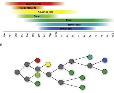
birthdating studies in the murine retina demonstrated that the different retinal cell
types are generated at distinct but overlapping time points throughout development
ganglion cells, followed closely by horizontal cells, amacrine cells, and cone
photoreceptors (Farah and Easter, 2005). Later-born cell types include rod
photoreceptors, bipolar cells, and Muller glia (Altshuler et al., 1991) (Figure 2A).
These birth orders are highly conserved across a multitude of different species,
including the chicken (Snow and Robson, 1994; Spence and Robson, 1989). Some
recent studies examining amacrine cells more closely have discovered that even
some subtypes have distinct birthdays, such that glycinergic amacrine cells are born
earlier than their GABAergic relatives (Cherry et al., 2009; Hu and Easter, 1999;
Voinescu et al., 2009).
In addition to birthdating, one can track the lineage of a single progenitor cell
– that is, all the different cell types it produces – by injecting a developing retina
with a low-titer retrovirus expressing a reporter, such as green fluorescent protein
(GFP), or other fluorescent dye to allow visualization. Investigations of progenitor
lineages demonstrated that retinal progenitor cells are multipotent throughout
development, or capable of generating multiple kinds of cells including neurons and
glia (Holt et al., 1988; Turner and Cepko, 1987; Turner et al., 1990) (See Figure 2B).
These progenitor cells show transient biases in the proportion of cell types they
generate, as seen through comparisons of the population of daughter cell types
generated at either embryonic or postnatal time points. Specifically, retroviral
injections of rat progenitor cells at birth resulted in labeling of rod photoreceptors,
bipolar cells, amacrine cells and Muller glia (Turner and Cepko, 1987). When the
injections were performed during the embryonic period, many progenitor cells
current understanding is that a given progenitor cell is capable of generating
several, if not all, of the cell types present in the mature retina. With this knowledge,
retinal development studies have focused on elucidating the influence of intrinsic
gene expression patterns and the extrinsic environment on each progenitor cells’
fate decision.
2.2 Competence in retinal cell fate determination
Based on the data from the lineage experiments, we know that individual
retinal progenitor cells are capable of generating an array of cell types. A progenitor
cell which is not yet determined to give rise to a particular cell type, but rather has
the capability to become any of a variety of retinal cells, is said to have the
competence to generate those cell types (Cepko et al., 1996). Retinal progenitor
cells are competent to generate a different set of cells at distinct and overlapping
timepoints (Figure 2). The probability that a progenitor cell will generate a
daughter cell of one type or another changes as development progresses (Figure 2).
One example is that early-born retinal cells such as ganglion cells or cone
photoreceptors are decreasingly likely to emerge from progenitors of increasingly
later stages. The proposed progression of retinal progenitor cells through distinct
states of competence is highly reminiscent of the generation of neurons from
neuroblast progenitor cells in Drosophila. These neuroblasts produce intermediate
cells called ganglion mother cells (GMCs) that in turn produce a stereotyped set of
neurons in a distinct order (Pearson and Doe, 2004). Particular transcription
the transition in neuron production (Pearson and Doe, 2004). Extension in the time
that Hunchback is expressed, for example, results in an extension in the generation
of the first-born neurons (Isshiki et al., 2001). Misexpression of Ikaros, the mouse
ortholog of Hunchback, can prolong the window during which early-born cells are
generated, while loss of Ikaros specifically leads to a loss of early-born cells (Elliott
et al., 2008). However, these effects were not as pronounced as those induced by
hunchback. Interestingly, a converse experiment misexpressing and conditionally
deleting Castor (Casz1), a gene that is present in mid- and late-stage RPCs and is
repressed by the appropriate Hunchback homologue in both Drosophila and mouse,
found that mouse Casz1 is both necessary and sufficient to inhibit generation of
early-born cells (Mattar et al., 2015). Currently it is unclear exactly which upstream
or downstream factors control these changes in retinal neuron production over
time.
The likelihood of a progenitor cell producing one cell type over another could
be determined by intrinsic factors (the genes expressed by that progenitor cell),
extrinsic factors (signals contributed by the progenitor’s environment), or some
combination of the two. The extent to which intrinsic and extrinsic signals influence
a progenitor cell’s fate decision is still a hotly debated topic. Several studies have
attempted to address the relative importance of these signals. In the first study,
embryonic retinal progenitor cells were isolated, mixed with an excess of postnatal
cells and their resulting cell fates analyzed. Importantly, no embryonically derived
progenitor cells were observed to prematurely adopt any postnatally generated cell
experiment. Progenitor cells derived from postnatal retinas did not adopt
early-generated cell fates when mixed with an excess of embryonic cells (Belliveau et al.,
2000). While there were slight changes in the relative proportions of some cells,
these experiments point to intrinsic signaling as the driving force behind a
progenitor cell’s ability to become a given cell type, in spite of any environmental
signals. In one final experiment, rat progenitor cells that were dissociated from one
another and cultured in vitro generated the normal proportions of the different
retinal cells with the correct timing (Cayouette et al., 2003). Again, these results
demonstrate that retinal cell fate decisions proceed normally even in the absence of
normal extracellular signals.
2.3 Models of retinal cell fate determination
A discussion of the influences that different intrinsic and extrinsic factors can
have on the acquisition of different retinal cell fates suggests that retinal progenitor
cells are enacting discrete genetic programs that lead to the reproducible
production of particular types of cells. These programs are represented in the
deterministic model of cell fate determination (Figure 3A). Perhaps the most
well-known example of such a deterministic organization of cell fate occurs in C. elegans.
Researchers have discovered the identity and number of all the cells in this
nematode and even have mapped out the lineages for all cells, as they do not change
from one animal to another (Hobert, 2010). While the case of C. elegans is an
extreme one, it has been presumed for some time that cell fate determination
cells enact a specific intrinsic program, at a specific time and specific position. The
results of that intrinsic cell fate program would be the production of a specific cell
type. This program would be the same each time this particular retinal neuron type
was produced. As development proceeds, the competence of the progenitor cells
would change, but the intrinsic programs they enact would be the same for any
particular cell type. However, more recent data suggests that not all the cell fate
decisions of retinal progenitor cells are deterministic, but instead there may be
more randomness to these decisions than was previously appreciated (Figure 3B).
Stochastic cell fate determination has been demonstrated in Drosophila eye
development, where progenitors are randomly recruited to clusters of eight
photoreceptors, or ommatidia, and adopt a photoreceptor fate based on their
position and timing of recruitment. Extracellular Notch/Delta signaling plays a
critical role in the differentiation of cells in the ommatidia. Progenitor cells are
initially indistinguishable until small changes in the cells’ reaction to Notch signals
initiate feedback loops to differentiate those cells further (Johnston and Desplan,
2008). Removal of the sevenup gene, which is repressed by Notch and is necessary
to generate R1 and R6 photoreceptors, prevents progenitor cells from developing
properly, and leaves them to decide randomly between an R7 or an R8
photoreceptor fate (Miller et al., 2008). The cells forced into these fates decide to
become either R7 or R8 regardless of spatial considerations, and express genes
specific to one photoreceptor type completely exclusively of the other (Miller et al.,
The gene networks involved in the cell fate decision-making process in
vertebrates are complex and dynamic, which impedes experiments designed to
differentiate between a stochastic and deterministic model. To begin to gain insight
into which process is operating, researchers first set out to understand the types of
divisions that progenitor cells undergo. These divisions can be classified as
proliferative (generating two new progenitors), self-renewing (generating one
progenitor cell and one differentiated retinal cell), and terminal (generating two
non-proliferative retinal cells). In zebrafish, marked lineages of progenitor cells
were monitored through multiple cell divisions and the resulting data used to
generate a mathematical model of retinal development. The best model to explain
the data was one where the progenitor cells produced progeny without adherence
to a strictly deterministic pattern (He et al., 2012). The model is not consistent with
a fully stochastic model either, but rather one in which progenitor cells make
“random” cell fate choices based on a weighted probability that changes throughout
development (Figure 3). Interestingly, this study also showed that the bHLH factor
ath5 can “tip the scales” in favor of ganglion cell production, linking a factor with a
wealth of previous literature to this new model of retinal development (He et al.,
2012). This probabilistic mode of cell fate determination is not unique to zebrafish.
When videomicroscopy was used to monitor rat progenitor cells and the cell fates
they produce over time, it was found that again the most consistent model of cell
fate acquisition was a more stochastic one (Gomes et al., 2011). Specifically, a
progenitor cell was able to generate a late-born neuron and then an early-born
stochastic probability that each progenitor cell could differentiate into any of a few
given cell types, as opposed to a more deterministic timed cell lineage.
3. “Omics” approaches to understanding retinal cell fate decisions
3.1 Whole retina studies
Classic studies designed to identify and characterize retinal cell fate
determinants traditionally focused on the role of a single factor or, in rare instances,
several factors simultaneously. However, it is undoubtedly combinations of genes
(networks) that work together in concert to produce the distinct types and subtypes
of retinal neurons. Therefore, in the search for intrinsic and extrinsic factors that
work together to drive the fate of retinal progenitor cells, it is important to leave no
stone unturned. Forward genetics, the technique by which an interesting phenotype
(such as microphthalmia or lack of optic nerve development) is discovered and
studied to determine its genetic root, is an extremely powerful approach to
determine genes that drastically affect large-scale developmental cascades.
However, many genes may be missed by forward genetics. A converse approach,
called reverse genetics, starts with a gene in mind and works backward to
determine the role that gene plays in a system. The process of reverse genetics may
seem less exciting at first, but it allows for a much more thorough understanding of
developmental factors. The key to a reverse genetics approach is knowing the
transcriptome – the full collection of mRNA transcripts that are expressed, whether
Many recent studies have tackled the problem of identifying all the mRNA
transcripts associated with different stages of retinal development and distinct
retinal cell populations.
Several groups have examined retinal gene expression using microarrays or
expressed sequence tag (EST) based approaches to catalog all the transcripts
expressed at distinct times during normal retinal development (Chowers et al.,
2003; Livesey et al., 2004; Swaroop and Zack, 2002). Additionally, comparisons of
transcriptomes between wildtype and mutant organisms can help to reveal
previously uncharacterized factors involved in the development of particular retinal
cells. For instance, one gene with particular importance to RGC genesis is Brn3b. In
the absence of Brn3b,RGC populations were decreased by 80% (Gan et al., 1996). A
screen for genes differentially expressed between embryonic day (E)14.5 wildtype
and Brn3b knockout mice revealed a host of candidate genes to be tested for their
functional roles in ganglion cell development (Mu et al., 2001). Comparisons have
also been made between the transcriptomes of wildtype mice and Math5-deficient
mice at multiple developmental time points. These microarray experiments
revealed a wealth of potential downstream targets of Math5 that may be important
in the cell fate determination of several early-generated retinal cell types (Mu et al.,
2005). As with all of these large-scale profiling studies, the future challenge is to
understand which of these genes are the critical cell fate regulators and how all of
To further expand our knowledge of gene expression, serial analysis of gene
expression, or SAGE, was employed at two day intervals throughout retinal
development (Blackshaw et al., 2004). This technique has the added advantage of
not relying solely on those genes that are spotted down on a microarray.
Additionally, this study further characterized the expression of >1000 genes by in
situ hybridization (Blackshaw et al., 2004). Mining this dataset has already
identified transcription factors and signaling molecules that could play key roles in
setting up distinct cell fate choices. As sequencing techniques have improved,
RNA-seq has now become a widely used profiling technique. Thus far it has only been
used to identify mRNAs expressed in the adult retina (Gamsiz et al., 2012), but it will
soon be utilized for multiple different time points. RNA-seq allows for the
identification of novel splice isoforms and long non-coding RNAs to add to the
repertoire of potential cell fate determinants (Wang et al., 2009) . These studies will
continue to produce candidate factors for study for many years to come.
3.2 Single cell studies
Since the developing retina contains a complex mixture of uncommitted
retinal progenitor cells and different cell types in various stages of fate
determination and maturation (Young, 1985), identifying programs that drive
distinct cell fates is challenging with classic whole tissue approaches. Given these
issues, single cell-based profiling techniques were adapted to retinal cells to achieve
the resolution necessary to examine gene networks in even rare cell types. After
needle and cDNA libraries were generated through amplification-based strategies
(Goetz and Trimarchi, 2012). The resulting microarray data from these single cell
libraries revealed a treasure trove of marker genes for mouse retinal progenitor
cells at different stages of development (Trimarchi et al., 2008) and, specifically, for
developing retinal ganglion and amacrine cells (Trimarchi et al., 2007). In addition
to the new markers, the single cell retinal profiling studies revealed a surprising
level of gene expression heterogeneity, even among retinal progenitor cells isolated
from the same embryonic time point (Trimarchi et al., 2008). Much of this observed
heterogeneity was observed to derive from the differential expression of
transcription factors from diverse sets of transcription factor families, including
those described in this review and many others. Despite this wealth of
expression-based information, precisely how these genes work together to produce each
specific type and subtype of retinal neuron is not fully understood. A combination of
sophisticated computational tools and manually-driven functional studies will be
necessary to generate a more complete picture of the role(s) for each of these genes
in retinal cell fate determination.
Since it was known that retinal progenitor cells change their competence as
they progress through developmental time (Cepko et al., 1996) and that this
progression appeared to be largely controlled by intrinsic mechanisms (Cayouette
et al., 2003), one would predict that clusters of genes would track specifically with
either early or late single retinal progenitor cells. While this was indeed found to be
the case, the number of genes and their expression patterns proved to be a surprise.
broadly expressed primarily in early embryonic progenitor cells. A handful of
additional genes showed expression that was restricted to early progenitor cells, but
each of these genes was only observed in a small subset of cells. What these results
mean in terms of the competence model of retinal development remains unclear.
Perhaps competence is not driven by changes in mRNAs expressed in progenitor
cells, but rather by changes in protein expression (see Ikaros), changes in miRNA
expression (Smirnova et al., 2005), or some combination of both. Alternatively, the
gene expression heterogeneity revealed by the single cell profiling could be pointing
to a more stochastic model of retinal development. No matter which model proves
to be ultimately correct, collecting more single cell profiles will help to sort through
the meaning of all the gene expression heterogeneity. For these future studies
either genetically encoded or electroporated fluorescent reporters (Cherry et al.,
2009; Matsuda and Cepko, 2007) will enable the isolation of more specific sets of
cells. This refinement, combined with the change from microarray technology to
RNA-seq technology, will expand the amount of information collected and the
number of single cells analyzed.
4. Intrinsic signals and retinal development
In addition to external signaling molecules, intrinsic factors expressed within
a given retinal progenitor cell can play critical roles in determining final cell fate
decisions. Most often, these intrinsically-expressed proteins are transcription
factors that bind specific DNA sequences and control the expression of cohorts of
work has focused on the cell-intrinsic mechanisms driving cell fate decisions, this
introduction will primarily examine a few of the myriad transcription factors that
have been shown to influence retinal cell fate and then discuss the roles of more
recently identified cell fate effectors.
4.1 Basic helix-loop-helix factors
Members of the basic helix-loop-helix (bHLH) transcription factor family
have been shown to be involved in multiple developmental processes, including
retinal neurogenesis (Lee, 1997). These proteins are composed of a helix-loop-helix
domain that contributes to dimerization with other bHLH factors, and a basic region
that binds to the E-box sequence (CANNTG) of DNA (Murre et al., 1989). In addition,
different bHLH proteins can either repress or activate the transcription of
downstream genes. Generally the bHLH activators promote neurogenesis, while the
repressors work to inhibit neuronal differentiation (Hatakeyama and Kageyama,
2004). Given that different combinations of all the bHLH factors are expressed in
different subsets of retinal progenitors (Brzezinski et al., 2011; Trimarchi et al.,
2008), their contributions to individual retinal cell fates have proven to be a bit
more complicated than was initially predicted.
4.1.1 Ath5
Ath5 belongs to a subfamily of bHLH transcription factors that is homologous
to the Drosophila atonal protein. Atonal is required for the specification of the
1995). Given this result, it was of great interest to test whether vertebrate
homologs of atonal could play similar roles. Overexpression of the Xenopus
homolog, Xath5, significantly increased the population of retinal ganglion cells and
led to fewer amacrine cells, bipolar cells and Muller glia when injected into the frog
(Kanekar et al., 1997). This effect appeared specific to Xath5 as overexpression of a
different bHLH factor, NeuroD, did not lead to a change in ganglion cell numbers, but
rather to an increase in amacrine and bipolar cells (Kanekar et al., 1997). However,
it is unclear whether this effect of Xath5 is generalizable or species specific.
Overexpression of the mouse homolog, Math5, in the same assay resulted in an
increase in bipolar cells and not an increase in ganglion cells (Brown et al., 1998).
Using viruses to misexpress either Math5 or the chicken homolog (Cath5) in
developing chicken retinas did lead to an increase in the number of cells expressing
ganglion markers (Liu et al., 2001). In total, misexpression of Ath5 in different
organisms pointed toward a role in retinal cell fate acquisition with a specific
emphasis on retinal ganglion cells. The exact nature of that cell fate role was not
fully clear as homologs from different species displayed different potencies.
To better understand the role of Math5 in cell fate determination, it helps to
know exactly which cells in the mature retina have a history of Math5 expression.
To trace the lineage of Math5 positive progenitor cells, cre recombinase was
inserted into the Math5 locus and these mice were crossed to a cre-dependent
reporter mouse for cell visualization. These mice demonstrated that the lineage of
Math5-expressing cells includes most of the ganglion cells and some photoreceptors,
Math5-cre mice to a GFP reporter confirmed the earlier lineage results and showed that
Math5-expressing cells contribute to both rod and cone photoreceptor populations
and to a subset of amacrine cells (GABAergic, cholinergic and AII subtypes)(Feng et
al., 2010). Given the multitude of cell types with a history of Math5 expression, a
simple model where Math5 instructively drives the formation of ganglion cells
seems unlikely.
The role of Math5 in retinal cell fate determination was further investigated
through the generation of Math5-deficient mice by several laboratories. In the
absence of Math5, many cell fate-related phenotypes were observed including some
that differed between the mice from distinct labs. The most consistent phenotype
was a severe reduction in ganglion cell populations (80-90% of ganglion cells lost)
(Brown et al., 2001; Wang, 2001). In fact, complete loss of ganglion cells is also
observed in the Lakritz zebrafish mutant for Ath5 (Kay et al., 2001). However,
additional scrutiny revealed other retinal phenotypes in the Math5 knockout mice.
In at least one study, the number of cone photoreceptors increased (Brown et al.,
2001). This observation suggested a possible simple cell fate switch from ganglion
cells with Math5 to cone photoreceptors without Math5. The situation has proven
to be more complicated than that simple model, though, as the amacrine cell
population is also affected in the absence of Math5. One study found that syntaxin, a
marker of all amacrine cells, was expressed normally in the absence of Math5
(Brown et al., 2001). Even with this normal expression, however, the numbers of
two subtypes of amacrine cells, AII and dopaminergic, were substantially decreased
different Math5 mutant mouse, syntaxin staining was increased and cholinergic
amacrine cells (as defined by ChAT staining) were also increased (Wang, 2001).
Altogether the results from the Math5-deficient mice demonstrate the importance of
this bHLH factor in cell fate acquisition in the early developing retina.
Recent Math5-related studies have focused on deciphering the complexity of
bHLH interactions and compensation. When Math5-null mice instead express the
related bHLH factors NeuroD1 or Math3 under the Math5 promoter, retinal ganglion
cell development is partially rescued to 40% and 10% of wildtype populations,
respectively (Mao et al., 2008). Conversely, insertion of Math5 into the NeuroD1
locus reprogrammed future amacrine cells into retinal ganglion cells (Mao et al.,
2013). However, this is not a universal feature of Math5 since it could not convert
rod photoreceptor precursor cells into ganglion cells when inserted into the Crx
gene locus (Prasov and Glaser, 2012). All bHLH factors were not created equal, as
replacement of Math5 with a different bHLH, Ascl1, failed to rescue ganglion cell
production (Hufnagel et al., 2013). These results demonstrate that bHLH factors
including Ath5 execute their functions in a context dependent manner. Under
certain conditions, Ath5 expression provides a permissive environment for the
production of ganglion cells, whereas under other conditions it fails to do so.
Unraveling the additional factors and precise conditions are challenges for future
4.1.2 NeuroD family
The NeuroD family of bHLH transcription factors is also important in eye
development; the various family members have a range of overlapping, though not
identical, expression patterns and effects. Overexpression of NeuroD1 leads to an
increase in rod photoreceptors at the expense of Muller glia (Morrow et al., 1999;
Ochocinska and Hitchcock, 2009). However, mice engineered to lack NeuroD1 only
show a slight increase in bipolar cells (Morrow et al., 1999). Amacrine cell
differentiation is delayed, but by adulthood the total number of amacrine cells is
normal in NeuroD1 deficient mice (Morrow et al., 1999). Generating a compound
mutant mouse where both NeuroD1 and another bHLH factor, Math3, are deleted
resulted in a complete loss of amacrine cells and an increase in both ganglion cells
and Muller glia (Inoue et al., 2002). Overexpression experiments, meanwhile, have
demonstrated that while these bHLH factors are necessary for amacrine cell fates,
they are not sufficient (Inoue et al., 2002). Other factors, either bHLH factors or
factors belonging to additional transcription factor families, are clearly necessary to
combine with these bHLHs to drive the cell fate determination of amacrine cells. In
an interesting twist on these overexpression experiments, NeuroD1 was recently
knocked into the Math5 locus in mice. This meant that there was no endogenous
Math5 present and NeuroD1 was now expressed under the control of Math5
regulatory sequences. When this is the case, NeuroD1 is capable of partially
rescuing the production of ganglion cells in a Math5-deficient mouse (Mao et al.,
2008). These results show that while NeuroD1 has evolved its own functions in
similarly as a completely distinct bHLH factor when expressed at a different time
and place.
Recently, other NeuroD family members also have been implicated in
retinal cell fate determination. Loss of NeuroD2 in mice led to a specific decrease in
AII amacrine cells while overexpression led to an increase in amacrine cells at the
expense of bipolar cells and Muller glia (Cherry et al., 2011). Surprisingly,
overexpression of NeuroD2 at postnatal day 0 (P0) led to an increase in the
production of ganglion cells beyond the time when retinal progenitor cells are
normally competent to produce these cells (Cherry et al., 2011). Removal of another
related family member, NeuroD6, also leads to a change in amacrine cell fate. In this
case, more glycinergic amacrine cells are produced at the expense of GABAergic
cells (Kay et al., 2011) . These experiments show that to fully understand the
generation of all retinal cells, studies of cell fate determination must be brought to
the level of small subtypes of retinal neurons.
4.1.3 Acsl1/Mash1
Acsl1, also referred to as Mash1, is the mammalian homolog of the
Drosophila achaete-scute like family of bHLH transcription factors. Mice deficient
for Acsl1 die at birth, making a thorough assessment of early and late-generated
retinal fates in vivo impossible without use of a conditional knockout mouse
(Guillemot et al., 1993). Experiments in which retinas were explanted and cultured
ex vivo revealed a possible cell fate connection between Ascl1 and bipolar cells
factor Math3 displayed a complete loss of bipolar cells and an increase in Muller glia
in explant culture experiments (Tomita et al., 2000). Taken together, these mouse
results point to a role for Ascl1 in bipolar cell fate determination, but since in vitro
culture experiments can be difficult to interpret in the absence of the in vivo
environment, further experimentation is warranted. Recently, some interesting
results have come to light regarding Ascl1 and its ability to promote neurogenesis at
the expense of gliogenesis. In the zebrafish, ascl1a is upregulated rapidly after
retina injury and is required for retinal regeneration (Fausett et al., 2008). In mouse
Muller glia, forced expression of Ascl1 in Muller glia led to a downregulation of glial
gene expression and an acquisition of the capacity to produce retinal neurons
(Pollak et al., 2013) . These observations reinforce the idea that insights gleaned
from retinal cell fate experiments will be crucial for informing efforts to achieve
regeneration of particular retinal cells or entire retinas.
To gain further insight into the role of Acsl1 in retinal cell fate acquisition,
lineage tracing studies were performed to determine which cells had a history of
Acsl1 expression. In general, all major cell classes in the retina were represented
except for the nearly complete lack of retinal ganglion cells derived from
Acsl1-positive progenitors (Brzezinski et al., 2011). Additionally, the other
early-generated retinal neurons (horizontal cell, cone photoreceptors and amacrine cells)
were over-represented in the Acsl1-lineage, while rod photoreceptors were
under-represented (Brzezinski et al., 2011). These results indicate that the heterogeneity
in transcription factor expression observed in retinal progenitor cells can affect the
4.1.4 Additional bHLH family members
Many other bHLH transcription factors are also expressed in the developing
retina (Blackshaw et al., 2004; Trimarchi et al., 2008) and are likely involved in
retinal cell fate determination. For instance, deletion of Bhlhb5 in the mouse results
in a decrease in GABAergic amacrine cells and a subset of bipolar cells (Feng et al.,
2006). Olig2, another bHLH from the Olig subfamily, is expressed in only a small
subset of retinal progenitor cells (Hafler et al., 2012). Cone photoreceptors,
horizontal cells, amacrine cells and bipolar cells all displayed a history of Olig2
expression. Overexpression of Olig2, however, did not convey any specific cell fate,
but rather led to a general cell cycle exit (Hafler et al., 2012). In addition, loss of
Olig2 did not lead to any observable cell fate defect (Hafler et al., 2012). These
results point to the fact that distinct bHLH factors have distinct roles in retinal cell
fate determination. Further studies will be required to fully elucidate any
overlapping and combinatorial roles these transcription factors might play.
Pancreas transcription factor 1a (Ptf1a) is a bHLH factor that had been linked
to cell fate acquisition in the developing pancreas (Kawaguchi et al., 2002). Lineage
tracing analyses in the retina showed that only horizontal and amacrine cells have a
history of expressing Ptf1a (Fujitani et al., 2006; Nakhai et al., 2007). Loss of Ptf1a in
the developing mouse retina led to a complete loss of horizontal cells, a significant
decrease in amacrine cells and a corresponding increase in ganglion cells (Fujitani et
al., 2006; Nakhai et al., 2007). Along these same lines, overexpression of Xenopus
cells (Dullin et al., 2007). Ptf1a plays a similar role in the developing mouse retina,
although while coincident misexpression of Ptf1a and the homeodomain
transcription factor Onecut1 is necessary and sufficient to promote a horizontal cell
fate, neither gene alone appears to be sufficient to generate ectopic horizontal cells
(Wu et al., 2013). These experiments demonstrate that this bHLH factor is a key
player in the cell fate determination of horizontal and amacrine cells.
4.2 Homeodomain-containing transcription factors
Homeotic genes are perhaps most well-known for the Drosophila mutants
where one body segment has transformed into another such that the mutant flies
can have a pair of legs emanating from their head (Gehring and Hiromi, 1986).
However, homeodomain-containing proteins can regulate many different
developmental processes including cell fate determination. These proteins contain a
widely conserved homeobox domain that is 180 bp in length and is responsible for
conferring sequence specific DNA binding activity on this set of transcriptional
regulators (Gehring et al., 1994). Homeodomain-containing transcription factors
are well known for their importance in the initial formation of the eye field itself
(Zuber et al., 2003), but I will not discuss these roles in detail here. Not
surprisingly, many homeodomain transcription factors are involved in the
determination of particular retinal fates and I will discuss several examples here to
4.2.1 Vsx2/ Chx10
Visual system homeobox 2 (Vsx2), formerly known as Chx10, is one member
of the homeodomain transcription factor family. Mice that harbor a mutation in the
Vsx2 gene display microphthalmia (small eye) (Burmeister et al., 1996), primarily
due to the upregulation of a cyclin-dependent kinase inhibitor, p27Kip1, in retinal
progenitor cells (Green et al., 2003) . In fact, removal of p27Kip1 in a Vsx2 mutant
mouse significantly rescues the small eye phenotype by restoring progenitor cell
proliferation (Green et al., 2003). However, these mice also showed that Vsx2 is
critically important for the production of bipolar cells, as they are absent in both
Vsx2 mouse models (Burmeister et al., 1996; Green et al., 2003). Vsx2 is not simply
instructive for bipolar cell production on its own. This is observed through Vsx2
overexpression experiments that led to an increase in Muller glia cells (Hatakeyama
et al., 2001). However, when Vsx2 was expressed in combination with either of two
bHLH proteins, Math3 or Mash1, an increase in bipolar interneurons was observed
(Hatakeyama et al., 2001). These experiments demonstrate that homeodomain
proteins play important roles in specific retinal cell fates and that combinations of
transcription factors from different families will prove critical for driving cell fate
acquisition in different retinal cell types.
4.2.2 Otx2
A second homeodomain transcription factor expressed during retinal
development is Otx2 (Baas et al., 2000). Conditional removal of mouse Otx2 in the
corresponding increase in several different types of amacrine cells (Koike et al.,
2007; Nishida et al., 2003). Misexpression of Otx2 in P0 rat retinas promotes a
photoreceptor cell fate at the expense of the other late-generated cells (amacrine,
bipolar and Muller glia) (Nishida et al., 2003). These results point to Otx2 as a key
inducer of the photoreceptor fate and, perhaps, as a repressor of the amacrine fate.
It is noteworthy that a highly related transcription factor, Crx, is not involved in
photoreceptor cell fate, but rather in the further downstream terminal
differentiation of photoreceptors (Chen et al., 1997; Furukawa et al., 1997)(Chen et
al., 1997; Furukawa et al., 1997). This indicates that there is an exquisite specificity
operating at the level of these cell fate-inducing transcription factors. In fact, it has
recently been demonstrated that another homeodomain transcription factor, Rax,
can activate the Otx2 promoter, while the Notch pathway has been implicated in the
repression of Otx2 (Muranishi et al., 2011). Deciphering the interplay of these
intrinsic mechanisms and extrinsic signaling pathways is an important future
direction for gaining a complete understanding of how retinal cell fate
determination is controlled.
4.2.3 Rax
Retinal homeobox 1 (Rax) is a homeodomain-containing transcription factor
that is essential for the development of the eye (Mathers et al., 1997). Retroviral
misexpression of Rax resulted in an inhibition of neurogenesis and a promotion of
Muller glia formation (Furukawa et al., 2000). This glial induction may be occurring
not known what role endogenous Rax is playing in retinal cell fate determination,
since removal of murine Rax results in a complete loss of eyes. A fuller
understanding of the role of Rax in cell fate determination awaits the
characterization of cell-type specific knockout mice. One potential hint as to the role
of Rax in individual cell types comes from studies on the zebrafish homologues.
Zebrafish contain three Rax-related genes (rx1, rx2 and rx3). Morpholino mediated
knockdown of either rx1 or rx2 during the period of photoreceptor production led
to a decrease in many photoreceptor-expressed mRNAs (Nelson et al., 2009).
Through the use of these “late-injected morphants” these studies were able to reveal
a possible role for rx genes in the acquisition or maintenance of the photoreceptor
fate.
4.2.4 Pax6
The paired-type homeobox transcription factor Pax6 has powerful functions
in eye development. It is essential for the development of the eye field and, when
overexpressed in Drosophila, generates ectopic eyes on wings, legs and antennae
(Grindley et al., 1995; Halder et al., 1995; Nornes et al., 1998). To bypass the early
requirement for Pax6 and to better understand its downstream cell fate effects, the
gene was conditionally inactivated in older retinal progenitor cells. In the absence
of Pax6, progenitor cells surprisingly produced only amacrine interneurons
(Marquardt et al., 2001). The mechanism by which Pax6 normally controls the
multipotency of progenitor cells is still unclear, but it most likely involves the
show to positively regulate the bHLH factor Math5 and negatively regulate the
photoreceptor transcription factor Crx (Oron-Karni et al., 2008; Riesenberg et al.,
2009). It will be important, as part of future studies, to fully define the gene
networks downstream of Pax6 and to link each of those networks to specific
functions of this important transcription factor.
4.2.5 Other homeodomain transcription factors
Other homeodomain-containing transcription factors also play significant
roles in retinal cell fate acquisition. When both of the Distal-less homeobox proteins
Dlx1/Dlx2 are removed from the retina, increased apoptosis accompanied a marked
thinning of the RGC layer (de Melo et al., 2005). These double-knockout mice are
embryonic lethal so it is difficult to discern the precise downstream targets of the
Dlxs and whether they play any additional roles in other cell types. Postnatal
misexpression of the homeodomain transcription factor Prox1 led to an increase in
horizontal cells (Dyer, 2003). Along the same lines, deletion of mouse Prox1 led to
the complete absence of horizontal cells demonstrating that this factor is both
necessary and sufficient for the production of horizontal cells (Dyer, 2003). These
experiments also showed a potential role for Prox1 in amacrine cells, rod
photoreceptors and Muller glia (Dyer, 2003). As with all the other factors involved
in retinal cell determination, it will be of interest to delineate the exact downstream
4.3 Other transcription factors and their effects on retinal cell fate
While the bHLH and HD families are two of the largest with identified roles in
retinal cell fate determination, there are many additional intrinsic factors that also
influence retinal cell fate. Many members of the forkhead/winged helix family of
transcription factors are expressed in the developing retina (Trimarchi et al., 2008).
Mice deficient for one of these members, Foxn4, show a near complete loss of
amacrine cells, a complete ablation of horizontal cells and an increase in
photoreceptor cells (Li et al., 2004). Misexpression of Foxn4 results in the
over-production of amacrine cells, showing that Foxn4 is both necessary and sufficient
for this retinal cell fate (Li et al., 2004). The downstream mechanisms through
which Foxn4 exerts its effects are still being worked out, but this factor has been
shown to impact numerous pathways, including upregulating bHLH and HD
transcription factors and influencing Notch activity through the regulation of a Delta
(Dll4) (Li et al., 2004; Luo et al., 2012). Given the plethora of Forkhead factors
expressed during retinal development, it will be of interest to determine the
phenotypes of all the others, alone or in combination.
Undoubtedly this introduction, as well as the wide range of studies not
covered herein, has only scratched the surface in our understanding of the roles
played by different transcription factors and their downstream networks in retinal
cell fate determination. While I have highlighted some of the larger transcription
factor families in this review, there are certainly additional factors that have been
added to the growing knowledge of the cell fate process. In the future, the biggest
challenge will be to integrate all of these distinct factors and their regulatory
networks into a comprehensive snapshot of how the cell fate programs are initiated
and executed to produce not only each retinal cell type, but their subtypes as well.
With this information in hand, researchers will be better able to produce or
regenerate any retinal cell types that may be dying or dysfunctional in different
retinal disorders.
5. Other influences on cell fate determination
5.1 Asymmetric versus symmetric cell divisions
We have discussed many intrinsic and extrinsic factors and the roles they
play in retinal cell fate decisions, but there are other aspects to the story. For
example, the position and activity of a progenitor cell during its progression through
the cell cycle may confer information about its final cell fate beyond its internal
composition and the nearby environmental factors. It is well documented that the
nuclei of cycling progenitor cells follow a predictable progression (Dyer and Cepko,
2001a). They migrate to the apical, or outer, surface of the retina to undergo mitosis.
As they move forward through the first gap (G1) phase, these progenitor cells shift
basally, toward the vitreal surface of the retina. At their basal nadir, progenitor cells
pass through G1 and start the S phase of the cell cycle. They then move apically
again in G2 phase before entering mitosis again at their apical peak. By studying the