Abstract: Background/Objectives: this study aims, first, to find soft materials like breast made by 3D printer, and, second, to model mammary duct, mammary gland, and pectoralis major muscle separately.
Methods/Statistical analysis: after smoothing the surface, we subtracted mammary gland tissue, mammary duct tissue, pectoralis major muscle tissue, and pectoralis minor muscle tissue, and converted them into STL for printing. It took seven days to pour and harden mold into a form. To get clear radiation images of mammary gland tissue and mammary duct tissue, we used contrast media.This study tried various contrast media to compare images.
Findings: The modeling is completed by merging mammary gland, mammary duct, pectoralis major muscle, and pectoralis minor muscle parts, and comparing two types of modeling to examine whether the types of modeling do not make any difference in anatomical locations of human body. And while pectoralis major muscle and pectoralis minor muscle tissues were not shown in radiation images, mammary duct tissue could be identified in the radiation image.
Improvements/Applications: we adopted the mold method to express fat among various breast tissues, and various contrast media to express mammary gland in breast.
Keywords: Mammography, 3D modeling, 3D print, silicon, mold
I.INTRODUCTION
With the change of diet and life styles, risk factors for breast cancer have increased, causing it to occur increasingly. For the last decade, breast cancer incidence rate has increased 3.5 times, becoming NO. 1 cancer for females in Korea[1]. Thus, increasing number of women periodically get mammography for early diagnosis of the disease. With the aging of population, people have become more interested in healthy life, which also increased the frequency of mammography. Women’s hospitals nationwide which provide mammography are busy in diagnostic season.
What radiological technologists doing mammography should not ignore is that, while radiation gives people
Revised Manuscript Received on May 22, 2019.
Dong-Hee Hong, Dept. of Radiology Science, Shinhan University, 95,
Hoam-ro, Uijeongbu-si, Gyeonggi-do, 11644, Republic of Korea
Hong-RyangJung, Dept. of Radiology Science, Hanseo University, 46,
Hanseo 1-ro, Daegok-ri, Haemi-myeon, Seosan-si,Chungcheongnam-do, 31962, Republic of Korea.
Cheong-Hwan Lim, Dept. of Radiology Science, Hanseo University, 46,
Hanseo 1-ro, Daegok-ri, Haemi-myeon, Seosan-si,Chungcheongnam-do, 31962, Republic of Korea. corresponding author E-mail : [email protected]*
benefits, radiation exposure can damage human body. Sometimes, it can cause serious damages to human body[2,3].Such a characteristics of radiation as double-edged sword makes clinical radiological technologists to be especially careful in posture of patients in taking mammography to prevent patients to be exposed to unnecessary radiation by getting it retaken.
To discover breast cancer early, mammography is the most important. Mammography is a test method designed to precisely examine forms and sizes of lumps, thickness of epidermis, enlargement, fiberization, and calcification of mammary gland. If a lesion is discovered by mammography, the patient is under more detailed investigation.
Depending on its purposes, mammography is divided into diagnostic and screening tests. Screening test is given to patients with no symptom. It is taken from two directions: (Cranio-Caudal; CC) and (Mediolateral-Oblique; MLO). Diagnostic test is additional test given to a patient when an abnormality is discovered in screening test. In addition to the test given to women with symptoms, mammography also includes all follow-ups with 6 months after surgery except for the post-surgery follow-up[4]. Accordingly, there are various methods of mammography, and radiological technologists need to learn all of these methods, and practice them to various patients.
In mammography, technologists give pressure to breast using pressure pad which is part of the apparatus to get images of breast. Breast pressure is painful to patient, but it is inevitable to shrink breast thickness and get good-quality images. In addition, pressure is useful to get clear images by detaching overlapped breast tissues, improve contrast by making brightness and darkness clearer, and reduce exposure dose[5]. As the degree to which pressure is applied is very important in taking mammography, technologists need to continuously practice it to find ways to reduce pain of patients and get high-quality images.
But, there are not many radiation departments which teach mammography in their regular curricula, and even if it is included in curriculum, accurate practice is not made. While breast is a sensitive part of body to be used for practice, there is no phantom to be used for practice. Now, there is Japanese-made phantom for mammography shooting practice. But, as it lacks mammary gland, mammary duct, and pectoralis major muscle,
it is difficult for technologist to recognize whether she
Comparative study on the Material and Method
of new 3D Printer when Making Phantom for
Mammography
takes a good posture to shoot mammography. It is consensus among female technologists who find jobs in mammography room, department of radiology that college or institution provides mammography training program.
3D printer which has recently become popular has merits as follows: It can be manufactured in a short period of time; personalized manufacturing is possible; it is cheap to assemble parts[6]. It is the apparatus to make a product by piling method known as additive manufacturing with materials like polymer (synthetic resins) and metal according to the model blue print[7].
But, if breast is printed with hard materials, it cannot be used as shooting phantom, which is only a sculpture to watch. Like soft breast, printed product should be soft and elastic. To realize this condition, material should not be piled with additive manufacturing. It needs to be made in other method. Now, 3D printing technology is used in various ways especially in health and medical fields[8].
Accordingly, this study aims, first, to find soft materials
like breast made by 3D printer, and, second, to model mammary duct, mammary gland, and pectoralis major muscle separately.
II.MATERIALSANDMETHODS
2.1. Materials
[image:2.595.140.468.290.440.2]The breast image used in this study was MRI DICOM Volume dat as shown in Figure 1, and the image was acquired by 0.1 mm Volume Scan using GE Signa HDxt 3.0T (GE Healthcare, Milwaukee, WI, USA). To generate STL conversion file, the acquired image was edited using the Terarecon (USA) 3D program. To get models with various structures, we used STL View (Module Works GmbH), and, to get more solid and clear separation of mammary duct, mammary gland, and pectoralis major muscle, we edited these three parts through the modeling structure using Mesh tools of the Rhinoceros Program (McNeel North America, USA).
Figure 1. MRI DICOM Volume data for modeling
2.2. Methods
To generate STL (STereoLithography) conversion file which can be printed and edited in 3D printer, we segmented the material into mammary gland tissue, mammary duct tissue, pectoralis major muscle tissue, pectoralis minor muscle tissue, and fat tissue. Then, after smoothing the surface, we subtracted mammary gland tissue, mammary
[image:2.595.104.507.566.666.2]duct tissue, pectoralis major muscle tissue, and pectoralis minor muscle tissue, and converted them into STL for printing. It took seven days to pour and harden mold into a form. To get clear radiation images of mammary gland tissue and mammary duct tissue, we used contrast media. As shown in fig 2 and 3, this study tried various contrast media to compare images.
Figure 3. breast parenchyma make by contrast medium. Number 1 and 2 and 3 and 4 location is different
contrast.
III.RESULTS
3.1. Breast Segmentation of Modeling
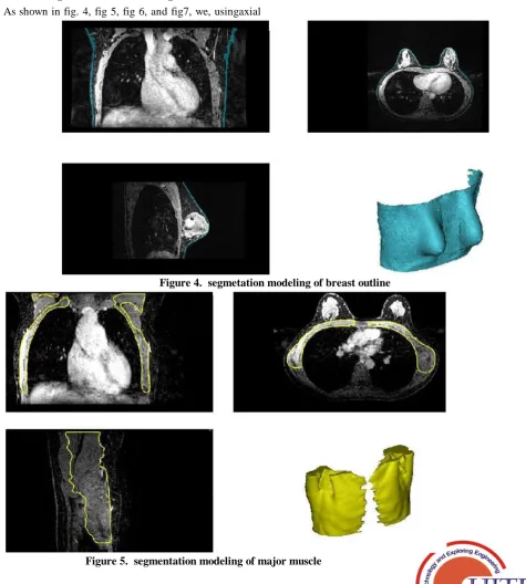
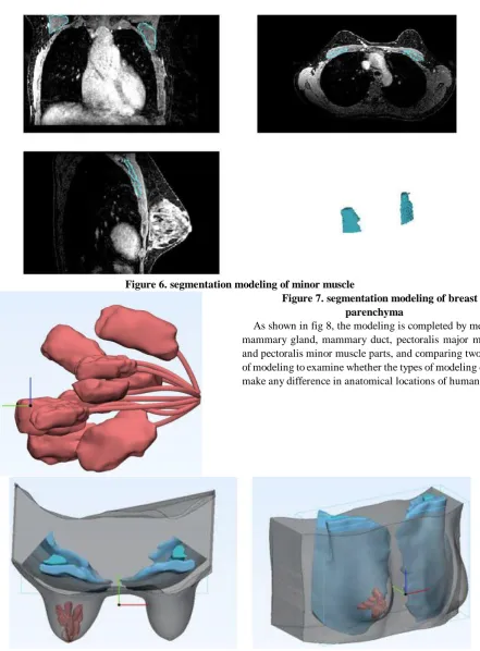
As shown in fig. 4, fig 5, fig 6, and fig7, we, usingaxial
view andsagittal view of DICOM volume data, segmented mammary gland tissue, mammary duct tissue, pectoralis major muscle tissue, and pectoralis minor muscle tissue in both breasts, and visually divided them. Using the manual segmentation technique, we completed modeling as the process of extracting the same values by strengths of DICOM images. Through meticulous work on 3D images, we made it by selecting mammary gland, mammary duct, pectoralis major muscle, and pectoralis minor muscle. Fat tissue modeling was made by setting it as the area excluding mammary gland, mammary duct, pectoralis major muscle, and pectoralis minor muscle parts in breast from the 3D surface image shown in DICOM image. Its upper limit is up to the border between collarbone and breast and its lower limit is up to the border of breast tissue.
[image:3.595.54.530.258.786.2]Figure 4. segmetation modeling of breast outline
Figure 6. segmentation modeling of minor muscle
Figure 7. segmentation modeling of breast parenchyma
As shown in fig 8, the modeling is completed by merging mammary gland, mammary duct, pectoralis major muscle, and pectoralis minor muscle parts, and comparing two types of modeling to examine whether the types of modeling do not make any difference in anatomical locations of human body.
Figure 8. STL conversion and breast segmentation tissue merge image 3.2. Contrast medium usingand mold modeling
Modeled breast fat tissue and real tissue went through conversion process to take forms through 3D printer. The modeling was converted to STL Binary file format (*.stl) which can convert the additive file to forms in 3D printer.
As shown in fig 9, identifying contrasts through radiation images, we tried different amounts of contrast media. And, we decided the ideal amount of contrast medium to express the contrast, and, based on it, we gave shape to mammary gland and pipe tissues.
As shown in fig 10, to embody soft material, we used mold technique. Fat tissue was expressed by mold modeling of 180 ㎜ x 131 ㎜ x 144 ㎜ size. Mammary gland and pipe tissues were made by flowing contrast media into the mold and letting the mold get hard. As a result, while pectoralis major muscle and pectoralis minor muscle tissues were not shown in radiation images, mammary duct tissue could be identified in the radiation image.
In mammography, there are structures whose images should be expressed with Cranio-Caudal view and Medio-Lateral oblique view[9]. In order to express accurately them by images, it is necessary to practice continuously using the phantom in which such structures are anatomically separated. By acquiring MRI images to completely separate mammary gland, mammary duct, pectoralis major muscle, and pectoralis minor muscle parts, and express them on radiation images, we realized 3D modeling[10]. MRI image has merits that without exposing patients to radiation, it is possible to get images of mammary gland, mammary duct, pectoralis major muscle, and pectoralis minor muscle tissues from patients. So, it is very useful in compartmentalized modeling.
Based on open source, we developed a 3D printer system to manufacture customized bone model for patients who need surgery[11]. While it has proved useful to manufacture human bone models, or various assistant tools and use them in surgery, the usefulness of soft materials like fat tissue of breast has not been proved. Therefore, we adopted the mold method to express fat among various breast tissues, and various contrast media to express mammary gland in breast.
V.CONCLUSION
As mammography needs high proficiency, schools and institutions have to train technologists on it. However, breast phantom has not been available. Thus, we made efforts to separate major organ tissues of breast and find soft material suitable to the characteristics of breast, as preliminary research to find a education phantom for breast model. Then, we succeeded to separate those breast organs by 3D printer, and we combined them with mold technique to express soft material of fat. And, we used contrast media to have high contrast of mammary gland and pipe tissues. We want to continue to contribute to improve phantoms of various organs which can help educating technologists.
ACKNOWLEDGMENT
This work was supported by the National Research
Foundation of Korea (NRF) grant (No.
NRF-2017R1C1B5017119) funded by the Korea government (MSIP).
REFERENCES
1. Ministry for Health, Welfare and Family Affairs. Annual Report of cancer incidence(2007), cancer prevalence(2007) and survival (1993-2007) in korea. 2009:15-6
2. Jeon GE, JinGH. A Survey of the Management of Patient Dose at Medical Center, Journal of the Korean Society of Radiology, 2009 Mar; 3(1):23-8 3. Kwon JW, Jeong JH, Jang KW, Lee JK. Medical exposure of Korean by diagnostic radiology and nuclear medicine examinations. J. korea association radiat. Prot., 2005;30(4):185-96
4. Kweon DC, Lee EM, Hong SM, Park P. Analysis of the compression force and thickness in the screening mammography. Korean Institute of Industrial Engineers, 2002: 606-10.
5. Norbert JP, Nishikawa RM, Whiting BR. The effect of breast positioning on breast compression in mammography:a pressure distribution perspective, Physics of Medical Imaging, 2012:83134M
6. Seoung YH. 3-Dimensional Printing for Mesh Types of Short Arm Castby Using Computed Tomography. the Journal of Korean Contents, 2015;15(1):308-15.
7. Choi EJ,Kim SA,Bae JY,Kwon YJ,Lee KH.A Study on the State-of-the-Art of 3D Printers, The Korea Society of Computer and Information. 2013; 21(2): 385-8.
8. Park SH,Park JH,Lee HJ,Lee NK.Current Status of Biomedical Applications using 3D Printing Technology, Journal of the Korean Society for Precision Engineering.2014;31(12):1067-76.
9. Wohler T. Wohlers Report 2013. Wohlers Associates Inc..2013:23-52. 10. Ahn DG, Application of 3D printing technology to mold industry, The
Korean society of mechanical engineers, 2014; 54(4):.46-51.