EFFECTIVENESS OF POSITIONAL RELEASE
TECHNIQUE VERSUS ISCHEMIC COMPRESSION
ON SEDENTARY WAY OF LIFE WITH UPPER
TRAPEZIUS TRIGGER POINTS
Dissertation submitted to
The Tamil Nadu Dr. M.G.R. Medical University
Chennai
In partial fulfillment of the requirements for the degree of
MASTER OF PHYSIOTHERAPY
(Advanced Physiotherapy in Orthopaedics)
Reg.No.271710001
MAY
–
2019
COLLEGE OF PHYSIOTHERAPY
CERTIFICATE
This is to certify that the dissertation work entitled “Effectiveness of Positional Release Technique versus Ischemic Compression on Sedentary
Way of life with Upper Trapezius Trigger Points” was carried out by the candidate bearing the Register No. 271710001 (MAY 2019) in College of Physiotherapy, SRIPMS, Coimbatore, affiliated to the Tamil Nadu Dr. M.G.R Medical University, Chennai towards partial fulfillment of the Master of Physiotherapy (Advanced Physiotherapy in Orthopaedics).
Prof. B. SANKAR MANI, MPT.,MBA., Principal, College of Physiotherapy,
Place: Coimbatore SRIPMS,
CERTIFICATE
This is to certify that the dissertation work entitled “Effectiveness of Positional Release Technique versus Ischemic Compression on Sedentary
way of life with Upper Trapezius Trigger Points” was carried out by the candidate bearing the Register No.271710001 (MAY 2019) in College of Physiotherapy, SRIPMS, Coimbatore, affiliated to the Tamil Nadu Dr. M.G.R Medical University, Chennai towards partial fulfillment of the Master of Physiotherapy (Advanced Physiotherapy in Orthopaedics) under my direct supervision and guidance.
Prof. K. SARAVANAN, MPT., (Orthopaedics) College of Physiotherapy, SRIPMS, Coimbatore – 641044.
CERTIFICATE
This is to certify that the dissertation work entitled Effectiveness of Positional Release Technique versus Ischemic Compression on Sedentary
way of life with Upper Trapezius Trigger Points” was carried out by the candidate bearing the Register No.271710001 (MAY 2019) College of Physiotherapy, SRIPMS, Coimbatore affiliated to The Tamil Nadu Dr. M.G.R Medical University, Chennai towards partial fulfillment of the requirements for the degree of Master of Physiotherapy (Advanced physiotherapy in Orthopaedics) was evaluated.
INTERNAL EXAMINER
EXTERNAL EXAMINER
Place:
ACKNOWLEDGEMENT
It is my privilege to express our deep sense of gratitude to the
LORD ALMIGHTY for showering his blessing and who has always been my source of strength and inspiration, throughout my life.
I dedicate the study to my Parents and my Family Members for providing their love and support in each and every step of my life.
This is my tribute to my study guide Prof. K. Saravanan, MPT (Orthopaedics), College of Physiotherapy, SRIPMS, Coimbatore, who enlightened me in the right path with patience. I am grateful to his
guidance, constant support and constructive criticism given at every stage
of our study and made the study successful.
My sincere gratitude to Professor Mr.B. Sankarmani, MPT, MBA., Principal, College of Physiotherapy, SRIPMS, Coimbatore, who had given me an opportunity to do the thesis work.
My special thanks to all Staffs of College of Physiotherapy, SRIPMS, for their timely help and valuable support and contribution which
help me to develop this study.
My profound debt of thanks to all the Non Teaching Staffs of college of physiotherapy, SRIPMS for their constant support.
I would like to acknowledge my all My Friends for their constant support in helping me to complete this study successfully.
CONTENTS
CHAPTER TITLE Page No.
1 INTRODUCTION 1
1.1 NEED FOR THE STUDY 5
1.2 OBJECTIVE 5
1.3 HYPOTHESIS 5
2 REVIEW OF LITERATURE 6
3 MATERIAL AND METHODOLOGY 11
3.1 MATERIAL 11
3.2 METHODOLOGY 11
3.2.1 Study setting 11
3.2.2 Study design 11
3.2.3 Sample size 11
3.2.4 Sample format 11
3.2.5 Study duration 12
3.2.6 Treatment duration 12
3.2.7 Selection criteria 12
3.2.8 Outcome measures 12
3.2.9 Interventions 13
3.2.10 Parameters 13
3.2.11 Tool for data collection 13
3.2.12 Statistical tool used 14
4 DATA ANALYSIS AND INTERPRETATION 15
5 DISCUSSION 37
6 CONCLUSION 39
REFERENCES 40
ABBREVIATIONS
PRT
-
Positional Release Technique
IC - Ischemic compression
VAS
-
Visual Analogue Scale
NBQ - Neck Bournemouth questionnaire
ROM
-
Range of Motion
CROM - Cervical Range of Motion
MTrP - Myofascial trigger point
US - Ultrasound
MWD - Microwave Diathermy
TENS - Transcutaneous electrical nerve stimulation
POC - Position of comfort
ATP - Adenosine triphosphate
Ach - Acetylcholine
ABSTRACT
Trigger point in upper trapezius is a common cause for neck pain,
reduced cervical range of motion, and affects functional activities.
Objective is to compare the effectiveness of Positional release technique
and Ischemic Compression on reducing pain, improving ROM, functional
activities and cognitive aspects of subjects with upper trapezius trigger
points. 30 subjects with upper trapezius trigger points were randomized into
two groups. Group A received Positional release technique and group B
received Ischemic compression technique. The total number of treatment
was for 6 days alternatively within two weeks. Outcome measure was
measured by Visual Analogue Scale (VAS), cervical range of motion
(CROM) and Neck Bournemouth Questionnaire. The mean values for VAS
in group A and group B were2.47 and 4.20 respectively, mean values of
cervical lateral flexion to the right was 36.93 and 31.86 and for cervical
lateral flexion to the left were 38.67 and 33.33 respectively. For cervical
extension mean was 64.07 and 60.13, for rotation to the right it was 84.33
and 81.33 and for rotation to the left mean values were 82.27 and 78.80
respectively, NBQ was 20.47 and 22.67 respectively. Positional release
technique was superior to Ischemic compression technique in managing
pain, ROM, functional disability and psychometric analysis in subjects with
upper trapezius trigger points.
1. INTRODUCTION
Neck pain as a clinical syndrome is common and can be seen in both
presence and the absence of history of trauma and or positive radiographic
findings. The cervical spine is the most intricate region of the spine, and so are the
muscles of this region. Muscles of the neck and shoulder region always function
as a unit, and there is no movement in the upper extremity that would not be
reflected in the neck musculature. Working posture with the neck flexion
increases the load moment three to four times on the neck causing spasm of the
neck muscles. Also working tasks that involve continuous arm movements always
generate a static load component on these muscles; the principal muscle to carry
this load is the trapezius. Neck pain constitutes a significant health care problem
affecting 45% to 54% of the general population.
A Myofascial trigger point (MTrP) is a hyperirritable spots, located within
a taut band of skeletal muscle that is painful on compression or on stretch and that
can give rise to typical motor, sensory and autonomic components. Motor aspects
included disturbed motor function, muscle weakness, muscle stiffness and
restricted range of motion. Sensory aspects include local tenderness, referral of
pain and peripheral and central sensitization.
Etiological factors are poorly understood and are usually multifactorial,
including poor posture, anxiety and depression, neck strain, Occupational injuries.
Sedentary lifestyle is also cause for neck pain, a person living a sedentary lifestyle
is often sitting or lying down while engaged in an activity like reading,
socializing, watching television, playing video games, or using a mobile
phone/computer for much of the day. There is substantial evidence that such
ergonomic risk factors as repetition, awkward posture, contact stress and force if
overcome workers biomechanical capabilities may lead to work-related
mechanical neck pain. The symptoms usually have postural or mechanical basis
Trigger point form in the muscle’s close to the motor end plate
(neuromuscular junction). Excess acetylcholine (Ach) is released at the synapse,
usually associated with overuse or strain, leading to release of calcium. Resulting
ischemia creates an o2 deficit and energy crisis without available ATP, calcium
ions, which are keeping the gate open for ach to keep flowing, cannot be removed.
A chemically sustained contracture (without motor potentials) is different from a
spasm (involuntary with motor potential). Actin and myosin filaments shorten in
the area of the motor end plate. A contracture “knot” forms the characteristic
trigger point nodule. The remainder of the sarcomeres of that fiber is stretched,
creating the palpable taut band.
The initial change in muscle that is associated with Myofascial pain seems
to be the development of the taut band, which is in term a motor abnormality.
Several mechanisms have been hypothesized on explain this motor abnormality, the most accepted one is the “ IntegratedHypothesis” first developed by Simmons Later expanded by Gravin and Simmons integrated hypothesis is a six link chain
that starts with the abnormal release of acetylcholine. This triggers an increase in
muscle fiber tension (formation of taut band). The taut band is thought to constrict
blood flow that leads to local hypoxia. The reduced oxygen disrupts mitochondrial
energy metabolism reducing ATP and leads to tissue distress and the release of
sensitizing substances. These sensitizing substances lead to pain by activation of
nociceptors and also lead to autonomic modulation that then potentiates the first
step: abnormal acetylcholine release.
Grewin expanded this hypothesis by adding more specific details. He
stated that sympathetic nervous system activity augments acetylcholine release
and that local hypo perfusion caused by the muscle contraction (taut band)
resulted in muscle ischemia or hypoxia leading to an acidification of the PH. The
prolonged ischemia also leads to muscle injury resulting in the release of
potassium, bradykinins, cytokines, ATP, and substance P which might stimulate
nociceptors in the muscle. The end result is the tenderness and pain observed in
Myofascial trigger point. Depolarization of nociceptive neurons causes the release
of calcitonin gene related peptide CGRP inhibits acetylcholine receptors and
the presence of these substance using microdyalisis techniques at trigger point
site. Evaluation of substance P, protons (H+), (GRP, bradykinin, serotonin, nor
epinephrine, TNF, interleukin, and cytokines were found in active trigger point
compared to normal muscle or even latent trigger point. The PH of the active
trigger point region was decreased as low as PH (normal PH value is 7, 4) causing
muscle pain and tenderness as well as a decrease in acetylcholine esterase activity
in sustained muscle contraction.
Trapezius being one has to act continuously to hold the head in upright
position thus prone for formation of latent trigger point, which with use of
inefficient posture like chin forward posture, emotional stress can get activated to
become active trp. Location of trigger point in upper trapezius is mid way between
c7 spinous process and acromion.
Various physiotherapy protocols have been advocated in the past like
rest, heat, ultrasound therapy (UST), microwave diathermy (MWD),
Positional release therapy (PRT) is a method of total body evaluation and
treatment using tender points (TPs) and a position of comfort (POC) to resolve the
associated dysfunction. PRT is an indirect (the body part moves away from the
resistance barrier, i.e, the direction of greatest ease) and passive (the
physiotherapist performs all the movement without help from the patient) method
of treatment. All three planes of movement are used to attain the position of
greatest comfort.
Ischemic compression a manual therapy technique works on same
principle of applying sustained pressure to the trigger point and easing the muscle
tension. The compression is gradually applied with the finger, thumb, elbow
relatively to how much the patient can tolerate and maintained for as long as 90
seconds.
Positional release therapy (PRT) is a method of total body evaluation and
treatment using tender points (TPs) and a position of comfort (POC) to resolve the
associated dysfunction. PRT is an indirect (the body part moves away from the
resistance barrier, i.e., the direction of greatest ease) and passive (the
physiotherapist performs all the movement without help from the patient) method
of treatment. All three planes of movement are used to attain the position of
greatest comfort.
Upper Trapezius and Trigger Points
According to Travell and Simons (1999) referred pain arises as often from
trigger points in upper trapezius as in any other muscles of the body.
According to Lundervold (1951) there is an increased EMG activity on
upper trapezius muscle in keyboard operators due to tensed upright posture, sitting
without a firm back support and typing with keyboard elevated, thus there is more
1.1 NEED FOR THE STUDY
The most common cause for neck pain is Myofascial triggers with
symptoms of pain, muscle stiffness, restricted ROM, tenderness, etc. Upper
trapezius is most affected muscle in neck pain. According to the recent researches
the treatment available are rest, heat application, Ultrasound therapy, Microwave
diathermy, Transcutaneous electrical nerve stimulation, spray and stretch and post
isometric relaxation etc, but these provide only a temporary effect. The main aim
of the study is to treat the cause with permanent effect of decreasing pain,
increasing range of motion, functional disability and psychometric analysis. This
can be achieved by integrating and comparing the recent techniques like
Positional releasing technique and Ischemic compression technique.
1.2 OBJECTIVE OF THE STUDY
• To evaluate the effectiveness of Positional Releasing technique on pain, ROM, functional disability and psychometric analysis.
• To evaluate the effectiveness of Ischemic Compression on pain, ROM,
functional disability and psychometric analysis.
• To compare the effectiveness of Ischemic compression and Positional releasing technique on pain, ROM, functional disability and psychometric
analysis.
1.3 HYPOTHESIS
Null Hypothesis
There is no significant difference between the “Positional Releasing
Technique and Ischemic Compression” on pain, ROM, functional disability and
psychometric analysis in patients with upper trapezius trigger points.
Alternative Hypothesis
2. REVIEW OF LITERATURE
➢ Shweta Anil Kulkarni et.al (2017) conducted a study to compare
effectiveness of Ischemic compression v/s Myofascial release on
myofascial trigger point of upper trapezius. They selected 30 patients
measure the pain intensity by VAS and Neck disability index. They
concluded that Ischemic compression shows greater effectiveness as
compared with myofascial release in treatment of trigger point pain of
upper trapezius.
➢ Priyanka Devang Rana et.al (2017) conducted a study to compare the
Effect of MET versus PRT in computer workers with upper trapezius
muscle spasm. They selected 60 patients those were measured VAS,
NDIS, cervical ROM were measured using universal goniometer and
manual muscle testing done. They concluded that PRT was more
statistically and clinically effective for decreasing VAS, NDI score and
improving ROM and MMT. PRT showed earlier pain relief as
compared to MET.
➢ Ameneh Amini et.al (2017) conducted a study to compare the effects of Manual passive muscle shortening and positional release therapy on latent
myofascial trigger point of the upper trapezius. They selected 30 female
students were measured VAS, pressure pain threshold and bilateral active
range of cervical lateral flexion were recorded. They concluded that both
MPMS and PRT were effective techniques in immediate pain relief of
upper trapezius MTrPS.
➢ Danilo Harudy Kamonseki et.al (2016) evaluates the Translation and Validation of Neck Bournemouth Questionnaire to Brazilian Portuguese.
They selected 61 volunteers presenting neck pain participated in this study.
They concluded that Neck Bournemouth Questionnaire was translated and
culturally adapted to Portuguese language, and it demonstrated to be valid
➢ Gurkan Gunaydin et.al (2016) evaluates the Reliability, Validity and
cross Cultural Adaptation of the Turkish version of the Bournemouth
Questionnaire. They selected 110 patients for low back pain. They
concluded that the BO is a valid and reliable tool for the Turkish
population.
➢ Tommaso Giri et.al (2015) In this study determined that the psychometric
properties of the NBQ in patients with chronic neck pain. They proved that
NBQ may provide useful clinical profiles and change score of subjects
with chronic neck pain.
➢ Ahmed Samir Mohamed Abdelhamid et.al (2015) conducted a study to
compare the Ischemic compression versus Traditional physical therapy in
treatment of chronic mechanical neck pain. They selected 40 patients were
pain evaluated by VAS, neck pain and disability scale (NPADS) and
cervical range of motion (CROM) were evaluated. They concluded that
ICT is effective and is better than TPTT in improving pain sensation,
disability and CROMS in patients with CMNP.
➢ G. Yatheendra Kumar et.al (2015) conducted a study to compare the
effectiveness of MET, IC and strain counter strain on upper trapezius
trigger point. They selected 45 patients were measured VAS for pain,
cervical lateral flexion range of motion and NDI. They concluded that
MET is effective in the treatment of upper trapezius trigger points.
➢ Sweety Charles Carralho et.al (2014) conducted a study to compare
effect of PRT in subjects with subacute trapezitis. They selected 40
subjects were measured VAS, NDI and cervical range of motion. They
concluded that PRT with trapezius stretching found to be significantly
➢ Tommaso Geri, Alessio Signori et.al (2014) conducted a study to
compare cross-cultural adaptation and validation of the Neck
Bournemouth Questionnaire in the Italian population. A total 108 subject
were selected 80 were women and 28 were men. They suggested NBQ as a
two factor structure whose construct validity and responsiveness are
moderate. The result change and important of the Psycho, Social ability of
the patient clinically important is deducted by the NBQ.
➢ Cagnie B, Dewitte V et.al (2013) conducted a study to compare the effect
of Ischemic compression on trigger point in the neck and shoulder muscles
in office workers: They measured Numeric rating scale, NDI, Neck
mobility(inclinometer) muscle strength(dynamometer). They concluded
that treatment of TpS for IC resulted in a significant improvement in
general neck and shoulder complaints, pressure pain sensitivity, mobility
and muscle strength in the short term in a small sample of office workers
with mildly severe chronic pain.
➢ Boonstra, Anne M et.al (2008) conducted a study to compare VAS for
disability in patients with chronic musculoskeletal pain. They selected 52
patients in the reliability study, 324 patients in the validity study. They
concluded the study was that the reliability of the VAS for disability is
moderate to good. Because of a weak correlation with other disability
instruments and a strong correlation with the VAS poor pain, however, its
validity is questionable.
➢ Gopal S Nambi et.al (2013) conducted a study to compare difference in
effect between IC and MET on upper trapezius myofascial trigger points.
They selected 30 patients measured pain intensity by VAS and range of
motion by universal goniometer. They concluded that MET with
ultrasound may be more effective in reducing pain and improve ROM in
➢ Jagatheesan Alagesan et.al (2012) conducted a study to compare the effect of PRT and Taping on unilateral upper trapezius tender points. They
selected 60 patients were measured pain by numeric pain rating scale and
active range of ipsilateral neck flexion measured by goniometer. They
concluded that conventional treatment with PRT and conventional
treatment with taping are equally effective in unilateral upper trapezius
tender points.
➢ Aguilera F. J.M Martin et.al (2009) conducted a study to compare the
effect of ultra sound and IC techniques for the treatment of upper trapezius
latent myofascial trigger point in healthy subjects. They selected 66
patients were measured active ROM of cervical rachis using cervical range
of motion instrument, basal electrical activity of muscle trapezius
measured with surface electromyography. They concluded that both
treatments have been shown to be effective in the treatment of latent
myofascial trigger points.
➢ Gay RE, Madson. TJ, et.al (2007). This prospective longitudinal study was compared the NDI and the NBQ. They are proved that both are
sensitive to change and would be efficient outcome tools in studies of
chronic neck pain.
➢ Hugh Gemmell et.al (2007) conducted a study to compare the effect of IC and trigger point pressure release on neck pain and upper trapezius trigger
points. They selected 60 patients were measured pain level and degree of
lateral flexion were assessed. They concluded that patient treated with IC
is the five time more likely to improve compared to a patient treated
with SUS.
➢ Olaogun M, Adedoyin et.al (2003) conducted a study to compare the Reliability and concurrent validity of Visual Analogue Scale and Modified
Verbal Rating Scale of pain Assessment in Nigeria. They were selected 27
➢ Bolton J E. Humphereys B.K. et.al (2002) conducted a study the
Bournemouth questionnaire, a short form comprehensive outcome
measure. Psychometric properties in neck pain patients. They selected 102
patients in, they concluded that NBQ the salient dimensions of the
biopsychosocial model of pain, is quick and easy to complete, and has
been shown to be reliable, valid and responsible to clinically significant
change in patients with nonspecific neck pain. Its use as an outcome
measure in clinical trial and outcomes research is recommended.
➢ Polly E, Bijur et.al (2001) conducted a study to compare the Reliability of
the visual analogue scale for measurement of acute pain. This was a
prospective convenience sample of adult with acute pain presenting. These
study findings indicate that the VAS is a highly reliable instrument for
3. MATERIAL AND METHODOLOGY
The purpose of this study was to record the effectiveness of the Positional
Releasing technique and Ischemic Compression in patients with treatment of
upper trapezius trigger points.
3.1 MATERIALS
• VAS scale
• Universal Goniometer
• Assessment chart
• Neck Bournemouth Questionnaire
• Couch and plinth
• Chair
3.2 METHODOLOGY
3.2.1 Study Setting
This study was carried out in the department of physiotherapy under
supervision of the staff in charge, at Sri Ramakrishna Hospital, SRIPMS
Coimbatore.
3.2.2 Study Design
It was a comparative study design
3.2.3 Sample Size
A total number of 50 patients were selected for the study. 20 of them were
excluded for various reasons. Out of 30 who full fill the inclusion criteria were
assigned in to two groups.
3.2.4 Sample Format
The study sampling was convenient sampling and were assigned in to two
3.2.5 Study Duration
The study duration was 6 months.
3.2.6 Treatment Duration
The treatment duration was 2 weeks. The patients was treated for 25
minutes once every alternative day, for 2 weeks.
3.2.7 Selection Criteria
Inclusion Criteria
• Age limit 20-30 years
• Both male and female
• VAS value of minimum 4
• Volunteers having mechanical neck pain with upper trapezius trigger points
• Duration of the pain 1 month
• Being non athletic
Exclusion Criteria
• History of trauma or fractures in cervical spine
• Cervical spine surgery
• Skin disease
• Peoples who are taking pain killers
• Neck and back deformity likes scoliosis, torticollis
• Signs of cervical radiculopathy or myopathy
• Hyper mobile joints
• Sensory disturbance in the trapezius area
3.2.8 Outcome Measures
• The visual analogue scale is a tool to measure the intensity of pain.
• The universal goniometer to evaluate the range of motion.
3.2.9 Interventions
Group A
➢ Moist heat
➢ Positional release technique
➢ Home exercise
Group B
➢ Moist heat
➢ Ischemic compression
➢ Home exercise
3.2.10 Parameters
The following parameters were assessed for analysis of the outcomes.
• Pain
• ROM
• Functional disability and psychometric analysis
3.2.11 Tools for Data Collection
The following tools were used for analysis of the outcomes.
• Visual analogue scale
• Universal Goniometer
3.2.12 Statistical Tool Used
The study was conducted with the two groups, group A and
group B.
Unpaired ‘t’test
The unpaired‘t’ test was used to compare the post-test values between the
two groups
Formula
S =√∑(𝑥1−𝑥1)2+∑(𝑥2−𝑥2)2
(𝑛1+𝑛2−2)
𝑡 =𝑥1− 𝑥𝑠 2√𝑛𝑛1𝑛2
1+ 𝑛2
Sd = standard deviation
𝑥1 = mean values of group A
𝑥2 = mean values of group B
𝑛1 = number of subjects in group A
4. DATA ANALYSIS AND INTERPRETATION
The pre test and post test values were taken and the reduction in pain,
improvement in Range of motion, functional disability and psychometric analysis
was evaluated.
• Visual Analogue Scale
• Universal Goniometer
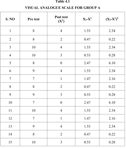
Table 4.1
VISUAL ANALOGUE SCALE FOR GROUP A
S. NO Pre test Post test
(X1) X1-X
1 (X1-X1)2
1 8 4 1.53 2.34
2 8 2 0.47 0.22
3 10 4 1.53 2.34
4 10 3 0.53 0.28
5 8 0 2.47 6.10
6 9 4 1.53 2.34
7 7 1 1.47 2.16
8 8 2 0.47 0.22
9 9 3 0.53 0.28
10 7 0 2.47 6.10
11 10 4 1.53 2.34
12 7 1 1.47 2.16
13 9 4 1.53 2.34
14 8 2 0.47 0.22
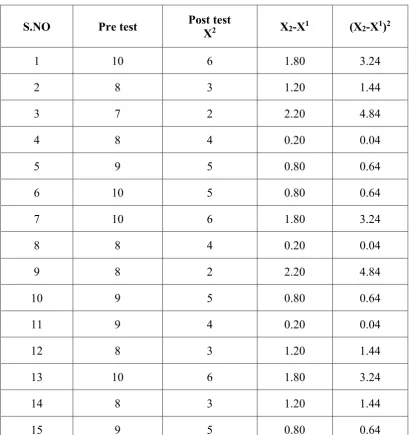
Table 4.2
VISUAL ANALOGUE SCALE FOR GROUP B
S.NO Pre test Post test
X2 X2-X1 (X2-X1)2
1 10 6 1.80 3.24
2 8 3 1.20 1.44
3 7 2 2.20 4.84
4 8 4 0.20 0.04
5 9 5 0.80 0.64
6 10 5 0.80 0.64
7 10 6 1.80 3.24
8 8 4 0.20 0.04
9 8 2 2.20 4.84
10 9 5 0.80 0.64
11 9 4 0.20 0.04
12 8 3 1.20 1.44
13 10 6 1.80 3.24
14 8 3 1.20 1.44
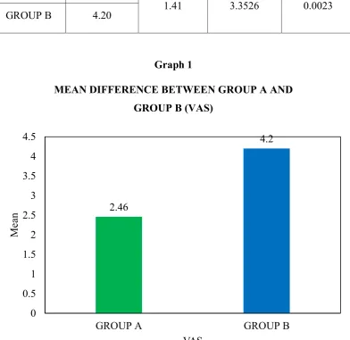
Table 4.3
MEAN DIFFERENCE BETWEEN GROUP A AND
GROUP B (VAS)
GROUP MEAN SD ‘t’ VALUE ‘p’ VALUE
GROUP A 2.46
1.41 3.3526 0.0023 GROUP B 4.20
Graph 1
MEAN DIFFERENCE BETWEEN GROUP A AND
GROUP B (VAS)
2.46
4.2
0 0.5 1 1.5 2 2.5 3 3.5 4 4.5
GROUP A GROUP B
M
ea
n
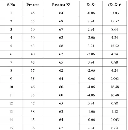
Table 4.4
EXTENSION CERVICAL ROM (DEGREES) FOR GROUP A
S.No Pre test Post test X2 X2-X1 (X2-X1)2
1 48 64 -0.06 0.003
2 55 68 3.94 15.52
3 50 67 2.94 8.64
4 50 62 -2.06 4.24
5 43 68 3.94 15.52
6 40 62 -2.06 4.24
7 45 65 0.94 0.88
8 37 62 -2.06 4.24
9 35 64 -0.06 0.003
10 46 60 -4.06 16.48
11 38 60 -4.06 16.48
12 47 65 0.94 0.88
13 38 63 -1.06 1.12
14 45 64 -0.06 0.003
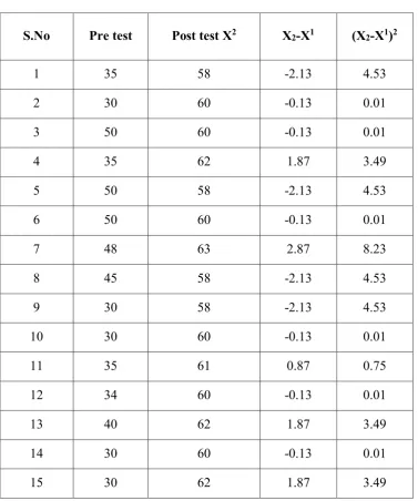
Table 4.5
EXTENSION CERVICAL ROM (DEGREES) GROUP B
S.No Pre test Post test X2 X2-X1 (X2-X1)2
1 35 58 -2.13 4.53
2 30 60 -0.13 0.01
3 50 60 -0.13 0.01
4 35 62 1.87 3.49
5 50 58 -2.13 4.53
6 50 60 -0.13 0.01
7 48 63 2.87 8.23
8 45 58 -2.13 4.53
9 30 58 -2.13 4.53
10 30 60 -0.13 0.01
11 35 61 0.87 0.75
12 34 60 -0.13 0.01
13 40 62 1.87 3.49
14 30 60 -0.13 0.01
TABLE 4.6
MEAN DIFFERENCE BETWEEN GROUP A AND GROUP B
CERVICAL EXTENSION
GROUP MEAN SD ‘t’ VALUE ‘p’ VALUE
GROUP A 64.07
2.19 4.9118 0.0001 GROUP B 60.13
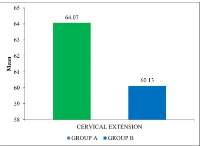
Graph 2
MEAN DIFFERENCE BETWEEN GROUP A AND GROUP B
CERVICAL EXTENSION
64.07
60.13
58 59 60 61 62 63 64 65
CERVICAL EXTENSION
M
e
an
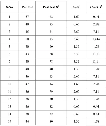
Table 4.7
CERVICAL ROTATION TO THE RIGHT (DEGREES)
GROUP A
S.No Pre test Post test X2 X2-X1 (X2-X1)2 1 30 85 0.65 0.44
2 50 83 1.33 1.78
3 35 84 0.33 0.11
4 54 82 2.33 5.44
5 50 85 0.67 0.44
6 40 84 0.33 0.11
7 45 85 0.67 0.44
8 35 86 0.33 0.11
9 30 84 0.33 0.11
10 30 83 1.33 1.78
11 35 85 0.67 0.44
12 34 86 0.33 0.11
13 40 84 0.33 0.11
14 30 85 0.67 0.44
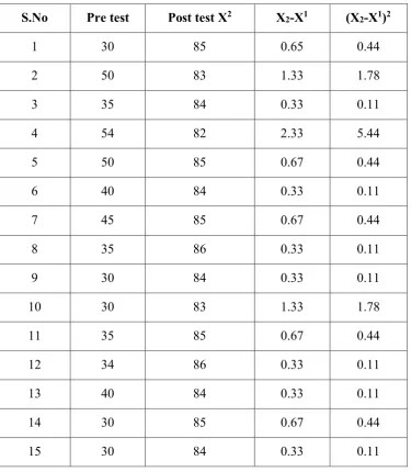
Table 4.8
CERVICAL ROTATION TO THE RIGHT (DEGREES)
GROUP B
S.No Pre test Post test X2 X2-X1 (X2-X1)2
1 37 82 1.67 0.44
2 48 83 0.67 2.78
3 45 84 3.67 7.11
4 50 85 3.67 13.44
5 30 80 1.33 1.78
6 43 78 3.33 11.11
7 40 78 3.33 11.11
8 40 80 1.33 1.78
9 36 83 2.67 7.11
10 47 84 1.67 2.78
11 36 79 2.67 7.11
12 38 80 1.33 1.78
13 46 82 0.67 0.44
14 38 82 0.67 0.44
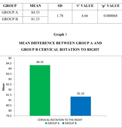
Table 4.9
MEAN DIFFERENCE BETWEEN GROUP A AND
GROUP B CERVICAL ROTATION TO RIGHT
GROUP MEAN SD ‘t’ VALUE ‘p’ VALUE
GROUP A 84.33
1.78 4.66 0.000068 GROUP B 81.33
Graph 3
MEAN DIFFERENCE BETWEEN GROUP A AND
GROUP B CERVICAL ROTATION TO RIGHT
84.33
81.33
79.5 80 80.5 81 81.5 82 82.5 83 83.5 84 84.5 85
CERVICAL ROTATION TO THE RIGHT
M
e
a
n
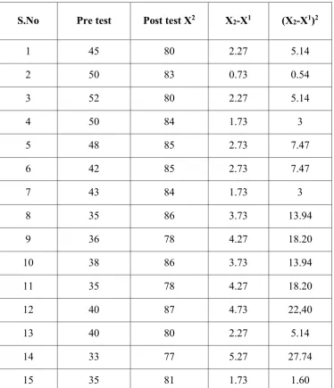
Table 4.10
CERVICAL ROTATION TO THE LEFT (DEGREES)
GROUP A
S.No Pre test Post test X2 X2-X1 (X2-X1)2
1 45 80 2.27 5.14
2 50 83 0.73 0.54
3 52 80 2.27 5.14
4 50 84 1.73 3
5 48 85 2.73 7.47
6 42 85 2.73 7.47
7 43 84 1.73 3
8 35 86 3.73 13.94
9 36 78 4.27 18.20
10 38 86 3.73 13.94
11 35 78 4.27 18.20
12 40 87 4.73 22,40
13 40 80 2.27 5.14
14 33 77 5.27 27.74
Table 4.11
CERVICAL ROTATION TO THE LEFT (DERGREES) GROUP B
S.No Pre test Post test X2 X2-X1 (X2-X1)2
1 40 82 3.20 10.24
2 48 80 1.20 1.44
3 45 80 1.20 1.44
4 35 79 0.20 0.04
5 33 78 0.80 0.64
6 40 78 0.80 0.64
7 40 75 3.80 14.44
8 38 75 3.80 14.44
9 42 83 4.20 17.64
10 44 82 3.20 10.24
11 37 76 2.80 7.84
12 38 79 0.20 0.04
13 42 83 4.20 17.64
14 36 79 0.20 0.04
Table 4.12
MEAN DIFFERENCE BETWEEN GROUP A AND GROUP B
CERVICAL ROTATION TO THE LEFT
MEAN SD ‘t’ VALUE ‘p’ VALUE
GROUP A 82.27
3.15 2.98 0.0058 GROUP B 78.80
Graph 4
MEAN DIFFERENCE BETWEEN GROUP A AND GROUP B
CERVICAL ROTATION TO THE LEFT
82.27
78.8
77 78 79 80 81 82 83
CERVIAL ROTATION TO THE LEFT
M
e
a
n
Table 4.13
CERVICAL LATERAL FLEXION TO THE RIGHT (DEGREES)
GROUPA
S.No Pre test Post test X2 X2-X1 (X2-X1)2
1 35 40 3.07 9.42
2 27 40 3.07 9.42
3 30 38 1.07 1.14
4 21 32 -4.93 24.30
5 22 37 0.07 0.004
6 26 32 -4.93 24.30
7 25 34 -2.93 8.58
8 33 34 -2.93 8.58
9 28 35 -1.93 3.72
10 22 34 -2.93 8.58
11 31 34 -2.93 8.58
12 29 38 1.07 1.14
13 35 40 3.07 9.42
14 39 42 5.07 25.70
Table 4.14
CERVICAL LATERAL FLEXION TO THE RIGHT (DEGREES)
GROUP B
S.No Pre test Post test X2 X2-X1 (X2-X1)2
1 23 32 0.14 0.01
2 19 30 -1.86 3.45
3 22 32 0.14 0.01
4 20 32 0.14 0.01
5 20 30 -1.86 3.45
6 21 32 0.14 0.01
7 20 34 2.14 4.57
8 21 33 1.14 1.29
9 23 33 1.14 1.29
10 22 32 0.14 0.01
11 22 28 -3.86 14.89
12 20 32 0.14 0.01
13 19 32 0.14 0.01
14 16 34 2.14 4.57
TABLE 4.15
MEAN DIFFERENCE BETWEEN GROUP A AND GROUP B FOR
CERVICAL LATERAL FLEXION TO THE RIGHT
GROUP MEAN SD ‘t’ VALUE ‘’p VALUE
GROUP A 36.93
2.84 4.7881 0.0001 GROUP B 31.86
Graph 5
MEAN DIFFERENCE BETWEEN GROUP A AND GROUP B FOR
CERVICAL LATERAL FLEXION TO THE RIGHT
36.93
31.86
29 30 31 32 33 34 35 36 37 38
CERVICAL LATERAL FLEXION RIGHT SIDE
M
e
an
Table 4.16
CERVICAL LATERAL FLEXION TO THE LEFT
(DEGREES) GROUP A
S.No Pre test Post test X2 X2-X1 (X2-X1)2
1 29 42 3.34 11.15
2 30 42 3.34 11.15
3 30 40 1.34 1.79
4 24 38 -0.66 0.43
5 23 38 -0.66 0.43
6 25 38 -0.66 0.43
7 26 34 -4.66 21.43
8 27 38 -0.66 0.43
9 28 40 1.34 1.79
10 22 42 3.34 11.15
11 30 38 -0.66 0.43
12 28 40 1.34 1.79
13 35 38 -0.66 0.43
14 28 38 -0.66 0.43
Table 4.17
CERVICAL LATERAL FLEXION TO THE LEFT (DEGREES)
GROUP B
S.No Pre test Post test X2 X2-X1 (X2-X1)2
1 23 40 6.67 44.48
2 25 40 6.67 44.48
3 22 34 0.67 0.44
4 20 34 0.67 0.44
5 20 30 -3.33 11.08
6 21 32 -1.33 1.76
7 22 34 0.67 0.44
8 20 30 -3.33 11.08
9 23 40 6.67 44.48
10 20 32 -1.33 1.76
11 22 30 -3.33 11.07
12 23 28 -5.33 28.40
13 19 30 -3.33 11.08
14 18 32 -1.33 1.76
Table 4.18
MEAN DIFFERENCE BETWEEN GROUP A AND GROUP B FOR
CERVICAL LATERAL FLEXION TO THE LEFT
GROUP MEAN SD ‘t’ VALUE ‘p’ VALUE
GROUP A 38.67
3.26 4.4721 0.0001 GROUP B 33.33
Graph 6
MEAN DIFFERENCE BETWEEN GROUP A AND GROUP B FOR
CERVICAL LATERAL FLEXION TO THE LEFT
38.67
33.33
30 31 32 33 34 35 36 37 38 39 40
CERVICAL LATERAL FLEXION
M
e
an
Table 4.19
NECK BOURNEMOUTH QUESTIONNAIRE FOR GROUP A
S. No Pre test Post test X1-X1 (X1-X1)2
1 54 22 1.54 2.37
2 52 24 3.54 12.53
3 50 22 1.54 2.37
4 55 18 -2.46 6.05
5 48 18 -2.46 6.05
6 46 17 -3.36 11.97
7 50 22 1.54 2.37
8 56 18 -2.46 6.05
9 52 20 -0.46 0.21
10 50 21 0.54 0.29
11 48 24 3.54 12.53
12 52 20 -0.46 0.21
13 48 18 -2.46 6.05
14 54 22 1.54 2.37
Table 4.20
NECK BOURNEMOUTH QUESTIONNAIRE FOR GROUP B
S.No Pre test Post test X2 X2-X1 (X2-X1)2
1 52 28 5.34 28.51
2 54 26 3.34 11.15
3 55 27 4.34 18.83
4 50 24 1.34 1.79
5 46 19 -3.66 13.39
6 48 22 -0.66 0.43
7 52 22 -0.66 0.43
8 50 25 2.34 5.47
9 48 22 -0.66 0.43
10 55 22 -0.66 0.43
11 49 19 -3.66 13.39
12 56 17 -5.66 32.03
13 52 24 1.34 1.79
14 48 22 -0.66 0.43
TABLE 4.21
MEAN DIFFERENCE BETWEEN GROUP A AND GROUP B
NECK BOURNEMOUTH QUESTIONNAIRE
GROUP MEAN SD ‘t’ VALUE ‘p’ VALUE
GROUP A 20.47
2.69 2.2372 0.033 GROUP B 22.67
Graph 7
MEAN DIFFERENCE BETWEEN GROUP A AND GROUP B
NECK BOURNEMOUTH QUESTIONNAIRE
20.47
22.67
19 19.5 20 20.5 21 21.5 22 22.5 23
NBQ
M
e
an
5. DISCUSSION
Upper trapezius is the most common postural muscle that tend to get
shorten leading to restricted neck mobility as they are most frequently used to
maintain posture. Mechanical neck pain a common problem within our society affecting individual’s physical and social functioning considerably and interfering with sufferer’s daily activities. Probably most of the patient suffering from pain and decreased neck activity is due to upper trapezius trigger points.
In present study pain and neck activity is taken as the parameters and they
were scored by using the visual analogue scale, goniometer and neck
Bournemouth questionnaire.
There are different treatments approaches that can be used for the upper
trapezius trigger points. In this study we used the Positional release technique and
Ischemic compression technique as the treatment approaches to full fill the aim of
study, to compare the effectiveness of both treatments in patients with neck pain
due to upper trapezius muscle trigger points.
Within two group analysis were revealed that there is a significant
decrease in Group A patient reported pain, range of motion and neck activity
scores when post test and post test were compared.
Mean score of group A for VAS was 2.47 and for group B was 4.20. Mean
score of group A and group B had difference in their value. But more reduce in
group A. SD and t value were 1.41 and 3.35 respectively. p value was 0.0023.
Extension CROM mean value for group A was 64.07 and for group B
60.13. Here also group A had higher value. 2.19 and 4.91were the SD and the t
value. P value was 0.0001.
In cervical rotation to the right, group A mean value was 84.33 and for
group B it was 81.33. Higher value are seen in group A. SD was 1.78 and t value
was 4.66 with p value of 0.000068. For cervical rotation to the left mean values
In cervical lateral flexion to the right mean value of group A was 36.93
and for group B it was 31.86. A higher value is seen in group A. SD and t value
were 2.84 and 4.78 respectively. P value was 0.0001. In cervical lateral flexion to
the left mean value of group A was 38.67 and for group B it was 33.33. A greater
value is found in group A. SD and t values were 3.26 and 4.47 respectively and a
p value of 0.0001.
For NBQ the mean value of group A was 20.47 and for B mean was 22.67.
From this group A had functional and cognitive improvement than group B. SD
and t value was 2.69 and 2.23 respectively. P value of 0.033.
On analyzing the statistical values there is a difference between the
parameters among group A and group B. The value was higher in group A.
During the time of follow up group B had recurrence of pain, decreased
CROM and activity limitation than group A which was only clinically identified.
This study gives an excellent view into the application of Positional
Release technique over Ischemic Compression technique for treating sedentary
6. CONCLUSION
Positional Release technique and Ischemic Compression technique was
found to be in reducing pain, ROM, functional disability and psychometric
analysis in patients with neck pain due to upper trapezius trigger points. However
according to statistical analysis Positional release technique was superior to
Ischemic compression technique.
Hence rejecting the null hypothesis and accepting the alternative
hypothesis. Thus the study concluded that there is a significant difference between
Positional Release technique versus Ischemic Compression on pain, range of
motion functional disability and psychometric analysis in upper trapezius trigger
points.
LIMITATIONS
• sample size taken was small
• Generalizability of the findings is limited by short term follow up used in this study.
• Patients may not follow the advice in a correct manner.
• Treatment was given only 2 weeks duration.
• Not consider the shoulder range of motion
RECOMMENDATIONS
• Study can be done with a larger sample size and longer study duration.
• In future treatment studies can be modified with other techniques and
REFERENCES
BOOKS
1) Magee, D,J. (2014). Orthopaedic physical assessment. (6th ed). Elsvier.
USA.
2) Chaurasia.B.D. (2010).Human anatomy regional and applied dissection
and clinical upper limb and thorax. (5th ed). CBS. India.
3) Travell &Simon’s. (1999). Myofascial pain and dysfunction: The trigger
point manual volume 1. Upper Half of Body. (2nded) Baltimore. Williams
&Wilkins: Pennsylvania.
4) Gray’s, H, (2008). The anatomical basis of clinical practice.
(40th ed). Elsevier.
5) Kapandji, A, (1988). The physiology of joints: Trunk and vertebral
column (2nd) ed. Churchill Livingstone.
JOURNALS
1) Priyanka Devang Ranna (2017). Effect of muscle energy technique
versus positional release technique in computer workers with upper
trapezius muscle spasm: A comparative study. International journal of
multidisciplinary Research and development.2349-5979.
2) AmenehAmini (2017). The Effects of manual passive muscle shortening
and Positional Release Therapy on Lateral Myofascial Trigger points of
the Upper trapezius: A double blind Randomized clinical Trial. Iran Red
Crescet med.
3) Shweta Anil Kulkarni (2017). Effectiveness of Ischemic compression
versus Myofascial release on Myofascial trigger point of upper trapezius.
International journal of allied medical sciences and clinical research
(IJAMSCR) 2347-6567.
4) Danilo Harudy Kamonseki (2017). Translation and Validation of Neck
Bournemouth Questionnaire to Brazilian Portuguese. Rev Bras
5) Gurkan Gunaydin (2016). Reliability, Validity, and Cross – cultural
Adaptation of the Turkish version of the Bournemouth Questionnaire.
Spine health Services Research.
6) Tommaso Geri. (2015). Rasch analysis of the Neck Bournemouth
Questionnaire to measure disability related to Chronic Neck Pain. Journal
of Rehabil Med 47:836-843.
7) G. Yatheendra Kumar (2015).Effectiveness of Muscle energy technique,
Ischemic Compression and Strain Counter Strain on Upper Trapezius
Trigger points. A comparative study. International journal of physical
education, sports and Health.
8) Ahmed Samir Mohamed Abdelhamid (2015). Ischemic Compression
versus Traditional Physical Therapy in Treatment of chronic mechanical
Neck Pin. International Journal of Advanced Research volume 3 Issue,
931-938.
9) Jay. P.Shah (2015). Myofascial Trigger points Then and Now: A
historical and scientific perspective. PMR Author manuscript 746-761.
10) Sweety Charles Carvalho (2014).Effect of Positional Release Technique
in subjects with subacuteTrapezitis. International Journal of physiotherapy
vol1,91-99.
11) Nehal Shah (2013). Comparative study of Myofascial release and cold
pack in upper Trapezius spasm. International Journal of health Science
and Research,ISSN:2249-9571.
12) Sahem A.M (2013). Positional Release Technique versus Manual pressure
Release on the Upper Trapezius muscle in patients with Myofascial pain
Dysfunction syndrome. Bull Fac. Ph. Thcairo Univ vol 18.
13) Gopal Nmbi (2013). Difference in effect between Ischemic Compression
and Muscle energy technique on Upper trapezius Myofascial trigger point:
Comparative study. Journal of Health and Allied Sciences.
14) Jagatheesan Alagesan (2012). Effect of Positional Release Therapy and
Taping on unilateral Upper trapezius Tender points. A randomized
15) Giburm Park (2010). Reliability and Usefulness of the pressure pain
threshold measurement in patients with Myofascial pain. Annals of
Rehabilitation medicine,471-701.
16) Montanez-Aguilera FJ (2010). Changes in the patients neck pain after
application of ischemic compression as a trigger point therapy journal of
back and musculoskeletal rehabilitation,23 (2).101-104.
17) Aguilera (2009). Immediate effect of ultrasound and Ischemic
Compression techniques for the treatment of trapezius latent Myofascial
trigger points in healthy subjects: A randomized controlled study journal
of manipulative and physiological therapeutics,515-520.
18) Hugh Gemmell (2008) Immediate effect of Ischemic compression and
trigger point pressure release on neck pain and upper trapezius trigger
points: A randomized controlled trial. Intl.elsevierhealth.com journal.
19) Paul Rankin (2006). The Bournemouth questionnaire as an outcome
measure in the rehabilitation of a person suffering with mechanical neck
and arm pain and concurrent charcot- marie-Tooth disease:a case report. J
can chiropr Assoc 50(3)
20) Peter Baldry (2002). Management of Myofascial trigger point pain.
Acupuncture in medicine 2-10.
WEB REFFERENCES
1 https://www.sportshealth.com
2 https://www.physiopaedia.com
3 https://www.spinehealth.com
4 https://wwwq.anatomy-physiotherapy.com
APPENDICES
APPENDIX - I
ASSESSMENT CHART
Subjective Assessment
Demographic data
Name:
Age:
Sex:
Occupation:
IP/OP number:
Address:
Date of evaluation:
Chief complaints:
History
Past medical history:
Present medical history:
Surgical history:
Drug history:
Personal history:
On Observation
Built:
Posture / postural changes (neck):
Any congenital abnormality:
Contracture/ deformity:
Any Inflammatory or malignant disease:
External appliances (cervical collar):
Respiratory muscle movements:
• On inspiration:
• On expiration:
On Palpation
Tenderness:
Warmth:
Tropical changes:
Trigger point:(upper trapezius)
▪ Criteria includes
o Palpable taut band
o Spot tenderness in the taut band o Referred pain as the “familiar pain” o Pincer flat technique for upper trapezius o Look for local twitch response
On Examination
❖ Vital Sign
• Blood Pressure (mmHg):
• Respiratory rate(/m):
• Pulse rate (/m):
• Heart rate (/m):
VAS scale:
0 5 10
Mild Moderate Severe
• Site of pain:
• Duration of pain:
• Quality of pain:
• Relieving factor:
• Severity of pain:
• Time of pain:
• Insufficient ADL activity due to pain:
❖ Range Of Motion
I. Range of motion - Neck
Movement Range Of Motion (Degree)
Neck flexion
Neck extension
Lateral flexion Right-Left
Rotation Right-left
Range of motion – Shoulder
Movement Range Of Motion (Degree)
Right Left
Flexion
Extension
Abduction
Adduction
❖ Sensation:
• Deep
• Superficial
❖ Neurological Examination
• Vertigo
• dizziness
• Neck pain radiating into arms and upper extremity
• Cervical radiculopathy,
• Cervical spondylosis
❖ Functional activity
❖ Using Neck Disability Index Scale
❖ Special test:
➢ Test for neurological symptoms:
I. Neurodynamic test
o ULTT 1
o ULTT2
o ULTT3
o ULTT4
II. Distraction and compression test
III. Spurling(Foraminal compression)
IV. Scalene cramp test
V. Shoulder depression test
VI. Romberg test
➢ Test for cervical instability:
I. Anterior shear or sagittal test
➢ Differential diagnosis: Facial neuralgia or cervical radiculopathy or other
trigger point
❖ Investigation
❖ Diagnosis
❖ Physiotherapy management
• Aims
• Goals
• Intervention plan
Group A- Positional Release Technique
Group B – Ischemic Compression
❖ Home advices
APPENDIC II
FOLLOW UP CHART
Patient name:
Age:
Sex:
Occupation:
Address:
In patient outpatient no:
Condition:
Chart:
Parameters Pretest value Posttest value Value after
3 weeks
VAS
Goniometer
APPENDIX III
TOOLS FOR MEASUREMENT
VISUAL ANALOGUE SCALE
The VAS score is determined by measuring in millimeters from left hand
end of the line to the point that the patient marks.
How severe is your pain today? Place a vertical mark on the line below to
indicate how bad you feel your pain is today.
No pain Very severe pain
1 2 3 4 5 6 7 8 9 10
NECK BOURNEMOUTH QUESTIONNAIRE
Neck Bournemouth questionnaire used to evaluate the impact of pain on
patient’s psyche and on socio- professional relationship for measuring the functional disability and psychometric evaluation.
Instruction: The following scales have been designed to find out about your
neck pain and how it is affecting you. Please answer all the scales, and mark the
one numbers on each scale that best described how you feel.
1. Over the past week, on average, how would you rate your neck pain?
No pain Worst pain possible
1 2 3 4 5 6 7 8 9 10
2. Over the past week, how much has your neck pain interfered with your daily
activities (housework, washing, dressing, lifting, reading and driving)?
3. Over the past week, how much has your neck pain interfered with your
ability to take part in recreational, social, and family activities?
No interference Unable to carry out
1 2 3 4 5 6 7 8 9 10
4 . Over the past week, how anxious (tense uptight, irritable, difficulty in
concentrating/relaxing) have you been feeling?
Not at all anxious extremely anxious
1 2 3 4 5 6 7 8 9 10
5 . Over the past week, how depressed (down –in-the dumps and ,in low spirits,
pessimistic, unhappy) have you been feeling?
Not at all depressed extremely depressed
1 2 3 4 5 6 7 8 9 10
6. Over the past week, how have you felt your work (both inside and outside
the home) has affected (or would affect) your neck pain?
Have made it no worse Have made it much worse
1 2 3 4 5 6 7 8 9 10
7. Over the past week, how much have you been able to control
(reduce/help) your neck pain on your own?
Completely control it No control whatsoever
1 2 3 4 5 6 7 8 9 10
0= Much better
5= no change
APPENDIX IV
MOIST HEAT PACKS
Moist heat packs is also applied in both groups for relieving pain. Gel packs is applied by wrapping in towel and placed over the affected upper trapezius muscle.
Position of the patient
Prone lying and the head turn around the unaffected side. Hands relaxed by the side.
Duration
10-15 minutes
POSITIONAL RELEASING TECHNIQUE
Positional release therapy is a very specialized technique focusing on treating protective muscle spasm in the body. This technique involves finding a tender point in the patient’s body(muscle, ligaments, tendons and joints) and then moving the patient’s body or body part away from the restricted motion barrier and towards the position of greatest comfort.
Once in this position of comfort, the point should no longer be tender. This precise position is held for a minimum of 90 seconds but can be held for several minutes. During this time period, the patient can feel heat, vibration, pulsation, and
can even reproduce their symptoms. Once the release is complete, the heat, vibration, pulsation and pain will diminish and there will be a sense of lengthening and relaxation in the tissue. Once the release is felt, the patient is slowly taken out of the position of comfort and the tissue should be relaxed. The tender point should either be completely gone or 70% better.
Mechanism of positional release technique
Positional release technique act on the muscle spindle mechanism and its
associated reflex mechanism(which control spasm) to promote a more normal firing
of the spindle and a more normal level of tension in the muscle which result in more
normal relationship within the various soft tissue surrounding the area. These
techniques work to reduce the hyperactivity of myotatic reflex arc and to reduce the
over whelming afferent nerve impulses within the arc that may lead to an overflow
of neurotransmitters into the associated dermatome, resulting in referred pain
(facilitated segment). The mechanism behind this technique is that the shortening
of the muscle sends a signal to the brain causing the muscle contraction to be
reduced. This technique is used for relief of somatic dysfunctions that are too acute
or too delicate to treat with other procedures.
Procedure
✓ Position of the patient: patient is in sitting position
✓ Position of the therapist: near to affected side of patient
✓ Technique: The therapist localizes the tender points along the trapezius.
Pressure is applied by pinching the muscle between the thumb and fingers.
The subjects head is laterally flexed toward the side of tender point (opposite
side rotation) then therapist grasp the subjects forearm and abduct shoulder
to approximately 90 degree and add slight flexion and extension to fine tune.
The ideal position of comfort achieved was held for a period of 90 seconds
and followed by a passive return of the body part to an anatomically neutral
ISCHEMIC COMPRESSION TECHNIQUE
Position of patient: Patient was placed supine on the couch with his head
fully on the surface of the couch, to reduce tension in the upper trapezius muscle.
Arm was positioned in slight abduction with the elbow bent and their hands resting
on their stomach.
Position of the therapist: To perform this IC to the upper trapezius,
therapist stands at the head of the couch.
Technique:
➢ At first, the therapist uses a pincher grasp moved throughout the fibers of
the upper trapezius and made note of the any active trigger points.
➢ To locate a trigger point, palpate the muscle to feel for a taut band (or) a
twitch response in the muscle belly. The common location of upper
trapezius trigger points is in the middle of the muscle belly, approximately
1 to 2 inches medial to the acromion process of the scapula.
➢ Once the trigger point is located, apply an IC by gradually applying
pressure to the trigger point with your thumb.
➢ While applying pressure, the patient will likely feel referred pain in a
question mark pattern(along the back of the neck, around the side of the
head, and then focused pain right behind the eye).
➢ Keep in communication with patient, to check whether the patient is staying
within the limits of his pain tolerance.
➢ Hold this technique for approximately 20 seconds to 1 minute, patient tells
you that pain has diminished, or until feels the muscle fiber begin to relax
under your pressure. once feel this release, gradually release pressure.
➢ All identified trigger points was treated. Then apply a few effleurage strokes
➢ This was repeated for three to five times for three sessions per week for 14
APPENDIX V
HOME EXERCISE PROGRAM
• Chin tucking exercise
• Active neck flexion and extension
• Lateral flexion
• Side to side rotation
• Shoulder shrugs
• Shoulder protraction and retraction
• Isometric exercise for neck
An ergonomic advice also conducted for each group and given home advice.
The ergonomics advice given depends upon patient work place and job.
❖ Active Exercises
Chin tucking exercises:
Sit in a chair with your back firmly supported by a wall or the back of the
chair. Make sure the back of your head, shoulders and upper back are against the
wall, you are looking straight ahead and the underside of your chin is level with the
floor. Slowly move your chin back and slightly down so your ears are in line with
your shoulders and you feel a stretch in the back of your neck. Hold for 10 seconds
and release. Do a couple of sets of 10 repetitions every day, if you can. You can
Active neck flexion and extension:
Cervical Flexion (bringing your chin to your chest) and Cervical Extension
(looking up to the ceiling). You want to start by tucking your chin in and gently
bringing your head forward and attempting to touch the chin to the chest. Next,
gently bend the head backwards as far as it will go and come to neutral position and
bend the head forward. Repeat this exercise 5 times. Forward head flexion is great
for those patients who suffer from hypertonic cervical paraspinals- which is
essentially pain in the back of the neck.
Lateral flexion exercise:
Bring your right ear to your right shoulder as far as you are able to. Do not
rotate or turn your head when you are doing this neck stretch. Come to neutral
Side to side rotations:
In this exercise you want to turn your head to the right as far as you possibly
can, trying to bring your chin over your shoulders. When you are doing this neck
exercise, do not bring up your shoulders. Hold this position for 3 to 5 seconds. Next
do this on the left side of the neck and repeat for 5 times.
• Shoulder shrugs:
Stand up straight in good posture. Keep your arms straight, shrug your
shoulders straight up toward your ears as high as you can. Hold this uppermost
position for one to two seconds. Do not roll your shoulders backward and lower
• Shoulder protraction and retraction:
Stand up straight in good posture. Keep your arms straight, move your
shoulder forwards and hold this uppermost position for one to two seconds. Then
come to neutral position. Then move your shoulder backward followed by neutral
position. Perform 5 to 10 repetitions.
❖ Isometric exercise for neck
• Neck Flexing:
Bend your neck slightly forward and put your hand on your forehead. Try
to bend your head forward while pushing back with your hand. Hold for 5-8
• Neck Extension:
Keep your up and your neck straight and place your hands at the back of
your head. Try to push your head backwards while pushing forward with your
hands. Hold for a count of 5-8 seconds.
Side Bending:
Keep your head straight and your chin level. Put your right hand on the right
side of your head. Try to bring your head down to your right shoulder while pushing
up with your right hand. Hold for 5-8 seconds. Repeat the Side Bending, but to the
• Rotation:
Put your left hand at chin level and turn your head slightly to the right. Put
your right hand on the right side of your face. Turn your head to the right while
pushing it back with your right hand. Hold for a count of 5-8 seconds.
❖ Self stretch
Positioning: You can do this sitting or standing. Always have your hand
on the shoulder you want to stretch to prevent it from moving up. The other hand
should be on top of your head with your fingers pointing towards the back. Your
neck should always remain in line with your back and the only body part that is
moving is your head.
• Forward stretch: Gently pull your head forward with your chin toward
your neck as if you were nodding. Hold this position for 10 to 15 seconds.
• Side stretch: Gently pull your head to the side so your ear approaches the
opposite shoulder. Switch sides. Hold this position for 10 to 15 seconds.
• Diagonal stretch: Gently pull your head diagonally forward so your chin
approaches the opposite shoulder. Hold this position for 10 to 15 seconds.
Repeat these stretches for the other side. Start with the forward stretch,
but this time, your hand should be on the opposite shoulder. Go through these
ERGONOMICS MODIFICATIONS AND HOME EXERCISES
ERGONOMICS MODIFICATIONS
Ergonomics is the process of designing or arranging work places, products
and systems so that they fit the people who use them. Ergonomics aims to improve
workspaces and environments to minimize risk of injury or harm. It is often
important to look at the work place ergonomics as part of treatment and prevention of
neck pain. Perhaps the placement of the desk, computer workstation and/or placement
of the computer monitor and keyboard can be improved to encourage improved neck
posture. The goal of an ergonomics program in industry is to adapt the workplace to a
specific worker, dependent on the job description, required tasks, and physical make
up