International Journal of
ELECTROCHEMICAL
SCIENCE
www.electrochemsci.orgShort Communication
Oriented T4 Bacteriophage Immobilization for Recognition of
Escherichia coli
in Capacitance Method
Danial Ashiani1, Amir Homayoun Keihan2, Jamal Rashidiani1, Fariba Dashtestani3, Khadijeh Eskandari1,*
1
Nanobiotechnology Research Center, Baqiyatallah University of Medical Sciences, Tehran, Iran 2 Molecular Biology Research Center, Baqiyatallah University of Medical Sciences, Tehran, Iran 3
Institutes of Biochemistry & Biophysics, University of Tehran *
E-mail: [email protected]
Received: 23 July2016 / Accepted: 11 September 2016/ Published: 10 November 2016
One of the important aspects in biotechnology is the development of a rapid and selective approachfor the identification and quantization of Escherichia coli (E. coli) bacteria. In this research, we fabricated a novel Label-free electrochemical E. coli biosensor based on oriented immobilization of T4 biotinylated bacteriophage as recognition receptor of E. coli on streptavidin immobilized on polyanilin (PANI) glassy carbon (GC) modified electrode. Labeled PANI was synthesized by chemical method and characterized by Raman and cyclic voltammetry (CVs) and field emission scanning electron microscope (FESEM). The bacteriophage/PANI/GC electrodes were applied as indicator electrodes for capacitance determination of E. coli bacteria. By fabricating this biosensor, E. coli was sensed in a linearrange and detection limit of 10-98 N/mL and 10 N/mL (N stands for number of E. coli) respectively. The proposed biosensor shows fast response time of ∼1 s and good selectivity over other bacteria. We successfully developed and implemented an E. coli biosensor based on oriented bacteriophage immobilization on PANI modified GC electrode. This biosensor is cost-effective, rapid, renewable and high selective for detection of E. coli bacteria. Also, this method can be extension to detection of other biological agents.
Keywords: E. coli, bacteriophageT4, capacitance, biosensor, PANI.
1. INTRODUCTION
Recently, microorganisms (viruses and bacteria) are generally found in the meal, soil, surroundings, aquatic environment and estuarine waters,and the intestinal tracts of human beings and animals[1].
Staphylococcus aureus are the most important food-borne pathogen bacteria, which are caused major borne illness [2]. Hence, it is really important to develop methods for detecting the food-borne pathogenic bacteria in food, water and air as its critical role in public health. While, cell culture and plating are standard microbiological methods, they are costly and time consuming. And also, most of conventional methods involve complex instrumentation and may not be used on-site [3]. Thus, developing biosensors for detection of pathogens as a fast and specific technique are growing [4,5,6].
One of the dangerous human pathogen is E. coli [7,8]. It causes life-threatening difficulties such as bloody diarrhea and renal failure [9]. Consequently, designing and constructing a rapid and specific biosensor for the detection and quantification of E. coli would be very important in analysis of food, environment and medical disorders. In this regard, several rapid and expensive systems were developed. These systems include as electric DNA chips, quartz crystal microbalance, amperometric biosensor, imaging ellipsometry, opto-fluidic ring resonator sensor, and surface plasmon resonance (SPR) [10,11].Biosensors are tools to determine levels of chemicals and microorganisms in a sample with high accuracy. They can be used in different branches of science like medicine, industry, agriculture, and etc [12].
In this work we have successfully fabricated a rapid and cost effective capacitive biosensor constructed by oriented bacteriophage modified glassy carbon (GC) electrode. Therefore, the layout of the biosensor is patterned as a two electrodes configuration. These electrodes were made by immobilization of oriented bacteriophage on polyaniline (PANI) via streptavidin on modified GC electrode (bacteriophage/streptavidin/PANI/GC). Consequently, the E. coli trapping by oriented bacteriophage on the PANI modified GC electrodes was reported by capacitance method. The proposed sensors show good characteristics such as; low detection limit, wide linear range, fast response time and good selectivity for E. coli detection.
2. MATERIALS AND METHODS 2.1. Apparatus and procedure
Raman spectroscopy was obtained by Nicolet Raman spectrometer. The morphology of the prepared bacteriophage/streptavidin/PANI/GC electrode was achieved by means of field emission scanning electron microscope (FESEM) (Hitachi S-4160, UK).
All of the capacitance evaluations were done by a LCR meter (GPS 3131B, Benchtop LCR Meter, Germany). Cyclic voltammetry (CVs) analysis were accomplished via an Autolab potentiostat/galvanostat (model 302N, Eco Chemie, Ultecht, Netherlands). The capacitance in this system was evaluated through two similar GC electrodes in 2 mm diameter (presented in Fig. 1) as a capacitor.
2.2. Reagents
LB Agar culture from Merck Germany, LB Broth culture from Merck Germany, Polyethylene glycole (PEG) 6000 from Merck, Germany, 1-Ethyl-3-(3-dimethylaminopropyl) carbodiimide (EDAC) with MW 155.24 from Sigma, United States of America, 2-(N-morpholino) ethanesulfonic acid (MES) buffer with MW 951.24 Sigma, United States of America, Dihydrogen phosphate (KH2PO4), dipotassium hydrogen phosphate (K2HPO4), hydrochloric acid (HCl) and hydrogen peroxide (H2O2) were obtained from Merck, Germany. A phosphate buffer solution (0.1 M, pH=7) was applied as supporting electrolyte. Ultrapure H2Oby a Millipore-Milli Q (18 MΩ cm, Barnstead, Dubuque, USA) system was used for all solution preparations. All the reagents were used as received, without any purification and all experiments were done at room temperature (25 0C). E. coli and bacteriophage were gotten from Molecular Biology Research Center, Baqiyatallah University of Medical Sciences, Tehran, Iran.
2.3. Preparation of bacteriophage/streptavidin/PANI/GC electrode
There were 3 steps for preparing bacteriophage/streptavidin/PANI/GC electrode. In the first step, biotinylated bacteriophage prepared as follow. NHS-Biotin dissolved in Dimethyl sulfoxide (DMSO) instantly in 22 mg/mL concentration. NHS-Biotin solution was added to the total volume of the bacteriophage10%, and stirred gently, and incubated at room temperature for 4 hours. In the second step, streptavidin immobilized on PANI modified GC electrode as the following order. Firstly, 3 μL of PANI (5% W/V) in DMSO solution was dropped on GC electrode. In the other vessel, 0.7 mg of streptavidin was dispersed in MES buffer (1.5 mL, 50 mM, pH= 6.1). Then, NHS (0.5 mL, 50 mg/mL) which was prepared in MES was added to the above suspension. While stirring suspension severely, 0.3 mL of fresh EDAC solution (10 mg/mLprepared in MES buffer) was combined quickly, and the consequence mixture was stirred at room temperature for 45 minutes repeatedly. After that, 20 μL of activated streptavidin immediately dropped on the PANI/GC electrode, and set the electrode stabile for 2 hours, to react slowly. Then, the streptavidin/PANI/GC electrode washed with MES buffer three times for removing the excess EDAC, NHS. In the third step, biotinylated bacteriophage, immobilized on streptavidin/PANI/GC electrode. For this purpose, 300 μL biotinylated bacteriophage solutions capped on streptavidin/PANI/GC electrode and keep away from light overnight at room temperature, then stabilized bacteriophage on streptavidin/PANI/GC electrode washed with phosphate-buffered saline (PBS) three times. In this stage oriented bacteriophage was attached on the GC electrode surface.
2.4. Procedure for E. coli detection on bacteriophage/streptavidin/PANI/GC electrode
Figure 1. Capacitance measurement illustration. The interaction between bacteriophage and E. coli followed on the electrodes modified through streptavidin /PANI on GC electrode.
3. RESULTS AND DISCUSSION
3.1. Characterization of Streptavidin immobilized on PANI by Raman Spectroscopy
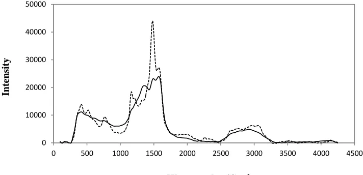
[image:4.596.186.424.90.241.2]The Raman spectra recorded for PANI and streptavidin/PANI are presented in Figure 2. The Raman spectrum of PANI show bands attributed to N-H group (1500 cm−1). For PANI, Raman scattering intensity at 1450 cm-1, related to the C-N group. Also, streptavidin displays a number of characteristic bands, in the 300-1750 cm-1 spectral range related to amino acid groups [13]. This spectrum proves that, the streptavidin was successfully immobilized on PANI/GC electrode.
Figure 2. Raman spectra for PANI (—) and streptavidin/PANI (…).
0 10000 20000 30000 40000 50000
0 500 1000 1500 2000 2500 3000 3500 4000 4500
Inte
ns
it
y
[image:4.596.116.472.493.665.2]3.2. The investigation of conductivity by Cyclic Voltammetry
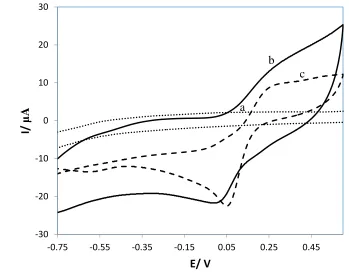
Cyclic voltammograms (CVs) were used to follow the electron conductivity of the bare GC, PANI/GC and bacteriophage/streptavidin/PANI/GC electrodes (Figure. 3a, 3b and 3c respectively). In this experiment, K3[Fe(CN)6] solution in PBS (0.1 M) is applied as redox probe. As shown in Fig. 3, the CVs of bare and modified electrodes, was seen in the potential from -0.75 to 0.5 V. The bare GC hasn't any pair redox peaks. The value of formal potential [E0=(Epc+Epa)/2] for PANI/GC and bacteriophage/streptavidin/PANI/GC modified electrodes were 0.2 and 0.15 V, respectively. This value of formal potential shows that, when the electrode surface immobilized by PANI, the current intensity was increased significantly. Since, PANI/GC electrode possess high conductivity, which could improve electron transferring activity between the electroactive species and the electrode surface in the solution[14]. Also, when the oriented biotinylated bacteriophage as an insulator, immobilized on PANI/GC electrode the current intensity was decreased.
Figure 3. Comparison between CVs. GC electrode (a), streptavidin/PANI/GC electrode (b) and bacteriophage/streptavidin/PANI/GC electrode (c) CVs is recorded in PBS solution (0.1 M, pH =7.2) with scan rate of 100 mV/s at air saturated condition.
3.3. Equivalent circuit fit for biosensor
Figure 1 illustrates the correlation between each immobilized layer on the GC electrode with the capacitance intensity. The insulating self-assembled monolayer pattern on the GC electrode caused a constant capacitance and prevents any faradic processes and the capacitance got the highest level (Figure 1). According to the image illustrated in Scheme 1, two planar bacteriophage modified GC electrodes stands as two capacitor plates and the buffer between them as an insulating layer. By
-30 -20 -10 0 10 20 30
-0.75 -0.55 -0.35 -0.15 0.05 0.25 0.45
I/
μ
A
E/ V
a b
[image:5.596.51.529.120.559.2] [image:5.596.119.468.285.561.2]developing each layer on the bacteriophage modified GC electrodes, a new capacitor value is reached by different dielectric coefficient, thickness and surface area. Therefore, by attachment of E. coli bacteria on the left and right bacteriophage modified GC electrodes (C1 and C2), the total capacitance was changed[15]. As presented in Figure 1, the total capacitance (Ct) at this level can be calculated through Eq. 1:
1/Ct=1/C1+1/C2 (1)
3.4. Calibration curve for E coli biosensor
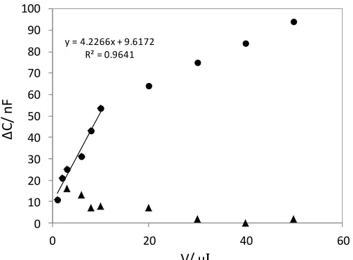
For measuring of E. coli the capacitance method was used. Figure 4 demonstrate the association of the change of capacitance besides E. coli number by bacteriophage modified GC electrodes. It is obvious that, the bare GC (without bacteriophage) fixed on electrodes do not show any response to E. coli concentration change. Also, oriented bacteriophage modified GC electrodes, has high sensitivity and wide concentration range, because modified electrode with bacteriophage immobilization creates a high sensitivity to area ratio for capacitance storage. In bacteriophage modified GC electrodes, E. coli was detected in a linear range from 3.2 × 10⁵ to 32 × 105 N/mL by the correlation coefficient of 0.998. The detection limit (DL) of this biosensor was calculated at signal to noise ratio of 3 (S/N=3) according to the Eq.2:
DL= 3.3 σ/S (2)
In the Eq. 2, σ stands for standard deviation of the response and S presents the slope of calibration curve [16]. According to Eq. 2, the detection limit was calculated equals to 12 N/mL. Finally, the analytical parameters of this biosensor were compared with other similar biosensors reported for detection of E. coli (Table 1).
y = 4.2266x + 9.6172 R² = 0.9641
0 10 20 30 40 50 60 70 80 90 100
0 20 40 60
Δ
C
/
nF
V/ μL
[image:6.596.171.426.524.712.2]Table 1. Comparison of the parameters obtained in this work with other reported in the literature
Species
Transduction Method Analytical
characteristics/sample
Ref
E.coli
EIS Antibody/polyaniline /Au electrode
Det.range102 CFU mL−1 of E. coli , detection limit of
107 CFU mL−1
[17]
E.coli
Amperometry DNA/Fe2O3@AuNP Det. range: up to 0.01 pM DNA
target; 500 cfumL_1; LOD: 5 cfumL
_1 /water
[18]
E.coli
DPV Cystine/amino probe DNA Det. range: 1×10_6– 1×10²⁰M pM DNA target; 60 s hybr.
time.
[19]
E.coli
Amperometry biotin/Strept-AP as reporter. LOD: 250 cfuµL_1 [20]
E.coli
DPV DNA/thiol/Au electrode LOD: 112 cfumL_1/milk [21]
E. coli
Capacitance bacteriophage/streptavidin/PA NI/GC electrode
Det:10-98 N/mL and 10 N/mL (N stands for number of E. coli), fast
response time of ∼1 s LOD: :20 Ml, aqueous
solution(PBS, PH=7)
D. Ashiani1, A. H. Keihan, J. Rashidiani 1, F. Dashtestani , K. Eskandari1 .
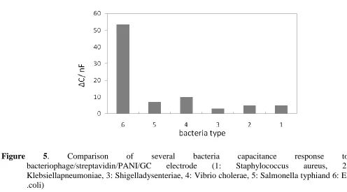
3.5. Selectivity of the E. coli biosensor
Figure 5. Comparison of several bacteria capacitance response to bacteriophage/streptavidin/PANI/GC electrode (1: Staphylococcus aureus, 2: Klebsiellapneumoniae, 3: Shigelladysenteriae, 4: Vibrio cholerae, 5: Salmonella typhiand 6: E .coli)
4. CONCLUSION
The investigation and preparation of biosensors based on E. coli is so essential for clinical diagnostic. Although biosensors with antibodies provide a quick and cheap method for detection of cells, spores, viruses and toxins, but they are limited in their specificity [22,23]. In addition, working with monoclonal antibodies requires the time and expertise related with the production of them [24]. Therefore, we propose inexpensive, label-free, fast, sustainable and renewable E. coli biosensors based on immobilization of oriented bacteriophage modified GC electrode and this electrode was used in capacitance method. Modified carbon electrode was caused to high capacitance signal by oriented bacteriophage was attached to the polyanilin surface [25].The oriented bacteriophage electrode presents convenient linear range, good detection limit and selectivity. The fabrication of this biosensor would be suitable for detecting other pathogens like E. coli or so on.
ACKNOWLEDGEMENTS
Authors are grateful to Nanobiotechnology Research Center, Baqiyatallah University of Medical Sciences for providing financial support to undertake this work.
References
1. D. Ivnitski, I. Abdel-Hamid, P. Atanasov, E. Wilkins, Biosensors and Bioelectronics,14 (1999) 599624.
2. M. Tietjen, D.Y. Fung, Critical reviews in microbiology, 21 (1995) 5383.
3. G.-J. Yang, J.-L. Huang, W.-J. Meng, M. Shen, X.-A. Jiao, Analytica chimica acta,647 (2009) 159166.
5. A. Mortari, L. Lorenzelli, Biosensors and Bioelectronics, 60 (2014) 821.
6. R. Singh, M.D. Mukherjee, G. Sumana, R.K. Gupta, S. Sood, B. Malhotra, Sensors and Actuators B: Chemical, 197 (2014) 385404.
7. R.A. Weiss, A.J. McMichael, REPRINT H, Health of People, 10 (2015) 431.
8. E.E. Gill, O.L. Franco, R. Hancock, Chemical biology & drug design, 85 (2015) 5678. 9. J. Wang, G. Rivas, X. Cai, Electroanalysis, 9 (1997) 395398.
10.C. Xiao, F. Jiang, B. Zhou, R. Li, Y. Liu, Science China Chemistry, 55 (2012) 19311939. 11.V. Singh, P. Jain, S. Dahiya, African Journal of Microbiology Research, 9 (2015) 18321839. 12.V. Romanov, V. Gribova, I. Galelyuka, O. Voronenko, International Journal" Information
Technologies & Knowledge" ITHEA.–Sofia, Bulgaria, 9 (2015) 310. 13.C. Fagnano, A. Torreggiani, G. Fini, Biospectroscopy, 2 (1996) 225232.
14.F. Dashtestani, H. Ghourchian, K. Eskandari, H.-A. Rafiee-Pour, Microchimica Acta, 182 (2015) 1045-1053.
15.E. Alipour, H. Ghourchian, S.M. Boutorabi, Analytical Methods, 5 (2013) 44484453. 16.A.M. Committee, Analyst, 112 (1987) 199204.
17.A.D. Chowdhury, A. Dea, C.R. Chaudhuri, K. Bandyopadhyay, P. Sen, Sensors and Actuators B, 171172 (2012) 916.
18.K. Li, Y. Lai, W. Zhang, L. Jin, Talanta, 84 (2011) 607.
19.C.M. Pandey, G. Sumana, K.N. Sood, B.D. Malhotra, Thin Solid Films, 519 (2010) 1178. 20.A. Walter, J. Wu, G.U. Flechsig, D.A. Haake, J. Wang, Analytica Chimica Acta, 689 (2011) 29. 21.C. Luo, Y. Lei, L. Yan, T. Yu, Q. Li, D. Zhang, S. Ding, H. Ju, Electroanalysis, 24 (2012) 1186. 22.J. Amani, S.A. Mirhosseini, A.A.I. Fooladi, Jundishapur journal of microbiology, 8 (2015). 23.T. Mirski, M. Bartoszcze, A. Bielawska-Drozd, P. Cieslik, A.J. Michalski, M. Niemcewicz, J.
Kocik, K. Chomiczewski, Annals of Agricultural and Environmental Medicine, 21 (2014).
24.P. Leonard, S. Hearty, J. Brennan, L. Dunne, J. Quinn, T. Chakraborty, R. O’Kennedy, Enzyme and Microbial Technology, 32 (2003) 313.
25.M. Janczuk, J. Niedziółka-Jönsson, K. Szot-Karpińska, Journal of Electroanalytical Chemistry, (2016).