GUTTA PERCHA, RESILON AND CPOINTS FOR
RETREATMENT USING TWO DIFFERENT ROTARY
RETRIEVAL SYSTEMS - AN INVITRO STUDY
Dissertation submitted to
The Tamil Nadu Dr M.G.R. Medical University
In Partial fulfillment of the degree of
MASTER OF DENTAL SURGERY
BRANCH IV
CONSERVATIVE DENTISTRY
AND ENDODONTICS
This is to certify that this dissertation titled “A comparative evaluation of retrievability of Gutta percha, Resilon and CPoints for retreatment using two different rotary retrieval systems - an invitro study” is a bonafide record of the work done by Dr. Aswathy Prasad, under our guidance during her post graduate study during the period of 2014-2017 under THE TAMILNADU Dr. M.G.R. MEDICAL UNIVERSITY, CHENNAI, in partial fulfillment for the degree of MASTER OF DENTAL SURGERY IN CONSERVATIVE DENTISTRY & ENDODONTICS, BRANCH IV. It has not been submitted (partial or full) for the award of any other degree or diploma.
Dr. RAJESH S, M.D.S Dr. MANO CHRISTAINE ANGELO, M.D.S
DECLARATION
I hereby declare that this dissertation “A COMPARATIVE EVALUATION OF RETRIEVABILITY OF GUTTA PERCHA, RESILON AND CPOINTS FOR RETREATMENT USING TWO DIFFERENT ROTARY RETRIEVAL SYSTEMS - AN INVITRO STUDY” is a bonafide record of work undertaken by me during the period 2014-2017 as a part of post graduate study. This dissertation, either in partial or in full, has not been submitted earlier for the award of any degree, diploma, fellowship or similar title of recognition.
Dr. Aswathy Prasad MDS Student
Dept. of Conservative Dentistry & Endodontics,
Sree Mookambika Institute of Dental Sciences
KULASEKHARAM
ENDORSEMENT BY THE PRINCIPAL/ HEAD OF THE
INSTITUTION
This is to certify that the dissertation titled “A COMPARATIVE EVALUATION OF RETRIEVABILITY OF GUTTA PERCHA, RESILON AND CPOINTS FOR RETREATMENT USING TWO DIFFERENT ROTARY RETRIEVAL SYSTEMS - AN INVITRO STUDY” is a bonafide research work done by Dr. ASWATHY PRASAD under the guidance of Dr. RAJESH S, M.D.S, Professor & HOD, Department of Conservative Dentistry and Endodontics, Sree Mookambika Institute of Dental Sciences, Kulasekharam.
Date: DR. ELIZABETH KOSHI; M.D.S
Principal
Sree Mookambika Institute of Dental Sciences,
VPM Hospital Complex, Padanilam,
Kanyakumari District,
You gave me life…You raised me with love and care…You stood by me when I was down… You gave me strength in my tough times… You lend me a hand when I was lost... You wiped my tears, when I was broken… you gave me the best in my life… I bow down in gratitude to the Almighty God
for giving me the best parents and teachers, and my dear ones.
Words are not enough to express my sincere gratitude towards all the
people who have helped me in completing this study. Without their guidance,
help, cooperation and encouragement I would not have made headway in this
study.
I take this opportunity to express my deep sense of gratitude to our
Chairman Dr. C.K.Velayuthan Nair, M.B.B.S, M.S., and to the Director
Dr.Rema.V.Nair, M.B.B.S., M.D, D.G.O, of our institution for their constant support and for providing facilities and infrastructure which was essential for me
to complete my study.
I would like to thank our trustees Dr. Vinu Gopinath, M.B.B.S, M.S, M.Ch and Dr. R.V.Mookambika, M.B.B.S, M.D, D.M., for their help and support to complete my study.
I take this opportunity to express my profound gratitude and sincere thanks
to my teacher and mentor, my guiding light Dr. Rajesh S, M.D.S. Professor, Guide and Head of the Department of Conservative Dentistry and Endodontics, for
his constant support for my dissertation work, motivation, enthusiasm and
guide Dr. Mano Christaine Angelo M.D.S, Department of Conservative Dentistry and Endodontics for his boundless patience, constructive criticisms,
timely help and constant advice all through my study as well as Post Graduation.
I would also like to acknowledge my heartfelt gratitude and respect to
Dr.Vijay Mathai M.D.S, Professor, Department of Conservative Dentistry and Endodontics for his valuable information, brilliant advices and consistent
encouragement throughout my study as well as Post Graduation.
I would like to express my gratitude to Dr. Vineet R.V. M.D.S and
Dr.Sarah Christopher M.D.S, who was my senior as well, as they have spared their valuable time for my thesis correction and was always approachable and
clarified my doubts and Dr. Vineesh K M.D.S Dr. Manu Unnikrishnan M.D.S, and Dr.Manoj Kumar T.S B.D.S for their valuable help and guidance.
I would also like to thank Dr. Sharad Shahai, Dr Ankita and the technician Mr Deepak Bhadur, of DMD Imaging and scanning center, Ghaziabad, Uttar Pradesh, for their help in the imaging and volume analysis for
my study.
I would like to extend my sincere gratitude to Mr. Sarath Babu K, M.Sc. (Medical) Pharmacology, Assistant Professor, Department of Pharmacology, Sree
Mookambika Medical College, who is a very friendly and approachable person,
for helping me with the statistical analysis of the study.
Dr.Betty Babu M.D.S, and Dr. Rahul for their timely advice and motivation. Without my seniors, I wouldn’t have been able to make my Post graduation life a
pleasant and unforgettable one. They helped me whenever I got worried in
academics as well as clinics and they were too perfect to be a senior and friend.
I would like to thank my batch mate Dr. Rejoy John for being a perfect co-postgraduate and friend in my post-graduation life and my junior colleagues
Dr. Sreelekshmy V.J, Dr. Soumya T.S, Dr Priya and Dr Sanjana for their ever-ready help and encouraging words. I appreciate all my friends who made
these three years memorable.
I would like to thank all the teaching and non-teaching staff for their help
and support throughout these three years.
Last but not the least I would like to thank my parents, my sister and my
husband and my dear daughter, for believing in me, for their unconditional love,
sacrifice and their emotional support in all my endeavours. Without whom I could
not have accomplished this.
Sl.No. INDEX PAGE NO
1. List of Abbreviations i
2. List of Tables ii - iii
3. List of Figures iv - vii
4. Abstract viii- x
5. Introduction 1 – 6
6. Aims & Objectives 7
7. Review of Literature 8 – 28
8. Materials & Methods 29 - 36
9. Results & Observations 37- 44
10. Discussion 45 - 65
11. Summary & Conclusion 66 – 68
12. Tables xi - xviii
13. Figures xix- xxxix
i
ANOVA – Analysis of Variance K file -Kerr file BC – Bioceramic Mm – millimeter CBCT – Cone Beam Computed
Tomography
NiTi – Nickel Titanium
EDTA – Ethylene diamine tetra acetic acid
NaOCl – Sodium hypochlorite
Eg – example Protaper UR – Protaper Universal Rotary files
Etc – etcetera Protaper R – Protaper Universal retreatment files
Fig – figure SE – Self Etch
GP – gutta percha SEM – Scanning Electron Microscope
HEMA – Hydroxyethyl methacrylate
SPSS – Statistical Package for Social Sciences
ii
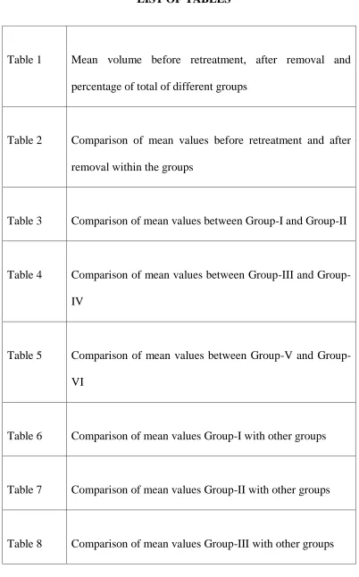
Table 1 Mean volume before retreatment, after removal and percentage of total of different groups
Table 2 Comparison of mean values before retreatment and after removal within the groups
Table 3 Comparison of mean values between Group-I and Group-II
Table 4 Comparison of mean values between III and Group-IV
Table 5 Comparison of mean values between V and Group-VI
Table 6 Comparison of mean values Group-I with other groups
Table 7 Comparison of mean values Group-II with other groups
iii
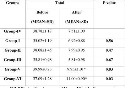
Table 9 Comparison of mean values Group-IV with other groups
Table 10 Comparison of mean values Group-V with other groups
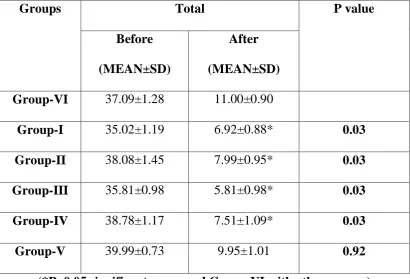
Table 11 Comparison of mean values Group-VI with other groups
Table 12 Multiple comparisons of mean values between the groups
iv
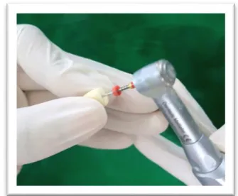
Fig 1 Armamentarium
Fig 2 Sixty extracted single rooted mandibular premolar teeth Fig 3 Decoronation
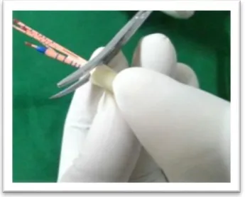
Fig 4 Working length determination
Fig 5 CBCT – CS9300 equipment (Carestream Healthcare India Pvt. Ltd.)
Fig 6 a Tooth preparation with Sx Protaper file Fig 6 b Tooth preparation with S1 Protaper file Fig 6 c Tooth preparation with S2 Protaper file Fig 6 d Tooth preparation with F1 Protaper file Fig 6 e Tooth preparation with F2 Protaper file Fig 7 a AH Plus Sealer dispensed on mixing pad Fig 7 b Sealer mixing
Fig 7 c Guttapercha Mastercone placement coated with sealer Fig 7 d Lateral condensation
Fig 7 e Completed obturation
Fig 8 a Dispensed sealer – RealSeal SE
Fig 8 b Resilon Master cone placement coated with sealer Fig 8 c Lateral condensation
v
Fig 9 a Dispensed sealer
Fig 9 b F2 size - verifier and Cpoint
Fig 9 c Application of Endosequence BC sealer Fig 9 d Check for fit with verifier
Fig 9 e Single cone obturation with CPoint and Endosequence Fig 10 CBCT image of group I before retreatment
Fig 11 CBCT image of group II before retreatment Fig 12 CBCT image of group III before retreatment Fig 13 CBCT image of group IV before retreatment Fig 14 CBCT image of group V before retreatment Fig 15 CBCT image of group VI before retreatment Fig 16 Protaper retreatment files – D1, D2, D3
Fig 17 a, b, c Retreatment of group I with Protaper retreatment files Fig 18 a, b, c Retreatment of group III with Protaper retreatment files Fig 19 a, b, c Retreatment of group IV Protaper retreatment files Fig 20 Mtwo retreatment files – R1 and R2
vi
Fig 26 CBCT images of group III after removal Fig 27 CBCT images of group IV after removal Fig 28 CBCT images of group V after removal Fig 29 CBCT images of group VI after removal
Fig 30 Graphical representation of Mean before retreatment, after removal and percentage of total of different groups Fig 31 Graphical representation of Comparison of mean values
before retreatment and after removal within the groups Fig 32 Graphical representation of Comparison of mean values
between Group-I and Group-II
Fig 33 Graphical representation of Comparison of mean values between Group-III and Group-IV
Fig 34 Graphical representation of Comparison of mean values between Group-V and Group-VI
Fig 35 Graphical representation of Comparison of mean values Group-I with other groups
Fig 36 Graphical representation of Comparison of mean values Group-II with other groups
vii
Group-IV with other groups
Fig 39 Graphical representation of Comparison of mean values Group-V with other groups
Fig 40 Graphical representation of Comparison of mean values Group-VI with other groups
Fig 41 Graphical representation of Multiple comparisons of mean values between the groups
viii
Introduction
The success of endodontic treatment depends on thorough debridement
of canal system, of infected or necrotic pulp tissue, microorganisms and
complete sealing of the canal space, thus preventing persistence of infection
and/or reinfection of the pulp cavity. A number of endodontic failure cases are
being reported to the dental clinics which require surgical or nonsurgical
retreatment. Nonsurgical retreatment requires regaining access to the entire
root canal system through complete removal of the core filling material and
sealer. For retreatment to be practical, the material should be removable from
the root canal space.
Aims and objectives
To compare the removal ability of Gutta percha, Resilon and CPoints,
for retreatment, using two rotary retreatment systems, Protaper and Mtwo and
comparing volume percentage of residual obturating material in the root canal
walls, after removal, using the Cone Beam Computed Tomography images.
Methodology
Sixty freshly extracted single rooted mandibular premolars which were
extracted for orthodontic purpose, were collected and were decoronated at the
cemento-enamel junction, approximately 15mm from apex. They were
ix
using Protaper rotary files and obturation of root canals using Gutta percha,
Resilon and CPoints, using their corresponding sealers, were performed for
twenty teeth each. The obturation material was removed with Protaper rotary
retreatment files for 10 teeth each from each group obturated with gutta-percha,
Resilon and CPoint. The rest were removed using Mtwo rotary retreatment
files. All teeth, after removal of obturation material was analysed using Cone
Beam Computed Tomography, for checking the cleanliness of root canals and
the remaining percentage volume of obturation material from each group was
calculated and compared.
Results and observations
The analysis of the results show that the mean volume percentage of the
residual material is least for RealSeal group with the removal by Protaper
retreatment file(16.35±2.69 %) and the highest volume percentage of residual
filling material was seen in the groups obturated by CPoint and removed by
Mtwo retreatment file system (29.67±2.34 %).
Conclusion
In this study, it is well demonstrated that the canals obturated with
CPoints were least retreatable and those with RealSeal were the easiest ones to
x
Clinical significance
Many retreatment cases are being reported to our clinics nowadays.
Root canal treatment failures occur when the treatment falls short of the
acceptable standards. It is usually associated with procedural errors in infected
tooth. It may be due to intra-radicular or extra-radicular infections or due to
overfilling of root canals or inadequate coronal seal.Retreatment has to be done
in such cases and hence the obturating materials used must be retrievable from
the root canals. It is a consensus that all filling material must be removed
during retreatment thus providing adequate disinfection of the root canal
system and favours conditions for new filling.
The three different materials in this study shows three different sealing
efficiency which affects the retrievability of the material from root canal.
Hence the retrievability of each obturating material from the canal walls was
evaluated by measuring the percentage volume of residual root canal obturating
material to the total volume of root canal after the obturating material is
removed by two retreatment file systems.
Keywords: CPoint, Guttapercha, Resilon, Retreatment, Protaper retreatment
1
The success of endodontic treatment depends on thorough debridement of root canal system, of infected or necrotic pulp tissue, microorganisms and complete sealing of the canal space, thus preventing persistence of infection and reinfection of the pulp cavity.1 The clinical success rate of endodontic treatment ranges between 50-90 %.2-3 This variability in the success rate of root canal treatment is determined by various factors like age of the patient, the tooth which is being treated, alterations in the natural anatomy of root canal by procedural errors, the ability in removal of coronal restoration for proper access to the pulp chamber and the ability to repair the pathologic and iatrogenic errors. Preoperative perforation, apical periodontitis and the quality of the filling material also determine the outcome of endodontic treatment.4,5,6 Numerous factors contribute to endodontic treatment failure which includes positive culture, broken instruments, canal over fillings, mechanical perforations, root fractures, presence of periradicular lesions and periodontal disease. A number of studies have also reported that endodontic treatment failures are greater in teeth that are associated with pre-treatment periradicular radiolucencies than in teeth without them.7
2
engine driven rotary files followed by cleaning, shaping and reobturation.8 Post-treatment disease might persist or emerge because of persistent bacteria in the root canal system as a consequence of insufficient cleaning, untreated canals, inadequate filling or coronal leakage.9 Cross-sectional studies have demonstrated persistent apical periodontitis associated with over 45% of endodontically treated teeth. Persistent apical periodontitis is caused mainly by root canal bacteria that survive primary treatment.10 It can be treated by means of orthograde retreatment. The primary purpose of the retreatment therapy is adequate cleaning and disinfection of the root canal system. During retreatment, it is essential to remove all the filling material, that hinders contact of irrigating solutions and intra canal dressings with the root canal walls, so that the residual microbial population can be eliminated and create favourable conditions for periradicular healing.11 To allow retreatment when indicated, the obturating material should be removable.10,11
Most widely accepted root canal filling material is gutta-percha (GP) in conjunction with a variety of sealers. However it has many drawbacks like poor sealing ability and its inability to further strengthen the teeth and thus reducing susceptibility to fracture. AH Plus is a thermoplastic, two-component paste root canal sealer based epoxy-amine resin, which permits removal of the material, if necessary, hence the most preferred.9,10,11
3
and was introduced in 2004. Many studies have proved that the performance and handling properties of Resilon is similar to that of Gutta percha.9,10,11,12 Resilon bonds to dentinal walls when used in conjunction with its root canal sealer, Epiphany/Real Seal SE and forms a ‘monoblock’ within the canal.13, 14 This prevents the chances of microleakage between core material-sealer interface and the sealer-dentin interface.15 The Resilon sealer is a dual-curable dental-resin composite sealer, which has a total filler content of about 70% of its weight, allowing its easy removal in retreatment cases. The fillers include calcium hydroxide, bariumsulfate, barium glass and silica. Resilon cones come in a range of sizes similar to gutta-percha cones. The Resilon bonding agent is a self-etching primer that contains sulfonic-acid terminated functional monomer, HEMA, water and a polymerization initiator.16
4
axially to adapt to the ramifications of the root canal. When hydrated in the root canal, CPoints expand, conforming to canal irregularities and pressing the companion hydrophilic sealer, into concavities, lateral portals of exit and the tubules of the dentin walls.18
K files and H files took longer time and were ineffective in the complete removal of obturation materials in the root canal.25,30,32 Ni-Ti files have the disadvantage of higher incidence of file fracture and apical extrusion of debris. Hence to overcome these drawbacks newer rotary retreatment systems like Protaper universal retreatment files were introduced.19,20 They have been used increasingly in root canal preparation and to remove filling materials because of their unique physical properties, resilience, high safety and cutting efficiency, providing efficient removal of obturation materials, maintenance of canal shape and shorter working time with limited usage of solvents.19,20
Protaper universal files with its rounded tip and increased flexibility makes it a good rotary file system for biomechanical preparation of root canals. It has three files for coronal preparation (SX, S1, S2) and five finishing files (F1, F2, F3, F4, F5) for apical preparation.11
5
middle third, has a length of 18mm and tip of 0.25mm with 0.08% taper. The D3 is used in apical third, has length of 22mm and tip of 0.20mm with 0.07% taper.11
The Mtwo instruments have an S-shaped cross-section, an increasing pitch length in the apical-coronal direction. The Mtwo retreatment kit consists of only two instruments with cutting tips, i.e., Mtwo R1 15/.05 and Mtwo R2 25/.05, designed to reach the apex. They too have an S-shaped cross-section as do the files of the basic sequence, but a shorter pitch length to enhance the advancement of the file into the filling material. These instruments are characterized by two cutting edges, which are claimed to cut dentine effectively. They also have the advantage of shaping the root canal in an under-prepared tooth, simultaneously.9
6
In the present study, the removal ability of Gutta percha, Resilon and C-point using Protaper retreatment files and Mtwo retreatment files is evaluated, by analysing the amount of remaining obturation material in the root canal after removal, using CBCT.
7
Aim
To compare the removal ability of Gutta percha, Resilon and CPoints, for retreatment, using two rotary retreatment systems – Protaper and Mtwo.
Objectives
1. To observe the removal ability of Gutta percha, Resilon and CPoints from root canals by two rotary retreatment file systems, after obturation. 2. To compare the volume percentage of residual obturating material in the
8
Bergenholtz et al.197922 studied the effects of endodontic retreatment on quality of seal and periapical healing and concluded that nonsurgical retreatment should be attempted if necessary before proceeding for surgical intervention.
Wilcox LR et al.19871 compared the retrievability of gutta-percha along
with different sealers and found out that none of the materials were completely removable from the canals.
Gilbert BO et al.198723in their review article has emphasised the need to remove the previous obturation material as the initial step, which when successfully accomplished, provides access to the root canal system so that treatment objectives can be carried out successfully.
Hülsmann M et al.199724 evaluated the efficacy of different methods
for gutta-percha removal and concluded that cleaner canals were achieved with the use of Hand files compared to the rotary methods using Gates Glidden.
Sae-Lim V et al.200025 studied the effectiveness of ProFile (.04 taper) rotary instruments in removal of obturating material and concluded that rotary instrumentation has been shown to be more effective than hand files in removing gutta-percha.
Imura N et al. 200026 compared the ability of two engine driven
9 using a stereomicroscope to assess the remaining filling residue and found that no method was effective in achieving a clean canal
Hülsmann M et al.200427 evaluate the efficacy, cleaning ability and safety of FlexMaster, GT Rotary, Protaper and Hedström files with and without solvents and found out that Protaper and Flexmaster were comparatively better than other system and the use of solvent allows easy and faster removal of obturation material.
Ezzie E et al.200628 studied the effect of retreatment techniques for a
resin-based root canal obturation material and inferred that Resilon is easily removable compared to gutta-percha
de Oliveira DP et al.200629 compared the remaining filling material and working time when removing gutta-percha/AH 26 and Resilon/Epiphany from root filled extracted teeth using chloroform and two different rotary systems (K3 and Liberator files). They came to the conclusion that Resilon/Epiphany was effectively removed with K3 or Liberator rotary files than gutta-percha.
10
Schirrmeister JF et al.200631 compared the effectiveness of hand files
and FlexMaster, Protaper, and RaCe rotary instruments for removing gutta-percha in curved root canals during retreatment by measuring the area of remaining obturation material and found that RaCe system is an efficient and safe in removing gutta-percha from curved root canals.
de Carvalho Maciel AC et al. 200632compared automated and manual
instrumentation techniques for removing filling material from root canal walls during root canal retreatment and deduced that photomicrographic method by epiluminescence was more effective than radiographic method and that rotary system was more effective than manual ones.
Gergi R et al.200733 analysed the efficacy of Protaper files and EndoRace files in gutta-percha removal and concluded that neither of the methods attained clean canals.
Cunha RS et al.200734 assessed the obturation removal and
re-instrumentation working time of canals filled with Resilon/Real Seal in comparison with canals filled with gutta-percha/AH Plus and inferred that Resilon was better removed from the canal than the gutta-percha cones and the AH Plus regardless of time factor.
11
Hassanloo A et al. 200736 in a study assessed the efficacy of retreatment
of canals filled with the Epiphany System with and without solvent, with reference to the extent of canal enlargement during retreatment. They concluded that Epiphany System was retreatable with and without chloroform, as compared to gutta-percha and AH Plus sealer.
Saad AY et al.200737 evaluated the effectiveness of removal of
Gutta-percha and its sealer by rotary instruments, Protaper and K3 in comparison with hand instrument, Hedstrom files by comparing the time required for removal and the apical extrusion of debris and came to the conclusion that Protaper and K3 took less time for removal than Hedsrom files and noticed not much difference in the apical extrusion of debris in any of the systems.
Govila S et al. 200738 in a review article summarised the application of
Cone beam computed tomography in acquiring three-dimensional images of periapical lesions and, mandibular canal. The review states that three dimensional anatomic and pathological observations improve the treatment outcome as well, since it allows single exposure there is slight decrease in the radiation exposure. Its inherent quickness in volumetric data acquisition and potential for reducing the cost of CT also has been mentioned.
12 system proved to be an efficient method of removing GP and sealer from maxillary anterior teeth.
Giuliani V et al.200839 studied the performance of the Protaper Universal System rotary retreatment system and of Profile 0.06 and hand instruments (K-file) in the removal of Gutta-percha and proved the rotary instruments to be fast and efficacious than hand instruments even though no system showed complete removal of obturation material.
Somma F et al.200840 in a study compared the effectiveness of the
Mtwo R, Protaper retreatment files and a Hedström manual technique in the removal gutta-percha, Resilon and EndoRez during retreatment and concluded that all instruments left remnants of filling material and debris on the root canal walls irrespective of the root filling material used. The use of retreatment Ni-Ti rotary files to remove filling material quickly should be followed by hand instrumentation to refine and complete its removal and to obtain better canal wall cleanliness especially in the apical third further increasing the size of apical preparation. They also found out that, to minimize apical extrusion of debris, Ni-Ti rotary instruments should be used 1 to 2 mm short of the working length.
13 faster removal than gutta-percha/AH Plus using a Gates Glidden drill with or without chloroform in both straight and curved canals.
Hammad M et al.200841 measured the remaining volume of different filling materials like gutta-percha and TubliSeal sealer, EndoRez points and EndoRez, RealSeal points and RealSeal sealer, gutta-percha point and GuttaFlow sealer after removing with Protaper retreatment files or hand K-files and they concluded that all tested filling materials were not completely removed during retreatment by using hand or rotary files and gutta-percha was more proficiently removed by using hand K-files.
Taşdemir T et al.200842 compared the efficacy of three rotary
instruments Protaper, R-Endo, Mtwo and hand instrument Hedström files, to remove gutta-percha and AH Plus sealer and identified that complete removal of materials did not occur with any of these instrument systems where as Protaper left less material when compared to the Mtwo instruments
Taşdemir T et al.200843 compared the ability of Mtwo and Mtwo
retreatment files in removing gutta-percha and Resilon and found that there was no significant difference between the two removal methods.
14
Só MV et al.200845 evaluated the efficacy of Protaper Universal rotary
retreatment system and hand files for filling material removal during retreatment and the influence of sealer type on the presence of filling debris in the re-instrumented canals and found out that none of the methods achieved clean canals and the apical third of canals to be the least retreatable area of canal.
Schäfer E et al 200846 compared the performance of Alpha-File,
FlexMaster, Mtwo, ProFile and RaCe by means of a computer-driven testing device and discerned that Mtwo and RaCe displayed the greatest cutting efficiency.
Çelik Ünal G et al.200947 compared the adeptness of conventional and ProFile, R-Endo or Protaper Universal retreatment files when removing gutta-percha root fillings in curved root canals and found out that Protaper retreatment file were less effective in removal of Gutta-percha than Profile and Hand files.
15 percha used with TubliSeal exhibited less voids and gaps than the other tested materials.
Anil Kumar G et al.200910 in this study evaluated the amount of residual filling material on the canal walls in root canals filled with gutta-percha or Resilon and retreated using the Protaper treatment files and inferred that even though gutta-percha and AH plus sealer do not adhere as well to the canal wall as epiphany, removal of epiphany left significantly less filling material than removal of gutta-percha and AH plus. The Resilon-Epiphany system is retreatable leaving cleaner dentinal walls in comparison to gutta-percha and AH Plus.
Zarei M et al. 200912 in a study to compare the retreatability of gutta-percha and Resilon, the amount of residual material and time of retreatment was determined in each group at three levels in the canal, using RaCe and came to conclusion that orthograde retreatment of Resilon was less efficient than that of gutta-percha leaving residual material in the canal.
16
Shrestha D et al.201049 in an article had reviewed the adhesive concept
of bonding of Resilon to root dentin and stated that it can be used as an alternative to gutta-percha even though the material lacks the required properties of an ideal root canal filling material. And also, mentioned that it is necessary to improve the mechanical and chemical properties of the material to achieve optimal bonding to root dentin, taking into consideration the heterogenicity in composition of dentin and the anatomical complexity. rotary system and gutta-percha took lesser time than for Resilon
Marfisi K et al.20109 evaluated the efficacy of Protaper Retreatment
files, Mtwo Retreatment files and Twisted Files in removal of gutta-percha and Resilon from straight root canals and proved that Mtwo Retreatment files required less time when compared to other instruments for removal of the root filling material. Removal of Resilon from the canal walls was significantly better than gutta-percha, irrespective of the rotary instruments used.
Khatavkar RA et al.201015 elaborates on the Resilon-Epiphany
obturation system and its bonding ability over gutta-percha making it a good obturating material and the paradigm shift in obturation with the introduction of newer obturation materials with improved properties than Gutta percha.
17 groups. In all the techniques, the apical third proved to be the least cleanable even though Endosolv R group achieved a comparatively cleaner surface.
Duarte MA et al.201051 compared the efficacy of manual and rotary Protaper retreatment files in removal of gutta-percha and its sealer with the help of microscopes and radiographs and concluded that the apical third of root canals was the most difficult to be cleaned and microscopic evaluation was comparatively efficient in the identification of remaining filling material in the canal.
Bramante CM et al.201052 evaluated the heat release, time required,
and cleaning efficacy of MTwo and Protaper Universal Retreatment systems in comparison with hand instrumentation and concluded that Protaper UR and MTwo R caused the greatest and lowest temperature increase on root surface, respectively even though all techniques left filling debris in the root canals
Luiz F F et al. 20118 ascertained the ability of ProFile, GT, Protaper,
Race and K3 rotary instruments compared with hand K-files for removal of gutta-percha during retreatment and concluded that GT, ProFile, Protaper and K3 were more effective in removing gutta-percha than manual and Hero instruments.
18 Retreatment system in the removal of filling material, regardless of the solvent solution used.
Reddy S et al. 201154 evaluated the cleanliness of root canal walls after retreatment using two rotary retreatment files (Protaper Universal retreatment system and R-Endo retreatment files) to hand instruments in severely curved canals obturated with gutta-percha and an epoxy resin based sealer and zincoxide eugenol based sealers and concluded that the R-Endo system removed the obturation material much better when compared to the Protaper retreatment file system, even though none of the systems used produced a thoroughly cleaned root canal.
Shanahan DJ et al.201155 has described Resilon obturation points after analysing various literatures related to the invitro studies with Resilon, mostly leakage-type studies. In this article, they have proposed Resilon as a replacement obturation material to the gold standard Gutta-percha obturation, considering its typical property of bonding to dentinal walls and ability to form ‘monoblock’ providing a perfect seal of root canal obturation.
Shenoy VU et al.201156 in a review article highlighted the advantages of Resilon over the traditionally used gutta-percha, emphasizing its adhesive property through monoblock formation.
Shwaliya et al.201157 compared the fracture resistance of teeth after
19 load at the fracture point revealed that Resilon system increased resistance to fracture than gutta-percha.
Al azzawi et al.201111 compared the removal efficacy of gutta-percha and Resilon using the Protaper retreatment files by visually examining the canals of split tooth and photographing and analysing in adobe photoshop software and found that canals obturated with gutta-percha had a cleaner canal when compared with Resilon obturated canals.
Marques da Silva B et al.201259 studied the ability of Protaper,
D-RaCe, and Mtwo retreatment files along with and without additional files of the respective systems and found out that there was no much difference in the removal of material even with the use of additional files.
Kfir A et al.201260 in a study evaluated the removal efficiency of
Gutta-percha and AH Plus using chloroform and Hedstrom files, SafeSider files, using a NiTi Peeso reamer and reciprocating file, with or without chloroform, or Protaper Universal retreatment files (D2, D3) with or without chloroform and assessed it using radiographs and microscopes. They concluded that none of the methods adopted created a clean canal and that the microscopic method of evaluation was superior to radiographic method in detecting the remaining filling material in the canal.
Kumar MS et al.201261 conducted a study to demontrate the efficacy of
20 required for removal of obturation material and found out that no technique achieved complete removal of material and Protaper was faster in removal compared to the hand file techniques.
Economides N et al.201262 compared the push-out bond strength of Smartseal with gutta-percha/AH26 to calculate the bond strength and found out that Smartseal and Gutta-percha showed similar bond strengths and adhesion to dentin irrespective of single cone or lateral condensation technique.
Mohebbi P et al.201263 – assessed the efficacy of Mtwo retreatment
files and Protaper retreatment files in removing the Resilon/Epiphany system with or without chloroform during retreatment and concluded that Protaper and Mtwo retreatment file systems were similar in removal efficacy for retreatment in the whole root canal system where as Protaper retreatment files along with solvents were more efficacious in removal of obturation material in the apical third of root canal.
Akhavan H et al.201264 did a microscopic evaluation of residual
gutta-percha and sealer in the root canal after removal using Mtwo and D-Race retreatment systems and inferred that both systems were equally efficient in removal of obturation material.
21 proved hand instrumentation to be better than the rotary systems in achieving cleaner canals.
Ma J et al. 201266 did a micro–computed tomography to estimate the amount of remaining root filling material in oval canals filled by gutta-percha in lateral condensation and continuous wave compaction using Protaper retreatment files with and without solvents and ascertained that none of the removal methods were efficient in obtaining clean canals and canals obturated with continuous wave compaction left more residue.
Dadresanfar et al.201267 compared the efficacy of Mtwo R and
Protaper retreatment files in removing the Resilon/Epiphany system with or without chloroform during retreatment using radiography; a stereomicroscope and SEM and found out that Protaper/solvent was better in the apical third; however, when considering the whole canal, Mtwo R and the Protaper D series had the similar efficacy.
Lotfi M et al.201368 reviewed existing literature from May 2004 to April 2012 which studied the physical and chemical properties of Resilon and they also reviewed some leakage studies to conclude that the resilon obturation material has the property to bind to the canal walls preventing leakage, also has antibacterial and antifungal properties, making it an acceptable material for obturation.
Pathivada L et al.201318 in an review article had introduced the newer
22 mentioned the hydrophilic property of this obturating points which can absorb surrounding moisture and expand resulting in filling of voids and spaces, helping in achieving a better seal of obturation.
Didato A et al.201369 studied the lateral hygroscopic expansion of CPoint with gutta-percha and found out that gutta-percha showed no time dependent expansion whereas CPoint did.22
Yadav P et al.201370 did an in vitro CT comparison of gutta-percha removal with two rotary systems and Hedstrom files by analyzing the images to calculate the remaining volume of obturation material in the canal after removal using Protaper retreatment files, Mtwo retreatment files or Hedstrom files and concluded that rotary files to be more efficient in removing obturation materials than manual files whereas no system was found to be effective in complete removal of the obturation material.
Wasnik et al.201371 compared the effectiveness in retreating
gutta-percha obturated root canals using Profile, with and without the aid of chloroform, to hand files with chloroform, with the help of, photograph after splitting the teeth longitudinally and came to the conclusion that although all the retreatment method were able to remove the obturation material, achieving clean canals were impossible.
İriboz E et al.201472 evaluated the effectiveness of the Protaper and
23 of resin based materials than gutta-percha and Protaper was faster in removal than Mtwo retreatment systems.
Arora S et al.201473 studied the homogeneity of filling in obturation of a novel polyamide polymer based obturating system, Smartseal system and Gutta-percha and sealer in simulated lateral canals. The study was done in calcified and decalcified samples of teeth where after obturation using Smart seal system and Gutta-percha they were observed using Cone beam computed tomography and digital radiography respectively, for the linear extension and area of the obturation material along with its sealer into lateral canals. They concluded that the polyamide polymer based obturation material showed better efficiency in obturation with the better adaptation and penetration of sealer into simulated lateral canals.
Dhillon JS et al.201474 demonstrated the efficacy of Pro Taper and Pro
Taper Retreatment instruments in the removal of gutta-percha during retreatment of straight root canals and found out that both Protaper files and Protaper retreatment files were equally effective in removing gutta percha during retreatment.
24
Asheibi F et al.201476 evaluated the effectiveness of ProTaper rotary
files with ProTaper retreatment and K-files in the removal of Resilon or gutta-percha (GP) from canals filled either by cold lateral condensation or thermal obturation using micro-CT. In roots filled with thermal obturation, Resilon was remaining more than gutta-percha and in obturation using thermal technique less material remained than cold condensation except Resilon retreated using ProTaper retreatment and K-files.
Niemczyk SP et al. 201477 in his article has explained the use of cone
beam computed tomography by illustrating the various clinical case scenarios, including anomalous root morphologies, additional canal spaces and insights into eccentric periapical pathologies. According to the study, the use of Cone beam computed tomography has become useful in retreatment situations where the normal anatomy of pulp chamber has been altered or destroyed and where conventional radiographs fail, for excavation in the proper dimension and direction to facilitate a more thorough treatment delivery.
de Azevêdo Rios M et al. 201478compared the retreatment efficiency of gutta-percha by two reciprocating rotary systems, WaveOne system and Reciproc system, in comparison to Protaper universal retreatment rotary systems, by measuring the remaining material in canal after removal, with the help of microscopic magnification and found out that both systems were as efficient as Protaper retreatment system in removing the obturation material.
Soares C et al.201579 reviewed articles published in the period from
25 root fillings and to compare the speed and efficacy of Resilon and gutta-percha root filling removal and concluded that the ProTaper (manual or rotatory) system in combination with chemical solvents is the most efficient method for removing Resilon root filling and retreatment of Resilon is more rapid and associated with less remnants of debris than Gutta-percha.
Prado RG et al.201580 compared the effectiveness of R-Endo, Mtwo
Retreatment, D-RaCe, and ProTaper by measuring the remaining material in the canal after removal and also the time for removal. They came to the conclusion that all systems were almost similar in cleaning the canal with the Mtwo system requiring lesser time for removal than R-Endo, D-RaCe, and ProTaper in sequence.
Gokturk H et al.201581 in a study evaluated the residual root canal
filling material after retreatment of root canals using stainless steel hand files, Mtwo R, R-Endo, ProTaper Universal Retreatment, and D-RaCe systems. They noticed the most residual gutta-percha and sealer in the apical third compared to the coronal and middle thirds. Nickel-titanium rotary retreatment instruments were faster than Hedström files in removal of obturation material, but had a higher risk of instruments fracture.
26 thirds. Retreatment of root canal filled with Real seal and Endo-Rez showed lesser remaining material than retreatment of gutta percha and AH plus sealer.
Hegde V et al.201583 compared the apical sealing ability of a novel Smart-Seal System, Resilon, and conventional Gutta-Percha system using a bacterial leakage model and concluded that the hydrophilic obturations of the root canal shows a better resistance to bacterial leakage as compared to hydrophobic obturations.
Hegde V et al.201584 evaluated the fracture resistance of roots obturated
with three hydrophilic systems - CPoint system, Resilon/Epiphany system, and EndoSequence BC sealer; and one hydrophobic gold standard gutta-percha/AH Plus system by recording the force required to fracture the obturation point from root canal, using a universal testing machine and concluded that all the hydrophilic obturation system showed higher fracture resistance than the hydrophobic gutta-percha obturation.
Singh R et al. 201585in a study conducted to compare the efficacy of R
endo retreatment files and Mtwo retreatment files with hand files, in removing gutta-percha from canals analysed using photograph, found that Mtwo and R Endo retreatment files to be more efficient than the hand instrumentation techniques.
Jaiswal et al. 201586 assessed the gutta-percha removal efficiency of
27 comparison with rotary files even though retreatment was possible in lesser time with rotary system.
Baig AR et al.201687 in a review article describred an ephemeral synopsis of smart seal, a hygroscopic obturating system with a reference of numerous studies associated with it. The sealing of root canal system is based on polymer technology with its hydrophilic property in which the material can absorb moisture from within the canal and associated sealer to expand within the canal system causing the adjunct sealer to be pushed in to the dentinal tubules and lateral canals which creates a better seal of the root canal space, thus making this material an acceptable material for root canal obturation.
Akbulut MB et al. 201688 studied the efficacy of Twisted File Adaptive, Reciproc, and ProTaper Universal Retreatment System instruments for retreatment using Cone beam computed tomography and also measuring the time required for removal with each systems and found out that root canal filling was more efficiently removed by using Reciproc and ProTaper UR instruments than TF Adaptive instruments and hand files and that ProTaper UR and Reciproc systems required shorter periods of time for retreatment.
28 retreated with Protaper retreatment systems and Mtwo retreatment system took lesser time for removal than Protaper retreatment systems.
29
MATERIALS USED IN THE STUDY
a. Saline - Baxter, India Pvt. Limited, Tamil Nadu, India.
b. 5.25% NaOCl - Azure Laboratories Pvt. Ltd., Maharastra, India. c. 17% EDTA - AvuePrep, Dental Avenue Pvt.Ltd., Maharastra, India. d. Protaper Gutta percha of F2 size and #20 & 25, 2% guttapercha
accessory cones - Dentsply Maillefer, Switzerland. e. AH Plus root canal sealer - Dentsply DeTrey, USA.
f. Resilon obturation points - #25, 6% and #20 & #25 2% as accessory cones - Sybron Endo, Orange, CA, USA
g. Real Seal SE sealer - Sybron Endo, Orange, CA, USA
h. CPoint – F2 size Propoint - EndoTechnologies, LLC, Shrewsbury, MA i. EndoSequence BC sealer - Brasseler, Savannah, Georgia, USA.
j. Cavit - 3M ESPE, Germany.
k. Paper points – Dentsply Maillefer, Switzerland.
Equipments/Instruments used in the study
a. Micromotor straight hand piece – NSK, Nakanishi, Japan. b. Diamond Disc – SS White, New Jersey, USA.
c. High Speed airotor hand piece – NSK, Nakanishi, Japan. d. Metal Scale – Marsman, India.
30
g. K files - No. 10, 15 - Mani Dental. Inc., Japan. h. X-Smart Plus – Dentsply, Maillefer, Switzerland.
i. Protaper NiTi rotary instruments SX, S1, S2, F1, F2 - Dentsply, Maillefer, Switzerland.
j. Spreaders 15, 20, 25 - Mani Dental. Inc., Japan.
k. Protaper retreatment file – D1, D2, D3 - Dentsply Maillefer, Switzerland.
l. MTwo retreatment files – R1, R2, R3 - VDW, Munich, Germany. m. CBCT – CS9300 equipment - Carestream Healthcare India Pvt. Ltd.
Inclusion criteria
- Single rooted mandibular premolars, freshly extracted for
orthodontic purposes, stored in saline and used within three months of extraction.Exclusion criteria
- Fractured teeth, more than one root canal, resorption, open apices, caries, obturated teeth, curved rooted teeth.METHODOLOGY
Specimen preparation
31
signs of internal resorption, calcification or previous endodontic therapy or caries, restoration or presence of dentin pins. Soft tissue and calculus were removed mechanically from the root surface. They were decoronated using a diamond disc (SS White, New Jersey, USA) to attain a 15mm. root length. Working length were determined with size #15 K-file (Mani Dental. Inc., Japan), by inserting the file into the canal until the tip of the file is just visible at the apical foramen and reducing 1mm from this length from coronal reference point to the tip. In all teeth root canal treatment was initiated. Root canal cleaning and shaping was done in crown down technique using Protaper Ni-Ti rotary system (Dentsply, Maillefer, Switzerland). Patency of the canal was maintained throughout the procedure by passing #10 K-file (Mani Dental. Inc., Japan) approximately 1mm through the apex.9,10,11
32
Groups used in the study
Tooth were randomly selected and divided into six groups of 10 teeth each.
Group I - obturated with Gutta percha and AH Plus and retreatment with Protaper retreatment file system
Group II - obturated with Gutta percha and AH Plus and retreatment with Mtwo retreatment file system
Group III - obturated with Resilon and Real Seal SE and retreatment with Protaper retreatment file system
Group IV - obturated with Resilon and Real Seal SE and retreatment with Mtwo retreatment file system
Group V - obturated with CPoint and Endosequence BC sealer and retreatment with Protaper retreatment file system
Group VI - obturated with CPoint and Endosequence BC sealer and retreatment with Mtwo retreatment file system
33
Real Seal SE sealer (Sybron Endo, Orange, CA, USA) with a previously selected Resilon master cone (Sybron Endo, Orange, CA, USA), lateral condensation was followed with accessory cones (Sybron Endo, Orange, CA, USA), immediately light-cured for 40 seconds and the excess is trimmed off using a bur.11,14,29 In Group V and Group VI canals were filled using the CPoint (EndoTechnologies, LLC, Shrewsbury, MA) self-sealing water expandable obturation pointalong with Endosequence BC sealer(Brasseler, Savannah, Georgia, USA.), after cleaning and shaping, without drying the canal17,18. Hydration from sealer and dentin-derived moisture helps the CPoints expand, conforming to canal irregularities, pressing the companion hydrophilic bioceramic sealer, Endosequence BC sealer into concavities, lateral portals of exit and the tubules of the dentin walls.108 Then the excess is trimmed off using a bur. The teeth were radiographed to confirm the adequacy of the root filling. After placing a temporary restoration of Cavit (3M ESPE, Germany), each tooth stored in a humidor at 37°C for 2 weeks to allow the sealer to set completely.9,11
CBCT Image Acquisition & Processing
All the specimens were imaged using Sirona CBCT machine from DMD imaging system, Dental and Maxillofacial diagnostics, Ghaziabad, Uttarpradesh.
34
kV, 2.5mA. A single scout image, i.e. lateral view was taken in accordance with the teeth position, and a 360° scan was acquired afterwards. The total scan time was 20s. The time required for the reconstruction of volumetric images after the sample's complete exposure was approx. 1 min.
Study images were reconstructed from the volumetric dataset, in planes perpendicular to the selected tooth axes. True and oblique axial, coronal and sagittal images with a thickness of 0.09mm and an interval of 0.09mm were obtained.
Obtained images were viewed using CS 3D imaging v3.5.7 software, Carestream Health. Inc, Internal Version 3.5.7.0(10/10/2014). Image assessment performed by a calibrated orthodontic post-graduate student and verified by a maxillofacial radiologist using the CBCT software tools.
For the assessment of volume of the root canal cavity in pre-treatment scan and residual endodontic restorative material in post-treatment scan, the scans were co-ordinated in all the three planes along the long axis of the tooth i.e. coronal, sagittal and axial plane to minimize any error.
Retreatment technique
:35
each type of instrument. For all teeth from Group I, III and V retreatment by removal of obturation materials were initiated using Protaper retreatment files (Dentsply Maillefer, Switzerland.), used in a brushing action according to the instructions of the manufacturer - D1 (30/.09) for removing materials from the coronal third, D2 (25/.08) for removing materials from the middle third, and D3 (20/.07) for removing material from the apical third.11,20 The working length were regained gradually using a pecking motion. The obturation materials from all the teeth from remaining groups – II, IV and VI were removed using Mtwo retreatment files (VDW, Munich, Germany) in a simultaneous technique to the working length until the wall of canal is smooth and clean till size R2 (size 25, 0.05 taper) in a brushing action with lateral pressing movements. Progression of the rotary file were performed by applying slight apical pressure and frequently removing the files to inspect the blades and clean the debris from the flutes.19,52 All the root canals were constantly irrigated with 2.5ml of 5.25% NaOCl, 10ml of EDTA solution and 5ml of NaOCl alternately with the final irrigation of 5 ml of saline, according to the irrigation protocol, in between each file change.9,11,115
36
Analysis of remaining filling material
using CBCT
The remaining filling material on canal walls were evaluated through CBCT. The. axial, frontal and sagittal sections at 1.25, 2.50, 3.75, 5, 6.25, 7.50, 8.75 and 10 mm were obtained after adjusting the appropriate parameters for scanning. Images were analysed and the amount of remaining filling material will be calculated with AutoCad software, version 2007 (Autodesk, San Rafael, CA, USA).
The volume of the canal and of the residual filling material were recorded, and the volume percentage of remaining filling material on canal walls was calculated with the following equation:
Volume % of remaining filling material = volume of remaining filling material/total volume of material in canal before removing × 100
37
RESULTS
Table 1 : Shows the mean volume of obturation material in the root canal before removal using retreatment file system, after removal using retreatment file system and the mean volume percentage of remaining filling material.
This table shows that the least volume percentage is in Group III (16.35±2.69%) followed by Group IV, followed by group I, II, V and VI, with the maximum volume percentage in Group VI (29.67±2.34%).
Table 2 : This table shows the comparison of mean values before retreatment and after removal of obturation material using retreatment file system within the six groups.
(P<0.05 significant compared before retreatment with after removal, within the groups)
There is statistically significant difference in the mean volume in all groups after removal of obturation material.
Table 3 : This table shows the comparison of mean values of volume of obturation material before retreatment and after removal between group I and II.
(P>0.05 no significant difference compared Group-I with Group-II)
38
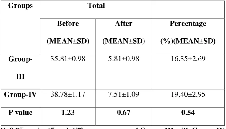
Table 4 : shows the comparison of mean values of volume of obturation material before retreatment and after removal using retreatment file systems between group III and IV.
(P>0.05 no significant difference compared Group-III with Group-IV)
There is no statistical significance in the mean values of volume of after removal between group III and IV.
Table 5 : shows the comparison of mean values of volume of obturation material before retreatment and after removal using retreatment file systems between group V and VI.
(P>0.05 no significant difference compared Group-V with Group-VI)
There is no statistical significance in the mean values of volume of after removal between group V and VI.
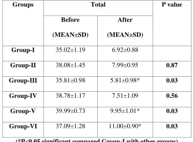
Table 6 : shows the comparison of mean values of volume of obturation material before retreatment and after removal between group I with all other groups.
(*P<0.05 significant - compared Group-I with other groups).
39
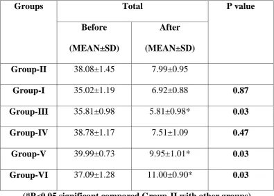
Table 7 : shows the comparison of mean values of volume of obturation material before retreatment and after removal between group II with all other groups.
(*P<0.05 significant compared Group-II with other groups)
There is no statistical significance in the values in comparison with Group I and IV whereas all other groups show statistically significant different results.
Table 8 : shows the comparison of mean values of volume of obturation material before retreatment and after removal between group III with all other groups.
(*P<0.05 significant compared Group-III with other groups)
There is no statistically significant differences in the mean values between group IV whereas all other groups show statistically significant different results.
Table 9 : shows the comparison of mean values of volume of obturation material before retreatment and after removal between group IV with all other groups.
(*P<0.05 significant compared Group-IV with other groups)
40
Table 10 : shows the comparison of mean values of volume of obturation material before retreatment and after removal between group V with all other groups.
(*P<0.05 significant compared Group-V with other groups)
There is no statistically significant differences in the mean values between group VI whereas there is statistically significant difference in the mean values with all the other groups.
Table 11 : shows the comparison of mean values of volume of obturation material before and after removal between group VI with all other groups.
(*P<0.05 significant compared Group-VI with other groups)
There is no statistically significant difference in the mean values between group V, whereas there is statistically significant difference in the mean values with all the other groups.
Table 12 : shows multiple comparison of mean volume values between all the groups
(*P<0.05 significant compared Group-I with other groups, #P<0.05 significant compared Group-II with other groups, $P<0.05 significant compared Group-III with other groups, ǁP<0.05 significant compared Group-IV with other groups)
41
Group I shows statistically significant difference in mean volume after removal with groups III, V, VI
Group II shows statistically significant difference in mean volume after removal with groups III, V, VI
Group III shows statistically significant difference in mean volume after removal with groups I, V, VI
Group IV shows statistically significant difference in mean volume after removal with groups V, VI
Group V shows statistically significant difference in mean volume after removal with groups I, II, III, IV
Group VI shows statistically significant difference in mean volume after removal with groups I, II, III, IV
Table-13 : shows the Multiple comparisons of mean total percentage values between the groups
(*P<0.05 significant compared Group-I with other groups, #P<0.05 significant compared Group-II with other groups, $P<0.05 significant compared Group-III with other groups, ǁP<0.05 significant compared Group-IV with other groups)
42
Group II shows statistically significant difference in mean volume percentage after removal with groups III, V, VI
Group III shows statistically significant difference in mean volume percentage after removal with groups I, V, VI
Group IV shows statistically significant difference in mean volume percentage after removal with groups V, VI
Group V shows statistically significant difference in mean volume percentage after removal with groups I, II, III, IV
Group VI shows statistically significant difference in mean volume percentage after removal with groups I, II, III, IV
OBSERVATIONS
The retrievability of three different obturation materials has been measured by calculating the volume percentage of the remaining material in the root canal after removal using the rotary retrieval systems.
The results show the highest retrievability for Group III (Resilon group removed using Protaper retreatment system) with the least mean volume percentage after removal as 16.35 ± 2.69 % and least retrievability for Group VI with the highest mean volume percentage as 29.67 ± 2.34 %.
43
Comparison between group I (obturated with Gutta-percha and AH Plus sealer removed by Protaper retreatment files) and II (obturated with Gutta-percha and AH Plus sealer removed by Mtwo rotary retreatment system) shows the mean volume percentage for group I was 19.71 ± 1.98% which was lesser than that of group II (21.00 ± 2.41%).
Comparison between group III (obturated with Resilon and RealSeal SE sealer removed by Protaper retreatment files) and IV (obturated with Resilon and RealSeal SE sealer removed by Mtwo rotary retreatment system) shows lesser mean volume percentage for group III (16.35 ± 2.69%) than IV (19.14 ± 2.95%).
Comparison between group V (obturated with CPoint and Endosequence bioceramic sealer removed by Protaper retreatment files) and VI (obturated with CPoint and Endosequence bioceramic sealer removed by Mtwo retreatment files) shows lesser mean volume percentage for group V (24.91 ± 2.66%) than VI (29.67 ± 2.34%).
There is no statistically significant difference in the mean volume percentage on comparison between group I and II, Group II and IV and Group V and VI.
44
removal than Mtwo retreatment file system removal even though there is no statistical significant difference in removal efficacy between the files.