MANDIBULAR MOLAR TOOTH REPLACEMENT
BY TWO NARROW DIAMETER DENTAL
IMPLANTS
Dissertation Submitted to
THE TAMILNADU Dr. M.G.R. MEDICAL UNIVERSITY
In partial fulfillment for the Degree of
MASTER OF DENTAL SURGERY
BRANCH III
his love, grace, mercy and wisdom which formed the foundation of my life and
all my work.
I wish to thank my father Mr M.jeyachandran and my mother
Mrs J.Sarala Devi for the sacrifices they made and for giving me a great
foundation in my life and for being the most wonderful parents. I thank my wife
S.kayathri Senthil kumar and my daughter S.Akshara for being the pillars of
my life and showering me there love, encouragement.
With deep satisfaction and immense pleasure, I present this work
undertaken as a Post Graduate student specializing in Oral and Maxillofacial
Surgery at Ragas Dental College and Hospital. I would like to acknowledge my
working on this dissertation which has been wonderful and enriching learning
experience.
I convey my heartfelt gratitude and my sincere thanks to my Head of the
department and my guide Professor Dr M Veerabahu, Department of Oral and
Maxillofacial Surgery, Ragas Dental College and Hospital, Chennai for his
exceptional guidance, tremendous encouragement, well timed suggestions,
concern and motivation providing me with his immense patience in brightening
in my thesis work and I am indebted towards you forever for all consideration
you have shown towards me. I would definitely cherish these memories
throughout my life.
I would like to extend my heartfelt gratitude to Professor Dr N S
Azhagarasan, Principal, Ragas Dental College and Hospital for allowing us to
use the scientific literature and research facilitates of the college and for
providing the platform to meet such wonderful academicians and people.
I owe enormous gratitude to my guide Professor Dr.D.Sankar for his
invaluable guidance and support throughout my course. He has always been a
source of provoking new thoughts in me. His loving and caring nature lightened
the burden of many hardships. It was an enriching experience to have spent
three years of my life under his guidance. He has enormous knowledge and tons
of experience with which he thought us with unconditional care. I shall forever
remain thankful to him for his valuable guidance and input throughout the
making of this dissertation.
I wish to convey my heartfelt thanks to Professor Dr B Vikraman, a
great teacher who has always been a source of inspiration. His way of looking
graduate life.
I would also thank my Professor Dr.Malini Jayaraj for everlasting
inspiration, constant encouragement, constructive criticism and valuable
suggestion conferred upon me throughout my postgraduate period.
I am greatly indebted to Dr Radhika Krishnan Anaesthesiologist, for
imparting and sharing her vast experience in the field of medicine. I thank her
for her valuable suggestion and constant encouragement through my
postgraduate course.
I am grateful and sincerely thankful to Dr A.Nathan, Dr Sathya Bama,
Dr Saneem and Dr Satish Readers, for their vehement personal interest, wish
and never-ending willingness to render generous help to me throughout my dissertation and post graduate with valuable advice.
I thank Dr Seema Alice Mathew, Dr James Bhagat, Dr Naren
Kumar, Dr K.M. Harish Senior lecturers for their guidance, scholarly
suggestion and whole-hearted support throughout my postgraduate course.
I thank Dr Venkatesh, Dr Aravindan KrishnaMoorthy Department of surgical oncology Cancer Institute Adayar, Chennai, and Dr Karthikeyan,
Dr Abinaya and Dr sakthivel for inspiring and encouraging me during my
postgraduate period. My heartfelt thanks to my dear friends, without whom my time at Ragas wouldn’t have been enjoyable.
I sincerely thank my batch mates, Dr Meeran Sharif, Dr Shiva Sharnu, Dr Narsimman, Dr Nirmal and Dr Nambi Nayaki for their
support, constructive criticism at every step and selfless co - operation during
my dissertation. I wish them a successful career ahead.
I offer my sincere thanks to my senior’s, Dr Sriram Choudary, Dr Giri
Chellapa,Dr Ravi Shankar, Dr Sriraman, Dr Nirmal tony, Dr Vivek, My
Juniors Dr Stephen raj kumar, Dr Manoj kumar, Dr Ajith, Dr Arun
vignesh,Dr Deepan,Dr Kishok, Dr Veera Ragavan, Dr Alka mathew, Dr Diana baburaj, Dr Arvind sai, for their encouragement and support during
the course.
I would like to convey my special thanks to Dr Meeran Sharif, Dr
Shiva Sharnu , Dr.Stephen Raj kumar, Dr manoj, , Dr Ajit and Dr Abinaya
for being main part and most helpful for finishing my dissertation on time.
I sincerely thank Mr Thavamani, for helping in editing and printing of
my thesis. I would also thank Theatre assistants Mr Venugopal, Sis. Malathi OT
staffs, Sis. Deepa, Sis. Laila, Sis. Leema, Sis. Mala for helping me throughout
AIM: The aim of the present study was to evaluate the outcome ofreplacement of single mandibular molar tooth with two narrow diameter implants in terms of evaluation of implant success rate, bone loss, soft tissue and hard tissue healing, oral hygiene maintenance, patient satisfaction and complications.
MATERIALS AND METHODS: The study was conducted in theDepartment
using a standard questionnaire.
RESULTS: Ten patients having partially edentulousness in either mandibular
first or second molar area had replaced with twenty narrow diameter implant. The average mesio-distal length of the edentulous space is 12.5mm ± 1mm, average buccolingual width is 6.3mm ± 0.7mm. All 20 implants placed were of 3mm diameter and the length of the implant ranged from 10 mm to 13 mm depending on the available length. Post-operative crestal bone loss at six month follow up (T1) was 0.52± 0.13mm, 0.57± 0.12mm for mesial and distal implant. Post-operative crestal bone loss at 12 month follow up (T2) was 1.05± 0.20mm, 1.08±0.23mm for mesial and distal implant respectively. Comparison of crestal bone loss at 6 months and 12 months was done using paired t test and it was statistically significant( p value >0.05) for mesial and distal implant. Comparison of crestal bone loss between mesial and distal implants at 6 months and 12 months is not statistically significant (p value <0.05).These measurements were made with the help of intra oral periapical radiograph film. Soft tissue and hard tissue wound healing was good in all our patients except in two patients who had mild gingival hyperplasia over the healing abutment.
oral hygiene.
CONCLUSION: In our study all our mandibular molar tooth replacedwith two
narrow diameter supported implant prosthesis had 100% success rate, with good soft tissue and hard tissue healing and good oral hygiene maintenance at one year follow up. None of our patients had either implant fracture or abutment screw loosening or any other complications. Therefore, the use of two narrow diameter implants to replace a single molar is a logical treatment solution to avoid prosthodontic complications.
KEYWORDS:
S.No
TITLE
PAGE
NO
1. INTRODUCTION 1
2. AIMS AND OBJECTIVE 6
4. REVIEW OF LITERATURE 7
5. MATERIALS AND METHODS 42
6. RESULTS 57
7 DISCUSSION 60
8 SUMMARY AND CONCLUSION 71
9 BIBLIOGRAPHY 72
NO
1. GENDER DISTRIBUTION
2. AGE DISTRIBUTION
3. IMPLANT SITE AND IMPLANT CHARACTERISTICS
INTERNATIONAL CONGRESS OF ORAL IMPLANTOLOGIST 4. ( ICOI) PISA IMPLANT QUALITY OF HEALTH SCALE:
5. VAS SCALE PAIN GRADING
6. WOUND HEALING INDEX, IMPLANT MOBILITY INDEX, MODIFIED BLEEDING INDEX, MODIFIED PLAQUE INDEX 7. PATIENT SATISFACTION INDEX
POST OPERATIVE CRESTAL BONE LOSS AT 6 MONTH 8. FOLLOW UP (T1)
POST OPERATIVE CRESTAL BONE LOSS IN 12 MONTHS 9. FOLLOW UP (T2)
COMPARISON OF CRESTAL BONE LOSS BETWEEN MESIAL
1. AND DISTAL IMPLANTS AT 6 MONTH
COMPARISON OF CRESTAL BONE LOSS OF MESIAL AND
INTRODUCTION
The permanent mandibular Molar is one of the first tooth to be lost over the lifetime of an individual, hence warranting a need for their replacement.33 The preferred choice for replacement of a missing tooth is by the use of a dental implant as vital tooth preparation necessary for bridge fabrication can be avoided thereby preserving tooth vitality. The placement of an implant to replace a missing molar presents diagnostic, surgical and prosthetic challenges such as an enlarged mesio-distal dimension and balanced distribution of occlusal forces. Poor bone density in the posterior molar regions could affect the short- and long-term implant success. Anatomical factors and nearby vital structures (i.e. maxillary sinus and mandibular canal), occlusal loads and the occlusal table which is always wider than the implant
diameter, should also be considered.44
Quality and density of the bone in the posterior molar regions can affect initial implant stability and load transfer to the bone. The most common single molar to be restored is the first mandibular molar, because this tooth is extracted usually35. Implant placement in the posterior mandibular area is a successful procedure over time. The reduced rate of complications in addition to the high long-term success rate make implant restoration a genuine solution to treat posterior partial edentulism24.
the root surface area is almost double as in contrast to the other teeth in the dentition. The mesio-distal extent of a mandibular molar be greater than that of most standard implants (3.75 to 4 mm), generate the possibility of functional overload resulting in the failure of the prosthetic components or the failure of the dental implants. Another result of these dimensional dissimilarity affecting molar restorations on standard implant is disadvantageous contours leading to poor esthetics and hygiene.
Therefore, the clinicians face a distinctive biomechanical challenge. In order to maintain the natural crown root ratio, implant diameter is often increased in the mandibular molar region for immediate loading, especially when the bone density is less or the chewing forces are greater. It is claimed that for the identical length, a wider diameter implant presents a greater surface area, thus bone to implant contact may be greater, thereby compensating for the lack of height or bone density. In recent years, enhancements in component stability have been derived from wider implant platforms, stronger screws, greater torque forces applied to retaining screws, larger hex designs on flat-top implants and the development of internal connections such as cones, internal hex and octagon configurations and combinations of these.20 These refinements have contributed to greater success with molar restoration.
bone receptor sites that are narrow buccolingually, and there have been reports of greater crestal bone loss compared to standard-diameter implant.7 Even after insertion of widest diameter implant, the existing crown root ratio is not achieved in all cases, especially when the bone height is less. Therefore, single implant-bearing molar restoration has historically presented a challenge in terms of form and function because a single implant does not provide the crown-to-root ratio that previously existed which may predispose the implant to over load and may lead to implant failure.
when using new available narrow diameter implants, 2 implants could be used even when the distance between the adjacent teeth is smaller. It has further been suggested by Davarpanah and others, Balshi and others, English and others and Bahat and Handelsman that the use of multiple implants may be the ideal solution for single-molar implant restorations.
To prospectively evaluate the clinical and radiological outcome of two implants supported single molar mandibular prosthesis in terms of
3. Implant success rate
4. Bone loss
5. Soft tissue and hard tissue healing
6. Oral hygiene maintenance
7. Patient satisfaction
8. Complications
1. TORGNY HARALDSON, GUNNAR E. CARLSSON &
BENGT INGERVALL(1979)discussed in detail about the functional state,bite
2.Skalak (1983) proposed that close apposition of bone to titanium is the essential feature that allows a transmission of stress from the implant to the bone without any appreciable relative motion or abrasion. The use of threaded screw provides a form of interlocking with the bone in shear or compression. A smooth cylindrical implant may require an adhesive bond for satisfactory performance ,but screw shape is able to work as long as the apposition of bone and implant is close ,whether or not true adhesive bond is developed.
3.Bass SL.Triplett RG (1991) evaluated the outcome of 1097
all 5-mm implants was 2.3%, and that for all double implants was 1.6%. The use of either 5-mm or double implants necessitates changes in surgical technique, and both are highly dependent for their success on proper surgical execution.
5.Thomas J .Balshi,Ramon E.Hernandez,(1996) compared one
for the single implant supported molar restoration, despite their high degree of mechanical problems. This study suggests that implant supported molars can be effective therapy, and the results confirm the biomechanical analysis that two implants provide more advantageous support than does one.
9. Thomas J. Balshi, Glenn J. Wolfinger (1997) described
two-implant-supported single molar replacement: Interdental Space Requirements and Comparison to Alternative Options. Posterior single-tooth implant restorations are subjected to an increased risk of bending overload. A high incidence of implant fracture has been reported when using a single standard 3.75-mm-diameter implant to support a molar restoration. The purpose of this article is to demonstrate the clinical feasibility of placing two implants to support a molar restoration and to compare this treatment option to the use of a single standard implant or a wide-diameter implant. Two osseointegrated dental implants used to support a molar restoration in interdental spaces as small as 10 mm is shown to be effective and predictable in 60 restorations over the past 7 years. The use of two implants provides more surface area for osseointegration and spreads the occlusal loading forces out over a wider area, reducing the potential bending forces that would otherwise exist in a single-implant molar restoration.
10. S. Ross Bryant, George A. Zarb,( 1998) evaluated the
osseointegration of oral implants in older and younger adults. Osseointegration
study aimed to test the hypothesis that there is no difference between older and younger adults in osseointegration success. A comparison was made between closely matched groups of 39 older adults who had 190 implants supporting 45 oral prostheses and 43 younger adults who had 184 implants supporting 45 oral prostheses. Patients were monitored for a period of 4 to 16 years after prosthetic loading. At the most recent follow-up, the cumulative implant success was 92.0% for the older group compared to 86.5% for the younger group. No statistical significance could be attributed to the difference in implant survival between the groups throughout the study period. Furthermore, the most common outcome for individual prosthetic sites was 100% implant success, and the original prosthetic design was maintained for as long as each patient was monitored in 41 of 45 prosthetic prescriptions for the older patients, and in 39 of 45 prescriptions for the younger patients.
8. Franck Renouard,Jean-Pierre Arnoux,David P. Sarment, (1999)
instead of 0.3 mm) . These features allow an implant 5 mm wide and 6 mm long to maintain the same area of bone contact as a 3.75 _ 10 mm implant. The absence of a smooth collar at the level of the hexagonal head eliminates the need to countersink the implant site and enables visual control of the depth of the implant . Although Langer et al2 have advocated the use of these implants in posterior areas, very little new information has been published since then.3 Therefore, the purpose of this paper is to report on 98 consecutively placed 5-mm-diameter implants without a smooth surface collar.
9. Devorah Schwartz-Arad, Naama Samet, and Nachum Samet
(1999) evaluated the single tooth replacement of molars. as experience with
in11 cases (14%), which included loosening of the abutment and/or the crown (9 cases), fracture of the abutment (1 case), and porcelain fracture (1 case). No incident of implant fracture occurred. Within the limits of this study, replacement of a single molar by a single implant is a valid and successful surgical treatment modality, with a high survival rate. Since Bränemark introduced osseointegrated implants more than 25 years ago, there has been an increased interest in the use of implants in partially edentulous patients. Replacement of a single tooth using a single osseointegrated implant (SOI) is an accepted and satisfactory treatment. It allows greater preservation of adjacent teeth and solves the potential problems caused by other alternative procedures.While there are many articles in the literature concerning replacement of a single anterior tooth using SOI, very few refer to its use in the molar area.
10. Y. SATO, N. SHINDOI, R. HOSOKAWA, K. TSUGA & Y.
AKAGAWA (2000) double implants have been thought to havebiomechanical
around the long axis (1.8-15 N .cm) whereas double implants had no torque. On the other hand, the vertical forces on the mesial double implant were both smaller (60% loaded at point C) and larger (140% ;loaded at point A) than the wide implant .Given the smaller surface area of the mesial double implant, this large force may generate much higher stress in peri implant bone. These results suggest that the biomechanical advantage of double implant for single molar replacement is questionable when the occlusal force is loaded at the occlusal surface near the contact point.
11. L. K. McCaul,W. M. M. Jenkins, and E. J. Kayet ( 2001)
in all age groups over 20 years, caries has become the commonest reason in contrast to 1984 when periodontal disease was the principal reason in patients over 40 years old. Caries and its sequelae remain the most important cause of tooth loss throughout adult life in Scotland and, therefore, caries prevention and maintenance of restorations are of great importance at all ages.
12. Michael Moscovitch,( 2001) evaluated the use of 2 implants to
restore a molar has been shown to eliminate problems associated with bone volume and prosthetic stability. One of the most significant barriers to the widespread use of this concept has been the limitation of the size of implants and their associated prosthetic components. This paper presents the use of 2 implants to replace a single molar using implants and prosthetic components in the Astra Tech Dental Implant System.
cementable restoration. Occlusal contacts were adjusted to conform to the patient’s acquired centric occlusion and lateral excursions. Access openings in the screw-retained restorations were sealed with a composite material. Cemented crowns were luted with a provisional cement . Radiographs were taken immediately postoperatively and, whenever possible, at 1-year intervals.
The postulated advantages of using 2 implants to support a molar restoration instead of a wide-diameter implant are several. There is wider support of the restoration in both the mesial-distal and the buccolingual dimensions. The dentist has greater flexibility to maximize placement in compromised bone receptor sites without perforation of the cortical plates, and thus there is better subsequent retention of crestal bone levels. The use of 2 implants diminishes the potential of the restoration to loosen under normal or parafunctional forces. The double implant may lessen the possibility of occlusal overload. It allows for greater flexibility in restorative style: cement or screw retained. The possibility of increased cost may be outweighed by the reduced likelihood of failure of the implant or the restoration based on the reported complications described earlier. Finally, the double implant requires no special components or procedures that are not normally used in other restorative applications.
13.Lara G. Bakaeen, Sheldon Winkler, ( 2001) conducted study to
(1) determine in vitro the effect of narrowing the buccolingual width of the
occlusal table on the untightening torque required to loosen gold prosthetic screws
loads, and (2) to compare the incidence of screw loosening and values of untightening torque of the screws among crowns supported by 1wide-diameter as opposed to 2 standard implants after loading in vitro. The restorations were divided into 4 groups (group 1, a narrow crown supported by one 5-mm wide-diameter implant; group 2, a narrow crown supported by 2 standard 3.75-mm– diameter implants; group 3, a wide crown supported by one5-mm wide-diameter implant; and group 4, a wide crown supported by 2 standard 3.75-mm–diameter implants). A custom-designed chewing machine was used to simulate the grinding phase of the masticatory cycle and lateral excursions.
untightening torque of gold screws was not affected by changing the width of the occlusal table of crowns supported by 2 implants.
14.S. Ross Bryant, George A. Zarb,( 2002) evaluated the outcomes
of implant prosthodontic treatment in older adults . Older adults are expected to account for an increasingly disproportionate number of individuals needing oral implant prostheses. However, this biotechnology was initially studied for predominantly middle-aged edentulous patients, not elderly people. High rates of success and minimal crestal bone loss have been reported for oral implants mainly in this group. The results of studies at the University of Toronto now clearly support earlier reports that older adults respond to oral implants in the same manner as younger adults, despite their tendency for systemic illness, including osteoporosis. However, unfavourable jawbone quantity and quality, particularly atrophy of the maxilla, impaired implant success. Furthermore, placement of implants in sites that had been edentulous for shorter periods was associated with greater crestal bone loss, a finding that may have implications for younger adults undergoing such treatment. The major decision-making challenge in managing depleted dentitions and complete edentulism in an aging society now lies in differentiating the treatment outcomes, especially patient-mediated assessments (including economic analyses), of the various prosthodontic options available for older adults.
15. Vicki C. Petropoulos,Glenn J. Wolfinger,Thomas J. Balshi,
(2004) described complications of mandibular molar replacement with a single
the surgical placement of a single implant and treatment following these complications. Both the surgical and prosthodontic procedures are described for the treatment of a 57-year-old man who had previously received a single implant for the replacement of a missing molar. Using 2 implants, 1 mesial and 1 distal to the previously placed single implant proved reliable. A logical treatment solution is to use 2 implants for the replacement of a single molar to avoid prosthodontic complications.
16. Eugenio Romeo,Diego Lops,Leonardo Amorfini (2005) studied
the Clinical and radiographic evaluation of small-diameter (3.3-mm) implants followed for 1–7 years. Implants with a small diameter may be used where bone width is reduced or in single-tooth gaps with limited mesio distal space, such as for the replacement of lateral maxillary or mandibular incisors. The purpose of the present longitudinal study was to compare the prognosis of narrow implants (3.3-mm-diameter) to standard (4.1-mmdiameter) implants. Over a 7-year period, 122 narrow implants were inserted in 68 patients to support 45 partial fixed prostheses (PFD) and 23 single-tooth prostheses (ST).
the mandible. Conversely,standard-diameter implants showed a cumulative survival rate of 96.8% in the maxilla and 97.9% in the mandible. The cumulative success rate was 97.6% in the maxilla and 93.8% in the mandible. Cumulative survival and success rates of small-diameter implants and standard-diameter implants were not statistically different (P40.05). Type 4 bone was a determining failure factor, while marginal bone loss was not influenced by the different implant diameters. The results suggest that small-diameter implants can be successfully used in the treatment of partially edentulous patients.
17. S. Jivraj and W. Chee (2006) described that differences in
anatomy and biomechanics make treatment of posterior quadrants with dental implants substantially different to that of anterior areas. Without implants, when posterior teeth were lost, treatment options included a long span fixed partial denture or a removable prosthesis, especially when no terminal abutment was available. Today, with the use of implants, options are available that allow preservation of unrestored teeth. When teeth are missing, implant supported restorations can be considered the treatment of choice from the perspective of occlusal support, preservation of adjacent teeth and avoidance of a removable partial denture.
18. Jeff Brink, Stephen J. Meraw, David P. Sarment ( 2006)
to changes in bone metabolism or transmission of masticatory forces. Understanding of long-term physiologic adjustment is critical to prevention of potential loss of osseointegration, especially because excessive occlusal forces lead to failure. To address this issue, wide diameter implants were introduced in part with the hope that greater total implant surface would offer mechanical resistance. Yet, there is little evidence that variation in diameter translates into a different bone response in the implant vicinity. Therefore, this study aimed at comparing the impact of implant diameter on surrounding bone. Twenty standard (3.75mm) and 20 wide (5mm) implants were placed using an animal model. Histo-morphometry was performed to establish initial bone density (IBD), bone to implant contact (BIC) and adjacent bone density (ABD).BIC was 71% and 73%, whereas ABD was 65% and 52%, for standard and wide implants, respectively. These differences were not statistically different (P40.05). Correlation with IBD was then investigated. BIC was not correlated with IBD. ABD was not correlated to IBD for standard implants (r2¼0.126), but it was correlated with wide implants (r2¼0.82). In addition, a 1 : 1 ratio between IBD and ABD was found for wide implants. It can be concluded, within the limits of this study, that ABD may be influenced by implant diameter, perhaps due to differences in force dissipation.
19. Liran Levin,Amir Laviv, and Devorah Schwartz-Arad (2006)
study was based on a consecutive cohort of 81 patients who received implants to replace a single molar between the years 1994 and 2004. Inclusion criteria for patients were having an implant replacing a molar between two natural teeth and follow-up data of at least 6 months. Data were recorded regarding the incidence of complications and success and survival rates of these implants. Results: The range of follow-up was from 6 to 125 months (mean: 36 months). Smoking was reported by 18.5% of patients. The replacement of a mandibular molar was more frequent (87.7%), with 25.9% of the implants placed immediately after tooth extraction. Two implants were used to replace a single molar in seven patients (8.6%). The failure rate was 7.4% (six implants failed: three had broken necks, and three failed because of infection or bone loss). Complications included suppuration in 11.1% of implants and a pocket around the implant in two patients (2.5%). No relation was found among failure, complications, timing of implant placement, and smoking habits. Conclusion: A single implant can serve as a good long-term and predictable treatment modality to replace a single molar with low complication and failure rates.
20. Len Tolstunov (2007) described the implant zones of the jaws:
of anatomy, blood supply, pattern of bone resorption, bone quality and quantity, need for bone grafting and other supplemental surgical procedures, and a location related implant success rate. The article discusses predisposing factors that can lead to early implant failures in different jaw zones. An implant location is investigated as one of these factors. A prior history of trauma to premaxillary region is described in the context of implant success in anterior maxilla. This zone is being referred by the author as the ‘‘traumatic zone.’’ The challenges of mandibular posterior implant reconstruction are presented in the context of blood supply to the mandible. A deficiency of vascularization in this region, especially in elderly and edentulous patients, lead the author to refer to this zone as the ‘‘ischemic zone.’’ The concept of relative ischemia of the posterior mandible that can develop with age and tooth loss is discussed. A thorough understanding of specifics of each functional implant zone should help to improve successes and prevent failures of dental implants.
21. Heather J. Conrad, John K. Schulte, and Mark C. Vallee,
( 2008) This clinical report describes 2 patient situations in which fractures
implant diameter, manufacturing defects, and galvanic activity. This article describes the management of implant and abutment fractures and discusses possible mechanisms of failure for the patient situations presented. Careful treatment planning and execution of implant therapy is necessary to minimize the risk of implant and component fractures.
22. C. ManganoF. Mangano, A. Piattelli G. Iezzi A (2009) this study
prosthetic complications were reported (0.65% of loosening at implant– abutment interface in SCs ).The use of Morse taper connection implants represents a successful procedure for the rehabilitation of partially and completely edentulous arches. The absence of an implant–abutment interface (micro gap) is associated with minimal crestal bone loss. The high mechanical stability significantly reduces prosthetic complications.
23. Young -kyun kim , Pil YoungYun, ( 2010) conducted study toevaluate
loading is also high. Therefore, to minimize the cantilever, implants must be placed
precisely and followed carefully and maintained for a long period of time
24. Moon-Sun Kim, Jae-Kwan Lee, Beom-Seok Chang (2011),
These findings indicate that a second molar single implant can increase masticatory function.
25. Brian J. Jackson (2011) discussed about utilization of small
diameter implants in limited osseous regions increases patients’ ability to choose implants as a viable restorative option. Although small diameter implants have been indicated in the incisor region for the maxilla and mandible primarily, their usage should be considered in select posterior regions. These 2 case reports demonstrate the incorporation of small diameter implants to replace missing mandibular posterior teeth. Small diameter (1.8– 3.0 mm diameter) implants have been widely accepted because they can be utilized in regions of the mouth that are deficient in arch length, as well as alveolar width. Although small diameter single-stage implants have been indicated mainly for the maxillary lateral incisors and the mandibular incisor region, another clinical situation may warrant their application. Loss of maxillary and mandibular molars results in a mesial-distal dimension that may be insufficient in length for the placement of 2 conventional, standard size implants (3.75 mm diameter). In addition, a single large implant (4.7 mm or 6.0 mm diameter) may demonstrate limitations caused by existing osseous structures or with regard to established implant occlusal principles.
stress under load to the crestal bone. This concept is consistent with the mathematical formula that stress is equal to force divided by area. Small diameter implants have reduced surface area compared with standard conventional implants. Therefore, when a force remains constant, overall stress to the crestal bone around small diameter implants will always be greater. It is the responsibility of the restorative dentist to minimize stress to the crestal bone to improve long-term success. The implant occlusal principles of prime importance are to develop a passive prosthesis with a reduced buccal-lingual dimension, direct the force of occlusion through the long axis of the abutments, and avoid eccentric interferences on the final prosthesis.
26. RS Bedi, Pardeep Verma,Poonam Goel, Puneet Kathutia
( 2011)conducted a study in 5 patients were two standard size implants were
used to replace one missing mandibular molar and compared with single wide diameter implant on the other side in the same patient on the basis of radiographic evaluation.
The patients were divided into two groups as follows:
Group I: Two standard-size implants (SSI) of 3.3 mm diameter and 11.5mm
length were placed in the right missing mandibular molar site.
Group II: In left mandibular molar edentulous site of the same patient, one
Comparative study was performed at each interval to detect and analyze the bony changes around the dental implants by making the following observations: 1.Marginal bone level (MBL).
2.Bone density (BD).
Accurate measurements of the bony changes were performed at standardized points on mesial and distal surfaces of all fifteen implants. Bone changes regarding bone quantity and quality were recorded. Measurements were taken as follows:
1. Assessment of marginal bone level (MBL) around the implants:
Mesial and distal bone height changes of implants were evaluated using the linear measurement system supplied by the digital OPG and digital intraoral sensor for periapical radiograph software. Measurement results were recorded in millimeters. The distance from the most apical part of the implant and the first point of bone-implant contact in cervical region mesially and distally were used to measure the bone level.
In Group I measurements, mean of mesial bone height and distal bone height for both the Standard Sized implants, were taken and tabulated. While in Group II similar measurements were taken in relation to the single WDI. 2. Assessment of the bone density around the implants:
dimension square areas were made just mesial and distal to the implant including the bone implant interface at the selected region of interest. The bone density measurement tool was selected and data recorded.
In the present study, the mean marginal bone level around implants showed that there was statistical significant decrease of marginal bone level comparing the values of immediate post-operative measurement and 9 months measurements in group I and group II. Comparison between marginal bone level in both groups showed statistical non-significant difference in both groups. However, the bone loss was greater in group II with WDI. Moscovitch et al results also show that Wide-diameter implants are limited in their ability to fit in bone recipient sites that are narrow buccolingually and there have been reports of greater crestal bone loss compared to standard-diameter implants15.
However, in the present study although no statistically significant differences in clinical and radiographic results were observed between both groups, yet two SSI implants were relatively superior to WDI. This is in agreement with Blatz et al who comprehensively, suggested the use of both techniques, however, they concluded that two SSI are better options to replace a single mandibular posterior molar and provide more surface area and better biomechanical properties than one WDI implant.
27. Ziv Mazor, AdiLorean ,Eitan Mijiritsky, and Liran Levin,
(2012) conducted a study to present results of single molar area rehabilitated
consecutive patients from 2 private practices between the years 2008 and 2009 had been evaluated. Patients who had a first molar single replaced by 2 narrow diameter implants (3 mm wide) were included in this case series. Patients’ demographics, site and implant characteristics, time of follow-up were recorded from the medical files. Overall, 33 patients received 66 implants replacing 33 missing first molars. Patients’ age ranged from 23 to 76 years with an average of 49.2 - 12.7 years. Most of the implants were used to replace a mandibular molar (76%) and 16 were used to replace 8 maxillary molars. In 2 patients, immediate implantation was performed. The mean distance between the adjacent teeth was 12.1 -1.0 mm. Follow-up time ranged from 10 to 18 months (average, 12.2 - 1.9 months). All implants survived the follow-up time. One implant presented with 1 mm of bone loss at 12-month follow-up. Replacing a single missing molar with 2 narrow diameter dental implants might serve as a viable treatment option providing good and predictable long-term results.
28. Vidya Kamalaksh Shenoy (2012) described the pretreatment
various factors at the pretreatment evaluation stage to set the groundwork for a dentist to consider implant as a restorative option.
29. B K Biswas , S Bag & S Pal (2012) described the biomechanical
This result can be used to design and evaluate any dental prosthesis so far its strength is concerned.
30. Andrea Mascolo, Paresh Patel (2012) described the technique of
splinted zirconia fixed partial denture supported by small diameter (mini implants) in the posterior mandible. Implant-supported fixed partial dentures can restore a patient’s missing posterior dentition. However, in sites that are atrophic, standard body end-osseous implants may not be properly contained by the available bone, thus violating the principle of encasing the implant in a minimum of 1 mm of bone. Alternatively, solid core one-piece, small-diameter (mini) dental implants can be used in highly selected sites with great circumspection. Mini dental implants have been successfully used to support fixed prosthesis that restore missing maxillary and mandibular incisors as well as mandibular posterior teeth. The purpose of this case letter is to demonstrate that splinted mini implants may successfully support a fixed zirconia partial denture in the posterior mandible in highly selected patients and with an appropriate prosthetic design. the use of 2 small diameter (mini) implants can reduce the cantilever effect created when using the procedure recommended by misch (4-mm implant for a 7-mm mesial distal width).
31. Maj Gen J.P. Singh, Col A.K. Gupta ,Col R.K. Dhiman ( 2013)
implant placement can be either functional or non-functional in nature. There is paucity of literature on the comparative evaluation of immediate functional and immediate non-functional loading of implants. This in-vivo study was undertaken to comparatively evaluate Immediate Functional Loading and Immediate Non-Functional Loading of Mono cortical implants with a follow-up period of 18 months. 50 partially edentulous cases were selected for the study. The cases were divided into two groups. In first group (Group-1), 25 implants were subjected to immediate functional loading. In second group (Group-2), 25 implants were subjected to immediate non- functional loading. The crestal bone loss, clinical stability and degree of osseointegration of these two groups were comparatively evaluated. The crestal bone loss in both groups was within acceptable limits.
32. Moustapha Saad, André Assaf (2013) they compared theoutcome of narrow diameter implant and lateral bone augmentation. For optimizing functional and esthetic implant therapy results, sufficient bone amount is required at the reception site. A reduced buccolingual ridge dimension may not allow the placement of a standard-diameter implant without the risk of implant thread exposure. In such situations, lateral bone augmentation procedures can be performed that would allow a restorative-driven placement of standard-diameter implants. Conversely, the use of narrow-standard-diameter implants (standard-diameter ≤ 3.5mm) could be another predictable solution to avoid any invasive surgical management. The aim of this review is to analyze the survival rate of narrow-diameter implants as well as the effectiveness of different techniques for lateral bone augmentation in improving implant clinical outcomes. The use of narrow-diameter implant as well as lateral bone augmentation are well documented in the literature as a treatment modality in reduced ridge width . Each treatment approach has its advantages and downsides. On one hand, narrow diameter implant is a simple and predictable treatment when used properly. On the other hand, hard tissue management improves implant survival rate together with soft tissue contour and phonetics. Moreover, lateral bone augmentations are sometimes required to optimize the sagittal intermaxillary relationship.
33. Eitan Mijiritsky, Ziv Mazor, Adi Lorean, and Liran Levin
(2013) conducted a study to evaluate the influence of implant length and
consecutive patients from 2 private practices between the years 2008 and 2011
had been evaluated .Patient demographics, site and implant characteristics, and
time of follow-up were recorded from the medical files. Overall, 3043 implants
were investigated. Overall survival rate was 98.7% with 39 implant failures
recorded. Survival rates for narrow- (,3.75 mm), regular- (3.75– 5 mm), and
wide- (.5 mm) diameter implants were 98.2%, 98.7%, and 98.5%, respectively
(P ¼ 0.89). Survival rates of short (,10 mm) and regular (10 mm and above)
implants were 97% and 98.7%, respectively (P ¼ 0.22). Conclusions: Implant
length and diameter were not found to be significant factors affecting implant
survival during the first 2 years of function in the present investigation of this
specific implant system by a single manufacturer. Further long term follow-up
studies are warranted because 2-years are only interim short-term results when
dealing with dental implants.
34. De Souza Tolentino L, Garcez-Filho J, Tormena M, Lima LA
and Araújo (2014) evaluated the outcome of Narrow Diameter Implants
(T0) and one year after loading (T1). Measurements were performed from implant shoulder to the first point of bone/implant contact. The differences in marginal bone change between the groups were analyzed by Student t-test for paired samples. A level of 95% of significance was adopted. A total of 42 implants were installed (21 RDIs and 21 NDIs). At the end of the follow-up period (12months of loading), implant success and survival rates of 100% were observed. The bone loss around implants atT0 was 0.41 (± 0.45) mm for NDIs and 0.47 (± 0.60) mm for RDIs and at T1 was 1.3 (± 0.3) mm for NDIs and 1.24 (±0.3) mm for RDIs. No statistically significant differences between the groups were found (p>0.05). This study demonstrated that RDIs and NDIs produced similar marginal bone alterations patterns after one year of loading, regardless the implant location, indicating that NDIs may be used in the posterior region of the jaws with single unit prostheses in selected patients.
35. Douglas R Monteiro, Emily V F Silva, (2015), conducted studyon
36. Dr. Mayur Kaushik, Dr. Sakshi Khattar (2016) described the treatment of thin tissue biotype around an implant using sub epithelial connective tissue graft. Implant dentistry has come a long way since 1965, with great improvements made to achieve primary implant stability and to improve bone-to-implant contact with the introduction of the concept of osteointegration. The focus has been shifted towards creating an esthetic restoration that is indistinguishable from natural teeth and is stable over time. Just as bone volume is crucial for ideal positioning of the implant, soft tissue volume predicts the ideal emergence profile and esthetics of the eventual implant restoration. The correct recognition of gingival biotypes is important for the treatment of planning process in restorative and implant dentistry. Patients with thin biotype are more prone to recession, inflammation, and compromised soft tissue response. This paper presents a case of management of the thin gingival biotype over the implant surface to a more favourable one using the sub epithelial connective tissue graft to achieve a more stable and esthetic result.
37. Zankhana Shah, Amar Shah, Priyanka Raiyani (2016)
described that Most frequent single molar to be replaced is the firstmandibular molar because this tooth is lost first. Implantation in the posterior area is a predictable procedure over time. The low rate of complications in addition to the high long‑ term success rate makes implant restoration a reliable solution to
case deals with the replacement of the lower right first molar having a previously failed root canal treatment with two narrow implants of diameter 3.5 mm and height 10 mm. Replacing a single missing molar with two narrow dental implants serves as a viable treatment option providing good and predictable long‑ term results.
38. Ho-Yong Song, Yoon-Hyuk Huh, Chan-Jin Park, Lee-Ra Cho
( 2016) Conducted a study to investigate the stress distribution of 2-short
concentration. 2SIs may be useful for achieving stable stress distribution on the surrounding bone and implant-abutment complex in the atrophic posterior maxilla.
39. Hadi Antoun, Pierre Cherfane, and Bouchra Sojodet (2016)
MATERIALS AND METHODS
This study was conducted at the department of Oral and maxillofacial surgery, Ragas dental college and hospital from December 2015 to December 2018. Ethical approval was obtained from the Institutional review board. Patients requiring replacement of single missing mandibular molar tooth were explained about both the single implant supported and double implant supported prosthesis. Patients who were willing to undergo the double implant supported molar replacement, were included in this prospective study. All the patients were systematically examined (Annexure III) preoperatively to rule out any systemic disorder or medically compromised condition or allergic reactions that will contradict the implant placement or post-operative medical management. The study protocol and the implant procedure were explained to the patient and informed consent (Annexure IV) was obtained prior to the procedure and the source data were collected accordingly.
INCLUSION CRITERIA
11. Patient with missing mandibular molar teeth with adjacent natural teeth and as well as natural teeth antagonist.
12. Good oral hygiene.
2 Sufficient residual bone volume to receive implants of minimum 3.0 mm in diameter and minimum 10 mm in length with mesio distal edentulous space of 12- 14 mm.
3 Minimum crown height space of 7mm.
EXCLUSION CRITERIA
(2) Presence of para-functional habits such as bruxism. (3) Chronic smokers.
(4) Patients under radiation therapy, chemotherapy, immunosuppressive drugs like corticosteroids
(5) Pregnancy.
(6) Insufficient bone quality or quantity, insufficient interarch space, poor oral status.
(7) Inflammatory and autoimmune conditions of the oral cavity.
DIAGNOSTIC PHASE:
Preoperatively all the patients were evaluated clinically and radiographically using (OPG) and intra-oral periapical views, to detect for
16. The absence of pathological lesion at the area of implant insertion, alveolar height above the inferior alveolar canal and condition of the adjoining teeth.
17. The condition of the bone and its suitability for implant placement. 18. Any root angulation in adjacent tooth, periodontal defects & amount of
interdental bone
19. The vertical height of bone to select the suitable implant length.
20. For all these patients bone mapping was done using sectioned impression casts to assess the width of bone available for selection of appropriate diameter of the Implant. Custom fabricated stents were fabricated to accurately locate the implant site and direction of insertion.
SURGICAL PHASE:
ARMAMENTARIUM:
2% Lignocaine with 1:80,000 Epinephrine
27 Gauze, 40 x 35mm disposable needle.
No 15 bard parker blade & handle with No 3 handle.
Molt no 9 Periosteal elevators.
Marking drill, Pilot burs with diameter of 2mm ,2.8mm ,Torque wrench , ratchet ,
Vernier caliper or Divider with Metal scale.
Narrow diameter endosseous implant which is 3 mm in diameter and varying length of 10mm,11.5 mm and 13mm were used in this study
3-0 silk suture material.
Surgical room was disinfected by fumigating the room with formaldehyde. Drills, implant components and the hand pieces were sterilized using an autoclave while ensuring peak performance and quality control. The patient is led into the surgery room to avoid contact with sterile items, and seated. Lighting unit handles were covered with sterile lead foil.
SURGICAL PROCEDURE:
reflection was done towards the lingual side to maintain blood supply to the bone.
The custom-made stent which was trailed before in patient’s mouth were kept in Glutaraldehyde solution for 2 hours before surgery. With the guidance of stent, a pilot drill was introduced into the bone, and two osteotomy sites were created. First on the mesial and the second on the distal side. They were taken to the desired depth. The sites were progressively enlarged and finished with the dedicated osteotomy drill. The profile gauge was inserted, and depth checked to ensure that the implant would sit just sub crestal. The dedicated tap was then introduced into the site to the depth established. Two mini implants of size 3.0 mm × 10 mm were carefully threaded into the prepared sites with minimum of 30 N/cm(fig.4). The flaps
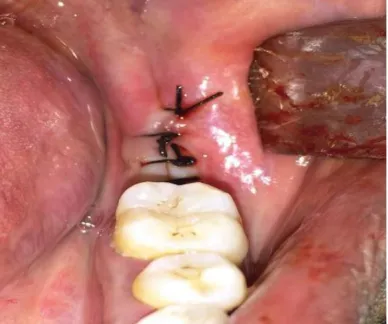
were closed with 3‑0 silk sutures.(fig.5)
POST SURGICAL PROTOCOL:
All patients were administered with a single dose of analgesic (inj.voveron 75mg) intramuscularly immediately after the procedure. They were prescribed with a regime of oral antibiotics cap.Amoxicillin 500mg TDS, Tab.Metronidazole 400mg TDS, analgesics Tab.Paracetamol 650mg TDS and antacid Tab.Ranitine hydrochloride 150mg BD before food for a period of 5 days.
Regular oral prophylaxis was advised. Suture
POST-OPERATIVE INSTRUCTIONS:
1. The prescription for the post-operative medication were handed over to the patient preoperatively and requested to keep in hand during the procedure.
2. Intermittent ice application for 48 hours.
3. Chlorhexidine rinses should be used gently three to four times daily for 2 weeks.
4. Brushing at the operative site should be discouraged for the first 24 hours. Then, a very soft brush (e.g., Oral B-20 or -30) can be used carefully for cleansing. Any dentifrice is satisfactory.
5. Eat very soft foods as tolerated. Mastication of food of challenging texture that might injure the operative site should be avoided. Plan a reasonable, nutritionally balanced diet. Good choices are soft boiled eggs, milk, ice cream, malts, boiled chicken and soup, cheeses, and junior foods.
6. For the first 24 postoperative hours, drink plenty of fluids: juice, soda, water, or milk.
7. Expect some amount of swelling, pain and discomfort. These are common and do not indicate infection or other problems. Sleep with your head well elevated.
9. Do not hesitate to telephone if any questions arise about your condition or the operation. In an emergency, you should call us at (telephone number).
Patients were recalled for follow up regularly at 1 week, 1 month and 3 months post-operative period. The two-stage surgical technique was chosen in this study for implant placement. In the first stage, the implants were placed and were left undisturbed for a healing period of three months for complete osseointegration.
PROSTHETIC PHASE:
be retightened, the prosthesis were considered mobile. If the screw was completely loose or fractured, this condition was registered separately.
The radiographs were taken 1 week after abutment connection, 6 and 12 months post operatively and once every 12 months thereafter. Radiographs were analyzed with respect to bone loss and change of density of the bone around the implants. The marginal bone height of each implant was measured mesially and distally by using the implant threads as the dimensional reference. The numerical mean of the mesial and distal measurements was used as a value for each implant. The marginal bone loss was calculated for each site.
IMPLANT EVALUATION
At the follow-up sessions, scheduled for 0, 6, 12 months after implant insertion, the following clinical parameters (primary and secondary endpoints of the study) were investigated.
I. International Congress of Oral Implantologist ( ICOI) Pisa Implant
quality of health scale:
Grading Group Clinical condition
1 Success (optimal No pain or tenderness upon function health) Zero mobility
Less than 2 mm radiographic bone loss from initial surgery
2 Satisfactory No pain on function Survival Zero mobility
2-4 mm radiographic bone loss No exudate history
3 Compromised May have sensitivity on function Survival No mobility
Bone loss more than 4 mm Probing depth more than 7mm May have Exudate history 4 Failure Pain on function
Mobility
Radiographic bone loss more than half the length of implant
Uncontrolled exudate No longer in mouth
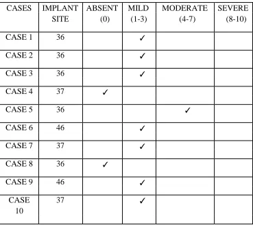
VAS scale Pain grading:
Pain score Grading
0 Absent
1-3 Mild
4-7 Moderate
8-10 Severe
III.Wound healing index:
SCORE DESCRIPTION
1 Uneventful wound healing with no gingival oedema,erythema,suppuration,patient discomfort or flap dehiscence.
2 Uneventful wound healing with slight gingival edema,patient discomfort,or flap dehiscence,but no suppuration.
IV. Implant mobility Index :
Scale Description
0 Absence of clinical mobility with 500 g in any direction.
1 Slight detectable horizontal movement.
2 Moderate visible horizontal mobility upto 0.5mm.
3 Severe horizontal movement greater than 0.5mm.
4 Visible moderate to severe horizontal and any visible vertical movement.
V. Modified plaque index:
Modified plaque index* (mPI), determined on the mesial, distal, buccal and palatal surface of the implants. For each implant, the mPI value was calculated based on the average of the four obtained values. The following scores were assigned on the basis of the amount of plaque:
Score 0: No plaque detected
Score 1: Plaque only recognized by running a probe across the marginal surface of the implant
Score 2: Plaque visible with the naked eye Score 3: Abundance of soft matter
Modified plaque index:
VI. Modified bleeding index:
Modified bleeding index* (mBI), assessed at the same surfaces, as an indicator of the existence and severity of peri-implant gingivitis. For each implant, the mBI value was calculated based on the average of the four obtained values:
Score 0: No bleeding running a periodontal probe along the gingival margin adjacent to the implant;
Score 1: Isolated bleeding spots evidenced;
Score 2: Blood forming a confluent line on the mucosal margin; Score 3: Profuse bleeding
Modified bleeding index:
Score 0 Score 1 Score 2 Score 3
VII. Patient satisfaction:
b. Are you satisfied with esthetics outcome of final prosthetic crown?
c. Are you having functional difficulty with final prosthesis?
d. Are you having difficulties in maintaining oral hygiene?
Patient satisfaction score :
Case Average score Grading
1-2 Not satisfied
3-4 Slightly satisfied
5-6 Moderately satisfied
VIII. Bone loss:
The distance between the implant shoulder and the first visible bone contact (DIB) in millimeters. To perform this evaluation, intraoral periapical radiographs were taken for each implant, with a rigid film-object X-ray source at the baseline (immediately after implant insertion) and at the follow-up sessions (0, 6, and12 months after implant insertion).
With these values, crestal bone level changes were registered as modifications in the distance from the implant shoulder to the bone level on the mesial and distal implant side. In order to correct dimensional distortion, the apparent dimension of each implant was measured on the radiograph and then compared with the real implant length. The radiographs were also analyzed for the presence or absence of continuous peri-implant radiolucencies.
IX. Complications:
Patients were also evaluated during the review for the presence of any of the following complications and the findings were recorded.
1. The presence or absence of pain or suppuration
2. The presence or absence of implant mobility tested manually using the handles of two dental mirrors
4. Soft tissue complications: 5. Prosthetic complications:
INSTRUMENT SETUP
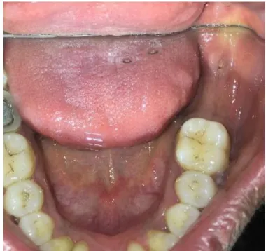
Fig. 1: PROFILE PHOTO FIG. 2: INTRA ORAL VIEW
FIG. 3: MUCOPERIOSTEAL FIG. 4: MESIAL AND DISTAL
[image:72.595.112.292.198.374.2] [image:72.595.316.488.468.643.2]FIG. 5: SUTURING DONE FIG. 6: HEALING ABUTMENT
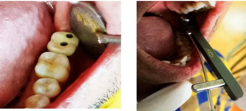
[image:73.595.97.293.456.648.2]FIG.8: ZIG TRIAL
[image:73.595.329.523.462.650.2]FIG. 10 : SCREW TIGHTENED FIG. 9:
SCREW RETAINED TO 20 N TORQUE
METAL CERAMIC PROSTHESIS
[image:74.595.107.499.220.398.2]
FIG. 12: OCCLUSAL ENTRY
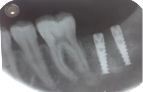
[image:74.595.105.298.220.391.2]FIG. 13: PREOPERATIVE IOPA
FIG. 14: IOPA AFTER IMPLANT PLACEMENT
[image:75.595.175.425.361.521.2]FIG. 16: PREOPERATIVE OPG
FIG. 17: OPG AFTER IMPLANT PLACEMENT
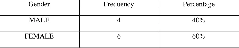
[image:76.595.102.366.363.524.2]Surgery at Ragas Dental College and Hospital, Tamilnadu. Patients of either sex, having partial edentulism in the posterior mandibular arch with narrow crestal width who required preferably implant based fixed prosthesis were included in this study. Ten patients (4 male and 6 female) received 20 implants for replacing either the first or second mandibular molar tooth (Table 1). Patients age ranged from 25 to 55 years with an average of 31.9 years (Table 2.). None of the patients had the habit of smoking at the time of implantation.
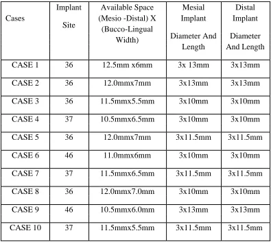
Implant location and implant characteristics are as follows: In five patients implants were placed in 36 region, three patients received implants at 37 region and in the other 2 patients implants were placed in 47 region. The average mesiodistal length of the edentulous space is 12.5mm ± 1mm, average buccolingual width is 6.3mm ± 0.7mm. All 20 implants placed were of 3mm diameter and the length of the implant ranged from 10 mm to 13 mm depending on the available length. (TABLE 3). All The implants belong to ADIN IMPLANT SYSTEMS with tapered Mors internal connection.
All the implants were loaded following two stage technique. Patients were evaluated at 6months and 12 months (average 6 months) according to ICOI criteria. On evaluation all implants were successful (Table 4.). All the implants were also evaluated for Implant mobility with two point scale and tap test. If the tap elicits a solid ring there is no mobility but if the sound is dull, the implant is not osseointegrated and surrounded by fibrous tissue. Among the 10 patient, none of the patient reported with clinically detectable implant mobility in horizontal and vertical direction. (TABLE 6)
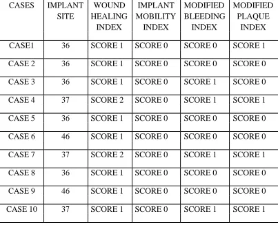
None of the implant patient experienced pain or tenderness on function (Table 5.). All ten implants showed zero mobility and less than 2mm radiographic bone loss from initial surgery. None of the implants showed any exudate (TABLE 6). Among the ten patients, eight patient had uneventful wound healing with no gingival oedema, erythema, suppuration, patient discomfort or flap dehiscence, and two patient had uneventful wound healing with slight gingival oedema, patient discomfort, or flap dehiscence, but no suppuration (TABLE 6). There was no peri – implant radiolucency present in any of the cases on routine radiographic follow up. On patient Satisfaction Index 3 patient were very satisfied and 3 patient were moderately satisfied and four patient were slightly satisfied.(TABLE 7)
detected on six patients and plaque was detected only on running the periodontal probe across the marginal surface of the implant in four patients.(TABLE 6). Modified bleeding index (mBI) and modified plaque index were assessed at the same surfaces, as an indicator of the existence and severity of peri-implant gingivitis. For each implant, the mBI value was calculated based on the average of the four obtained values:score 0,score 1 ,score 2 ,and score 3.out of 10 patients ,six patient had, no bleeding on running a periodontal probe along the gingival margin adjacent to the implant and 4 patients reported with isolated bleeding spots.(TABLE 6)
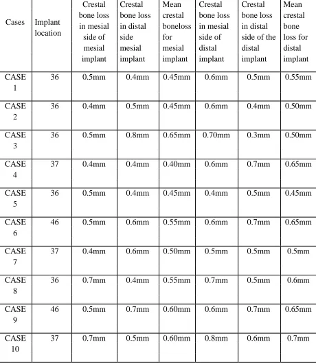
Post operative crestal bone loss at six month follow up (T1) revealed bone loss of, 0.52±0.13mm, 0.57±0.12mm for mesial and distal implant.(TABLE 8). Post operative crestal bone loss at 12 month follow up
CASE 2
PROFILE PHOTO INTRA ORAL VIEW
MUCOPERIOSTEAL STENT PLACEMENT
PARALLELING PIN MESIAL AND DISTAL
PLACEMENT IMPLANT PLACEMENT
SUTURING DONE HEALING ABUTMENT
SCREW RETAINED METAL
SUTURING DONE CERAMIC PROSTHESIS
SCREW TIGHTENED OCCLUSAL ENTRY
TO 20 N TORQUE CLOSED WITH
PREOPERATIVE IOPA
IOPA AFTER IMPLANT PLACEMENT
PREOPERATIVE OPG
OPG AFTER IMPLANT PLACEMENT
PROFILE PHOTO INTRA ORAL VIEW
MUCOPERIOSTEAL IMPLANT SITE
IMPLANT PLACEMENT MESIAL AND DISTAL IMPLANT PLACEMENT
HEALING ABUTMENT
SUTURING DONE ZIG TRIAL VERIFICATION
SCREW RETAINED METAL OCCLUSION
PREOPERATIVE IOPA IOPA AFTER IMPLANT PLACEMENT
ZIG TRIAL IOPA AFTER PROSTHESIS
OPG AFTER IMPLANT PLACEMENT
MALE 4 40%
FEMALE 6 60%
TABLE 2: AGE DISTRIBUTION
Age (years) Number of patients Percentage
18-25 1 10%
26-33 7 70%
34-41 1 10%
42-49 0 0
50-57 1 10%
[image:90.595.105.495.80.159.2]TABLE 3: IMPLANT SITE AND IMPLANT DIMENSIONS
Implant Available Space Mesial Distal Cases
Site (Mesio -Distal) X Implant Implant (Bucco-Lingual
Diameter And Diameter Width)
Length And Length
CASE 1 36 12.5mm x6mm 3x 13mm 3x13mm
CASE 2 36 12.0mmx7mm 3x13mm 3x13mm
CASE 3 36 11.5mmx5.5mm 3x10mm 3x10mm
CASE 4 37 10.5mmx6.5mm 3x10mm 3x10mm
CASE 5 36 12.0mmx7mm 3x11.5mm 3x11.5mm
CASE 6 46 11.0mmx6mm 3x10mm 3x10mm
CASE 7 37 11.5mmx6.5mm 3x11.5mm 3x11.5mm
CASE 8 36 12.0mmx7.0mm 3x10mm 3x10mm
CASE 9 46 10.5mmx6.0mm 3x13mm 3x13mm
TABLE 4: 1. INTERNATIONAL CONGRESS OF ORAL
IMPLANTOLOGIST (ICOI) PISA IMPLANT QUALITY OF
HEALTH SCALE
CASES IMPLANT GRADE 1 GRADE 2 GRADE 3 GRADE 4 SITE
CASE 1 36 ✓
CASE 2 36 ✓
CASE 3 36 ✓
CASE 4 37 ✓
CASE 5 36 ✓
CASE 6 46 ✓
CASE 7 37 ✓
CASE 8 36 ✓
CASE 9 46 ✓