S H O R T R E P O R T
Open Access
Post-exercise left ventricular dysfunction
measured after a long-duration cycling event
Enrique Serrano Ostariz
1, Marta López Ramón
2, Daniel Cremades Arroyos
1, Silvia Izquierdo Álvarez
3*,
Pilar Catalán Edo
1, Cristina Baquer Sahún
4and Alejandro Legaz Arrese
1Abstract
Background:In this research, an extension to our previous work published in theClinical Journal of Sports Medicine in 2009, we studied subjects that differed in terms of age and training status and assessed the impact of prolonged exercise on systolic and left ventricular diastolic function and cardiac biomarkers levels, recognized as identifiers of cardiac damage and dysfunction. We also assessed the possible influence of event duration, exercise intensity and weight loss (dehydration) on left ventricular diastolic function.
Findings:Ninety-one male cyclists were assessed by echocardiography and serum biomarkers before and after the 2005 Quebrantahuesos cycling event (206 km long and with an accumulated slope of 3800 m). Cardiac function was assessed by echocardiography and cardiac biomarkers were assessed in blood serum. Echocardiograms measured left ventricular internal dimension during diastole and systole, left ventricular posterior wall thickness during diastole, interventricular septum thickness during diastole, left ventricular ejection fraction and diastolic filling. The heart rate of 50 cyclists was also monitored during the race to evaluate exercise intensity.
Echocardiograph results indicated that left ventricular diastolic and systolic function decreased after the race, with systolic function reduced to a significant degree. Left ventricular ejection fraction was below 55% in 29 cyclists. The decrease in left ventricular systolic and diastolic function did not correlate with age, training status, race duration, weight loss or exercise intensity.
Conclusions:Left ventricular systolic and diastolic function was reduced and cardiac biomarkers were increased after the cycling event, but the mechanisms behind such outcomes remain unclear.
Keywords:Cycling event, Cardiac biomarkers, Prolonged exercise, Strenuous exercise
Findings Background
This study is a further analysis of data collected for a previous work [1]. One potential effect of prolonged strenuous exercise is exercise-induced cardiac fatigue, described as an alteration to ventricular systolic or diastolic function following such exercise [2,3]. Findings reported for numerous ventricular dysfunction studies have often been inconsistent [3-8], with disparities pos-sibly due to small sample sizes, differences in exercise intensity and duration and great variability in the training
status of the study subjects. As for cardiac-specific biomarkers of damage and dysfunction, a growing number of studies have demonstrated that prolonged and strenuous exercise raises cardiac troponin T and I (cTnT and cTnI, respectively) and N-terminal pro-brain natriuretic peptide (NT-proBNP) levels [9-14]. The link between systolic and diastolic ventricular dysfunction and the increase in bio-markers is not clear, however [15-18]. Most studies of high-intensity prolonged exercise resulting in increased cardiac biomarkers have been performed in relatively old subjects, but increases are not so evident for elite or professional sportspeople [19,20].
Specific exercise parameters (training status and exercise intensity and duration) that may affect observed changes in biomarkers and ventricular systolic and diastolic func-tion are poorly described and the mechanisms involved are not well understood. To the best of our knowledge,
* Correspondence:[email protected] 3
Servicio de Bioquímica Clínica Planta 3ª, Edificio de Consultas Externas, Hospital Universitario Miguel Servet, Calle Padre Arrupe, s/n, Zaragoza 50009, Spain
Full list of author information is available at the end of the article
the influence of exercise intensity on left ventricular function has not yet been determined, as we were un-able to locate any studies of ventricular dysfunction in cyclists of different ages and training levels for a long-duration cycling event (6 hours or more). Therefore, for a long-duration cycling test, we studied, following a similar study [19], the influence of age, level of training, exercise intensity, event duration and weight loss (dehy-dration) on ventricular function and their association with cardiac biomarkers.
Methods
Participants and study design
The study protocol was approved by the Research Ethics Committee of the Government of Aragón (Spain). Amateur male cyclists scheduled to participate in the 2005 Quebrantahuesos Cycle Race (UCI Golden Bike series) were invited to participate in the study via the offi-cial race organizer’s website [1]. The first 95 volunteers were recruited and their written consent was obtained (note that sample size was limited by the finishing-line resources available). Since 4 cyclists dropped out during the race, the study finally included 91 cyclists, profiled as follows in terms of mean ± standard deviation (SD): age, 40 ± 9 years; height, 1.76 ± 0.07 m; weight, 74.1 ± 8.4 kg; and training experience, 12 ± 7 years [1]. The race work-load (total distance 206 km for an accumulated slope of 3800 m) was comparable to that of the tougher mountain stages of the Tour de France. Ambient temperature during the test ranged between 16°C and 32.5°C.
Subjects completed a general questionnaire, to ascertain personal and cycling history, and a log referring to cycle training in the previous 6 months (4358 ± 1731 km). Cy-clists were assessed the day before, and approximately 20 minutes after, the cycling event [1]. Cyclists consumed fluid during the event, but, in accordance with guidelines by White et al. [21], consumption was not allowed until after post-race data was collected so as not to influence left ventricular loading.
Pre- and post-race assessment of the 91 cyclists in-cluded the following: echocardiography, body mass, heart rate (HR), and blood pressure measurements and a 12-lead electrocardiogram. HR for 50 of the cyclists was also measured during the race using a Polar HR monitor (Polar Team System, Polar Electro Oy, Finland) and the data was downloaded using Polar Precision Performance software (version 3.0) [1]. Maximum HR was calculated using the standard equation HR (max) = (220-age). Exercise intensity was defined by the HR ratio as HR (average)/HR (max) [22]. Finally, training impulse (TRIMP) values, which are used as an integra-tive marker of exercise load during competition, were calculated from event duration and the average HR using the formula described by Banister [23].
Echocardiograph procedures
Participants underwent a resting echocardiographic exam-ination in the left lateral decubitus position. One experi-enced sonographer performed all the measurements using a commercially available system (ATL ultrasound HDI 5000) and a 2.5 MHz phased array transducer. Two-dimensionally guided M-mode echocardiograms were obtained from the left parasternal long-axis view. These recordings, made according to American Society of Echo-cardiography recommendations [24], were used to obtain the following measurements: left ventricular internal dimension during diastole and systole (LVIDd and LVIDs, respectively), left ventricular posterior wall thickness during diastole (LVPWd) and interventricular septum thickness during diastole (IVSDd). Left ventricular ejection fraction (LVEF, %) was determined by calculating left ventricular volumes according to Teichholz et al. [25]. Doppler echocardiography was used to assess diastolic function. An apical four-chamber view was obtained for a maximized diameter of the mitral annulus. With the sample volume cursor aligned parallel to flow at the mitral annulus level, inflow velocities were interrogated by pulsed-wave Doppler. Minor adjustments were made to the transducer to ensure optimal spectral display, that is, the highest velocity for the least spectral dispersion. The velocity curves were digitized through the darkest grey scale and averages were calculated for the measurements obtained. Peak early and late diastolic filling (E and A, re-spectively) were measured and the corresponding ratios calculated. HR was determined by limb-lead electrocardi-ography combined with echocardielectrocardi-ography. Blood pressure was simultaneously measured using standard auscultation procedures.
Blood sampling procedures
Procedures were as described in the previous work published [1]. The upper reference limit (URL) for cTnI (defined as the 99th percentile of healthy participants) was less than 0.04μg L-1 [26]. NT-proBNP levels were measured using an electrochemiluminescence sandwich immunoassay (Elecsys ProBNP, Roche Diagnostics) with the Roche 2010 system. The URL was set at 125 ng L-1 [27]. Haematocrit concentrations were measured using an automated analyser (Sysmex K-1000; Sysmex GmbH, Langenfeld, Germany).
Statistical analyses
Pearson and Spearman correlation coefficients were cal-culated as standard. Multivariate linear regression was used to assess the relationships between left ventricular diastolic function—measured as the dependent variable—and age, weight loss, training status, event duration and exercise in-tensity as independent predictors. The level of significance was set toP< 0.05.
Results
All 91 cyclists lost weight (74.2 ± 8.6 kg pre-race weight versus 71.8 ± 8.6 kg post-race weight; P< 0.001) [1]. Post-race systolic blood pressure was lower (from 124 ± 12 mm Hg to 110 ± 11 mm Hg;P< 0.001) [1]. No cyclist required medical attention.
Echocardiographic data
Baseline echocardiographic examinations revealed no re-markable pathological findings. No cyclist had LVPWd or IVSDd values of more than 12 mm. Mean LVIDd was 51.2 ± 4.0 mm, mean LVEF (%) was 67.7 ± 8.2 and the mean E/A ratio was 1.6 ± 0.4.
The cycling event exercise resulted in decreased LVIDd and increased LVIDs in the participants but pro-duced no change in the LVPWd or IVSDd (Table 1). The LVEF (%) was reduced from 67.7 ± 8.2 to 59.7 ± 9.4 (P< 0.001) and, in 29 cyclists, post-race LVEF was below 55%.
Early transmitral diastolic filling velocities were reduced and late transmitral diastolic filling velocities were in-creased, resulting in a reduced E/A ratio (Table 1). The
post-race increase in HR was not significantly associated with alterations in the LVEF and the E/A ratio. The LVIDd was not related to any of the observed functional changes. Overall, there were no significant associations between al-terations in cardiac function (systolic or diastolic) and age, training status, event duration, weight loss, exercise inten-sity (the HR (average)/HR (max) ratio or TRIMP values) and NT-proBNP. When we considered multivariate re-gression, the model was not significant (systolic: R2= 0.147,P= 0.267; diastolic: R2= 0.091,P= 0.599).
Biochemical markers
Post-race cTnI and NT-proBNP levels were significantly raised, with 43% of participants exhibiting cTnI levels above 0.04 μg L-1 and with 65% of the participants exhibiting NT-proBNP levels above 125 ng L-1 [1]. The increases, however, did not correlate with post-race cardiac function.
Discussion
The results of this study confirm those of previous studies (running and ironman) in demonstrating that prolonged strenuous exercise reduces systolic and diastolic function [6,21,28-30] and increases the cardiac-specific markers cTnI and NT-proBNP [5,10,13,14,30,31].
The decrease in post-race LVEF supports the sugges-tion that systolic changes only develop during long-duration events [5,12,15,21,32,33].
No previous studies have been conducted for amateur cyclists of different ages and training levels or for long-duration cycling events (more than 6 hours). The long duration of the event may explain the deterioration in ventricular function in most cyclists in our study, as this finding coincides with the deterioration reported in studies for other longer duration tests.
The decrease in LVEF in the presence of decreased sys-tolic blood pressure reflects depressed cardiac contractil-ity. The lack of correlation between weight changes, LVIDd and diastolic function measurements suggests that the depression in diastolic function represents a true re-duction in left ventricular function. Furthermore, although post-race HR increased significantly compared to pre-race HR, the absence of any significant correlation between HR and any Doppler-derived index of diastolic function supports the presence of a change in diastolic function.
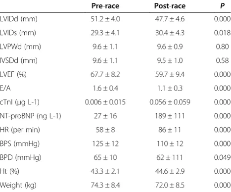
[image:3.595.57.291.479.673.2]Although our results agree with previously reported results, they should be interpreted with care. We report a weight loss of over 3%, which can be interpreted as a high degree of dehydration. We used weight changes and LVIDd as cardiac preload indicators; using weight loss as a preload indicator is open to question, however, as preload may be maintained by internal fluid shifts from the extravascular to intravascular space [29]. Table 1 Pre and post-race data (mean ± standard
deviation)
Pre-race Post-race P
LVIDd (mm) 51.2 ± 4.0 47.7 ± 4.6 0.000
LVIDs (mm) 29.3 ± 4.1 30.4 ± 4.3 0.018
LVPWd (mm) 9.6 ± 1.1 9.6 ± 0.9 0.80
IVSDd (mm) 9.6 ± 1.1 9.5 ± 1.0 0.58
LVEF (%) 67.7 ± 8.2 59.7 ± 9.4 0.000
E/A 1.6 ± 0.4 1.1 ± 0.3 0.000
cTnI (μg L-1) 0.006 ± 0.015 0.056 ± 0.059 0.000 NT-proBNP (ng L-1) 27 ± 16 189 ± 111 0.000
HR (per min) 58 ± 8 86 ± 11 0.000
BPS (mmHg) 125 ± 12 110 ± 12 0.000
BPD (mmHg) 65 ± 10 62 ± 111 0.049
Ht (%) 43.3 ± 2.1 44.6 ± 2.9 0.000
Weight (kg) 74.3 ± 8.4 72.0 ± 8.5 0.000
Moreover, although LVIDd may have been used as a measure of left ventricular preload in some studies [6,30,34], it is important to note that this parameter has limited use as a preload surrogate, given that the internal diameter of the left ventricle is inherent to the calculation of the LVEF.
Although our study has limitations because the pa-rameters used to measure diastolic function are load-dependent, our findings agree with other studies that reported a reduction in diastolic function using a less load-dependent measure of diastolic function (tissue Doppler imaging and 2D strain analysis). Changes in diastolic function following exercise were unrelated to changes in preload-related indexes (LVIDd and haemato-crit) and HR. Even in a controlled laboratory environment, where fluid ingestion was monitored throughout exercise and loading conditions were restored during the post-exercise assessment period, a decline in left ventricular diastolic function was observed. This suggests that the al-terations in left ventricular relaxation are probably related to other underlying mechanisms [35].
Hence, as per Shave et al. [6], investigators may wish to use tissue Doppler imaging or 2D strain analysis to gain a better understanding of global left ventricular function after prolonged exercise, independently of left ventricular loading conditions.
The significant increase in highly specific cardiac in-jury biomarkers like cTnI and NT-proBNP observed in this study indicates that myocardial damage may have occurred as a result of prolonged strenuous exercise [1]. Damage to myocardial cells may cause the changes in car-diac function reported in several studies [21,22,36,37]. Nevertheless, it is not clear whether the changes in func-tion or the release of biomarkers represent dysfuncfunc-tion or represent damage [37]. Moreover, according to most pre-vious studies [6,28,37,38], the release of biomarkers and the changes in left ventricular function observed after prolonged strenuous exercise may be two concomitant, yet independent, phenomena.
Conclusions
A long-duration cycling event led to a reduction in left ventricular systolic and diastolic function in cyclists and the unrelated appearance of elevated serum markers of cardiac myocardial cell damage. Nonetheless, the mecha-nisms behind these outcomes remain unknown. It is likely that factors other than myocardial damage contributed to the decrease in left ventricular function after intense exercise.
Abbreviations
cTnI:Cardiac troponin I; IVSDd: Interventricular septum thickness during diastole; LVEF: Left ventricular ejection fraction; LVIDd: Left ventricular internal dimension during diastole; LVIDs: Left ventricular internal dimension
during systole; LVPWd: Left ventricular posterior wall thickness during diastole; NT-proBNP: N-terminal proBNP.
Competing interests
The authors declare that they have no competing interests.
Authors’contributions
ESO conceived the study, participated in its design and coordination and helped draft the manuscript. MLR carried out tests, participated in designing the study and performed the statistical analysis. DCA carried out tests and assays and participated in designing the study. SIA helped draft the manuscript, revised it critically for intellectual content and gave final approval of the version to be published. PCE carried out the assays and participated in designing the study. CB and ALA participated in the sequence alignment and helped draft the manuscript. All authors read and approved the final manuscript.
Acknowledgements
Grateful thanks to participants in this study. Ailish M J Maher revised the English in a version of the manuscript.
Author details
1Physical Education and Sports Section, Universidad de Zaragoza, Zaragoza,
Spain.2Cardiology Service, Hospital Universitario Miguel Servet, Zaragoza,
Spain.3Servicio de Bioquímica Clínica Planta 3ª, Edificio de Consultas
Externas, Hospital Universitario Miguel Servet, Calle Padre Arrupe, s/n, Zaragoza 50009, Spain.4Hospital Clínico Universitario Lozano Blesa, Zaragoza,
Spain.
Received: 26 November 2012 Accepted: 21 May 2013 Published: 26 May 2013
References
1. Serrano E, Legaz A, Terreros JL, López M, Cremades D, Carranza LE, Izquierdo S, Bocos P:Cardiac biomarkers and exercise duration and intensity during a cycle-touring event.Clin J Sport Med2009,19:293–299. 2. Douglas PS, O’Toole ML, Hiller WD, Hackney K, Reichek N:Cardiac fatigue
after prolonged exercise.Circulation1987,76:1206–1213. 3. Oxborough D, Birch K, Shave R, George K:“Exercise-induced cardiac
fatigue”–a review of the echocardiographic literature.Echocardiography
2010,27:1130–1140.
4. Lucia A, Serratosa L, Saborido A, Pardo J, Boraita A, Morán M, Bandrés F, Megías A, Chicharro JL:Short-term effects of marathon running: No evidence of cardiac dysfunction.Med Sci Sports Exerc1999,31:1414–1421. 5. Shave R, Dawson E, Whyte G, George K, Gaze D, Collinson P:Altered
cardiac function and minimal cardiac damage during prolonged exercise.Med Sci Sports Exerc2004,36:1098–1103.
6. Whyte G, George K, Shave R, Dawson E, Stephenson C, Edwards B, Gaze D, Oxborough D, Forster J, Simspon R:Impact of marathon running on cardiac structure and function in recreational runners.Clin Sci (Lond)
2005,108:73–80.
7. Hassan MY, Noakes TD, Berlyn P, Shave R, George K:Preload maintenance protects against a depression in left ventricular systolic, but not diastolic, function immediately after ultraendurance exercise.
Br J Sport Med2006,40:536–540.
8. Middeleton N, Shave R, George K, Whyte G, Hart E, Atkinson G:Left ventricular function immediately following prolonged exercise: A meta-analysis.Med Sci Sports Exerc2006,38:681–687.
9. Herrmann M, Scharhag J, Miclea M, Urhausen A, Herrmann W, Kindermann W:Post-race kinetics of cardiac troponin T and I and Nterminal pro-brain natriuretic peptide in marathon runners.Clin Chem2003,49:831–834. 10. Vidotto C, Tschan H, Atamaniuk J, Pokan R, Bachl N, Müller MM:Responses
of N-terminal pro-brain natriuretic peptide (NT-proBNP) and cardiac troponin I (cTnI) to competitive endurance exercise in recreational athletes.Int J Sports Med2005,26:645–650.
11. Shave R, George KP, Atkinson G, Hart E, Middleton N, Whyte G, Gaze D, Collinson PO:Exercise-induced cardiac troponin T release: a meta-analysis.Med Sci Sports Exerc2007,39:2099–2106.
13. Serrano-Ostáriz E, Terreros-Blanco JL, Legaz-Arrese A, George K, Shave R, Bocos-Terraz P, Izquierdo-Álvarez S, Bancalero JL, Echavarri JM, Quilez J, Aragonés MT, Carranza-García LE:The impact of exercise duration and intensity on the release of cardiac biomarkers.Scand J Med Sci Sports
2011,2:244–249.
14. Legaz-Arrese A, George K, Carranza-García LE, Munguía-Izquierdo D, Moros-García T, Serrano-Ostáriz E:The impact of exercise intensity on the release of cardiac biomarkers in marathon runners.Eur J Appl Physiol2011, 111:2961–2967.
15. Rifai N, Douglas PS, O’Toole M, Rimm E, Ginsburg GS:Cardiac troponin T and I, echocardiographic wall motion analyses, and ejection fractions in athletes participating in the Hawaii Ironman Triathlon.Am J Cardiol1999, 83:1085–1089.
16. Neilan TG, Januzzi JL, Lee-Lewandrowski E, Ton-Nu TT, Yoerger DM, Jassal DS, Lewandrowski KB, Siegel AJ, Marshall JE, Douglas PS, Lawlor D, Picard MH, Wood MJ:Myocardial injury and ventricular dysfunction related to training levels among nonelite participants in the Boston Marathon.
Circulation2006,114:2325–2333.
17. Wilson M, O’Hanlon R, Prasad S, Oxborough D, Godfrey R, Alpendurada F, Smith G, Wong J, Basavarajaiah S, Shama S, Nevill A, Gaze D, George K, Whyte G:Biological markers of cardiac damage are not related to measures of cardiac systolic and diastolic function using cardiovascular magnetic resonance and echocardiography after an acute bout of prolonged endurance exercise.Br J Sports Med2011,45:780–784. 18. Mousavi N, Czarnecki N, Kumar K, Fallah-Rad N, Lytwyn M, Han SY, Francis A,
Walker JR, Kirkpatrick ID, Neilan TG, Sharma S, Jassal DS:Relation of biomarkers and cardiac magnetic resonance imaging after marathon running.Am J Cardiol2009,103:1467–1472.
19. König D, Schumacher YO, Heinrich L, Schmid A, Berg A, Dickhuth HH: Myocardial stress after competitive exercise in professional road cyclists.
Med Sci Sports Exerc2003,35:1679–1683.
20. Carranza LE, George K, Serrano E, Casado R, Caballero AL, Legaz A:Cardiac biomarker response to intermittent exercise bouts.Int J Sports Med2011, 32:327–331.
21. Whyte GP, George K, Sharma S, Lumley S, Gates P, Prasad K, McKenna WJ: Cardiac fatigue following prolonged endurance exercise of differing distances.Med Sci Sports Exerc2000,32:1067–1072.
22. Neumayr G, Pfister R, Mitterbauer G, Gaenzer H, Sturm W, Eibl G, Hoertnagl H:Exercise intensity of cycle-touring events.Int J Sports Med2002, 23:505–509.
23. Banister EW:Modeling elite athletic performance. InPhysiological Testing of Elite Athletes.Edited by Green HJ, McDougal JD, Wenger H. Champaign: Human Kinetics; 1991:403–424.
24. Sahn DJ, DeMaria A, Kisslo J, Weyman A:Recommendations regarding quantitation in M-mode echocardiography: results of a survey of echocardiographic measurements.Circulation1978,58:1072–1083. 25. Teicholz LE, Kreulen T, Herman MV, Gorlin R:Problems in
echocardiographic volume determinations: echocardiographic-angiographic correlations in the presence or absence of asynergy.
Am J Cardiol1976,37:7–11.
26. Apple FS, Quist HE, Doyle PJ, Otto AP, Murakami MM:Plasma 99th percentile reference limits for cardiac troponin and creatine kinase MB mass for use with European Society of Cardiology/American College of Cardiology consensus recommendations.Clin Chem2003,49:1331–1336. 27. Al-Barjas M, Nair D, Ayrton P, Morris R, Davar J:How can the role of N
terminal pro B Natriuretic Peptide (NT-proBNP) be optimised in heart failure screening? A prospective observational comparative study.
Eur Heart J Fail2004,3(Suppl. 1):51.
28. Shave RE, Dawson E, Whyte G, George K, Ball D, Gaze DC, Collinson PO: Evidence of exercise-induced cardiac dysfunction and elevated cTnT in separate cohorts competing in an ultra-endurance mountain marathon race.Int J Sports Med2002,23:489–494.
29. Oxborough D, Shave R, Middleton N, Whyte G, Forster J, George K:The impact of marathon running upon ventricular function as assessed by 2D, Doppler, and tissue-Doppler echocardiography.Echocardiography
2006,23:635–641.
30. Middleton N, Shave R, George K, Whyte G, Simpson R, Florida-James G, Gaze D:Impact of repeated prolonged exercise bouts on cardiac function and biomarkers.Med Sci Sports Exerc2007,39:83–90.
31. Koller A, Sumann G, Griesmacher A, Falkensammer G, Klingler A, Fliri G, Greie S, Schobersberger W:Cardiac troponins after a downhill marathon.
Int J Cardiol2008,129:449–452.
32. Tulloh L, Robinson D, Patel A, Ware A, Prendergast C, Sullivan D, Pressley L: Raised troponin T and echocardiographic abnormalities after prolonged strenuous exercise–the Australian Ironman Triathlon.Br J Sports Med
2006,40:605–609.
33. Ketelhut R, Losem CJ, Messerli FH:Is a decrease in arterial pressure during longterm aerobic exercise caused by a fall in cardiac pump function?
Am Heart J1994,127:567–571.
34. Percy RF, Conetta DA, Miller AB:Echocardiographic assessment of the left ventricle of endurance athletes just before and after exercise.
Am J Cardiol1990,65:1140–1144.
35. Banks L, Sasson Z, Busato M, Goodman JM:Impaired left and right ventricular function following prolonged exercise in young athletes: influence of exercise intensity and responses to dobutamine stress.
J Appl Physiol2010,108:112–119.
36. George K, Whyte G, Stephenson C, Shave R, Dawson E, Edwards B, Gaze D, Collinson P:Postexercise left ventricular function and cTnT in recreational marathon runners.Med Sci Sports Exerc2004,36:1709–1715.
37. Siegel AJ, Lewandrowski EL, Chun KY, Sholar MB, Fischman AJ, Lewandrowski KB:Changes in cardiac markers including B-natriuretic peptide in runners after the Boston marathon.Am J Cardiol2001, 88:920–923.
38. Scharhag J, Urhausen A, Schneider G, Herrmann M, Schumacher K, Haschke M, Krieg A, Meyer T, Herrmann W, Kindermann W:Reproducibility and clinical significance of exercise-induced increases in cardiac troponins and N-terminal pro brain natriuretic peptide in endurance athletes.
Eur J Cardiovasc Prev Rehabil2006,13:388–397.
doi:10.1186/1756-0500-6-211
Cite this article as:Serrano Ostarizet al.:Post-exercise left ventricular dysfunction measured after a long-duration cycling event.BMC Research Notes20136:211.
Submit your next manuscript to BioMed Central and take full advantage of:
• Convenient online submission
• Thorough peer review
• No space constraints or color figure charges
• Immediate publication on acceptance
• Inclusion in PubMed, CAS, Scopus and Google Scholar
• Research which is freely available for redistribution