commentary
review
reports
primary research
DOT = diffuse optical tomography; DPDW = diffuse photon density wave; ICG = indocyanine green; MRI = magnetic resonance imaging; NIR = near infrared.
Introduction
Tissue visualization with light is probably the most common imaging practice in medicine and medical research. Histori-cally, visual inspection of a patient or observation of tissues using optical microscopes has been widely used to assess structure and function. Those two examples use the human retina as the recording medium. Generally, medical optical imaging encompasses a large set of imaging technologies that use light from the ultraviolet to the infrared region to image tissue optical characteristics. Light offers wave-length-dependent interactions with tissue that yield unique contrast mechanisms for imaging; scattering, absorption, and fluorescence of intrinsic and extrinsic tissue elements reveal information on structure, physiology, biochemistry, and molecular function.
Because of the important information that light reveals, optical imaging has found many applications for in vivo
tissue measurements. Optical imaging has been used to probe surface structures, such as the functional activation of the exposed brain regions [1], skin cancers [2,3], and those revealed by endoscopic procedures [3,4], but it has also been used to investigate noninvasively the internal function of large organs such as the breast [5–7] and the unexposed brain [8,9]. There are, however, fundamental differences between optical imaging of surface structures and of large organs. Optical imaging, a high-resolution technique for surface imaging, becomes an imaging method with millimeter-scale resolution when probing large organs. This is because tissue scatters light signifi-cantly. Hence, photons that propagate inside tissue do not follow straight paths as do X-ray photons, but rather they diffuse and follow random paths [10••]. This process impairs resolution. Tissue absorption may also be a com-plication in imaging large tissues, depending on the wave-length used and the target organ. For this reason the near
Review
Probing physiology and molecular function using optical
imaging: applications to breast cancer
Vasilis Ntziachristos*
†and Britton Chance
†*Department of Bioengineering and †Department of Biochemistry/Biophysics, University of Pennsylvania, Philadelphia, Pennsylvania, USA
Correspondence: Vasilis Ntziachristos, Center For Molecular Imaging Research, Massachusetts General Hospital & Harvard Medical School, Bldg 149, 13thStreet 5406, Charlestown, MA 02129-2060, USA. Tel: +1 617 726 5788; fax: +1 617 726 5708; e-mail: [email protected]
Abstract
The present review addresses the capacity of optical imaging to resolve functional and molecular characteristics of breast cancer. We focus on recent developments in optical imaging that allow three-dimensional reconstruction of optical signatures in the human breast using diffuse optical tomography (DOT). These technologic advances allow the noninvasive, in vivo imaging and quantification of oxygenated and deoxygenated hemoglobin and of contrast agents that target the physiologic and molecular functions of tumors. Hence, malignancy differentiation can be based on a novel set of functional features that are complementary to current radiologic imaging methods. These features could enhance diagnostic accuracy, lower the current state-of-the-art detection limits, and play a vital role in therapeutic strategy and monitoring.
Keywords:contrast agents, diffuse optical tomography, spectral imaging Received: 2 October 2000
Revisions requested: 30 October 2000 Revisions received: 6 November 2000 Accepted: 8 November 2000 Published: 29 November 2000
Breast Cancer Res2001, 3:41–46
This article may contain supplementary data which can only be found online at http://breast-cancer-research.com/content/3/1/046
infrared (NIR) part of the spectrum is typically selected for imaging large organs because tissue exhibits low absorp-tion in the NIR and allows light of safe power to penetrate to depths of several centimeters.
Optical imaging may have a major role in breast cancer research and detection, despite its low resolution, by assessing functional and molecular cancer characteristics. Intrinsically, the main light absorbers of the breast in the NIR window are oxyhemoglobin and deoxyhemoglobin. Hence, the optical technique is a unique noninvasive tech-nology for imaging and quantifying vascularization, and especially oxygen saturation of breast tumors. These fea-tures are associated with angiogenesis and hypoxia, which are two correlates of breast malignancy. Further-more, there is an intensified effort to produce extrinsic absorbing and fluorescent probes, especially for the NIR region, that target physiologic and genetic responses [11–13]. These probes could increase cancer contrast and target specific gene expression that could eventually improve early detection limits and specificity, but also help in the design of optimum treatments and in assessing treatment efficacy.
The unique features of the optical method, along with its high sensitivity for detecting photons and use of nonioniz-ing radiation, renders optical imagnonioniz-ing a technology that could complement existing breast imaging techniques for cancer detection and characterization. The compatibility of the technology with most other radiologic imaging tech-niques allows the creation of combined modalities for simultaneous breast examinations that yield a superior feature set. Furthermore, optical methods are economic and can acquire data continuously; hence, they may be used for real-time monitoring.
In the following commentary, a brief historical perspective on breast cancer optical imaging is presented and current advances in this field are discussed. Specific focus is given to the infant clinical steps of DOT, a method that uses light and that can image and quantify tissue optical properties (and thus function) in three dimensions. The combination of the technique with novel vascular and mol-ecular contrast agents is also discussed, and the possibil-ity of coupling the optical method with other medical modalities for improving the information content of the composite examination is outlined.
Transillumination
Breast cancer detection using optical imaging is not a novel idea; it dates back to 1929 when Cutler [14] shined light through the pendant breast to observe the absorption pattern on the other side. This method was termed ‘trans-illumination’ or ‘diaphanography’. Although cancerous lesions with increased vascularization were detected, certain other benign formations with increased hemoglobin
content also yielded absorption contrast. Because no absorption quantification or high-resolution architectural information was available, the method did not offer suffi-cient specificity for clinical utility. Technologic break-throughs during the late 1970s and in the 1980s, specifically the use of video cameras in the visible or NIR parts of the spectrum, revived interest in transillumination. However, the basic limitations that Cutler encountered, regarding differentiating between malignant and benign lesions, were not significantly improved by the use of better recording media. Furthermore, reports on the sensitivity of the method varied significantly. Sensitivity values as high as 96% have been reported [15,16], but several studies [5,17,18] found transillumination to yield sensitivity around 60% and to be significantly inferior to X-ray mammography.
Transillumination was revisited during the 1990s, employ-ing further advances in source and detection technology. Laser light and photon pulses in the picosecond or femp-tosecond range have been employed in breast imaging [19]. This technique allows the formation of images using photons that arrive at the detector at selected time windows relative to the time at which the incident photon pulse was injected into the medium. In this manner, photons that have undergone minimum scattering and therefore arrive earlier at the detector can be selected to produce higher resolution images, but this method usually operates at low signal : noise ratio. Other time windows have also been investigated [19]. Additionally, laser light of modulated intensity has been used [6] to correct for light attenuation variations seen on the projected images (‘shadowgrams’) caused by breast thickness variations. A craniocaudal and oblique view that was obtained at 690 nm from a 72-year-old patient with an invasive ductal carcinoma using this method is shown in Fig. 1 [20]. The carcinoma was 2.5 cm in diameter and appears in both views with high contrast, probably because of increased hemoglobin content. Patterns of surface vessels are also apparent on the images.
Sensitivity and specificity measures using these advanced transillumination approaches have not yet appeared in the literature. Probably the most promising characteristics of these approaches, however, are the fol-lowing: the use of multiple distinct wavelengths that could enhance sensitivity and specificity on the basis of spectral signatures; and the use of a set of assumptions combined with theoretic models that could provide a quantified estimation of the geometric and optical para-meters of the lesion measured [19,21].
Diffuse optical tomography
commentary
review
reports
primary research
quantified tomographic images of the internal optical properties of organs. The technique employs technologic and mathematical advances and, depending on the tech-nology employed, can yield quantified, three-dimensional maps of absorption, scattering, vascularization, oxygena-tion, and contrast agent uptake in either fluorescence or absorption mode.
In comparison with transillumination, the technique offers superior quantification accuracy, independent determina-tion of absorpdetermina-tion, scattering and fluorescence lifetime and yield, three-dimensional imaging capability, and lesion size determination because of the multiple-projection infor-mation content and inclusive theoretic approaches it uses. Generally, DOT uses a theoretic model (typically a numeric or analytic solution of the diffusion equation [22••]) to describe the propagation of photons into diffuse media and to predict the measurements of the experimen-tal arrangement (forward problem). Then, inversion methods, which are based on this forward model, recon-struct the optical properties of the breast under investiga-tion by operating on a set of light measurements that are taken through this tissue. If the method employs measure-ments at multiple distinct wavelengths, spectral informa-tion can also be obtained.
Although constant intensity light can be used for DOT, especially when only changes in tissue absorption or fluo-rescence are to be imaged, there are certain advantages with the use of short photon pulses (in the picosecond range or less) or light of modulated intensity. DOT employs the information gained by advanced photon tech-nology in a more comprehensive and meaningful way than does transillumination. Photon pulses or light of modulated intensity direct photon waves into diffuse media (called diffuse photon density waves or [DPDWs] [23]). The use of DPDWs in the megahertz range can separate, and inde-pendently image and quantify the distribution of tissue absorption and scattering properties [23]. In fluorescence mode, the use of DPDW can be used to image fluo-rophore concentration and lifetime in three dimensions. The use of photon pulses is equivalent (via Fourier trans-formation) to measurements with light that is modulated at multiple frequencies (practically up to 1 GHz). The use of multiple frequencies can be simultaneously combined in one reconstruction scheme to increase the information content and the accuracy of the inversion [24].
DOT has recently been applied to clinical imaging of the breast, and several prototype breast optical tomographic systems have been developed that operate using photon pulses [24], light of modulated intensity [25] or light of constant intensity [26]. Our group has demonstrated quantified DOT images of uptake of the NIR contrast agent indocyanine green (ICG) from breast lesions in a study that was performed simultaneously with
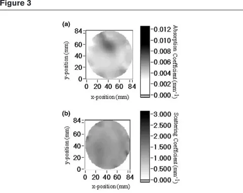
gadolinium-enhanced magnetic resonance imaging (MRI) for valida-tion [7]. Fig. 2 shows a result obtained from a 70-year-old patient with a 0.8 cm infiltrating ductal carcinoma. Fig. 2a shows a sagittal magnetic resonance image after gadolin-ium contrast enhancement (shown in color) passing through the center of the cancerous lesion. Fig. 2b depicts a coronal DOT image of the absorption coefficient increase due to ICG administration. This image is perpen-dicular to the plane in Fig. 2a, and was obtained at 830 nm for the volume of interest indicated on Fig. 2a with the dashed line box. Fig. 2c shows a magnetic resonance coronal reslicing of the volume of interest with the same dimensions as Fig. 2b. Furthermore tomographic images of intrinsic contrast have been also demonstrated [27]. Figure 3 displays quantified coronal images of the absorp-tion and reduced scattering coefficients obtained from the breast of a patient with a well-localized 3.4-cm fibroade-noma in the upper central region of her breast at 754 nm.
Optical imaging of intrinsic and extrinsic
breast cancer activity
Imaging of intrinsic contrast
The capacity of optical imaging, and especially DOT, to image and quantify intrinsic and extrinsic tissue optical properties is discussed above. Intrinsic optical contrast offers significant functional information. Imaging of scatter-ing may be associated with structural characteristics and the concentrations of organelles. More importantly, imaging of the absorption coefficient at appropriately selected wavelengths can quantify the concentrations of water and oxyhemoglobin and deoxyhemoglobin of breast tumors, and obtain measures of hemoglobin concentration Figure 1
and hypoxia. Therefore, the optical method is unique in assessing and quantifying those important functional tissue and cancer characteristics. Correlation of intrinsic signals and malignancy has been demonstrated [28••]. The detection limits and the diagnostic capacity of any combination of the intrinsic features on a screening popu-lation remain to be seen, because the limited number of patients examined by DOT thus far does not allow such factors to be identified.
Imaging of contrast agents
Similar to other clinical imaging modalities, a novel element that can enhance the potential applications of optical imaging is the use of contrast agents. In the NIR range the most widely used contrast agent is ICG, because it is a safe, US Food and Drug Administration-approved NIR absorbing and fluorescing dye. ICG is an intravascular contrast agent that may extravasate through vessels of high permeability, such as cancerous vessels. Therefore, ICG imaging of the breast mainly probes per-meability and vascularization [7]. Other ICG derivatives with extravascular distribution mechanisms have recently been considered [11] for diagnostic purposes. Several other imaging methods can target such features with the use of appropriate contrast agents. Similar to nuclear
imaging, however, the optical method generally can detect very small concentrations of chromophores or fluoro-phores, but without using ionizing radiation and at a reduced cost. Therefore, optical methods may still have significant advantages in breast cancer detection using such extrinsic contrast agents.
Imaging molecular activity and gene expression
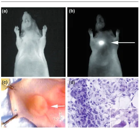
[image:4.612.315.555.89.280.2]A new advance that holds great promise for breast cancer research is the recent development of optical probes for molecular imaging, specifically in the NIR range. Fluorescent dyes that target specific tumor recep-tors [12], or that are activated (fluoresce) by tumor-asso-ciated enzymes (such as cathepsins and matrix metalloproteinases) [13], have been shown to identify their molecular targets in vivo. The latter probes probably hold the greatest promise, because they are quenched in the absence of the targeted enzymatic activity, yielding highly specific fluorescence signals.
[image:4.612.57.298.95.220.2]Fig. 4 depicts the light image and the contrast-enhanced fluorescence image of a LX-1 tumor implanted into the mammary fat pad of a nude mouse. The contrast agent used was a synthetic graft copolymer with Cy5.5-quenched fluorochromes, which becomes activated in the presence of cathepsin D. This NIR fluorescent probe demonstrated a 12-fold signal increase in cancers. Figure 2
DOT of ICG enhancement obtained simultaneously with gadolinium-enhanced MRI of the same breast affected by an 8-mm ductal carcinoma. (a)Sagittal magnetic resonance image of the breast (in grayscale) superimposed with the gadolinium enhancement (in color). The cancerous lesion appears enhanced in the center of the image because of gadolinium administration. (b)Coronal DOT image of the absorption coefficient change due to ICG distribution. This image is perpendicular to the plane of the MRI image in (a) and is obtained for the volume of interest indicated on (a) with the dashed line box. (c) Functional magnetic resonance coronal reslicing of the volume of interest with the same dimensions as (b). Gadolinium enhancement is averaged over the volume of interest and appears in color. The cancer appears in the right upper corner of the coronal image and has high spatial congruence with its appearance on the DOT image. A secondary lesion on the left, middle part of the image also coregisters well with the DOT image. A third lesion, which appears on the lower boundary of the DOT image, is probably an artifact. Although single slices through the breast volume are shown here, both MRI and DOT retrieve three-dimensional information of the volume of investigation. Published with permission from Ntziachristos et al [7]; © 2000 National Academy of Sciences, USA.
Figure 3
Using this technology, appropriately engineered fluores-cent probes can be selectively activated by endogenous or transferred gene expression. The combination of such probes with optical imaging may yield a unique, highly sensitive technology for in vivo and real-time imaging of the expression patterns for various enzymes, which are crucially involved in tumor formation and metastasis. Various breast cancer cell lines have been identified to over-express specific enzymes such as matrix metalloproteinases [29], which are not over-expressed in normal cells.
Therefore, the impact of developing molecular–optical imaging, and in particular molecular–DOT, of the breast is potentially enormous. First, selected molecular activity can be achieved with high sensitivity, because back-ground fluorescence is quenched. Second, cancers could be detected at their molecular onset, before anatomic changes become apparent. Therefore, thera-pies can be initiated at a very early stage, which is the single most important strategy in achieving high survival rates. Third, specific cancer parameters such as growth kinetics, angiogenesis growth factors, tumor cell markers, and genetic alterations could be studied without perturbing the tumor environment. Finally, this additional information could aid in the development of novel targeted drugs and therapies, and could allow assessment of their efficacy at the molecular level. The importance of this imaging strategy is further amplified by considering that photon technology can detect single photons, so that it can resolve fluorescent molecules at nanomolar to picomolar concentrations, and requires instrumentation that is of relatively low cost and that uses nonionizing radiation.
Multimodality imaging
Another exciting application of optical technology is the combination of optical imagers with other imaging modalities. Light guidance using optical fibers makes optical imaging compatible with many other radiologic methods [24], such as mammography, ultrasound, MRI, and positron emission tomography, among others. The development of hybrid modalities offers the potential of simultaneously scanning the breast, under identical physiologic and geometric conditions. The optical method offers several complementary features to those of established medical imaging methods, mainly through targeting oxyhemoglobin and deoxyhemoglobin, but also through the study of molecular events and gene expres-sion, as discussed above. This can produce an increased number of features that may augment the diagnostic value of any single technique alone. Addition-ally, high-resolution information taken from another imaging modality can be implemented into the DOT inversion scheme to improve the quantification accuracy of the optical method.
Conclusion
Imaging of function and molecular activity is at the frontier of current research efforts to detect and study cancer non-invasively. Optical imaging offers complementary features to those of established radiologic imaging techniques, pri-marily the quantitative imaging of hemoglobin saturation and concentration, and the selective imaging of specific gene expression with high sensitivity, because back-ground signals can be suppressed using enzyme-acti-vated fluorescence probes. Similarly to other technologies, the method can also characterize vascular-ization, permeability, and a plethora of contrast agents with high sensitivity, without using harmful radiation and probably at lesser cost.
Current trends in optical imaging focus on the construc-tion imagers that yield an increased data-set of optical measurements per examination, so that higher resolution and quantification accuracy is achieved. The use of multi-ple wavelengths in order to capitalize on spectral informa-tion and to allow the quantificainforma-tion of other tissue chromophores apart from oxyhemoglobin and deoxyhemo-globin is also being pursued. More accurate and more effi-cient forward and inversion problems for improving the quantification accuracy and reconstruction speed are also
commentary
review
reports
[image:5.612.313.557.96.321.2]primary research
Figure 4
being investigated. Finally, the abilities of various contrast agents and probes to assess different functional and mol-ecular cancer characteristics are being explored. We believe that optical imaging will play a vital role in our further understanding of carcinogenesis, in early detection of cancer, and in the design of effective treatments.
Aknowledgements
We are grateful to Brian Pogue, Sergio Fantini, and Ralph Weissleder for providing exciting results, and to Christoph Bremer and Christina Benou for useful discussions.
References
Articles of particular interest have been highlighted as:
• of special interest
•• of outstanding interest
1. Cannestra AF, Bookheimer SY, Pouratian N, O’Farrell A, Sicotte N, Martin NA, Becker D, Rubino G, Toga AW: Temporal and topographical characterization of language cortices using intraoperative optical intrinsic signals.Neuroimage2000, 12: 41–54.
2. Yang P, Farkas DL, Kirkwood JM, Abernethy JL, Edington HD, Becker D: Macroscopic spectral imaging and gene expression analysis of the early stages of melanoma.Mol Med1999, 5: 785–794.
3. Svanberg K, Wang I, Colleen S, Idvall I, Ingvar C, Rydell R, Jocham D, Diddens H, Bown S, Gregory G, Montan S, Anders-son-Engels S, Svanberg S: Clinical multi-colour fluorescence imaging of malignant tumours: initial experience.Acta Radiol 1998, 39:2–9.
4. Stepp H, Sroka R, Baumgartner R: Fluorescence endoscopy of gastrointestinal diseases: basic principles, techniques, and clinical experience.Endoscopy1998, 30:379–386.
5. Sickles EA: Breast cancer detection with transillumination and mammography.Am J Roentgenol1984, 142:841–844. 6. Franceschini MA, Moesta KT, Fantini S, Gaida G, Gratton E, Jess
H, Mantulin WW, Seeber M, Schlag PM, Kaschke M: Frequency-domain techniques enhance optical mammography: initial clinical results.Proc Natl Acad Sci USA1997, 94:6468–6473. 7. Ntziachristos V, Yodh AG, Schnall M, Chance B: Concurrent MRI
and diffuse optical tomography of breast after indocyanine green enhancement.Proc Natl Acad Sci USA2000, 97:2767– 2772.
8. Benaron DA, Hintz SR, Villringer A, Boas D, Kleinschmidt A, Frahm J, Hirth C, Obrig H, van Houten JC, Kermit EL, Cheong WF, Stevenson DK: Noninvasive functional imaging of human brain using light.J Cereb Blood Flow Metab2000, 20:469–477. 9. Villringer A, Chance B: Non-invasive optical spectroscopy and
imaging of human brain function.Trends Neurosci1997, 20: 435–442.
10. Yodh AG, Chance B: Spectroscopy and imaging with diffusing
•• light.Physics Today1995, 48:34–40.
This is a concise overview of diffuse tissue optics as applied to diffuse optical spectroscopy and DOT.
11. Licha K, Riefke B, Ntziachristos V, Becker A, Chance B, Semmler W: Hydrophilic cyanine dye as contrast agents for near-infrared tumor imaging: synthesis, photophysical properties and spectroscopic in vivo characterization.Photochem Photo-biol2000, 72:392–398.
12. Achilefu S, Dorshow RB, Bugaj JE, Rajagopalan R: Novel recep-tor-targeted fluorescent contrast agents for in vivo tumor imaging.Invest Radiol2000, 35:479–485.
13. Weissleder R, Tung CH, Mahmood U, Bogdanov A: In vivo imaging of tumors with protease-activated near-infrared fluo-rescent probes.Nature Biotechnol1999, 17:375–378. 14. Cutler M: Transillumination as an aid in the diagnosis of
breast lesions.Surg Gynecol Obstet1929, 48:721–729. 15. Bundred N, Levack P, Watmough DJ, Watmough JA: Preliminary
results using computerized telediaphanography for investi-gating breast disease.Br J Hosp Med1987, 37:70–71. 16. Lafreniere R, Ashkar FS, Ketcham AS: Infrared light scanning of
the breast.Am Surg1986, 52:123–128.
17. Brenner RJ: X-ray mammography and diaphanography in screening for breast cancer.J Reprod Med1982, 27:679–684. 18. Drexler B, Davis JL, Schofield G: Diaphanography in the
diagno-sis of breast cancer.Radiology1985, 157:41–44.
19. Grosenick D, Wabnitz H, Rinneberg HH, Moesta KT, Schlag PM: Development od a time domain optical mammograph and first in vivo applications.Appl Opt1999, 38:2927–2943. 20. Fantini S, Heffer EL, Franceschini MA, Götz L, Heinig A,
Heywang-Köbrunner S, Schütz O, Siebold H: Optical mammog-raphy with intensity-modulated light. In: Proceedings Volume from In Vivo Optical Imaging Workshop; September 16–17 1999. Edited by Gandjbakhche A. Bethesda, MD: National Insti-tutes of Health; 2000:in press.
21. Fantini S, Walker SA, Franceschini MA, Kaschke M, Schlag PM, Moesta KT: Assessment of the size, position, and optical prop-erties of breast tumors in vivo by noninvasive optical methods.Appl Opt1998, 37:1982–1989.
22. Arridge SR: Optical tomography in medical imaging. Inverse
•• Problems1999, 15:R41–R93.
This is a complete review of methods for DOT.
23. O’Leary MA, Boas DA, Chance B, Yodh AG: Experimental images of heterogeneous turbid media by frequency-domain diffusing-photon tomography.Opt Lett1995, 20:426–428. 24. Ntziachristos V, Ma XH, Chance B: Time-correlated single
photon counting imager for simultaneous magnetic reso-nance and near-infrared mammography. Rev Sci Instrum 1998, 69:4221–4233.
25. Pogue BW, Testorf M, McBride T, Osterberg U, Paulsen K: Instrumentation and design of a frequency-domain diffuse optical tomography imager for breast cancer detection. Opt Express1997, 1:391–403.
26. Hoogenraad JH, van der Mark MB, Colak SB, ‘t Hooft GW, van der Linden ES: First results from the Philips optical mammo-scope. In: Photon Propagation of tissues III. Edited by Benaron DA, Chance B, Ferrari M. SPIE: The International Society for Optical Engineering; 1997, 3194:184–190.
27. Pogue BW, McBride TO, Osterman S, Poplack S, Osterberg U, Paulsen KD: Quantitative hemoglobin tomography with diffuse near-infrared spectroscopy: pilot results in the breast. Radiol-ogy2000, 218:in press.
28. Tromberg BJ, Shah N, Lanning R, Cerussi A, Espinoza J, Pham T,
•• Svaasand L, Butler J: Non-invasive in vivo characterization of breast tumors using photon migration spectroscopy. Neopla-sia2000, 2:26–40.
This is a comprehensive review of the potential biomedical applications of diffuse optics in characterizing breast tumors on the basis of intrinsic contrast.